Article

The Effect of Glucosinolates on the Growth and Development of Helicoverpa armigera Larvae and the Expression of Midgut Sulfatase Genes

1
Xinjiang Key Laboratory of Biological Resources and Genetic Engineering, College of Life Science and Technology, Xinjiang University, Urumqi 830046, China
2
Agriculture Ministry Key Laboratory of Integrated Pest Management on Crops in East China/State & Local Joint Engineering Research Center of Green Pesticide Invention and Application, Department of Entomology, College of Plant Protection, Nanjing Agricultural University, Nanjing 210095, China
3
Synthetic Biology Laboratory, School of Future Technology, Xinjiang University, Urumqi 830046, China
*
Authors to whom correspondence should be addressed.
These authors contributed equally to this work.
Submission received: 13 November 2021 / Revised: 11 January 2022 / Accepted: 21 January 2022 / Published: 25 January 2022
(This article belongs to the Special Issue Advances in Pesticide Discovery and Application)

Abstract

:
The plant–pest interaction and its mechanisms are a novel research direction for pest control. They provide molecular targets for developing new pesticides and targeted control measures to control insect herbivores. Glucosinolate is a large family of secondary substances found in cruciferous plants that are harmful to herbivorous insects. Specialist herbivores have developed specific anti-defense genes and detoxifying mechanisms against glucosinolate from the host plant, but how generalist herbivores respond to glucosinolate at the molecular level is unknown. In this study, we investigated the effects of different glucosinolate concentrations on the growth and development of Helicoverpa armigera. Moreover, the expression of sulfatase genes (HaSulfs) was also checked following exposure to glucosinolate concentrations. The developmental duration of larvae and pre-pupa of H. armigera was significantly increased by 14.79–25.03% after feeding glucosinolate compared to the control. Quantitative Real-Time PCR (RT-qPCR) was carried out to analyze the expression of HaSulf family genes in the midgut of fifth instar larvae of H. armigera. The results showed that the upregulated expression patterns of HaSulf family genes were diversified after feeding at different concentrations. The expression level of HaSulf was detected with the HaSulf antibody. Only the glucosinolate-fed larvae had a visible target band and were mainly distributed in the midgut wall. Taken together, glucosinolate can significantly affect the growth and development of H. armigera larvae. It can induce the expression of HaSulf in the midgut of H. armigera at gene and protein levels. This study could be useful to understand the development of plant-derived insecticides resistance in H. armigera.

1. Introduction

The cotton bollworm, Helicoverpa armigera Hübner (Lepidoptera: Noctuidae), is a worldwide pest that causes severe damage to economic crops [1]. At present, chemical insecticides [2,3] and transgenic Bacillus thuringiensis (Bt) crops [4,5] are widely used to control them, but their long-term application has led to the gradual resistance of H. armigera. For many years, people have been guiding the rational use of pesticides to overcome or delay the development of resistance. However, ignoring the mutual restriction between chemical control and plants, plant secondary substances and pests may accelerate the development of pest resistance and reduction of crop resistance, leading to pest outbreaks [6,7,8]. The interaction of plant secondary metabolites and pests and their mechanisms is a major topic in scientific research. It is important to promote the selection of pest-targeting genes and the development of green pesticides [9,10,11,12].
Glucosinolate (GS) is a secondary metabolite of nitrogen and sulfur-containing anions widely present in cruciferous plants [13,14]. Glucosinolate can be catalyzed by myrosinase to produce a series of toxic hydrolysis products, including isothiocyanate (ITC), nitrile, thiocyanate, and other active substances. Some of these products poison the pests or inhibit them from laying eggs, thereby avoiding or reducing plant damage [15]. At present, there are more than 120 types of glucosinolate found in nature. The types and content of glucosinolate in cruciferous plants are the most abundant [16]. Studies have found that different varieties of cruciferous plants have different types of glucosinolate. The composition and content of glucosinolate vary greatly between parts of the same plant and at various stages of development [17]. This variation in glucosinolate quantity and composition also helps cruciferous vegetable crops to survive pests and other environmental stresses [18].
Herbivorous insects have been hampered to some extent by the diversity of the plant’s complex and delicate glucosinolate-myrosinase defense mechanisms. However, cruciferous vegetable pests can feed host plants as part of a long-term co-evolutionary process. Several anti-defense mechanisms deal with glucosinolate-myrosinase defense mechanisms in plants [19,20,21,22,23,24]. The cabbage stem flea beetle (Psylliodes chrysocephala) and the specialist pest Plutella xylostella may desulfurize host plant glucosinolates via their glucosinolate sulfatase (GSS) [25,26]. As a result, the host plant myrosinase cannot recognize and hydrolyze the substrate glucosinolate and produce toxic secondary metabolic defense substances [27,28,29]. Sulfatase is also found in generalist herbivores. The expression of Heliothis virescens sulfatases was upregulated after feeding wild-type Arabidopsis thaliana, while the larvae weight increased significantly on the mutant A. thaliana without glucosinolate [30]. Sulfatase is an evolutionarily conservative enzyme found in both prokaryotic and eukaryotic organisms. The sulfatase activity is initiated by the sulfatase modification factor (SUMF1), which modifies a cysteine residue in the active center of sulfatase to formylglycine (FGLY) [31,32,33]. The functions of the SULF and SUMF genes in insects have not been thoroughly studied. The insect genome sequences provide an opportunity to examine the genes involved in the detoxification of plant glucosinolate [34]. In this study, eight HaSulfs and one HaSumf1 were identified from the H. armigera genome. It was found that feeding H. armigera larvae on different concentrations of glucosinolate (above 4 ppm concentration range) can considerably restrict the growth and development of the larvae and can promote the expression of HaSulfs in the midgut tissue of the H. armigera. Our findings provide a theoretical foundation for developing new green pesticides and targeted control measures for H. armigera.

2. Materials and Methods

2.1. Materials

The sinigrin hydrate (>99.0%, TLC Grade), Extraction kit (TransZol Up Plus RNA Kit), Reverse Transcription Kit (TransScript One-Step gDNA Removal and cDNA Synthesis Super Mix), and Fluorescence Quantitative kit TransStart® Green qPCR SuperMix were purchased from Beijing TransGen Biotech. The ethanol (100%), trichloromethane, and RNase-free water were all domestic chemical reagents. The primer synthesis and sequencing were performed by Sangon Biotech (Shanghai) Co., Ltd.
The H. armigera strain used in this study was obtained from the Key Laboratory of Bioresource Genetic Engineering of Xinjiang University, China. Helicoverpa armigera larvae were reared on artificial breeding. The adults were fed with a honey solution at 27 ± 2 °C, 60% ± 10% relative humidity, and L:D = 14: 10 h in a greenhouse without exposure to insecticides.

2.2. Bioassay

In bioassays experiments, seven glucosinolate concentrations (0, 2, 4, 20, 40, 80, 160 ppm) were used. Hundred-second instar larvae of same age were used for each concentration. The developmental duration from larva to pupae and survival rate under different glucosinolate concentrations were observed. The weight of the fifth instar larvae and pupa were checked regularly.

2.3. Identification and Phylogenetic Tree Construction of HaSulf

The sulftases sequence was downloaded by searching the conserved protein domain database at NCBI (http://0-www-ncbi-nlm-nih-gov.brum.beds.ac.uk/ (accessed on 1 June 2017)). MEGA7 software was used to align all sequences and build a phylogenetic tree using the maximum likelihood technique. Bootstrap values for 1000 replicates were calculated with MEGA7. The BLAST approach was used to identify SULFs and SUMFs genes from the genomes of H. armigera, P. xylostella, and Bombyx mori. Through https://jaspar.genereg.net/analysis, the transcription binding site of the promoter region was predicted [35].

2.4. Expression of HaSulfs and HaSumf1

The RNA-seq data of HaSulfs and HaSumf1 were downloaded from the NCBI database (https://0-www-ncbi-nlm-nih-gov.brum.beds.ac.uk/ (accessed on 1 June 2017)). The RT-qPCR was performed using SYBR-green fluorescence and gene-specific primers to determine the expression levels of HaSulfs and HaSumf1 fed with different glucosinolate concentrations. Five biological and two technical replicates were used in RT-qPCR.
The midgut of fifth instar H. armigera larvae fed with different concentrations of glucosinolates was dissected and collected (24–48 h after molting). The TransZol Up Plus RNA Kit was used to extract the total RNA from the midgut following the recommended protocol. cDNAs were synthesized using 1.0 μg total RNA by PrimeScript RT reagent kit with gDNA Eraser (Takara Biotechnology, Dalian, China). RT-qPCR reactions were performed using TransStart®Green qPCR SuperMix reagent in a Real-Time PCR System (Applied Biosystems, US Applied Biosystems, Waltham, MA, USA) following the manufacturer’s instructions. PCR conditions were 94 °C for 30 s, 40 cycles of 94 °C for 5 s, 60 °C for 15 s, and 72 °C for 10 s, then 95 °C for 15 s and 60 °C for 1 min.
The relative expression levels were calculated using 2−ΔΔCt method. The ribosomal protein L15 (HaRPL15) gene was used as an internal reference. The primers sequences for RT-qPCR are listed in Table 1.

2.5. Determination of Protein Production in the Midgut

The proteins were extracted from the midgut wall and midgut contents of the fifth instar larvae of H. armigera when treated with 4 ppm glucosinolates (24–48 h after molting). In addition, 4 ppm of gossypol and methyl jasmonate were fed to H. armigera as controls for xenobiotics, and the production of HaSulf protein was measured. The Western blot step was performed as follows: (1) SDS-PAGE electrophoresis was used to separate the different protein samples, which was then transferred to a PVDF membrane; (2) samples were blocked with 5% skimmed milk powder overnight at 4 °C, then washed 3 times with PBST for 5 min each time; (3) the primary antibody was incubated as follows: The primary antibody was diluted at a ratio of 1:10 and blocked for 2 h at 37 °C. The primary antibody removed and washed three times with PBST for five minutes each time; (4) the secondary antibody was incubated as follows: 2% skimmed milk powder was used, diluted (1:100) with goat anti-mouse secondary antibody (HRP-labeled), incubated at 37 °C for 2 h, and washed 3 times with PBS for 5 min each time; (5) DAB color development: 50 µL of freshly prepared color development solution was added dropwise to the membrane, darkened the color development for 10 min, and then immediately washed with distilled water to stop the reaction.

2.6. Statistical Analysis

The relative mRNA expression levels of genes and the growth and development data were analyzed by one-way analysis of variance (ANOVA) using SPSS v. 19 (SPSS Inc., Chicago, IL, USA). P < 0.05 was considered significant for all treatments. All the figures were generated using GraphPad Prism 5.0.

3. Results

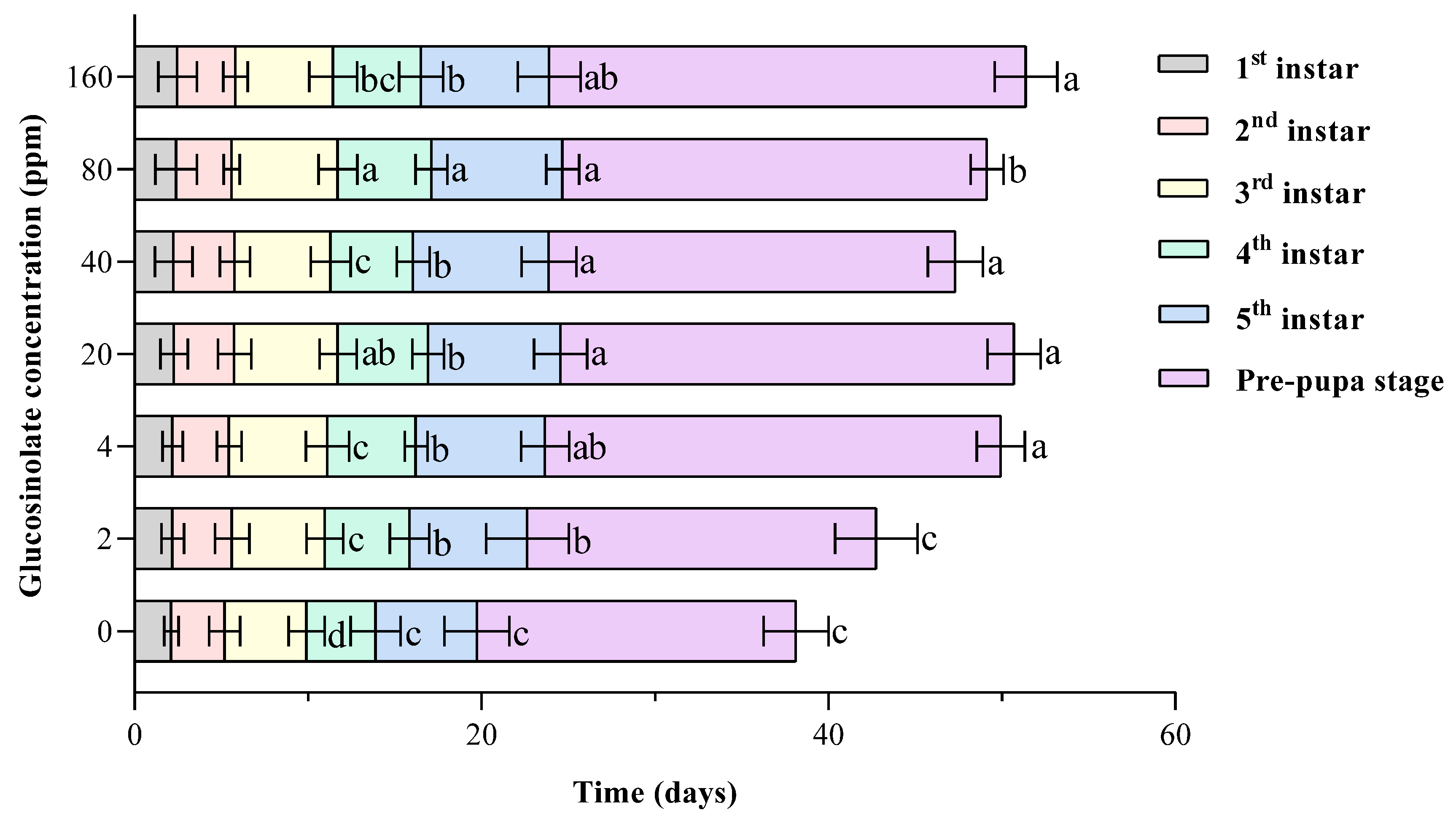
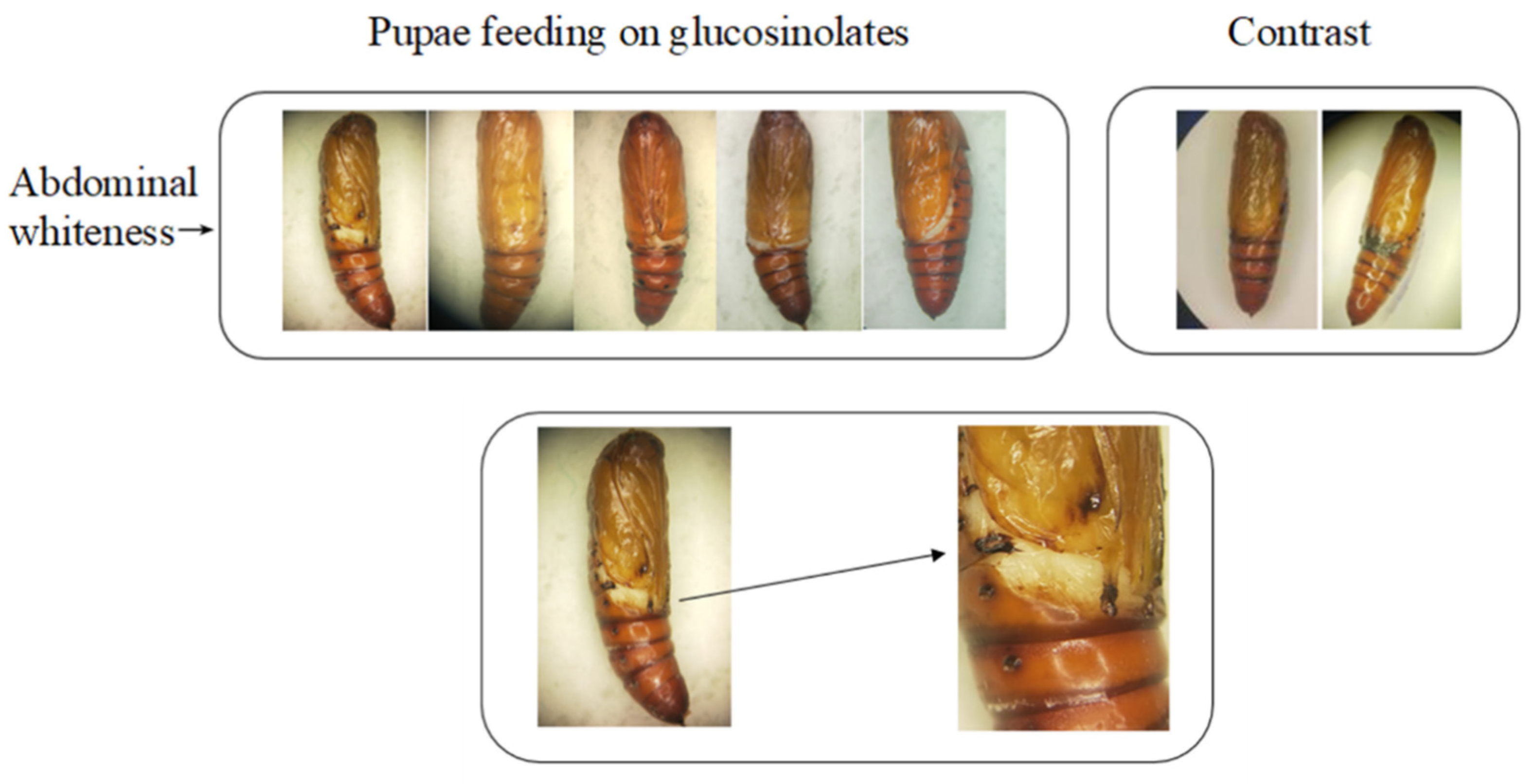
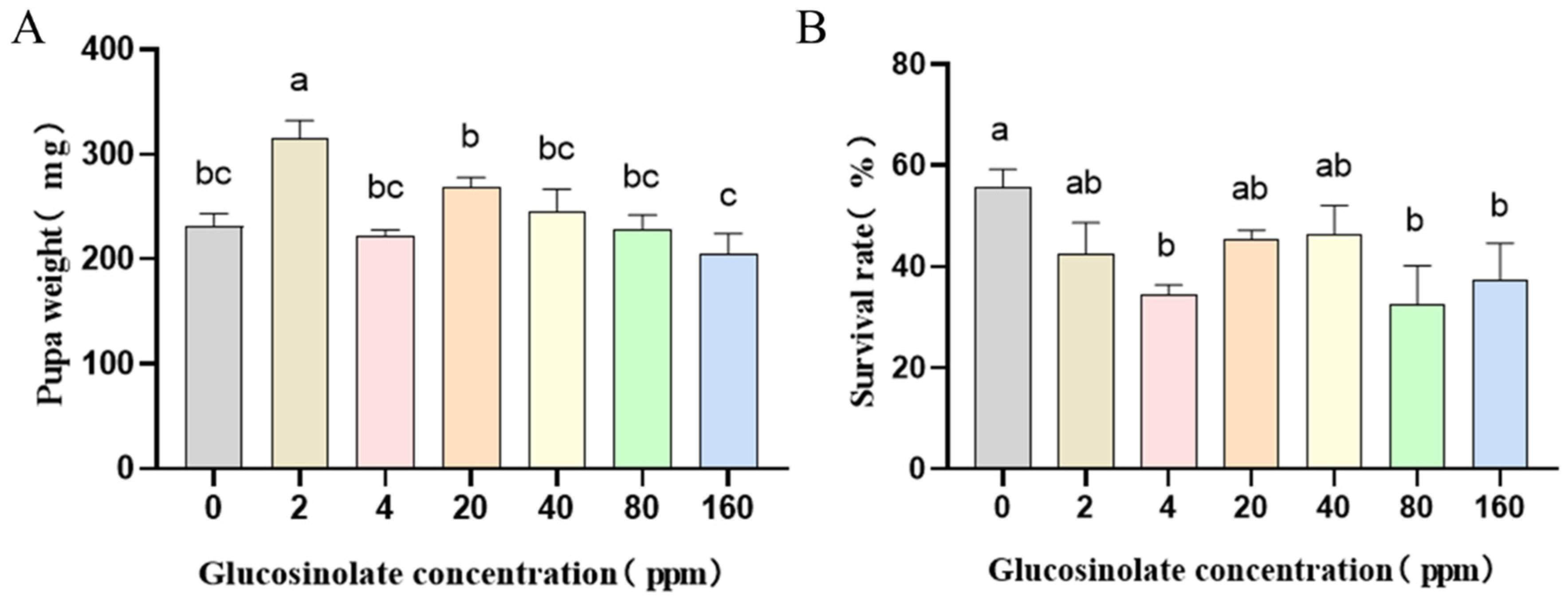
3.1. Effects of Different Concentrations of Glucosinolate on the Growth and Development of H. armigera

Different concentration gradients of glucosinolates (0–160 ppm) are added to the artificial diet to feed the second instar larvae of H. armigera. The biological parameters, including the developmental stages of larvae, pupae weight, and pupal rate, were observed. After feeding glucosinolate, the development duration of H. armigera was significantly delayed in the fifth instar and the pre-pupal stage. The development duration of H. armigera larvae fed with different glucosinolate concentrations significantly (p < 0.05) increased by 14.79–25.03% (22.66–24.68 d) as compared to the control group (19.74 d) (Figure 1). H. armigera pupae fed glucosinolate have deformed pupae and incomplete abdominal wrapping (Figure 2). Furthermore, the pupa weight results showed that low concentrations of glucosinolate (2 ppm) had a positive impact on H. armigera growth and development as compared to the control group (p < 0.05) (Figure 3A). The survival rate of the pupae of H. armigera fed with 4, 40, and 160 ppm glucosinolate was significantly decreased (32%–37%) as compared to the control group (56%) (Figure 3B).
The results of development duration, growth, and pupal weight showed that the response of the H. armigera to glucosinolate is not entirely linear with the glucosinolate concentrations. The pupa weight of H. armigera is concentration-dependent in the range of 20 to 160 ppm. The survival rate of H. armigera pupae is concentration-dependent in the range of 0 to 4 ppm (Figure 3). Furthermore, the pupa survival rate of H. armigera fed with 80 and 160 ppm glucosinolate was significantly lower than the control group (p < 0.05) (Figure 3B).

3.2. Identification of HaSulf Gene and Protein Prediction

Eight HaSulfs and one HaSumf1 gene were identified, and a bioinformatics analysis of these sequences was carried out (Table 2, Table 3 and Table 4). The sulfatase gene family comprises four branches, namely, SulfA, B, C, and D [36]. In the evolutionary tree, HaSulf1, HaSulf2, and HaSulf4 are on the C branch, HaSulf5a and HaSulf5b are on the A branch, HaSulf6 is on the D branch, and HaSulf7 and HaSulf8 are on the B branch (Figure 4). The length of most predicted HaSulfs ranges from 290 to 615 amino acids (aa), of which 1 ultralong HaSulf7 gene contains 1403 amino acids. The gene positions range from 3,168,620 to 3241586, showing a high degree of structural complexity (Table 2). It is predicted that there are big differences in each protein’s isoelectric point, extinction coefficient, instability coefficient, fat index, and hydrophilicity. The isoelectric points of most HaSulfs are 5.19 to 6.63, among which the isoelectric points of HaSulf1, HaSulf6, and HaSulf7 are 8.86, 9.38, and 9.13, respectively, which are significantly higher than other proteins. The extinction coefficient of most HaSulfs ranges from 82,110 to 135595, while the extinction coefficient of HaSulf5a is 54680, which is substantially lower than other proteins. The extinction coefficient of HaSulf7 is 180710, which is significantly higher than other proteins. The instability coefficients of most proteins are predicted to be 33.03 to 45.91, and the instability coefficient of HaSulf7 is 52.71, which is significantly higher than other proteins (Table 3). It is predicted that the fat index of most proteins ranges from 76.85 to 91.04. However, the fat indices of HaSumf1 and HaSulf7 are 55.28 and 62.37, respectively. The hydropathic index of most HaSulfs is −0.163 to −0.690, and the hydropathic index of HaSulf7 is −0.985, which is quite different from other proteins. By predicting the transcription binding site of the promoter region, it is interesting to find that HaSumf1 has high similarity with the transcription factors of HaSulf2, HaSulf4, and HaSulf5a in the sulfatase gene family (Table 4), and we also found that HaSulf2 and HaSulf4 are co-located on the SulfC evolutionary branch [36]. This phenomenon provides an important theoretical basis for subsequent studies on the regulation mechanism of sulfatase genes and their modifier genes.

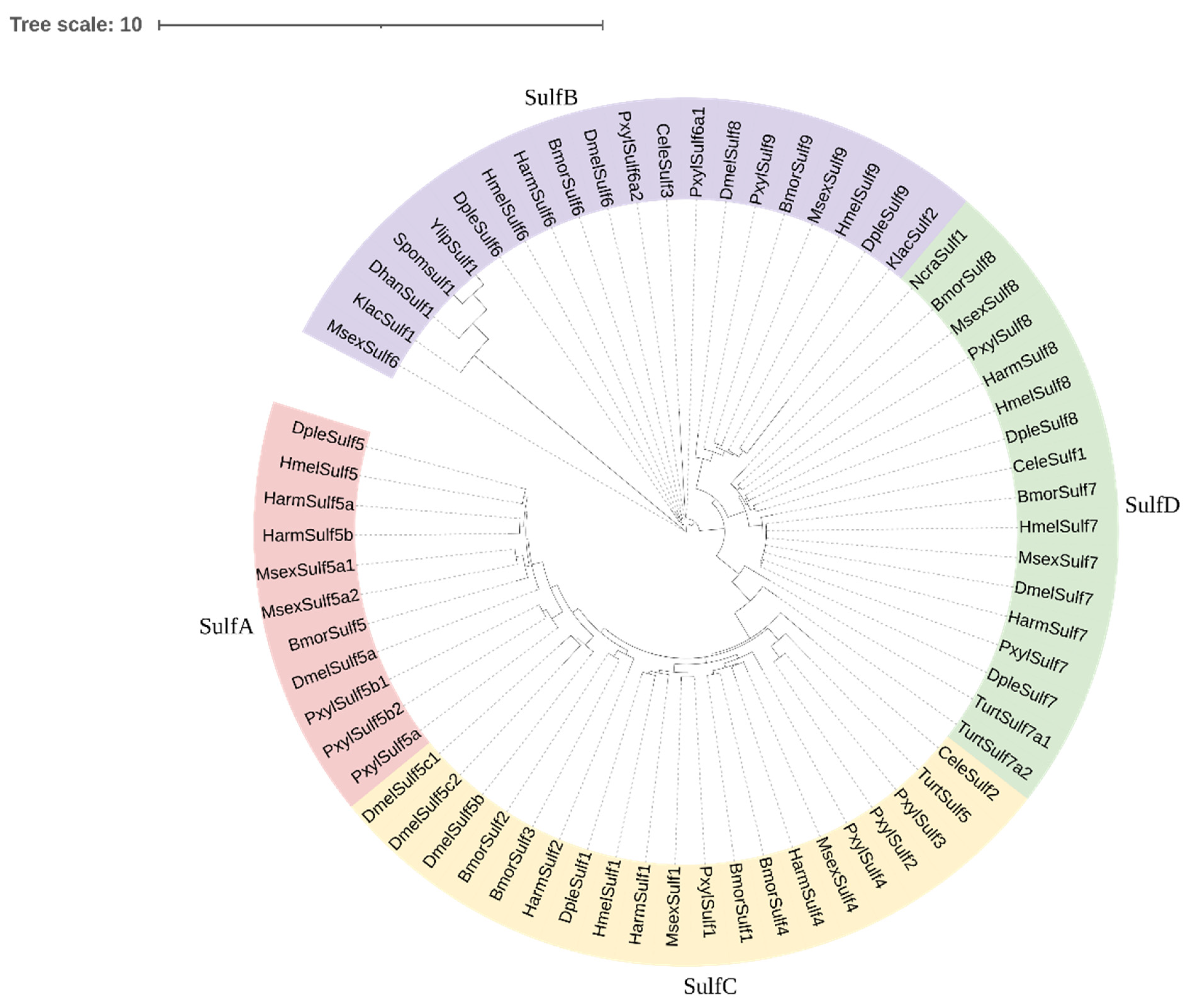
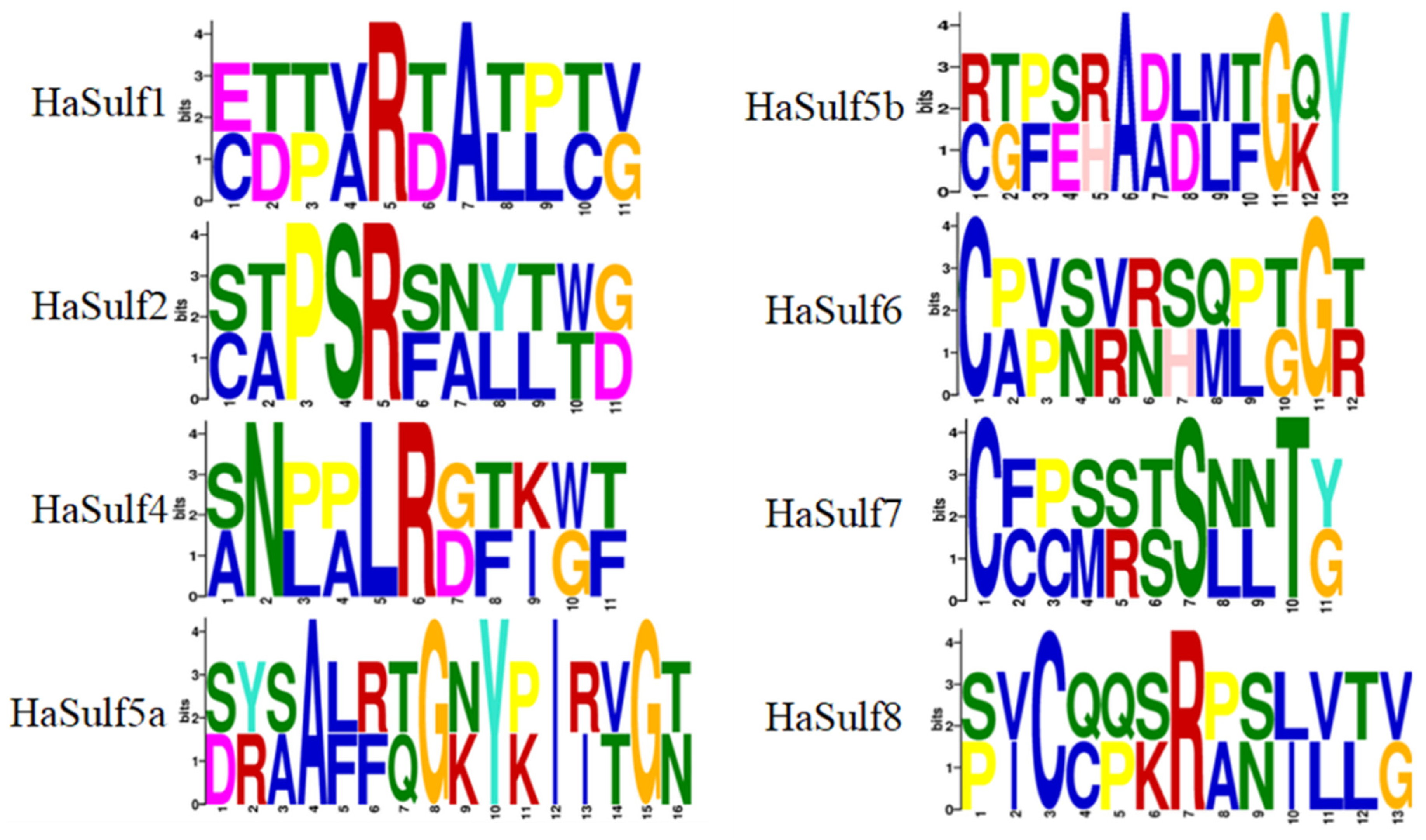
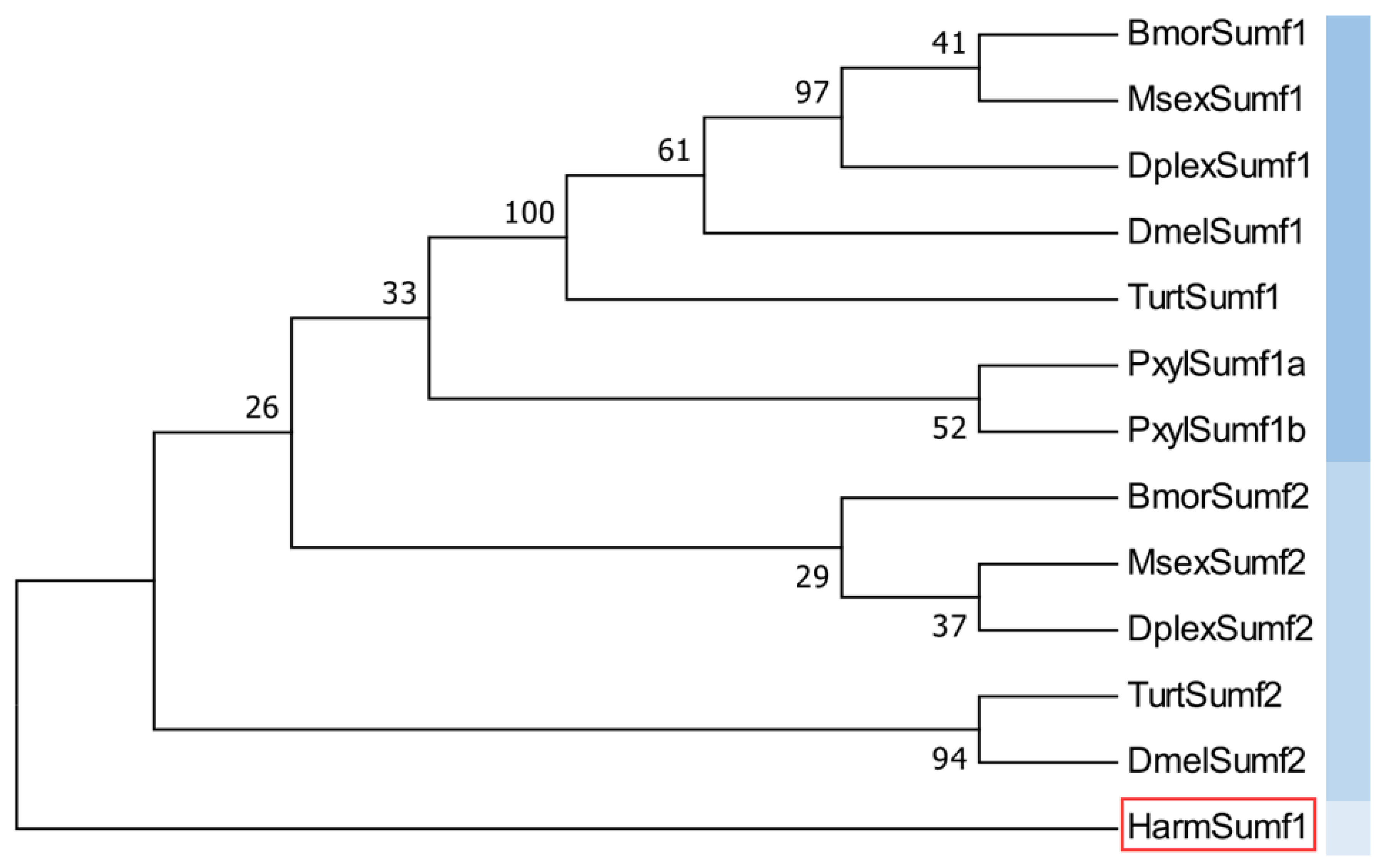
3.3. Prediction of Conserved Domains of HaSulfs and Construction of Phylogenetic Tree

Twenty different conserved motifs were identified from the eight HaSulfs of the H. armigera (Figure 5). Previous research has shown that C/SXPXRXTG is the shortest catalytic active center sequence that can maintain activity following HaSulf translation modification [37,38]. Different HaSulf sequences show substantial similarity at this site. We constructed an evolutionary tree to better understand the evolutionary relationship of H. armigera HaSulf. The HaSulf of H. armigera has homology with other insects, but there are some differences between different HaSulfs (Figure 4 and Figure 5).

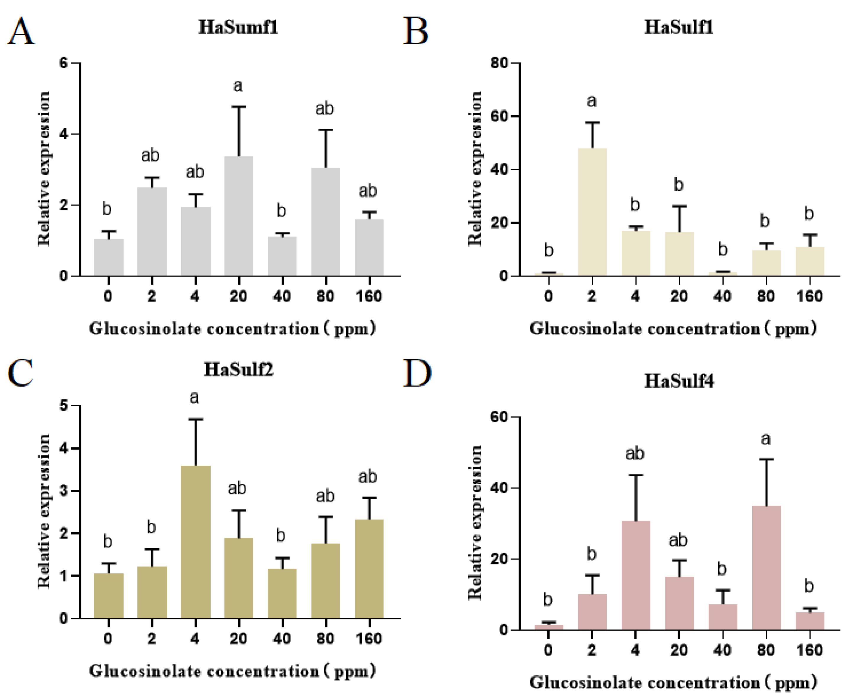
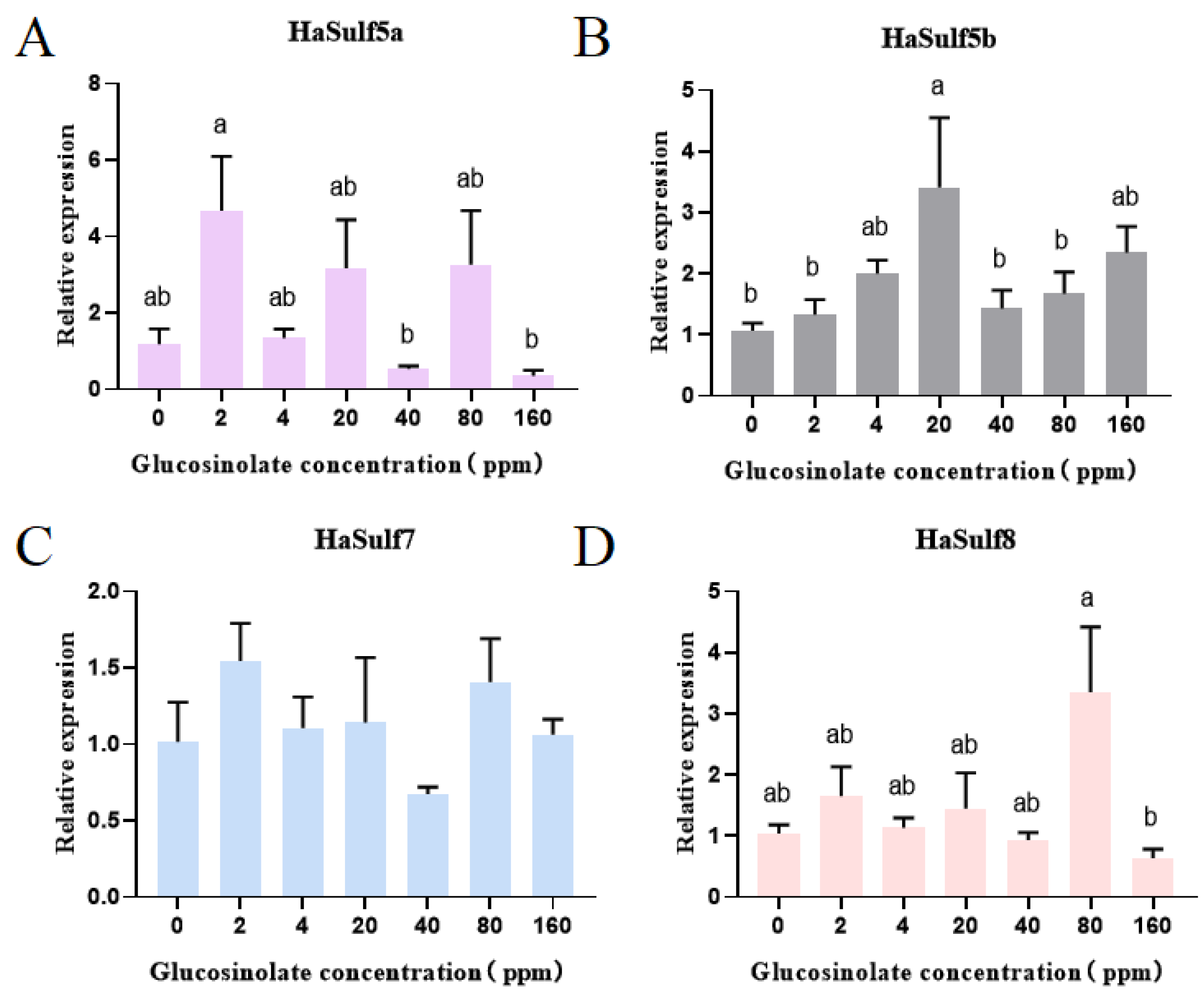
3.4. RT-qPCR Analysis of the Effects of Different Concentrations of Glucosinolate on the HaSulf Genes of H. armigera

Different concentrations of glucosinolate (0–160 ppm) were fed to the second instar larvae of H. armigera. At the fifth instar (24–48 h after molting), RT-qPCR was used to analyze the expression levels of the HaSulfs gene family in the midgut of H. armigera. The results showed that, except for HaSulf7, glucosinolate significantly induced other HaSulf genes’ expression (Figure 6 and Figure 7). The HaSumf1 gene fed with 40 ppm glucosinolate had no difference compared with the control group. Furthermore, the HaSumf1 gene was up-regulated to varying degrees when fed different concentrations of glucosinolate (Figure 6). The expression of the HaSulf1 gene was highly upregulated (48.1 times) when fed 2 ppm glucosinolate (Figure 6). The expression of the HaSulf2 gene was significantly increased (3.6 times) when fed 4 ppm glucosinolate (p < 0.05). The expression of HaSulf4 gene fed with 80 ppm glucosinolate was significantly upregulated (34.9 times) compared to the control. The HaSulf5a gene fed with 2 ppm glucosinolate was upregulated 4.7 times more compared to the control (Figure 6). However, the HaSulf5a gene fed with 40 and 160 ppm glucosinolate was downregulated to a certain extent. A significant difference was observed in the expression level of the HaSulf5a gene when H. armigera was fed with 2, 40, and 160 ppm glucosinolate (p < 0.05). The expression of the HaSulf5b gene was highly upregulated (3.4 times) at a 20 ppm concentration of glucosinolate (Figure 7). Compared with the control group, the HaSulf8 gene fed with 80 ppm glucosinolate has a 3.3 times greater expression. HaSulf8 fed with 160 ppm glucosinolate had a certain degree of downregulation compared to the control group. A significant difference was observed when the HaSulf8 gene was fed with 80 and 160 ppm glucosinolate (p < 0.05). There was no significant difference in the expression of HaSulf7 gene under different concentrations of glucosinolate (Figure 7).

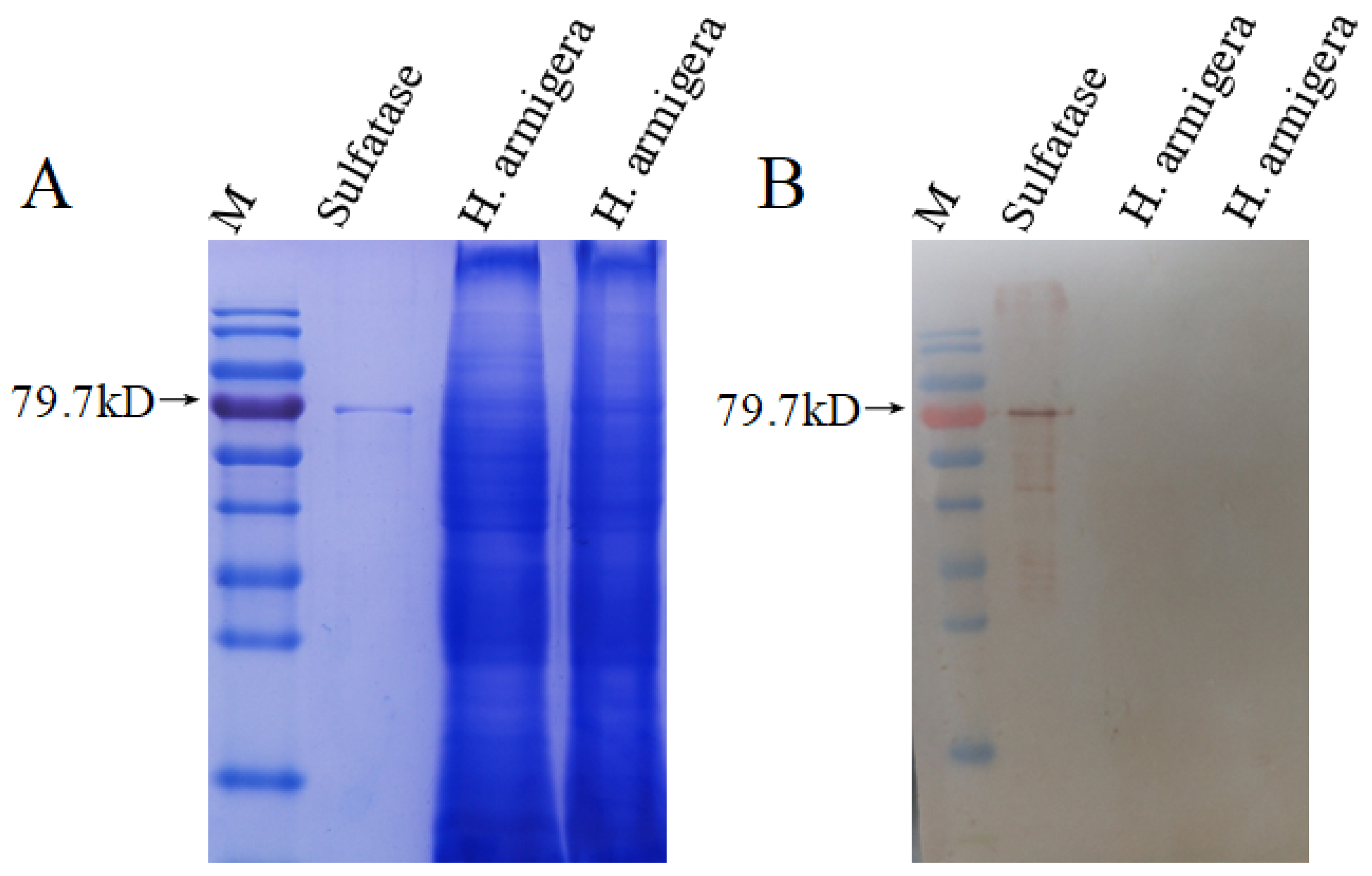
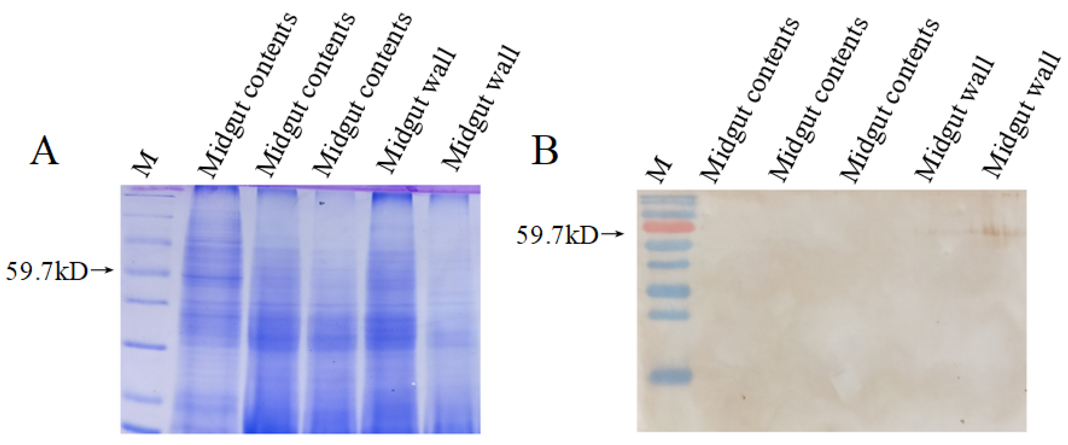
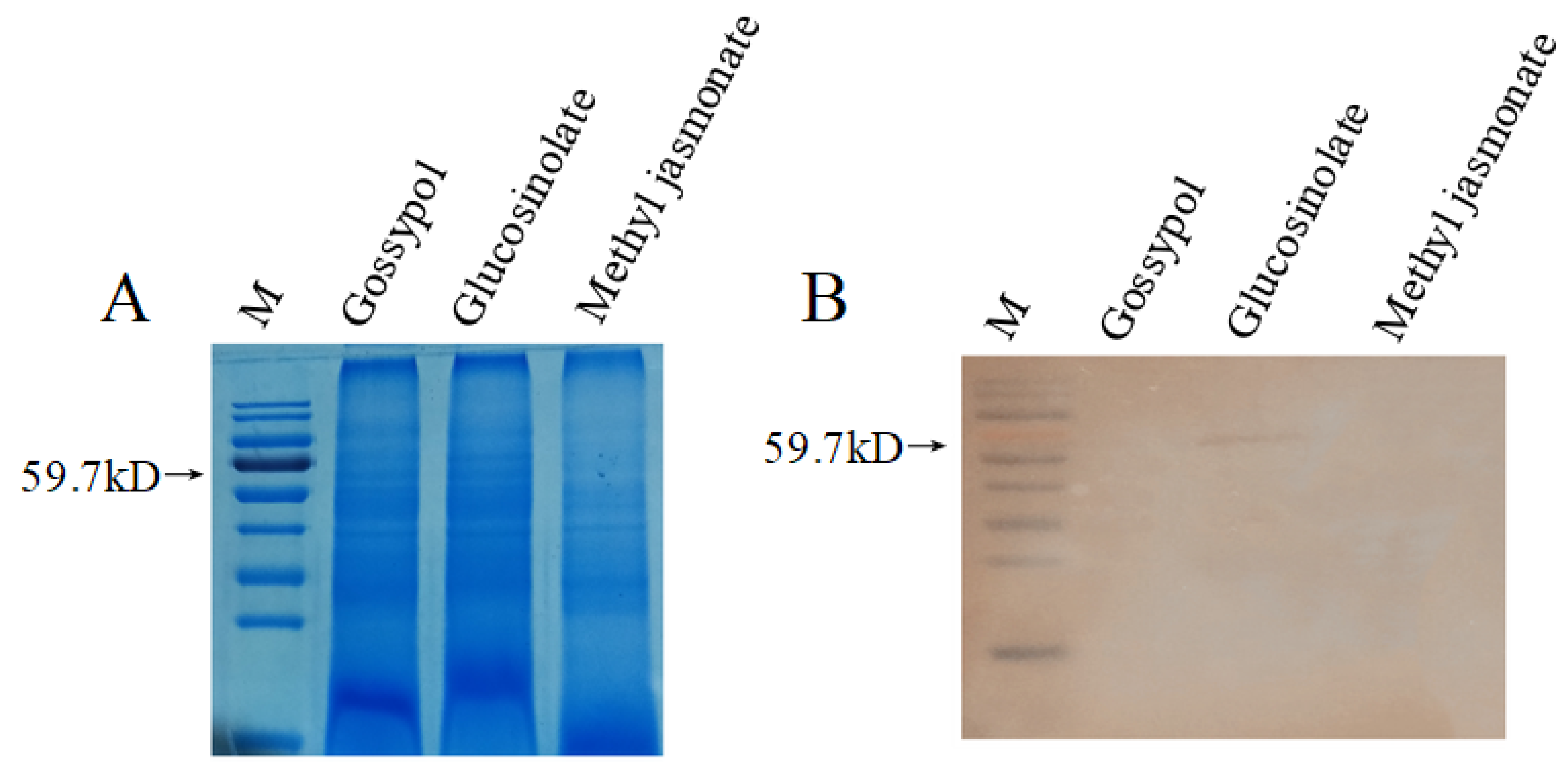
3.5. Western Blot Analysis of HaSulf Production at the Protein Level

The production of HaSulf was also observed at the protein level using the Western blot technology. The midguts of H. armigera (fifth instar) were dissected, and the proteins were extracted. The HaSulf antibody was used to detect the protein production level of HaSulf. In vitro induced and purified HaSulf protein was used as a positive control. The results showed that the prokaryotic expressed and purified HaSulf had a clear target band, with an Mr size of about 79,700 (Figure 8). However, the midgut protein of the H. armigera did not detect a visible target band (Figure 8).
Subsequently, the H. armigera larvae (fifth instar) fed with 4 ppm glucosinolate were collected. After dissection, the midgut and midgut contents were collected. The proteins were extracted from all samples. The target band was visible on the midgut wall, with an Mr size of around 59700, which was consistent with the size of the target band. No corresponding band was found in the contents, as detected by the HaSulf antibody (Figure 9). Furthermore, the fifth instar H. armigera was fed with 4 ppm gossypol, glucosinolate, and methyl jasmonate. After 24 h, the midgut was dissected, and the protein was extracted. Then, the protein production level of HaSulf was detected by HaSulf antibody. The results showed that only H. armigera larvae had clear bands after feeding glucosinolate. It was also hypothesized that plant glucosinolate induced HaSulf in H. armigera (Figure 10).

4. Discussion

HaSulf genes play multiple roles in insects, including growth, development, and detoxification metabolism. The HaSulf family genes are closely linked with the anti-plant defense system. Understanding the expression level of the HaSulf gene family in H. armigera is crucial to elucidate the relationship between HaSulf and glucosinolate. Therefore, simulating the difference in the glucosinolate content of insects feeding on the same plant with different glucosinolate concentrations might reduce the complexity of insects feeding on different plants.
At present, the sulfatase gene has not been studied deeply in insects. The increasing number of insect genome sequencing provides an opportunity to analyze the insect genes involved in the detoxification of plant glucosinolates from the whole genome. Sulfatase is a hydrolase that can decompose sulfate from various substrates, including glycosaminoglycans, sulfolipids, and steroid sulfates [38]. The function of insect sulfatase is related to the gradual adaptation of the host plant’s defense system in the co-evolutionary arms race of cruciferous plants. The specialist pest P. xylostella can use its glucosinolate sulfatase (GSSs) to competitively combine with the glucosinolates of the host plant to quickly desulfurize glucosinolates, so that the host plant myrosinase cannot recognize and hydrolyze the substrate glucosinolates and cannot produce toxic secondary metabolic defense substances [39,40]. Generalist pests also contain sulfatases, however, the function of sulfatase genes and the molecular mechanism of how to detoxify host plant glucosinolates in generalist pests are still unclear. Our previous study identified eight HaSulf genes and one modifier gene HaSumf1. The HaSulf has a certain degree of homology with other insect HaSulf enzymes (Figure 4). The HaSUMF1 is similar to the SUMF2 of Tetranychus urticae and Drosophila melanogaster through evolutionary trees (Figure 11). Different HaSulf sequences show substantial similarity at the C/SXPXRXTG site (Figure 5). Each amino acid column has a distinct height, showing variances in the amount of amino acid information and conservation of each HaSulf. Differences in amino acids and conserved motifs may affect the biological functions of sulfatase and the binding and hydrolytic activity of substrates to a certain extent [40]. Moreover, HaSulfs have different isoelectric points, extinction coefficients, instability coefficients, fat indices, hydrophilicity, and conservative motifs with different effects. The length of most predicted HaSulfs ranges from 290 to 615 amino acids (aa), of which one ultralong HaSulf7 gene contains 1403 amino acids. The gene positions range from 3,168,620 to 3,241,586, showing a high degree of structural complexity. From the similarity of the sulfatase genes between H. armigera and other insects, we speculate that H. armigera sulfatase also detoxifies host plant glucosinolates [41]. The sequence expansion and differences in the H. armigera sulfatase family members may be related to their feeding habits. The sulfatase family members have a certain degree of synergy and new functionalization to adapt to the secondary metabolites of different host plants [42].
Furthermore, RT-qPCR results showed that the HaSulf1 and HaSulf5a genes were up-regulated in the midgut of H. armigera fed with 2 ppm glucosinolate, the HaSumf1 and HaSulf5b genes were upregulated at 20 ppm, while the HaSulf4 and HaSulf8 genes at 80 ppm glucosinolate (Figure 6 and Figure 7). Based on these results, it is hypothesized that these genes may have a cooperative expression [36]. The HaSulf7 gene may be relatively conservative, therefore, no significant difference was observed. The HaSumf1, HaSulf2, HaSulf4, HaSulf5a, and HaSulf8 genes in the H. armigera fed 80 ppm glucosinolate were up-regulated to varying degrees compared with 40 ppm glucosinolate (Figure 6 and Figure 7). It is hypothesized that the H. armigera may activate other defense mechanisms when fed 40 and 80 ppm glucosinolate. RT-qPCR results showed that the expression level of HaSulf was not consistent when fed with different glucosinolate concentrations (Figure 6 and Figure 7). Therefore, it is speculated that different HaSulf genes play a crucial role in various concentrations. Ren J et al. studied the expression levels of different sulfatase genes in Bemisia tabaci and found that the same sulfatase gene is differentially expressed in different subspecies. This different differential expression phenomenon may be related to the feeding habits of insects [42]. Manivannan A and others also measured the expression of the sulfatase gene. In vivo silencing of the B. tabaci GSS gene expression via RNA interference led to lower levels of desulfoglucosinolates in honeydew [41]. The biological function of H. armigera sulfatase requires HaSumf1 to activate and HaSumf1 also has different responses to different concentrations of glucosinolates (Figure 7), which may also be one of the reasons for the different expression of sulfatase. The cooperative expression of sulfatase genes seems to be consistent with the results in the evolutionary tree (Figure 6 and Figure 7). The sulfatase gene family comprises four branches, namely, SulfA, B, C, and D [36]. In the evolutionary tree, HaSulf1, HaSulf2, and HaSulf4 are on the C branch, HaSulf5a and HaSulf5b are on the A branch, HaSulf6 is on the D branch, and HaSulf7 and HaSulf8 are on the B branch (Figure 4). The differential expression of sulfatase genes in H. armigera may be related to the hydrolysis and binding activity of the substrate [40].
The HaSulf antibody was used to detect the expression level of sulfatase protein. In vitro induced and purified HaSulfs were used as a positive control (Figure 8). It is found that the production of sulfatase protein was only detectable in the midgut wall of H. armigera after glucosinolate feeding (Figure 9). Subsequently, we used different secondary metabolites gossypol and methyl jasmonate to demonstrate that the production of sulfatase protein detected in the midgut wall of H. armigera is not due to biotic stress (Figure 10). Fabian et al. reported that the sulfatases of the generalist moth H. virescens were induced by glucosinolates, and further biochemical experiments are needed to confirm [30]. This study reported that Hasulfs genes and protein levels are both induced by glucosinolates. We found that feeding low concentrations of glucosinolates can increase the production of H. armigera sulfatase, but we have not yet found a regulatory relationship between glucosinolate concentration and sulfatase production. Therefore, it is speculated that glucosinolates can induce the production of sulfatase protein in the midgut wall of H. armigera, but this induction phenomenon is not a concentration-dependent regulatory relationship, which requires further biochemical experiments to verify. Chen et al. found that after knocking out the sulfatase gene of P. xylostella through CRISPR/Cas9 technology, the egg hatching rate and larval survival rate of the mutant P. xylostella were significantly reduced, and the survival time of the larval stage was prolonged [40]. Agnihotri et al. showed that the developmental duration, survival rate, body length, and weight were significantly decreased when H. armigera was fed with 0–500 ppm glucosinolate. Moreover, the trypsin, chymotrypsin, cathepsin, and total protease activities were also reduced considerably [43]. The inhibition of digestive proteases by glucosinolate may cause insects to starve, leading to nutritional deficiencies, reduced free amino acids, and decreased metabolic energy [38]. It indicates that glucosinolate intake may affect the sulfatase content of the H. armigera, which in turn affects the growth and development of the H. armigera [40]. In our research, we also found that the glucosinolate-fed H. armigera had varying degrees of developmental delay compared to the control group (Figure 1). Moreover, the pupa weight was decreased, and the mortality rate increased (Figure 3). It is hypothesized that when the H. armigera is fed glucosinolate, it consumes energy to produce HaSulf to detoxify glucosinolate, indicating that HaSulf may affect the growth and development of insects. These combined effects of glucosinolate consumption result in growth retardation, developmental defects, and, ultimately, death of insects. The results showed whiteness in the abdomens of H. armigera fed glucosinolate, confirming that glucosinolate influenced H. armigera growth and development (Figure 2).
Low concentrations of glucosinolate (2 ppm) have a stimulating effect on the growth and development of H. armigera (Figure 1). However, the response of H. armigera to glucosinolate and its concentrations is not linear. The pupa weight of H. armigera is concentration-dependent in the range of 20–160 ppm, while the survival rate of pupae is in the range of 0 to 4 ppm (Figure 3B). The pupa weight of H. armigera fed with 80 and 160 ppm glucosinolate was not substantially different from the control group (Figure 3A). However, the pupa survival rate was considerably lower than the control group (p < 0.05). Therefore, it is speculated that glucosinolates may have affected the energy storage during the metamorphosis and development of the H. armigera pupa or another set of anti-defense mechanisms [13,15]. P. chrysocephala is a specialist pest that feeds on cruciferous plants. It can detoxify the glucosinolate-myrosinase defense system by desulfurization. However, testing found that desulfurized glucosinolates do not account for a high proportion of glucosinolates in P. chrysocephala, and it can use a variety of anti-defense mechanisms to resist the host plant’s glucosinolate-myrosinase defense system [44]. Combined with the results of qPCR experiments, it is found that different sulfatase genes have different response patterns to glucosinolates. Therefore, it is speculated that H. armigera sulfatase is not the specific detoxification mechanism of glucosinolates.

5. Conclusions

Taken together, the glucosinolate damaged the physiological functions, growth, and development process of H. armigera when fed with different concentrations. Our results showed that HaSulf gene family is involved in glucosinolate detoxification and may be linked to H. armigera growth and development. Further research on this phenomenon could show how the generalist pest H. armigera adapts its physiological activities to feed on plants containing various plant defense compounds. Therefore, an in-depth functional study of HaSulf is essential to determine the underlying molecular mechanism in H. armigera.

Author Contributions

X.M. and L.J. planned and designed this study; X.L., F.C., C.L., G.K. and S.Z. performed calculation analysis, performed experiments, and generated numbers; X.M., X.L. and L.J. contributed to the development and/or revision of the manuscript; C.L. and Y.Z. contributed to sample preparation; X.L. conducted qRT-PCR experiments and data analysis; X.M., X.L., L.J., C.L. and Y.Z. participated in the discussion. All authors have read and agreed to the published version of the manuscript.

Funding

This research was funded by the Natural Science Foundation of the Autonomous Region (2020D01C060), the Young Doctoral Science and Technology Talent Training Program of the Autonomous Region (2019660012), and the open project of the Fujian-Taiwan National Key Laboratory of Crop Pest Ecological Prevention and Control (SKL2018003), the National Natural Science Foundation of China (32060620), the Tianshan Innovation Team Project in Xinjiang Autonomous Region (2020D14022), and the research start-up fund of Xinjiang University (2018660010).

Data Availability Statement

Not applicable.

Conflicts of Interest

The authors declare no conflict of interest.

References

  1. Tay, W.T.; Soria, M.F.; Walsh, T.; Thomazoni, D.; Silvie, P.; Behere, G.; Anderson, C.; Downes, S. A Brave New World for an Old World Pest: Helicoverpa armigera (Lepidoptera: Noctuidae) in Brazil. PLoS ONE 2013, 8, e80134. [Google Scholar] [CrossRef] [PubMed] [Green Version]
  2. Gong, P.; Li, X.; Wang, C.; Zhu, S.; Li, Q.; Zhang, Y.; Li, X.; Li, G.; Liu, E.; Gao, H.; et al. The Sensitivity of Field Populations of Metopolophium dirhodum (Walker) (Hemiptera: Aphididae) to Seven Insecticides in Northern China. Agronomy 2021, 11, 1556. [Google Scholar] [CrossRef]
  3. Falk, K.L.; Kastner, J.; Bodenhausen, N.; Schramm, K.; Paetz, C.; Vassão, D.G.; Reichelt, M.; Von Knorre, D.; Bergelson, J.; Erb, M.; et al. The role of glucosinolates and the jasmonic acid pathway in resistance of Arabidopsis thaliana against molluscan herbivores. Mol. Ecol. 2014, 23, 1188–1203. [Google Scholar] [CrossRef] [Green Version]
  4. Lü, L.; Luo, J.; Zhang, S.; Yu, Q.; Ma, L.; Liu, X.; Wang, C.; Ma, X.; Ma, Y.; Cui, J. Efficiency of cotton bollworm (Helicoverpa armigera Hübner) control of different Bt cotton varieties in North China. J. Cotton Res. 2018, 1, 4. [Google Scholar] [CrossRef]
  5. Wei, J.; Liang, G.; Wang, B.; Zhong, F.; Chen, L.; Khaing, M.M.; Zhang, J.; Guo, Y.; Wu, K.; Tabashnik, B.E. Activation of Bt Protoxin Cry1Ac in Resistant and Susceptible Cotton Bollworm. PLoS ONE 2016, 11, e0156560. [Google Scholar] [CrossRef] [PubMed] [Green Version]
  6. Hopkins, R.J.; van Dam, N.M.; van Loon, J.J. Role of Glucosinolates in Insect-Plant Relationships and Multitrophic Interactions. Annu. Rev. Èntomol. 2009, 54, 57–83. [Google Scholar] [CrossRef] [PubMed]
  7. Wittstock, U.; Gershenzon, J. Constitutive plant toxins and their role in defense against herbivores and pathogens. Curr. Opin. Plant Biol. 2002, 5, 300–307. [Google Scholar] [CrossRef]
  8. Liu, J.; Abbas, I.; Noor, R. Development of Deep Learning-Based Variable Rate Agrochemical Spraying System for Targeted Weeds Control in Strawberry Crop. Agronomy 2021, 11, 1480. [Google Scholar] [CrossRef]
  9. Ahuja, I.; Rohloff, J.; Bones, A.M. Defence mechanisms of Brassicaceae: Implications for plant-insect interactions and potential for integrated pest management. A review. Sustain. Agric. Vol. 2010, 30, 311–348. [Google Scholar] [CrossRef] [Green Version]
  10. Giamoustaris, A.; Mithen, R. The effect of modifying the glucosinolate content of leaves of oilseed rape (Brassica napus ssp. oleifera) on its interaction with specialist and generalist pests. Ann. Appl. Biol. 1995, 126, 347–363. [Google Scholar] [CrossRef]
  11. Grbić, M.; Van Leeuwen, T.; Clark, R.M.; Rombauts, S.; Rouzé, P.; Grbić, V.; Osborne, E.J.; Dermauw, W.; Ngoc, P.C.T.; Ortego, F.; et al. The genome of Tetranychus urticae reveals herbivorous pest adaptations. Nature 2011, 479, 487–492. [Google Scholar] [CrossRef] [PubMed] [Green Version]
  12. Sarfraz, M.; Dosdall, L.M.; Keddie, B.A. Diamondback moth–host plant interactions: Implications for pest management. Crop Prot. 2005, 25, 625–639. [Google Scholar] [CrossRef]
  13. Grubb, C.D.; Abel, S. Glucosinolate metabolism and its control. Trends Plant Sci. 2006, 11, 89–100. [Google Scholar] [CrossRef] [PubMed]
  14. Wittstock, U.; Halkier, B.A. Glucosinolate research in the Arabidopsis era. Trends Plant Sci. 2002, 7, 263–270. [Google Scholar] [CrossRef]
  15. Kliebenstein, D.; Kroymann, J.; Mitchell-Olds, T. The glucosinolate–myrosinase system in an ecological and evolutionary context. Curr. Opin. Plant Biol. 2005, 8, 264–271. [Google Scholar] [CrossRef]
  16. Fahey, J.W.; Zalcmann, A.T.; Talalay, P. The chemical diversity and distribution of glucosinolates and isothiocyanates among plants. Phytochemistry 2001, 56, 5–51. [Google Scholar] [CrossRef]
  17. Badenes-Pérez, F.R.; Reichelt, M.; Gershenzon, J.; Heckel, D.G. Phylloplane location of glucosinolates in Barbarea spp. (Brassicaceae) and misleading assessment of host suitability by a specialist herbivore. New Phytol. 2010, 189, 549–556. [Google Scholar] [CrossRef]
  18. Poelman, E.H.; Dam, N.M.; Loon, J.J.A.; Vet, L.E.M.; Dicke, M. Chemical diversity in Brassica oleracea affects biodiversity of insect herbivores. Ecology 2009, 90, 1863–1877. [Google Scholar] [CrossRef] [Green Version]
  19. Ahuja, I.; De Vos, R.C.H.; Rohloff, J.; Stoopen, G.M.; Halle, K.K.; Ahmad, S.J.N.; Hoang, L.; Hall, R.; Bones, A.M. Arabidopsis myrosinases link the glucosinolate-myrosinase system and the cuticle. Sci. Rep. 2016, 6, 38990. [Google Scholar] [CrossRef] [Green Version]
  20. Ali, J.; Agrawal, A.A. Specialist versus generalist insect herbivores and plant defense. Trends Plant Sci. 2012, 17, 293–302. [Google Scholar] [CrossRef]
  21. de Vos, M.; Kim, J.H.; Jander, G. Biochemistry and molecular biology of Arabidopsis–aphid interactions. BioEssays 2007, 29, 871–883. [Google Scholar] [CrossRef] [PubMed]
  22. Heckel, D.G. Insect detoxification and sequestration strategies. Annu. Plant Rev. 2014, 47, 77–114. [Google Scholar] [CrossRef]
  23. Müller, C. Interactions between glucosinolate- and myrosinase-containing plants and the sawfly Athalia rosae. Phytochem. Rev. 2008, 8, 121–134. [Google Scholar] [CrossRef]
  24. Winde, I.; Wittstock, U. Insect herbivore counteradaptations to the plant glucosinolate–myrosinase system. Phytochemistry 2011, 72, 1566–1575. [Google Scholar] [CrossRef]
  25. Beran, F.; Pauchet, Y.; Kunert, G.; Reichelt, M.; Wielsch, N.; Vogel, H.; Reinecke, A.; Svatos, A.; Mewis, I.; Schmid, D.; et al. Phyllotreta striolata flea beetles use host plant defense compounds to create their own glucosinolate-myrosinase system. Proc. Natl. Acad. Sci. USA 2014, 111, 7349–7354. [Google Scholar] [CrossRef] [Green Version]
  26. Ahn, S.-J.; Betzin, F.; Gikonyo, M.W.; Yang, Z.-L.; Köllner, T.G.; Beran, F. Identification and evolution of glucosinolate sulfatases in a specialist flea beetle. Sci. Rep. 2019, 9, 15725. [Google Scholar] [CrossRef]
  27. Chen, G.; Liu, H.; Mo, B.-C.; Hu, J.; Liu, S.-Q.; Bustos-Segura, C.; Xue, J.; Wang, X. Growth and Development of Helicoverpa armigera (Lepidoptera: Noctuidae) Larvae Infected by Heliothis virescens ascovirus 3i (HvAV-3i). Front. Physiol. 2020, 11, 93. [Google Scholar] [CrossRef]
  28. Kumar, S. Plant secondary metabolites (PSMs) of Brassicaceae and their role in plant defense against insect herbivores—A review. J. Appl. Nat. Sci. 2017, 9, 508–519. [Google Scholar] [CrossRef] [Green Version]
  29. Schramm, K.; Vassão, D.G.; Reichelt, M.; Gershenzon, J.; Wittstock, U. Metabolism of glucosinolate-derived isothiocyanates to glutathione conjugates in generalist lepidopteran herbivores. Insect Biochem. Mol. Biol. 2012, 42, 174–182. [Google Scholar] [CrossRef] [PubMed]
  30. Schweizer, F.; Heidel-Fischer, H.; Vogel, H.; Reymond, P. Arabidopsis glucosinolates trigger a contrasting transcriptomic response in a generalist and a specialist herbivore. Insect Biochem. Mol. Biol. 2017, 85, 21–31. [Google Scholar] [CrossRef] [Green Version]
  31. Cosma, M.P.; Pepe, S.; Annunziata, I.; Newbold, R.F.; Grompe, M.; Parenti, G.; Ballabio, A. The Multiple Sulfatase Deficiency Gene Encodes an Essential and Limiting Factor for the Activity of Sulfatases. Cell 2003, 113, 445–456. [Google Scholar] [CrossRef]
  32. Fraldi, A.; Biffi, A.; Lombardi, A.; Visigalli, I.; Pepe, S.; Settembre, C.; Nusco, E.; Auricchio, A.; Naldini, L.; Ballabio, A.; et al. SUMF1 enhances sulfatase activities in vivo in five sulfatase deficiencies. Biochem. J. 2007, 403, 305–312. [Google Scholar] [CrossRef] [PubMed] [Green Version]
  33. Ma, X.-L.; He, W.-Y.; Chen, W.; Xu, X.-J.; Qi, W.-P.; Zou, M.-M.; You, Y.-C.; Baxter, S.W.; Wang, P.; You, M.-S. Structure and expression of sulfatase and sulfatase modifying factor genes in the diamondback moth, Plutella xylostella. (L.). Insect Sci. 2017, 25, 946–958. [Google Scholar] [CrossRef] [PubMed]
  34. Claudianos, C.; Ranson, H.; Johnson, R.M.; Biswas, S.; Schuler, M.A.; Berenbaum, M.R.; Feyereisen, R.; Oakeshott, J.G. A deficit of detoxification enzymes: Pesticide sensitivity and environmental response in the honeybee. Insect Mol. Biol. 2006, 15, 615–636. [Google Scholar] [CrossRef] [Green Version]
  35. Fornes, O.; Castro-Mondragon, J.A.; Khan, A.; Van Der Lee, R.; Zhang, X.; Richmond, P.A.; Modi, B.; Correard, S.; Gheorghe, M.; Baranašić, D.; et al. JASPAR 2020: Update of the open-access database of transcription factor binding profiles. Nucleic Acids Res. 2019, 48, D87–D92. [Google Scholar] [CrossRef]
  36. Heidel-Fischer, H.M.; Kirsch, R.; Reichelt, M.; Ahn, S.-J.; Wielsch, N.; Baxter, S.W.; Heckel, D.; Vogel, H.; Kroymann, J. An Insect Counteradaptation against Host Plant Defenses Evolved through Concerted Neofunctionalization. Mol. Biol. Evol. 2019, 36, 930–941. [Google Scholar] [CrossRef]
  37. Sardiello, M.; Annunziata, I.; Roma, G.; Ballabio, A. Sulfatases and sulfatase modifying factors: An exclusive and promiscuous relationship. Hum. Mol. Genet. 2005, 14, 3203–3217. [Google Scholar] [CrossRef] [Green Version]
  38. Schlotawa, L.; Steinfeld, R.; von Figura, K.; Dierks, T.; Gärtner, J. Molecular analysis of SUMF1 mutations: Stability and residual activity of mutant formylglycine-generating enzyme determine disease severity in multiple sulfatase deficiency. Hum. Mutat. 2007, 29, 205. [Google Scholar] [CrossRef]
  39. Parenti, G.; Meroni, G.; Ballabio, A. The sulfatase gene family. Curr. Opin. Genet. Dev. 1997, 7, 386–391. [Google Scholar] [CrossRef]
  40. Chen, W.; Dong, Y.; Saqib, H.S.A.; Vasseur, L.; Zhou, W.; Zheng, L.; Lai, Y.; Ma, X.; Lin, L.; Xu, X.; et al. Functions of duplicated glucosinolate sulfatases in the development and host adaptation of Plutella xylostella. Insect Biochem. Mol. Biol. 2020, 119, 103316. [Google Scholar] [CrossRef]
  41. Manivannan, A.; Israni, B.; Luck, K.; Götz, M.; Seibel, E.; Easson, M.; Kirsch, R.; Reichelt, M.; Stein, B.; Winter, S.; et al. Identification of a sulfatase that detoxifies glucosinolates in the phloem-feeding insect Bemisia tabaci and prefers indolic glucosinolates. Front. Plant Sci. 2021, 12, 671286. [Google Scholar] [CrossRef] [PubMed]
  42. Ren, J.; Peng, Z.; Yang, Z.; Tian, L.; Liu, S.; Wang, S.; Wu, Q.; Xie, W.; Zhang, Y. Genome-wide identification and analysis of sulfatase and sulfatase modifying factor genes in Bemisia tabaci (Hemiptera: Aleyrodidae). Insect Sci. 2021, 28, 1541–1552. [Google Scholar] [CrossRef] [PubMed]
  43. Agnihotri, A.R.; Hulagabali, C.V.; Adhav, A.S.; Joshi, R.S. Mechanistic insight in potential dual role of sinigrin against Helicoverpa armigera. Phytochemistry 2017, 145, 121–127. [Google Scholar] [CrossRef] [PubMed]
  44. Beran, F.; Sporer, T.; Paetz, C.; Ahn, S.-J.; Betzin, F.; Kunert, G.; Shekhov, A.; Vassão, D.G.; Bartram, S.; Lorenz, S.; et al. One Pathway Is Not Enough: The Cabbage Stem Flea Beetle Psylliodes chrysocephala Uses Multiple Strategies to Overcome the Glucosinolate-Myrosinase Defense in Its Host Plants. Front. Plant Sci. 2018, 9, 1754. [Google Scholar] [CrossRef]
Figure 1. Growth and development of cotton bollworm larvae. The development period of the H. armigera at different instars under different concentrations of glucosinolate. Error bars indicate standard errors of the means (SE) (n = 100). Letters indicate values that are statistically different from the control group (p < 0.05).
Agronomy 12 00306 g001
Figure 2. Phonotype of H. armigera pupa. The pupae of H. armigera fed with glucosinolate and the control group without glucosinolate.
Agronomy 12 00306 g002
Figure 3. The weight and survival rate of H. armigera pupae under different concentrations of glucosinolate: (A) Weight of H. armigera pupae; (B) Survival rate of H. armigera pupae; Error bars indicate the standard error of the mean (SE) (n = 100). Lowercase letters indicate significant difference at p < 0.05 (One-way ANOVA).
Agronomy 12 00306 g003
Figure 4. Phylogenetic tree of HaSulf amino acid sequence.
Agronomy 12 00306 g004
Figure 5. The core catalytic sequence of HaSulfs.
Agronomy 12 00306 g005
Figure 6. The effect of different concentrations of glucosinolate on the expression of HaSulfs gene family. The picture shows the expression level of HaSulfs gene family after feeding different concentrations of glucosinolate. (A) The relative gene expression of HaSumf1. (B) The relative gene expression of HaSulf1. (C) Relative gene expression of HaSulf2. (D) The relative gene expression of HaSulf4. Error bars indicate the standard error of the mean (SEM) (n = 5). Lowercase letters indicate significant difference at p < 0.05 (One-way ANOVA).
Agronomy 12 00306 g006
Figure 7. The effect of different concentrations of glucosinolate on the expression of HaSulfs gene family. The picture shows the expression level of HaSulfs gene family after feeding different concentrations of glucosinolate. (A) The relative gene expression of HaSulf5a. (B) The relative gene expression of HaSulf5b. (C) The relative gene expression of HaSulf7. (D) The relative gene expression of HaSulf8. Error bars indicate the standard error of the mean (SEM) (n = 5). Lowercase letters indicate significant difference at p < 0.05 (One-way ANOVA).
Agronomy 12 00306 g007
Figure 8. SDS-PAGE and Western blot analysis of sulfatase protein expressed in vitro and cotton bollworm protein. (A) SDS-PAGE analysis of sulfatase protein expressed in vitro and cotton bollworm protein; (B) Detection of sulfatase and cotton bollworm protein in vitro by Western blot analysis using anti-sulfatase antiserum. M: Protein marker.
Agronomy 12 00306 g008
Figure 9. SDS-PAGE and Western blot analysis of cotton bollworm midgut wall and midgut contents. (A) SDS-PAGE analysis of cotton bollworm midgut wall and midgut contents; (B) Detection of cotton bollworm midgut wall and midgut contents in vitro by Western blot analysis using anti-sulfatase antiserum. M: Protein marker.
Agronomy 12 00306 g009
Figure 10. SDS-PAGE and Western blot analysis of cotton bollworm fed with gossypol, glucosinolate, and methyl jasmonate. (A) SDS-PAGE analysis of cotton bollworm fed with gossypol, glucosinolate, and methyl jasmonate; (B) Detection of cotton bollworm fed with gossypol, glucosinolate, and methyl jasmonate by Western blot. M: Protein marker.
Agronomy 12 00306 g010
Figure 11. Phylogenetic tree of the amino acid sequence of HaSulf modifying factor (HaSumf1).
Agronomy 12 00306 g011
Table 1. The primers used for RT-qPCR.
Gene NamePrimer (5′-3′)
HaRPS15 qPCR FCTGAGGTCGATGAAACTCTC
HaRPS15 qPCR RCTCCATGAGTTGCTCATTG
HaSumf1 qPCR FGCCAAAGATGGTTATGAAGG
HaSumf1 qPCR RATTCCCACACATTGCCAG
HaSulf1 qPCR FGTTCGCTTCCGACAATGGAG
HaSulf1 qPCR RGCATCAACCCTTGCCAAACT
HaSulf2 qPCR FCGGTGATATAGTTGCAGCTTT
HaSulf2 qPCR RCTCCACAATAACCCGACAACC
HaSulf5a qPCR FATGGAGTCCGCTACTGAAG
HaSulf5a qPCR RACACTAACATCGCCACCAG
HaSulf5b qPCR FGTTGCTTCCACAGTTCCTG
HaSulf5b qPCR RCGAATCCTCGGTTCATAGG
Table 2. Identification of HaSumf1 and HaSulfs.
Protein
Name
Gene
ID
Gene Size (bp)ORF
(bp)
Protein
(AA)
LocationAssembly
HaSumf111037522922461020339NW_018395484.1 (276539..279127)Harm_1.0 (GCF_002156985.1)
HaSulf111037508631561848615NW_018395392.1 (3407398..3426461)Harm_1.0 (GCF_002156985.1)
HaSulf211037478324111608535NW_018395392.1 (3399889..3404850)Harm_1.0 (GCF_002156985.1)
HaSulf411037515928771659552NW_018395392.1 (3370118..3375945)Harm_1.0 (GCF_002156985.1)
HaSulf5a1103815661327873290NW_018395780.1 (177570..180958)Harm_1.0 (GCF_002156985.1)
HaSulf5b11037440416441617538NW_018395467.1 (310504..316591)Harm_1.0 (GCF_002156985.1)
HaSulf611037099118061524507NW_018395415.1 (788596..790955)Harm_1.0 (GCF_002156985.1)
HaSulf7110373702562742121403NW_018395390.1 (3168620..3241586)Harm_1.0 (GCF_002156985.1)
HaSulf811037365718691491496NW_018395449.1 (312589..321088)Harm_1.0 (GCF_002156985.1)
Table 3. Various physical and chemical parameters of HaSumf1 and HaSulfs.
Protein
Name
Molecular
Weight
Theoretical pIExtinction Coefficients
(M−1 cm−1) *
Estimated Half-Life (hours) **Instability IndexAliphatic
Index
Grand Average of Hydropathicity (GRAVY)
HaSumf138,590.955.6482,1103038.17(stable)55.28−0.690
HaSulf168,881.908.86135,5953038.58(stable)83.85−0.338
HaSulf259,732.775.19104,6703036.61(stable)89.29−0.163
HaSulf467,365.486.07104,9903035.13(stable)90.15−0.204
HaSulf5a32,028.266.4554,6803041.02(unstable)87.83−0.358
HaSulf5b59,969.676.6399,2803033.03(stable)91.04−0.214
HaSulf658,420.099.3889,0753041.02(unstable)87.28−0.360
HaSulf7162,832.019.13180,7103052.71(unstable)62.37−0.985
HaSulf855,756.015.7485,4403045.91(unstable)76.85−0.347
* Abs 0.1% (=1 g/L), assuming all pairs of Cys residues form cystines. ** The N-terminal of the sequence considered is M (Met). The estimated half-life is 30 h (mammalian reticulocytes, in vitro); >20 h (yeast, in vivo); >10 h (Escherichia coli, in vivo). https://web.expasy.org/protparam/ (accessed on 3 January 2022).
Table 4. Promoter transcription binding site prediction.
Matrix IDNameScoreRelative ScoreSequence IDStartEndStrandPredicted Sequence
MA0049.1MA0049.1.hb11.9755250.9738739070180799HaSumf1574583+GCAAAAAAAA
MA0015.1MA0015.1.Cf210.6314210.9114732446482743HaSulf1346355+GTATATGTTT
MA0049.1MA0049.1.hb10.6129540.9318941952719153HaSulf27887+GAGAAAAAA
MA0049.1MA0049.1.hb11.2910260.9527850486969089HaSulf4510519+GAACAAAAA
MA0049.1MA0049.1.hb11.1390230.9481019493665909HaSulf5a10721081+GAATAAAAAT
MA0015.1MA0015.1.Cf211.8326570.9412119301944221HaSulf5b6372+ATATATGTGT
MA0010.1MA0010.1.br12.4893170.9206608060709957HaSulf6399412+AAAATAAACAAAAG
MA0012.1MA0012.1.br8.7264850.8802668436514HaSulf7978988+TCAACTATAAC
MA0015.1MA0015.1.Cf28.4445390.857333168027844HaSulf83342+ATAAATGTAC
Publisher’s Note: MDPI stays neutral with regard to jurisdictional claims in published maps and institutional affiliations.

