Article

Evaluation of the Antioxidant and Wound-Healing Properties of Extracts from Different Parts of Hylocereus polyrhizus

1
Department of Beauty & Health Care, Min-Hwei Junior College of Health Care Management, Tainan 73658, Taiwan
2
Department of Cosmetic Science, Providence University, Taichung 43301, Taiwan
3
Department of Applied Chemistry, Providence University, Taichung 43301, Taiwan
4
Department of Cosmetic Science, Chia Nan University of Pharmacy & Science, Tainan 71710, Taiwan
5
Bachelor Program in Cosmeceutical and Biotech Industry, Chia Nan University of Pharmacy & Science, Tainan 71710, Taiwan
6
Xtremes Pure Company, Taipei 10652, Taiwan
*
Author to whom correspondence should be addressed.
Submission received: 13 December 2018 / Revised: 1 January 2019 / Accepted: 4 January 2019 / Published: 9 January 2019

Abstract

:
Hylocereus polyrhizus cultivation started in Taiwan around the 1980s. The pulp of the fruit is edible and contains small, black, and soft seeds. The peel of the fruits are covered with bracts. The H. polyrhizus fruit is known to be rich in nutrients and minerals. To evaluate the potential applications of the agricultural wastes of H. polyrhizus, the stem, peel, and flower of H. polyrhizus were extracted with solutions of ethanol and water mixed in different ratios. Data was collected for the H. polyrhizus extract including the yield of total phenolics, the total flavonoids, and antioxidant activity, as determined by the 2-2-diphenyl-1-picrylhydrazyl (DPPH) and 2,2′-azino-bis (3-ethylbenzothiazoline-6-sulphonic acid) (ABTS) radical scavenging assay. The protective effects of H. polyrhizus extract on DNA was investigated using an assay with the pUC19 plasmid. The cell proliferation and migration effects were evaluated in the NIH-3T3 fibroblast cell line. The greatest yield of extract from the stem of H. polyrhizus was 44.70 ± 1.77% which was obtained using 50% aqueous ethanol and the greatest yield of extract from the peel was 43.47% using distilled water. The stem extract, which was prepared with 95% aqueous ethanol, had the highest composition of phenolics and flavonoids as well as the best DPPH radical scavenging activity. The stem extract had excellent ABTS radical scavenging activity as well. The stem, peel, and flower extracts, which were prepared using 95% aqueous ethanol, showed excellent results in protecting themselves from DNA damage, similar to the effect of 0.3 mg/mL ferulic acid. None of the extracts were able to promote cell proliferation at concentrations of 250 μg/mL to 2,000 μg/mL in a 24 h period. The 1000 μg/mL stem and flower extracts in 95% aqueous ethanol promoted considerable cell migration after a 24 h period.

1. Introduction

There are many species of Hylocereus cultured throughout Central America, the West Indies, and western Mexico. Among these species, Hylocereus polyrhizus (red pitaya/red dragon fruit), Hylocereus undatus (white pitaya/white dragon fruit), and Hylocereus megalanthus (yellow pitaya/ yellow dragon fruit) are the three major species that are commercially cultivated worldwide. Cultivation of H. polyrhizus attained popularity in Taiwan around the 1980s. The pulp of the fruit is edible and contains small, black, and soft seeds. The peel of the fruits is covered with bracts. The H. polyrhizus is known to be rich in nutrients and minerals [1,2,3]. H. polyrhizus is also known to be rich in phyto-albumins and betalains, which are highly valued for their antioxidant properties. Betalains also serve as natural food colorants which makes them useful within the food industry [4,5].
Numerous reports have shown that fresh fruits and vegetables have excellent antioxidant constituents and thus they may confer protection against chronic diseases caused by oxidative stress, such as cardiovascular disorders and some kinds of cancer [6,7,8,9,10]. Phenolic and flavonoid compounds have been reported to exhibit antioxidant properties [11].
When DNA strands break inside cells it can lead to cytotoxicity, mutagenesis, carcinogenesis, and some genetic disorders [12]. Exposure of plasmid DNA to H2O2 and UV radiation may result in the cleavage of phosphodiester chains in supercoiled DNA which, therefore, makes the DNA assume a more relaxed and open-circular form. Further cleavages within the DNA structure, occurring near the first breakage point, results in a linear double-stranded DNA molecule. Therefore, transformation of DNA into its open-circular form indicates single-strand breakage. Transformation of DNA into its linear form indicates double-strand breakage [13].
Wound-healing is an orderly process of events that re-establishes the integrity of damaged tissue [14]. This process may get impaired when cells secret reactive oxygen species (ROS) [15]. ROS can degrade extracellular matrix proteases and damage fibroblasts and keratinocytes [16].
Recently, many agricultural waste products have attracted great attention as potential sources of bioactive polyphenols, which have potential applications in the pharmaceutical, cosmetic, and food industries. Studies regarding grape waste have mainly focused on the flavonoids produced by grape seeds [17,18,19]. The seed and peel of a grape have also been evaluated as potential sources of polyphenols in addition to its stem [20,21,22,23,24]. However, there is a lack of data on the economic feasibility of using these plant parts which thereby limits their adoption and utilization [17]. In order to evaluate the potential application of the agricultural wastes of H. polyrhizus, the stem, peel, and flower of H. polyrhizus were hot air-dried and extracted with ethanol-water solutions of different concentrations. The compositions of total phenolics, flavonoids, protein, and sugar were measured in these plant part extracts. Their antioxidant properties, including 2-2-diphenyl-1-picrylhydrazyl (DPPH) and 2,2′-azino-bis (3-ethylbenzothiazoline-6-sulphonic acid) (ABTS) radical scavenging activities, were also analyzed. In addition, DNA damage protection and cell migration abilities, promoted by the extracts, of these body parts were also evaluated. Finally, the correlation of the active components and the biological properties of these different plant parts were analyzed in this study.

2. Materials and Methods

2.1. Materials

Quercetin, glucose, gallic acid, ferulic acid, trolox (6-hydroxy-2,5,7,8-tetramethylchroman-2-carboxylic acid), Folin and Ciocalteau′s phenol reagent, DPPH (2,2-diphenyl-1-picrylhydrazyl), MTT (3-(4,5-Dimethylthiazol-2-yl)-2,5-diphenyltetrazolium bromide), ABTS (2,2′-azino-bis(3-ethylbenzothiazoline-6-sulfonic acid)), peroxidase type VI from horseradish, and all other chemicals and reagents were purchased from Sigma-Aldrich Co. (St. Louis, MO, USA). Trypsin-EDTA, non-essential amino acid, Dulbecco’s modified Eagle medium (DMEM), Dulbecco’s phosphate-buffered saline, fetal bovine serum (FBS), Tris-acetate-EDTA (TAE) buffer, and Coomassie (Bradford) protein assay kits were purchased from Thermo Fisher Scientific Inc. (Waltham, MA, USA). The pUC119 plasmid was purchased from TaKaRa Bio Inc. (Tokyo, Japan). NIH-3T3 fibroblast cell strain was purchased from the Bioresource Collection and Research Center (BCRC; Hsinchu, Taiwan).

2.2. Plant Sample Collection and Preparation

The stem and peel of H. polyrhizus were collected in January 2016 from Liu-Ying district (Tainan, Taiwan). Whole fresh flowers of H. polyrhizus were purchased in May 2015 from a local farm in the Chi-Shan district (Kaohsiung, Taiwan).
Stem and peel samples of H. polyrhizus were washed and cut into small pieces. Flowers of H. polyrhizus were bisected vertically. All the samples were dried in a 50 °C oven and the dehydrated stem (10.11 ± 1.84% when compared to the original weight), peel (10.19 ± 2.43%), and flower (8.63 ± 0.65%) were obtained. The dehydrated samples were powdered using an ultrafine pulverizer (MacroGreat, Yunlin, Taiwan), placed in a closed vial, and stored in a freezer at −80 °C.

2.3. Extract Preparation

Dehydrated sample powder (10.0g) was added to a conical flask. Added to this conical flask mixture was a 50% or 95% aqueous ethanol solution. The sample was then subjected to vigorous mixing using an ultrasonic vibration box and sonicated for 20 min. Subsequently, the sample was maintained at 25 °C for 48 h with stirring. After 48 h of extraction, the sample was centrifuged at 3000× g for 5 min and the supernatant was collected. The precipitate was extracted again and the supernatants were combined. The obtained crude extract was subjected to a vacuum concentrator to remove the remaining solvent. This concentrated crude extract was then freeze-dried. The crude extract powder was sealed in a vial and stored in a freezer at −80 °C for later use.

2.4. Composition Analysis

The total phenolic content of the extract was determined using the Folin-Ciocalteu reaction, as described previously [25]. Gallic acid was used as the standard for the calibration. The contents of phenolic compounds were expressed as μg gallic acid equivalent (GAE)/mg extract. The total flavonoid content was determined according to the aluminum chloride colorimetric method [26]. Quercetin was used as the standard for the calibration curve. The total flavonoid content was determined and the results expressed as μg quercetin equivalents (QE)/mg extract. The total protein content of the sample was measured using the Bradford method, with bovine serum albumin (BSA) protein used as the standard. The amount of total sugar in the sample was determined by the phenol-sulfuric acid method [27]. The absorbance was measured at 490 nm. D-glucose was used to construct a standard curve.

2.5. Antioxidant Activity

The DPPH assay was performed according to the method developed by Wen et al. [28]. The methanolic solution of DPPH and the sample were mixed in a 96-well micro-plate in the dark at room temperature. After incubation for 30 min, the absorbance was measured at 492 nm using an ELISA reader. The ABTS assay was based on the method of van den Berg et al. [29] and modified slightly by Kim et al. [30]. The blue-green ABTS solution was cooled to room temperature and diluted with a PBS buffer. Next, 20 mL of standard or sample solution was mixed with 980 mL of the ABTS solution and incubated at 37 °C for 10 min. The decrease in absorbance was monitored at 734 nm. The standard curves for the DPPH and ABTS assays were obtained by measuring the ABTS and DPPH scavenging activities of trolox.

2.6. Cell Proliferation

NIH-3T3 cells were cultured in Dulbecco’s modified Eagle’s medium, with 10% fetal bovine serum (FBS), in a humidified incubator with 5% CO2 at 37 °C. Cell viability assays were performed using 3-(4,5-dimethylthiazol-2-yl)-2, 5-diphenyltetrazolium bromide (MTT) as described previously [31].

2.7. DNA Damage Protection Assay

The ability of compounds to protect pUC19 plasmid DNA against strand breakage was investigated as previously described [32]. Plasmid DNA was replicated and amplified in Escherichia coli. NIH-3T3 cells were collected by centrifugation, and plasmid DNA was extracted using a plasmid DNA extraction kit (Viogene, Sunnyvale, CA, USA). Plasmid DNA was then oxidized by UV treatment and H2O2 in the presence of red pitaya extract and evaluated using 1% agarose gel electrophoresis.

2.8. Scratch Assay

The stimulatory effect of the extract on the migration of NIH-3T3 cells was determined by Balekar et al. [33]. The cells were seeded at a density of 5 × 10⁵ cells/well into a 24-well plate containing DMEM culture medium supplemented with 10% FBS and incubated overnight at 37 °C in a humidified 5% CO2 atmosphere. After incubation, DMEM was completely removed and the adherent cell layer was scratched with a sterile yellow pipette tip. Cellular debris was removed by washing the plate with phosphate buffer saline (PBS). The cells were treated using DMEM with 10% FBS which contained various extracts of H. polyrhizus. The cells were incubated at 37 °C in a humidified 5% CO2 atmosphere for 24 h, following which, using the built-in camera of a microscope under 40× magnification, image recording of the scratch area was carried out at two time points, videlicet, immediately after scratching the cells at 0 h and at 24 h after incubation with the extracts and the control.

2.9. Statistical Analysis

All the analytic measurements were performed, at a minimum, in triplicate. The data were expressed as means ± SD. The data obtained in this study were subjected to statistical analysis using analysis of variance (ANOVA) and the mean values were compared using Duncan’s multiple range test (DMRT) in SPSS software version 24. p-values less than 0.05 were considered as statistically significant.

3. Results

3.1. Effects of Extraction Solutions of Different Parts of H. polyrhizus on Extraction Yield

As shown in Table 1, both ethanol-to-water ratio and part of H. polyrhizus significantly affected the extraction yield. The 95% aqueous ethanol solution had the lowest extraction yield for all the different parts of H. polyrhizus tested. The decrease in ethanol-to-water ratio from 95:5 to 50:50 resulted in higher extraction yield from the stem, peel, and flowers of H. polyrhizus. Further reduction in the ethanol-to-water ratio from 50:50 to 0:100 resulted in the highest extraction yield from the peel (43.47%) and flower (26.90%). However, the highest extraction yield for the stem was 44.70%, obtained using 50% aqueous ethanol. The highest extraction yield for the peel was 43.47% using distilled water.

3.2. Composition of the Extracts from H. polyrhizus

Phenolic and flavonoid compounds are abundant in plants. These compounds have antioxidant properties and free radical scavenging abilities. The different extracts obtained had different compositions of phenolic and flavonoid compounds. According to the results presented in Table 2, distilled water was not a good solvent for the extraction of phenolic compounds from the different parts of H. polyrhizus. In contrast, 95% aqueous ethanol was the best extraction solvent for phenolic compounds from these parts. The stem extract in 95% aqueous ethanol had the highest phenolic composition (8.16%) in this study.
The conditions for flavonoid extractions from different H. polyrhizus parts were similar to those of phenolic compound extraction. The highest flavonoid composition (0.87%) was obtained from the stem extract of H. polyrhizus in 95% and 50% aqueous ethanol solutions.
The extraction of the proteins were carried out differently from the extraction of phenolic and flavonoid compounds. Distilled water was the best solvent for the extraction of proteins from different H. polyrhizus parts. Both the stem extract in distilled water and the flower extract in 50% aqueous ethanol had the highest protein composition in this study. The total sugar composition differed across the various H. polyrhizus extracts in our study.

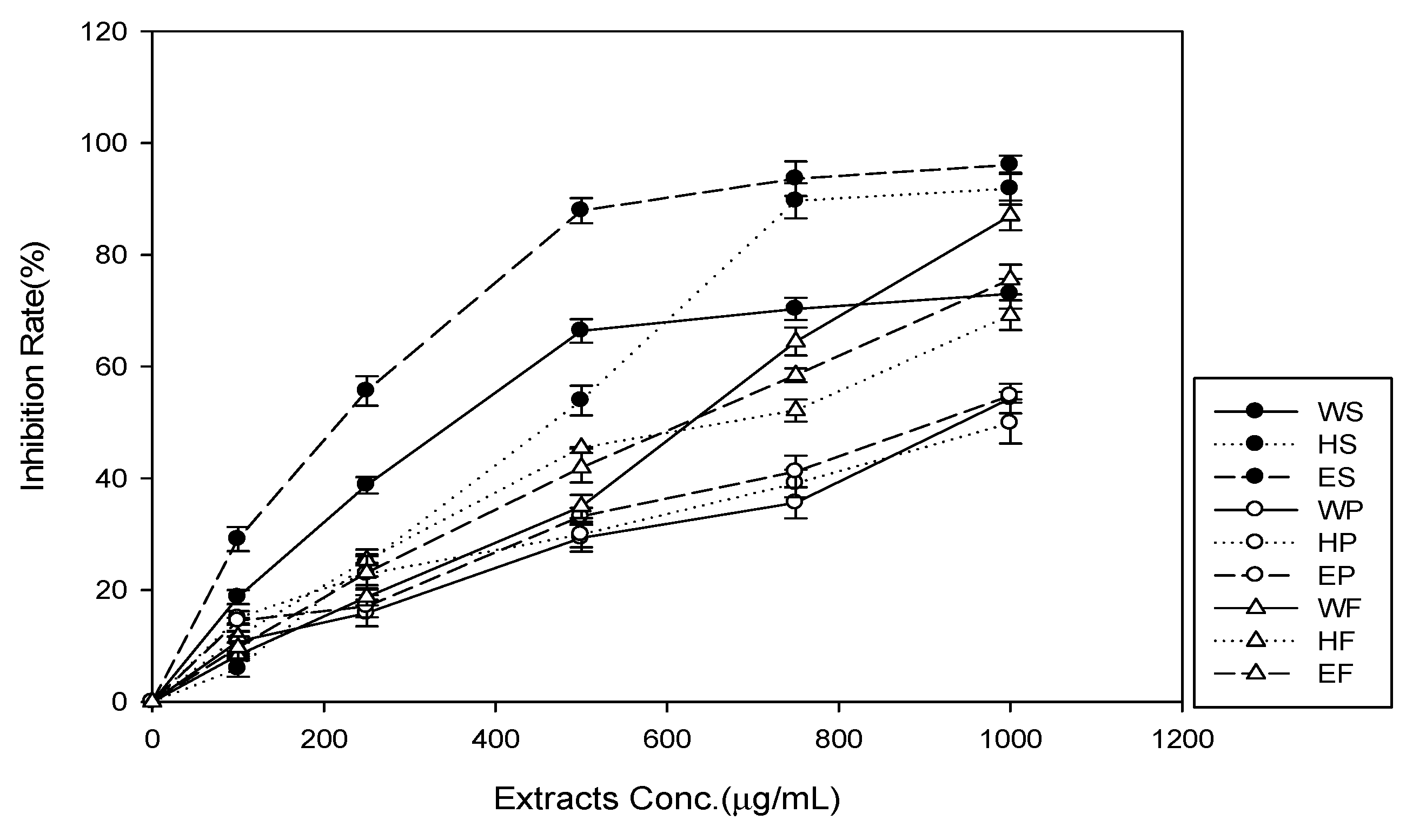
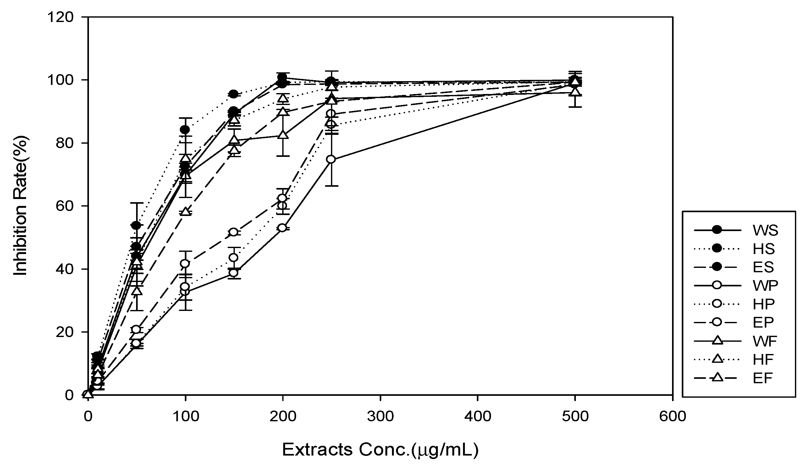
3.3. Antioxidant Properties

The DPPH and ABTS radical scavenging activities of different H. polyrhizus extracts are shown in Figure 1 and Figure 2, and Table 3. The DPPH and ABTS radical scavenging activities of the H. polyrhizus extracts were all greater than 50% at the highest concentration. The stem extract in 95% aqueous ethanol had the highest DPPH radical scavenging activity (IC50 = 224 µg/mL). The peel extracts had no good DPPH radical scavenging activities. In the case of ABTS radical scavenging activity, the stem extract in 50% aqueous ethanol had the highest ABTS radical scavenging activity (IC50 = 46 µg/mL). In summary, the stem extract showed a good potential in terms of free radical scavenging activities.

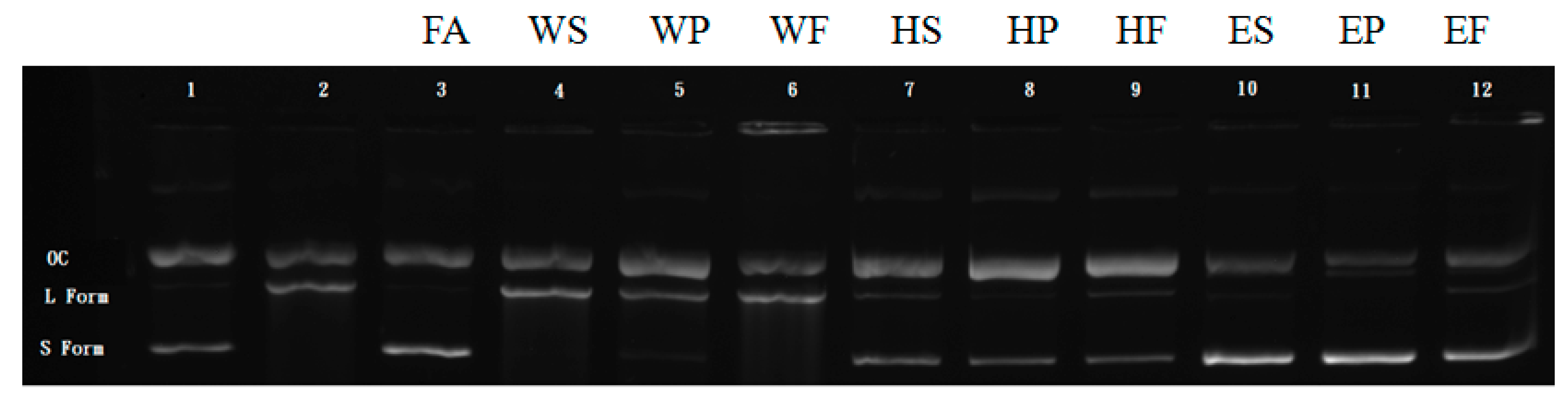
3.4. Protection against DNA Damage

The protective effects of H. polyrhizus extracts on DNA were investigated using an assay with the pUC19 plasmid (Figure 3). DNA strand breaks were induced in pUC19 using H2O2 and UV radiation (Figure 3, lane 2). The H. polyrhizus extract in distilled water had no protective effects against the pUC19 DNA strand breaks (Figure 3, lane 4–6). However, the H. polyrhizus extracts in 50% (Figure 3, lane 7–9) and 95% (Figure 3, lane 10–12) ethanol showed a significant reduction in the formation of nicked DNA and increased the native form of DNA. The 95% extracts exhibited the best protective effects against DNA damage. This was true for the extracts from all plant parts, i.e., stem, peel, and flower, with similar results. The protective effects of the 95% ethanol extracts were similar to the effect of 0.3 mg/mL ferulic acid (Figure 3, lane 3).

3.5. Cell Proliferation and Viability

The effect of H. polyrhizus extract on cell proliferation and viability was evaluated in the NIH-3T3 fibroblast cell line at different concentrations after 24 h with an MTT assay. As shown in Table 4, none of the extracts were able to significantly promote cell proliferation after 24 h at concentrations of 250 μg/mL to 2000 μg/mL. The 2000 μg/mL concentration of the peel extract in distilled water, as well as that of stem and flower extracts in 95% aqueous ethanol, showed significant cytotoxic activity.

3.6. Cell Migration (Scratch Assay)

The cell migration ability was tested with the NIH-3T3 fibroblast cell line using the scratch assay. Considering the higher concentration extracts exhibited cytotoxic ability, the 1000 μg/mL extracts were selected to pursue with the subsequent wound healing assays. Cell migration was determined after 24 h of exposure to the cell culture medium with various extract. The stem and flower extracts in 95% aqueous ethanol showed obvious cell migration after 24 h.

4. Discussion

Many natural ingredients are traditionally extracted with organic solvents. However, most of the organic solvents are recognized as toxic chemicals and the extraction conditions are strict. The recovery of total phenolics from citrus samples extracted using ethanol and methanol were similar [34]. However, methanol is believed to be toxic and the extraction condition is stricter. The food-grade ethanol is, therefore, more acceptable for use in food industry.
The antioxidant properties of fruits (peels) and pulps of H. polyrhizus and H. undatus had been investigated previously. By considering the free radical scavenging activity and ferrous ion chelating activity the pulp of H. undatus had the highest antioxidant properties [35]. The total phenolic content of the seed of H. polyrhizus was 13.56 ± 2.04 [1]. It was higher than the phenolics extract from the stem, peel, and flower of H. polyrhizus.
The Folin-Ciocalteu assay has been used in food and agricultural industries for many years to determine the phenolic content of plant products. However, this assay should be regarded as an assay of total antioxidant capacity rather than total phenolic content, as the reagent reacts with other antioxidants such as amino acids, proteins, vitamins, and thiols [36]. The content of phenolics and flavonoids are also related to tissue repair properties [37]. In fact, literature evidences suggest that both phenolics and flavonoids promote the fibroblast proliferation and the cell migration up to the formation of new blood vessels and capillaries [38].
Phenolic acids have protective effects on DNA against the toxic and mutagenic effects of UV and H2O2. For example, ferulic acid was found as a good active phytochemical that protects the DNA from damage in the presence of UV and H2O2. Similarly, caffeic, rosmarinic, vanillic, and cinnamic acids also have protective properties [13]. Although the separate phenolic acid contents were not measured in our experiments, the total phenolic contents were evaluated. The extracts prepared using 95% aqueous ethanol had the highest total phenolic content. It was consistent with the protective effects for DNA reported in this study.
Two kinds of DNA oxidative damage assays have been used in the literature. The hydroxyl radical generated from the Fenton reaction may react with DNA molecules [39,40]. In the site-specific assay, ferrous ion can bind to DNA and produce hydroxyl radicals at the binding site [41]. The major protective mechanism is by means of the chelating ability. In the non-site-specific assay EDTA molecules form a complex with ferrous ions and then hydroxyl radicals are generated in the solution. The major protective mechanism here is the hydroxyl radical scavenging. In our experiment, hydroxyl radicals were generated from UV radiation in the presence of H2O2. Thus, the non-site-specific assay was more appropriate. The major protective mechanism of H. polyrhizus extract was found to be the hydroxyl radical scavenging.
There are two important processes during the tissue formation phase in wound-healing: Cell proliferation and migration. The scratch assay is an imitative form that stimulates a wound in-vitro and evaluates the cell migration rate. When the cell monolayer is disrupted cell–cell interactions disappear and the secretion of growth factors and cytokines is increased at the wound edge, initiating migration and proliferation of cells [16]. Interestingly, none of the extracts were able to promote cell proliferation at concentrations of 250 μg/mL to 2000 μg/mL over a 24 h period. This effect is affirmatory for the wound-healing process because fibroblasts are important in wound contraction.

5. Conclusions

Our study showed that extracts from the stem of H. polyrhizus had excellent DPPH and ABTS radical scavenging activity. The stem, peel, and flower extracts in 95% aqueous ethanol showed excellent DNA damage protection effects as well. The 1000 μg/mL stem and flower extracts in 95% aqueous ethanol promoted cell migration properties. These results demonstrate that the stem, peel, and flower of H. polyrhizus are sources of antioxidant polyphenolics and have potential applications in the pharmaceutical, cosmetic, and food industries.

Author Contributions

C.-H.Y., C.-G.L., Y.T., K.-F.H., and Y.-C.H. designed the study. Y.T., W.-L.C., C.-Y.C., and C.-H.Y. wrote the paper. Y.T., W.-L.C., and C.-Y.C. performed the experiments. C.-H.Y., C.-G.L., and K.-F.H. supervised the study. All authors made substantial contributions to the discussion of data and approved the final manuscript.

Funding

This research was supported by the National Science Council of the Republic of China, grant number NSC 107-2313-B-126-001-MY3.

Conflicts of Interest

The authors declare no conflict of interest.

References

  1. Adnan, L.; Osman, A.; Hamid, A.A. Antioxidant activity of different extract of red pitaya (Hylocereus polyrhizus) seed. Int. J. Food Prop. 2011, 14, 1171–1181. [Google Scholar] [CrossRef]
  2. Le-Bellec, F.; Vaillant, F.; Imbert, E. Pitahaya (Hylocereus spp.): A New Crop, a Market with a Future. Fruits 2006, 61, 237–250. [Google Scholar] [CrossRef]
  3. Wu, L.C.; Hsu, H.W.; Chen, Y.C.; Chiu, C.C.; Lin, Y.I.; Ho, J.A. Antioxidant and antiproliferative activities of red pitaya. Food Chem. 2006, 95, 319–327. [Google Scholar] [CrossRef]
  4. Gengatharan, A.; Dykes, G.A.; Choo, W.S. The effect of pH treatment and refrigerated storage on natural colourant preparations (betacyanins) from red pitahaya and their potential application in yoghurt. LWT-Food Sci. Tech. 2017, 80, 437–445. [Google Scholar] [CrossRef]
  5. Ashwini Gengatharan, A.; Dykes, G.A.; Choo, W.S. Stability of betacyanin from red pitahaya (Hylocereus polyrhizus) and its potential application as a natural colourant in milk. Int. J. Food Sci. Tech. 2016, 51, 427–434. [Google Scholar] [CrossRef]
  6. Soccio, M.; Laus, M.N.; Alfarano, M.; Dalfino, G.; Panunzio, M.F.; Donato Pastore, D. Antioxidant/Oxidant Balance as a novel approach to evaluate the effect on serum of long-term intake of plant antioxidant-rich foods. J. Funct. Food. 2018, 40, 778–784. [Google Scholar] [CrossRef]
  7. Costa-Rodrigues, J.; Pinho, O.; Monteiro, P.R.R. Can lycopene be considered an effective protection against cardiovascular disease? Food Chem. 2018, 245, 1148–1153. [Google Scholar] [CrossRef]
  8. Sadeghi Ekbatan, S.; Li, X.-Q.; Ghorbani, M.; Azadi, B.; Kubow, S. Chlorogenic Acid and Its Microbial Metabolites Exert Anti-Proliferative Effects, S-Phase Cell-Cycle Arrest and Apoptosis in Human Colon Cancer Caco-2 Cells. Int. J. Mol. Sci. 2018, 19, 723. [Google Scholar] [CrossRef]
  9. Ivana Vrhovac Madunić, I.V.; Madunić, J.; Antunović, M.; Paradžik, M.; Garaj-Vrhovac, V.; Breljak, D.; Marijanović, I.; Gajski, G. Apigenin, a dietary flavonoid, induces apoptosis, DNA damage, and oxidative stress in human breast cancer MCF-7 and MDA MB-231 cells. Naunyn. Schmiedebergs. Arch. Pharmacol. 2018, 391, 537–550. [Google Scholar] [CrossRef]
  10. Reboredo-Rodríguez, P.; González-Barreiro, C.; Cancho-Grande, B.; Forbes-Hernández, T.Y.; Gasparrini, M.; Afrin, S.; Cianciosi, D.; Carrasco-Pancorbo, A.; Simal-Gándara, J.; Giampieri, F.; et al. Characterization of phenolic extracts from Brava extra virgin olive oils and their cytotoxic effects on MCF-7 breast cancer cells. Food Chem. Toxicol. 2018, 119, 73–85. [Google Scholar] [CrossRef]
  11. Du, W.X.; Olsen, C.W.; Avena-Bustillos, R.J.; Friedman, M.; McHugh, T.H. Physical and antibacterial properties of edible films formulated with apple skin polyphenols. J. Food Sci. 2011, 76, 149–155. [Google Scholar] [CrossRef]
  12. Moskovitz, J.; Yim, K.A.; Choke, P.B. Free radicals and disease. Arch. Biochem. Biophys. 2002, 397, 354–359. [Google Scholar] [CrossRef]
  13. Sevgi, K.; Tepe, B.; Sarikurkcu, C. Antioxidant and DNA damage protection potentials of selected phenolic acids. Food Chem. Toxicol. 2015, 77, 12–21. [Google Scholar] [CrossRef]
  14. Kokane, D.D.; More, R.Y.; Kale, M.B.; Nehete, M.N.; Mehendale, P.C.; Gadgoli, C.H. Evaluation of wound healing activity of root of Mimosa pudica. J. Ethnopharmacol. 2009, 124, 311–315. [Google Scholar] [CrossRef]
  15. Gangwar, M.; Gautam, M.K.; Ghildiyal, S.; Nath, G.; Goel, R.K. Mallotus philippinensis Muell. Arg fruit glandular hairs extract promotes wound healing on different wound model in rats. BMC Complem. Altern. M. 2015, 15, 123. [Google Scholar] [CrossRef]
  16. Pitz, H.S.; Pereira, A.; Blasius, M.B.; Voytena, A.P.L.; Affonso, R.C.L.; Fanan, S.; Trevisan, A.C.D.; Ribeiro-do-Valle, R.M.; Maraschin, M. In vitro evaluation of the antioxidant activity and wound healing properties of Jaboticaba (Plinia peruviana) fruit peel hydroalcoholic Extract. Oxid. Med. Cell Longev. 2016, 2016, 3403586. [Google Scholar] [CrossRef]
  17. Makris, D.P.; Boskou, G.; Andrikopoulos, N.K. Polyphenolic content and in vitro antioxidant characteristics of wine industry and other agri-food solid waste extracts. J. Food Compost. Anal. 2007, 20, 125–132. [Google Scholar] [CrossRef]
  18. Guendez, R.; Kallithraka, S.; Makris, D.P.; Kefalas, P. An analytical survey of the polyphenols of seeds of varieties of grape (Vitis vinifera sp.) cultivated in Greece: Implications for exploitation as a source of value-added phytochemicals. Phytochem. Anal. 2005, 16, 17–23. [Google Scholar] [CrossRef]
  19. Yilmaz, Y.; Toledo, R.T. Health aspects of functional grape seed constituents. Trend Food Sci. Technol. 2004, 15, 422–433. [Google Scholar] [CrossRef]
  20. Pinelo, M.; Rubilar, M.; Jerez, M.; Sineiro, J.; Nunez, J.M. Effect of solvent, temperature, and solvent-to-solid ratio on the total phenolic content and antiradical activity of extracts from different components of grape pomace. J. Agric. Food Chem. 2005, 53, 2111–2117. [Google Scholar] [CrossRef]
  21. Kammerer, D.; Claus, A.; Schieber, A.; Carle, A. A novel process for the recovery of polyphenols from grape (Vitis vinifera L.) pomace. J. Food Sci. 2005, 70, C157–C163. [Google Scholar] [CrossRef]
  22. Louli, V.; Ragoussis, N.; Magoulas, K. Recovery of phenolic antioxidants from wine industry by-products. Bioresour. Technol. 2004, 92, 201–208. [Google Scholar] [CrossRef]
  23. Alonso, A.M.; Guille’n, D.A.; Barroso, C.G.; Puertas, B.; Garcı’a, A. Determination of antioxidant activity of wine byproducts and its correlation with polyphenolic content. J. Agric. Food Chem. 2002, 50, 5832–5836. [Google Scholar] [CrossRef]
  24. Souquet, J.M.; Labarbe, B.; Le Guerneve’, C.; Cheynier, V.; Moutounet, M. Phenolic composition of grape stems. J. Agric. Food Chem. 2000, 48, 1076–1080. [Google Scholar] [CrossRef]
  25. Chiang, H.M.; Chen, H.C.; Lin, T.J.; Shih, I.C.; Wen, K.C. Michelia alba extract attenuates UVB-induced expression of matrix metalloproteinases via MAP kinase pathway in human dermal fibroblast. Food Chem. Toxicol. 2012, 50, 4260–4269. [Google Scholar] [CrossRef]
  26. Lin, J.Y.; Tang, C.Y. Determination of total phenolic and flavonoid contents in selected fruits and vegetables, as well as their stimulatory effects on mouse splenocyte proliferation. Food Chem. 2007, 101, 140–147. [Google Scholar] [CrossRef]
  27. Masuko, T.; Minami, A.; Iwasaki, N.; Majima, T.; Nishimura, S.; Lee, Y.C. Carbohydrate analysis by a phenol-sulfuric acid method in microplate format. Anal. Biochem. 2005, 339, 69–72. [Google Scholar] [CrossRef]
  28. Wen, K.-C.; Chiu, H.-H.; Fan, P.-C.; Chen, C.-W.; Wu, S.-M.; Chang, J.-H.; Chiang, H.-M. Antioxidant Activity of Ixora parviflora in a Cell/Cell-Free System and in UV-Exposed Human Fibroblasts. Molecules 2011, 16, 5735–5752. [Google Scholar] [CrossRef]
  29. Van den Berg, R.; Haenen, G.R.; van den Berg, H.; Bast, A. Applicability of an improved Trolox equivalent antioxidant capacity (TEAC) assay for evaluation of antioxidant capacity measurements of mixtures. Food Chem. 1999, 66, 511–517. [Google Scholar] [CrossRef]
  30. Kim, D.-O.; Chun, O.K.; Kim, Y.J.; Moon, H.Y.; Lee, C.Y. Quantification of polyphenolics and their antioxidant capacity in fresh plums. J. Agric. Food Chem. 2003, 51, 6509–6515. [Google Scholar] [CrossRef]
  31. Chiang, H.M.; Lin, T.J.; Chiu, C.Y.; Chang, C.W.; Hsu, K.C.; Fan, P.C.; Wen, K.C. Coffea arabica extract and its constituents prevent photoaging by suppressing MMPs expression and MAP kinase pathway. Food Chem. Toxicol. 2011, 49, 309–318. [Google Scholar] [CrossRef]
  32. Russo, A.; Cardile, V.; Lombardo, L.; Vanella, L.; Acquaviva, R. Genistin inhibits UV light-induced plasmid DNA damage and cell growth in human melanoma cells. J. Nutr. Biochem. 2006, 17, 103–108. [Google Scholar] [CrossRef]
  33. Balekar, N.; Katkam, G.; Nakpheng, T.; Jehtae, K.; Srichana, T. Evaluation of the wound healing potential of Wedelia trilobata (L.) leaves. J. Ethnopharmacol. 2012, 141, 817–824. [Google Scholar] [CrossRef]
  34. Li, B.B.; Smith, B.; Hossain, Md.M. Extraction of phenolics from citrus peels I. Solvent extraction method. Sep. Purif. Technol. 2006, 48, 182–188. [Google Scholar] [CrossRef]
  35. Choo, W.S.; Yong, W.K. Antioxidant properties of two species of Hylocereus fruits. Adv. Appl. Sci. Res. 2011, 2, 418–425. [Google Scholar]
  36. Everette, J.D.; Bryant, Q.M.; Green, A.M.; Abbey, Y.A.; Wangila, G.W.; Walker, R.B. Thorough study of reactivity of various compound classes toward the folin-Ciocalteu reagent. J. Agric. Food Chem. 2010, 58, 8139–8144. [Google Scholar] [CrossRef]
  37. Amri, B.; Martino, E.; Vitulo, F.; Corana, F.; Kaâb, L.-B.; Rui, M.; Rossi, D.; Mori, M.; Rossi, S.; Collina, S. Marrubium vulgare L. leave extract: Phytochemical composition, antioxidant and wound healing properties. Molecules 2017, 22, 1851. [Google Scholar] [CrossRef]
  38. Villanueva, J.R.; Esteban, J.M. An insight into a blockbuster phytomedicine; Marrubium vulgare L. Herb. More of a myth than a reality? Phytother. Res. 2016, 30, 1551–1558. [Google Scholar]
  39. Dizdaroglu, T.P.A.; Jaruga, P.; Birincioglu, M.; Rodriguez, H. Free radical induced damage to DNA: Mechanisms and measurement. Free Radic. Biol. Med. 2002, 32, 1102–1115. [Google Scholar] [CrossRef]
  40. Valko, M.; Izakovic, M.; Mazur, M.; Rhodes, C.J.; Telser, J. Role of oxygen radicals in DNA damage and cancer incidence. Mol. Cell Biochem. 2004, 266, 37–56. [Google Scholar] [CrossRef]
  41. Aruoma, O.I. Methodological consideration for characterizing potential antioxidant actions of bioactive components in plant foods. Mutat. Res. 2003, 523, 9–20. [Google Scholar] [CrossRef]
Figure 1. The antioxidant activity of crude extracts of Hylocereus polyrhizus parts, as measured by 2-2-diphenyl-1-picrylhydrazyl (DPPH) radical scavenging assay. WS, distilled water extract of stem; WP, distilled water extract of peel; WF, distilled water extract of flower; HS, 50% aqueous ethanol extract of stem; HP, 50% aqueous ethanol extract of peel; HF, 50% aqueous ethanol extract of flower; ES, 95% aqueous ethanol extract of stem; EP, 95% aqueous ethanol extract of peel; and, EF, 95% aqueous ethanol extract of flower.
Agronomy 09 00027 g001
Figure 2. The antioxidant activity of crude extracts of Hylocereus polyrhizus parts, as measured by the 2,2′-azino-bis (3-ethylbenzothiazoline-6-sulphonic acid) (ABTS) radical scavenging assay. WS, distilled water extract of stem; WP, distilled water extract of peel; WF, distilled water extract of flower; HS, 50% aqueous ethanol extract of stem; HP, 50% aqueous ethanol extract of peel; HF, 50% aqueous ethanol extract of flower; ES, 95% aqueous ethanol extract of stem; EP, 95% aqueous ethanol extract of peel; and, EF, 95% aqueous ethanol extract of flower.
Agronomy 09 00027 g002
Figure 3. DNA damage protection assay results for crude extracts of Hylocereus polyrhizus parts. All wells contained 1µg pUC119 plasmid. Well 1 and 2 did not have any test sample and served as (+/−) controls. Well 3 had 0.3 mg/mL ferulic acid (FA). Well 4–12 were added with 3mg/mL distilled water extract of stem (WS), distilled water extract of peel (WP), distilled water extract of flower (WF), 50% aqueous ethanol extract of stem (HS), 50% aqueous ethanol extract of peel (HP), 50% aqueous ethanol extract of flower (HF), 95% aqueous ethanol extract of stem (ES), 95% aqueous ethanol extract of peel (EP), and 95% aqueous ethanol extract of flower (EF), respectively. Well 1 was treated with 5 µL 0.3% H2O2. Well 2–9 were treated with 5 µL 0.3% H2O2 and 30 mJ/cm2 Ultraviolet B. OC denotes DNA in open circular form. L denotes DNA in linear form. S denotes DNA in supercoiled circular form.
Agronomy 09 00027 g003
Table 1. The extraction yield for the different parts of Hylocereus polyrhizus.
SampleSolventExtraction Yield (%)
Stem95% aqueous Ethanol3.85 ± 1.11 a
50% aqueous Ethanol44.70 ± 1.77 f
Distilled Water20.80 ± 2.75 c
Peel95% aqueous Ethanol4.21 ± 1.48 a
50% aqueous Ethanol31.90 ± 1.20 e
Distilled Water43.47 ± 1.95 f
Flower95% aqueous Ethanol7.43 ± 1.23 b
50% aqueous Ethanol21.72 ± 1.66 c
Distilled Water26.90 ± 2.56 d
Data are the mean ± SD (n = 3). Means with different letters are significantly different at p ≤ 0.05 according to Duncan’s multiple range test (DMRT).
Table 2. The composition of the extracts from Hylocereus polyrhizus.
SampleSolventContent (%)
Phenolic (GAE)Flavonoid (QE)Protein (AE)Glucose (GE)
95% Aqueous Ethanol8.16 ± 0.45 e0.87 ± 0.08 c8.56 ± 0.94 b40.15 ± 2.15 a
Stem50% Aqueous Ethanol5.50 ± 0.37 b0.87 ± 0.05 c12.14 ± 1.62 d,e53.91 ± 3.49 b
Distilled Water4.91 0.72 a,b0.38 ± 0.06 a12.98 ± 1.23 e35.38 ± 1.90 a
95% Aqueous Ethanol7.28 ± 0.49 d0.59 ± 0.06 b7.84 ± 0.45 b50.45 ± 2.12 b
Peel50% Aqueous Ethanol5.59 ± 0.56 b,c0.34 ± 0.05 a8.97 ± 0.85 b40.56 ± 3.41 a
Distilled Water4.50 ± 0.25 a0.40 ± 0.07 a9.44 ± 0.63 b,c83.88 ± 3.43 d
95% Aqueous Ethanol6.41 ± 0.68 c0.53 ± 0.06 b5.49 ± 0.88 a67.61 ± 4.28 c
Flower50% Aqueous Ethanol5.77 ± 0.15 b,c0.42 ± 0.03 a11.00 ± 1.36 c,d69.01 ± 3.91 c
Distilled Water4.11 ± 0.10 a0.39 ± 0.05 a12.36 ± 1.01 d,e36.34 ± 1.72 a
Each value is presented as mean ± SD (n = 3). GAE: gallic acid equivalent; QE: quercetin equivalent; AE: bovine serum albumin equivalent; and, GE: glucose equivalent. Means with different letters are significantly different at p ≤ 0.05 according to DMRT.
Table 3. The IC50 value of radical scavenging for crude extracts from Hylocereus polyrhizus parts as measured via the 2-2-diphenyl-1-picrylhydrazyl (DPPH) scavenging assay and the 2,2′-azino-bis (3-ethylbenzothiazoline-6-sulphonic acid) (ABTS) scavenging assay.
SampleSolventIC50 Value(µg/ml)
DPPHABTS
95% Aqueous Ethanol224.00 ± 14.81 b59.05 ± 3.49 d
Stem50% Aqueous Ethanol441.85 ± 16.13 d46.05 ± 3.07 b
Distilled Water351.83 ± 23.27 c52.51 ± 2.54 c
95% Aqueous Ethanol994.25 ± 29.63 h144.25 ± 4.65 g
Peel50% Aqueous Ethanol951.00 ± 26.84 g164.77 ± 5.14 h
Distilled Water908.60 ± 32.49 g186.49 ± 5.34 i
95% Aqueous Ethanol655.57 ± 29.50 f83.45 ± 2.77 f
Flower50% Aqueous Ethanol579.13 ± 28.92 e61.61 ± 3.14 d
Distilled Water575.67 ± 30.15 e68.49 ± 2.56 e
Trolox5.89 ± 0.17 a6.11 ± 0.11 a
Each value is presented as mean ± SD (n = 3). Means with different letters within the same column are significantly different at p ≤ 0.05 according to DMRT.
Table 4. The percentage of cell survival for crude extracts from Hylocereus polyrhizus parts as detected using the 3-(4,5-Dimethylthiazol-2-yl)-2,5-diphenyltetrazolium bromide (MTT) cell proliferation assay.
µg/mLDistilled Water Extract50% Aqueous Ethanol Extract95% Aqueous Ethanol Extract
StemPeelFlowerStemPeelFlowerStemPeelFlower
25099.28 ± 6.5098.82 ± 1.7797.45 ± 8.8597.03 ± 2.57105.29 ± 9.2991.30 ± 8.0694.60 ± 4.50101.50 ± 1.8895.23 ± 4.17
500107.27 ± 3.2597.05 ± 4.1692.01 ± 8.5796.34 ± 2.3596.21 ± 2.8886.05 ± 5.3894.61 ± 5.4991.91 ± 7.4894.68 ± 7.80
750100.89 ± 4.2790.96 ± 8.0188.54 ± 8.3495.63 ± 3.1792.99 ± 7.6990.10 ± 7.6583.22 ± 6.2597.04 ± 5.5890.57 ± 8.45
1000108.90 ± 8.9885.96 ± 8.55111.97 ± 2.3993.99 ± 4.9196.85 ± 8.6894.51 ± 6.6884.55 ± 9.9796.60 ± 9.1693.82 ± 7.69
2000118.27 ± 5.0376.50 ± 3.71 *120.42 ± 6.0596.59 ± 9.58106.88 ± 3.6586.27 ± 4.5575.81 ± 2.60 *94.33 ± 4.8979.47 ± 7.15 *
Each value is presented as mean ± SD (n = 3). *, p < 0.05, compared with the untreated cells (control).

