Article

Molecular Characterization of Choroideremia-Associated Deletions Reveals an Unexpected Regulation of CHM Gene Transcription

by
Tiziana Fioretti
1,†,
Valentina Di Iorio
2,†,
Barbara Lombardo
1,3,
Francesca De Falco
1,
Armando Cevenini
1,3,
Fabio Cattaneo
3,
Francesco Testa
2,
Lucio Pastore
1,3,
Francesca Simonelli
2 and
Gabriella Esposito
1,3,*
1
CEINGE-Biotecnologie Avanzate s.c. a r.l., Via G. Salvatore 486, 80145 Naples, Italy
2
Eye Clinic, Multidisciplinary Department of Medical, Surgical and Dental Sciences, University of Campania Luigi Vanvitelli, Via S. Pansini 5, 80131 Naples, Italy
3
Department of Molecular Medicine and Medical Biotechnologies, University of Naples Federico II, Via S. Pansini 5, 80131 Naples, Italy
*
Author to whom correspondence should be addressed.
These authors contributed equally to this work.
Submission received: 15 June 2021 / Revised: 16 July 2021 / Accepted: 19 July 2021 / Published: 22 July 2021
(This article belongs to the Special Issue Genetics in Inherited Retinal Diseases)

Abstract

:
Choroideremia (CHM) is a X-linked recessive chorioretinal dystrophy due to deficiency of the CHM gene product, i.e., Rab escort protein isoform 1 (REP1). To date, gene therapy for CHM has shown variable effectiveness, likely because the underlying pathogenic mechanisms as well as genotype-phenotype correlation are not yet fully known. Small nucleotide variants leading to premature termination codons (PTCs) are a major cause of CHM, but about 20% of patients has CHM gene deletions. To improve understanding of the disease mechanisms, we analyzed molecular features of seven deletions involving the CHM gene sequence. We mapped the deletion breakpoints by using polymerase chain reaction, sequencing and array comparative genomic hybridization; to identify rearrangement-promoting DNA sequences, we analyzed genomic architecture surrounding the breakpoint regions. Moreover, in some CHM patients with different mutation types, we measured transcript level of CHM and of CHML, encoding the REP2 isoform. Scattered along the whole CHM gene and in close proximity to the deletion breakpoints we found numerous repeat elements that generate a locus-specific rearrangement hot spot. Unexpectedly, patients with non-PTC variants had increased expression of the aberrant CHM mRNA; CHML expression was higher than normal in a patient lacking CHM and its putative regulatory sequences. This latest evidence suggests that mechanisms regulating CHM and CHML gene expression are worthy of further study, because their full knowledge could be also useful for developing effective therapies for this hitherto untreatable inherited retinal degeneration.

1. Introduction

Choroideremia (CHM; MIM #303100) is an X-linked recessive condition characterized by slow progressive degeneration of choroid, photoreceptors and retinal pigmented epithelium. Its prevalence is estimated at 1 in 50,000 males. Affected males develop night blindness in their teenage years, which is followed by loss of peripheral vision and complete blindness at middle age [1]. Very relevant for clinical diagnosis is the evidence that carrier females may exhibit a wide spectrum of disease severity. Indeed, although they are mainly asymptomatic or mildly symptomatic, with funduscopic examination often showing patchy areas of chorioretinal atrophy, a minority of cases presents a severe phenotype, with retinal and choroidal atrophy similar to that observed in affected males [2,3].
CHM is caused by sequence alteration of the CHM gene (HGNC:1940; MIM * 300390), which spans a genomic region of about 190 kb on chromosome Xq 21.2 and contains 15 exons [4]. This gene encodes the ubiquitously expressed Rab Escort Protein 1 (REP1) that, similarly to the closely related isoform REP2 (MIM * 118825), is an essential component of the Rab geranyl-geranyl transferase II complex [5,6], which catalyzes prenylation of ras-related Rab GTPases, a group of proteins with key role in of intracellular membrane trafficking regulation [7,8]. Although REP1 deficiency in CHM results in reduced prenylation of a subset of Rabs and affects multiple intracellular trafficking pathways in different cell types, no clinical consequence due to such deficiency has been reported in tissues other than the eye. However, derangement of vesicular trafficking, exocytosis and secretion have been observed in CHM fibroblasts and monocytes, in addition to crystals in peripheral blood lymphocytes and significant abnormalities in plasma fatty acid and red blood cell membrane [9,10].
Currently, prenatal molecular diagnosis is one of the possible prevention options for at risk families because CHM is still an untreatable inherited retinal degeneration [11,12,13,14]. Indeed, although gene therapy has entered late-phase clinical trials, it has shown variable effectiveness, likely because the underlying pathogenic mechanisms as well as genotype–phenotype correlation with respect to onset of symptoms, decline in visual acuity and visual fields are not yet fully known, unlike other retinal dystrophies [13,14,15]. Therefore, any study aimed at elucidating molecular mechanisms underlying CHM could provide useful insight for the development of targeted therapies [16].
More than 420 unique CHM pathogenic sequence variants that often abolish REP1 synthesis have been identified in CHM patients (Leiden Open Variant Database—LOVD, https://databases.lovd.nl/shared/genes/CHM; Human gene mutation database, HGMD®, accessed on 16 July 2021) [16,17,18,19]. About 20% of patients have chromosomal deletions of variable size that remove single CHM exons or eliminate CHM and various contiguous genes [19]. No apparent correlation has been found between the size of the deletion and the severity of CHM, however, a few patients with deletions ranging between 5 and 12 Mb had syndromic CHM [20,21,22,23,24].
The relatively high incidence of genomic deletions indicates that CHM locus is a hot spot of genomic rearrangements, likely due to the local genomic structure [25]. To date, only a few studies mapped CHM deletion breakpoints (BPs) and analyzed local genomic architectural features that might be associated with the generation of this type of copy number variation [4,21]. Therefore, we used a combination of molecular analysis, i.e., polymerase chain reaction (PCR), Sanger’s sequencing and array comparative genomic hybridization (aCGH), to map seven genomic deletions that we identified in unrelated Italian CHM families. We analyzed genomic architecture of the rearranged sequences and, in a few patients, we measured transcript levels of the aberrant CHM and of the closely related CHML gene (HGNC:1941), encoding the REP2 isoform.

2. Materials and Methods

We studied 11 patients (9 males, 2 females) from 7 Italian families affected by CHM with deletions involving CHM gene sequences. The diagnosis in males and females was based on clinical and instrumental data [11,26,27]. Diagnostic criteria included a history of night blindness and the typical appearance of the fundus, namely, peripheral pigmentary retinopathy with areas of pigment epithelial and choroidal atrophy [11,26,27]. General anamnesis did not indicate any other significant pathological condition other than CHM. Informed consent was signed by all the patients who approved to undergo the molecular diagnosis, which was performed according to the guidelines for genetic tests approved by the Italian Ministry of Health and by a local Institutional Ethics Committee. Patients also agreed to the use of their clinical and molecular data for scientific research purposes, in anonymous form. Analysis was carried out in accordance with the principles of Declaration of Helsinki.
Genomic DNA was extracted from peripheral blood leukocytes with the Nucleon™ BACC2 kit (GE Healthcare Italia, Milan, Italy). All the 15 CHM exons and their flanking regions were amplified by PCR as reported elsewhere [11]. To map intragenic deletion BP, we amplified intronic sequences before and after the deleted exons, according to a previously described strategy [28,29]. Long-range PCR was carried out with the Roche Expand Long Template PCR System (Roche, Mannheim, Germany) to amplify the CHM intragenic deletion BPs of family 6 (Fw 5′-CTGATGTCCAGCTGTAGTCTC-3′; Rev 5′-GTAAGTGACAGTCCAGTGTGG-3′) and family 7 (Fw 5′- GGAAGAGGGTAATGAAGTAACG -3′; Rev 5′- CAATGACATCAGAGACAGCC -3′). Fragments spanning the deletion BPs were analyzed by Sanger sequencing and the resulting sequences analyzed by BLAST (http://blast.ncbi.nlm.nih.gov/Blast.cgi, accessed on 21 March 2019) using reference sequences of the Xq21 locus in the human genome assembly 38 (GRCh38/hg38 Human Assembly). To test X-inactivation in carrier females, we carried out methylation-dependent amplification of the polymorphic triplet repeats at the androgen receptor HUMARA locus [30].
Total RNA was isolated from peripheral blood leukocytes by using the QIAamp RNA blood mini kit (Italy-QIAGEN S.p.A., Milan, Italy); 100 ng of RNA were reverse transcribed with SuperScript III (Invitrogen, Waltham, MA, USA) and random-hexamers, as previously performed [11]. Real time PCR was carried out in iCycler iQ Real Time PCR Thermal Cycler (Bio-Rad Laboratories S.r.l., Segrate, Italy) with gene-specific (for CHM or CHML) primers, by using SYBRTM Green Master Mix (Bio-Rad Laboratories S.r.l., Segrate, Italy). Reactions was carried out in triplicate (with duplicate samples for each experiment) in iCycler™ version 3.021 (Bio-Rad Laboratories S.r.l., Segrate, Italy) and the fluorescent signal intensity was recorded and analyzed with the iCycler™ iQ Optical System software v3.0a (Bio-Rad Laboratories S.r.l., Segrate, Italy). Relative gene expression was normalized to GAPDH and ACTB reference genes and determined using the 2−ΔΔCt method [11,31]. Average values from at least three independent experiments for each sample were graphically reported as fold change and statistical analysis was performed by one-way Student’s t-test (for independent samples). To analyze the CHM transcript, we used primers REP1F: 5′-ATGGCGGATACTCTCCCTTCG-3′ and REP1R 5′-GCTACTATGGAGGAAACTGGG-3′; for CHML, primers were REP2F 5′-ATGGCGGACAATCTTCCCACAG-3′ and REP2R 5′-CTATGGAGGAAACTGGGCTAG-3′. Mutation numbering is based on the reference genomic and transcript sequences of CHM (NG_009874.2, NM_000390.3).
To perform array comparative genomic hybridization (aCGH) analysis, genomic DNA was analyzed with the Human Genome CGH Microarray kit 4 × 180 K (Agilent Technologies Italia SpA, Milan, Italy). The reference DNA was a pool of genomic DNA from seven normal control samples (Promega, Madison, WI, USA). The aCGH we used contains 170,334 oligonucleotide probes (60-mer), which cover the whole genome with an average spatial resolution of 13 kb. DNA digestion, labeling and hybridization were performed according to the manufacturer’s protocols. Microarrays were scanned on an Agilent G2565CA scanner and image files were quantified using Agilent’s Feature Extraction software [V10.10.1.1]; data were visualized with Agilent’s Genomic WorkBench Standard Edition [V6.5.0.58] (Agilent Technologies Italia SpA, Milan, Italy) [32,33,34].
Bioinformatic analysis of CHM genomic sequences were carried out by RepeatMasker [www.repeatmasker.org] and by consulting the Human (Homo sapiens) Genome Browser Gateway of the University of California, Santa Cruz (UCSC) (GRCh38/hg38 Human Assembly; http://genome-euro.ucsc.edu/cgi-bin/hgGateway, accessed on 16 May 2019) [29]. Repeat elements around and within the deleted sequences were aligned by BLAST.

3. Results

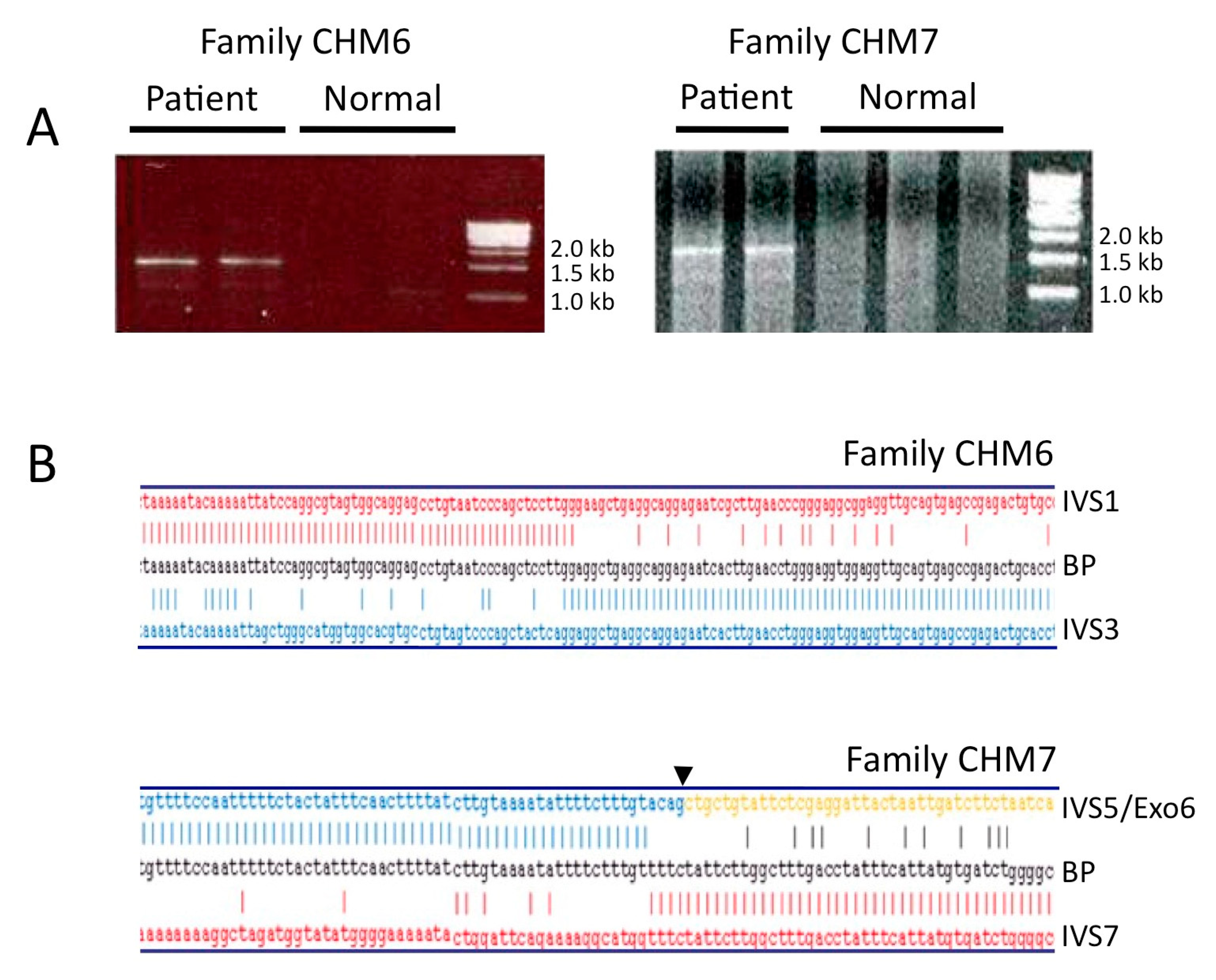
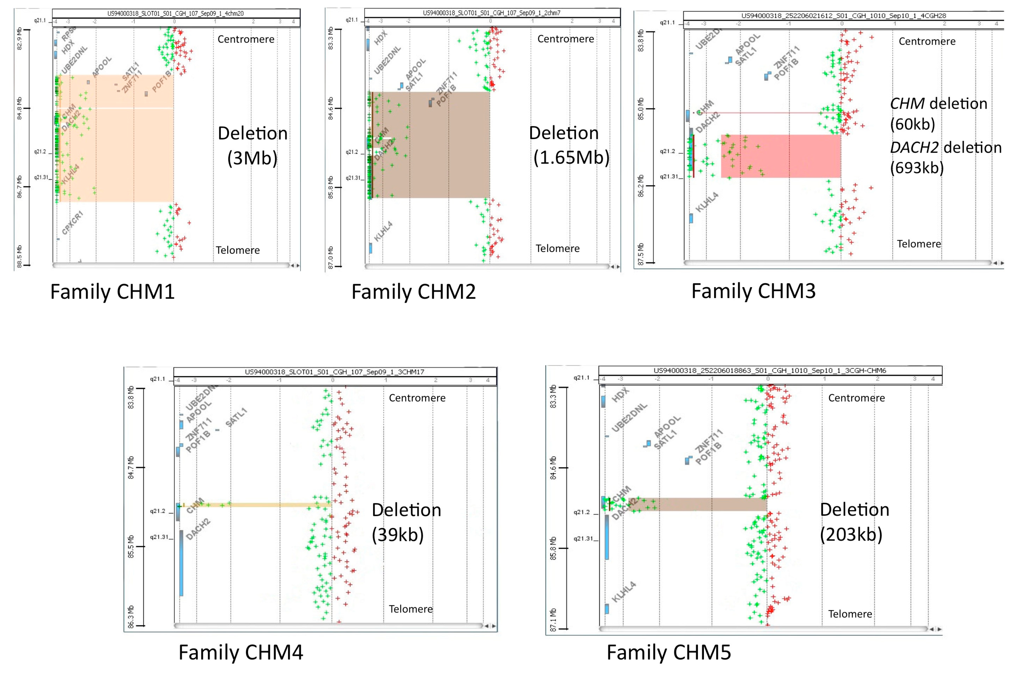
3.1. Molecular Characterization of CHM Deletions

Table 1 lists the deletions identified in the probands (from 7 unrelated families), who belong to a cohort of CHM patients described earlier [11,26].
To roughly delimit the extent of the intragenic deletions we found in families CHM6 and CHM7 (Table 1), we amplified CHM-specific intronic sequences in the respective affected males and in normal controls. In both cases, we limited the deletion BP within genomic regions no longer than 5 kb and therefore we were then able to amplify, by long-range PCR, the BP junction regions (Figure 1A). Sequence analysis of the resulting amplicons demonstrated that the genomic deletion removing exons 2 and 3 in family CHM6 was 55,962 bp long (Figure 1B, top panel); in family CHM7, a 1635 bp long deletion removed exons 6 and 7 (Figure 1B, bottom panel). In both cases, the deletion caused a shift in the CHM transcript reading frame that likely gave rise to a premature termination codon (PTC) [11].
DNA analysis previously performed in family CHM3 revealed the deletion of exon 12; however, leukocytes of affected males lacked the CHM transcript [11]. In agreement with the variable spectrum of disease severity observed in CHM carriers [3,27], a 76 year old carrier female affected by a severe phenotype and a 32 year old asymptomatic carrier were also present in this family. The phenotype of the elderly carrier was more severe than that of her affected relatives, so that an incorrect diagnosis of retinitis pigmentosa was initially made. During the familial segregation study, we analyzed RNA extracted from leukocytes of these two females by RT-PCR and sequencing and in both cases we only found the wild type CHM transcript. Interestingly, in the elderly carrier, methylation-specific PCR pattern at the HUMARA locus was consistent with a random X-inactivation (Supplementary Figure S1), thereby suggesting that her severe phenotype was not due to a somatic unbalanced lyonization favoring expression of the X chromosome that carried the mutated CHM gene. This result is in line with previous data that do not support a correlation between X chromosome inactivation status and abnormal retinal phenotype in CHM carrier females [35].
We previously reported that, in family CHM4, the genomic deletion removing CHM exons 10–15 produced an aberrant transcript that retained exons 1–9 and part of intron 9, presumably leading to a truncated REP1 (p.Cys416Ter) [11,26]. In family CHM5, the genomic deletion removed CHM exons 1–12 and extended upstream the putative CHM promoter region [36]; accordingly, RT-PCR did not detect any specific transcript in leukocytes of the male proband of CHM5 family.
To define the extent of the widest deletions identified in our families, we performed aCGH analysis (Figure 2), which is able to successfully detect in a single experiment copy number changes across the whole genome [32,33,34]. In family CHM1, array limited the deletion to about 3 Mb that removed UBE2DN (pseudogene), APOOL, SATL1, ZNF711, POF1B, CHM, DACH2 and KLHL4 genes. In family CHM2, the deletion was about 1.65 Mb long and encompassed SATL1, ZNF711, POF1B, CHM and DACH2. In family CHM4, the deletion that removed exons 10–15 was about 34 kb long, started in IVS9 and ended in the 3′UTR of CHM, just before the polyadenylation signal. In family CHM5, aCGH showed that the deletion removed about 153 kb of CHM, including exons 1–12 and about 33 kb of its 5′ flanking sequences. In family CHM3, aCGH confirmed the deletion of CHM exon 12 and surprisingly revealed an additional structural variation, i.e., a deletion that started in intron 1 and removed all the downstream sequences of DACH2, a CHM contiguous gene.

3.2. Analysis of the Genomic Architecture Flanking the BP Junctions

Once the approximate genomic positions of our BPs were determined, we analyzed junction-sequence signatures to explore why the CHM locus and the surrounding genomic regions are so prone to rearrangements. First, we performed in silico analysis of the Xq21.2 locus to search for repeated sequences potentially leading to genomic instability. RepeatMasker and UCSC genome browser-based analysis of the largest deleted region (chrX:84,831,307–87,941,186) in our patients revealed 4473 interspersed repeats, mainly long interspersed elements (i.e., LINE1, LINE2), short interspersed elements (i.e., Alu, MIR), long terminal repeats, single tandem repeats. Along the X chromosome, the CHM gene (chrX: 85,861,180–86,047,562; NC_000023.11) has inverted orientation (minus strand).
Numerous and different types of repeated elements are also scattered along the whole CHM gene and in close proximity to the breakpoints of the deletions detected in our families (Supplementary Figure S2).
Table 2 summarizes high identity interspersed repeats that lie within the genomic regions proximal and distal to the deletion BPs of our CHM patients and their relative percentage of sequence identity, as determined by RepeatMasker and BLAST analysis, respectively. Moreover, we used BLAST to look for sequence identity in the regions surrounding the BPs of each deletion. Notably, the BP junction regions of the five largest deletions contained sequence stretches of various extents (from 13–80 to 200–500 bp long) that share from 65% to 100% identities (Table 2, last column).
Lastly, we compared by BLAST the whole sequences of CHM (NC_000023.11; chrX:85861180–86047562) and DACH2 (NC_000023.11; chrX:86148451–86832602) and found that the two genes contain at least 12 large sequence stretches (ranging from ~2000 to ~5000 bp) that share from 77% to 92% of identities.

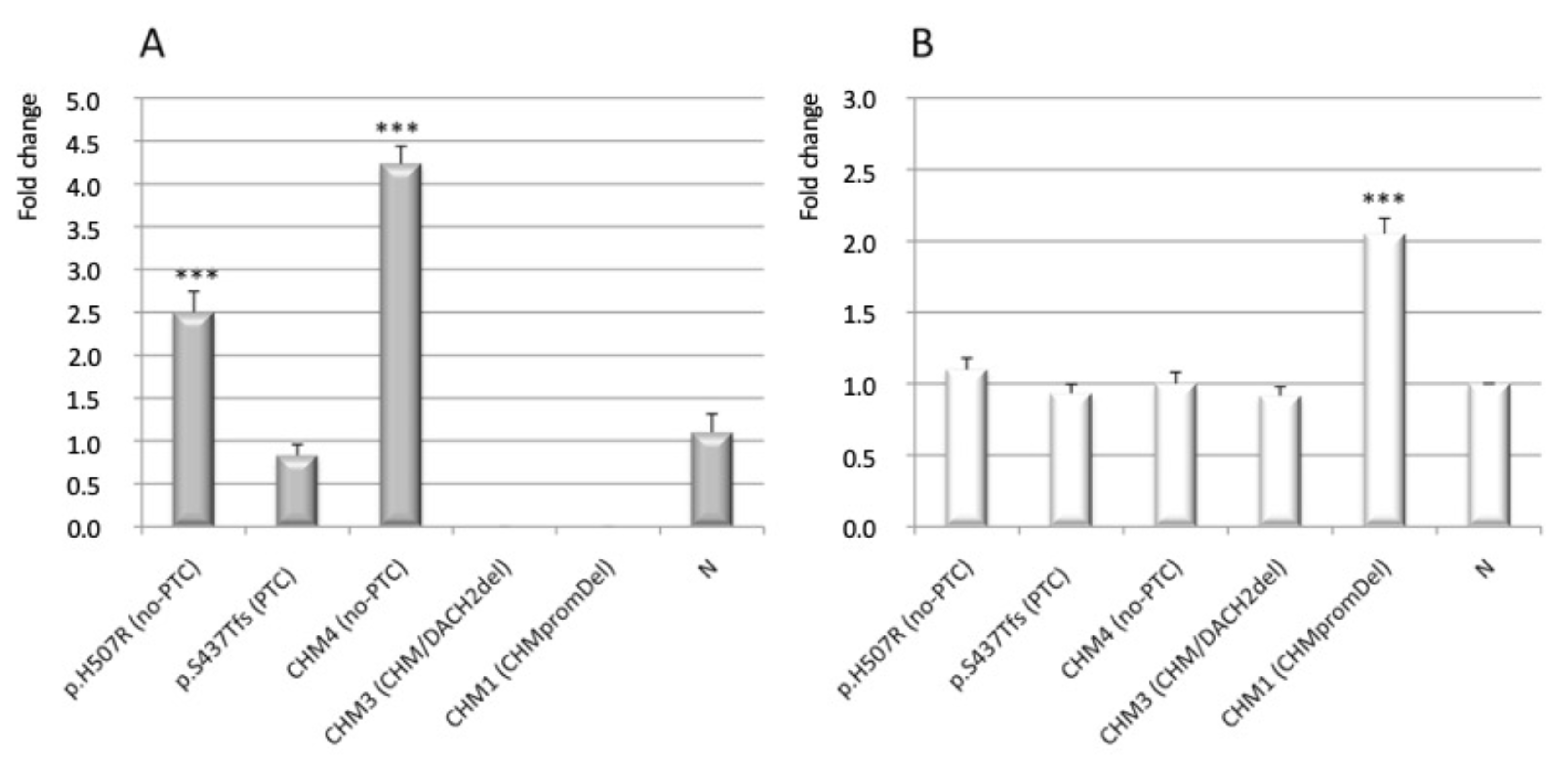
3.3. Analysis of CHM and CHML Transcript Level

To further study molecular consequences of the mapped deletions, in the three male patients for whom RNA was available (namely CHM1, CHM3, CHM4), we analyzed residual CHM transcript level. Results obtained from the deleted patients were compared to those obtained from selected reference males, i.e., a patient with a typical PTC frameshift variant (p.Ser437Thrfs), one with a no-PTC variant, namely the missense p.His507Arg, and three normal males. Of note, also the deletion sparing the CHM polyadenilation site (p.Cys416Ter), which was detected in the CHM4 proband, is a no-PTC variant. We confirmed absence of CHM expression in the male patient of family CHM3 (with the CHM/DACH2 complex deletion), whereas mRNA level of the patient with the PTC variant p.Ser437Thrfs was comparable to the normal (Figure 3A). Surprisingly, in leukocytes of both patients having the no-PTC variants p.His507Arg and p.Cys416Ter, the respective aberrant CHM transcript levels were 2.5 and 4.0 fold higher than both normal and PTC mRNAs (Figure 3A).
As literature reports that the phenotypic variation in CHM may in part be explained by the degree to which the absence of REP1 can be compensated by other prenylation proteins such as REP2 [14], we also analyzed expression of CHML, encoding the REP2 isoform, in the same patient group. Notably, CHML mRNA expression was comparable to normal in the CHM3 and CHM4 probands and in the affected and unaffected reference subjects (see above), but was unexpectedly higher (2.0 fold) than normal in the analyzed proband of family CHM1, who had a deletion removing the whole CHM gene and its flanking regions (Figure 3B).

4. Discussion

Pathogenic variants in the CHM gene result in complete loss of REP1, which is a typical feature of CHM cells [10,16,17,18,19]. Genomic deletions involving CHM are loss-of-function defects that have been detected in about 15–20% of CHM patients [19,20,21,22,23]. Here, we report molecular characterization of seven large deletions that remove in part or all the CHM gene in Italian patients with choroideremia (Table 1). Similar deletions are listed in the CHM-specific LOVD database, which in the July 2021 update reports 84 different ones: 28% of them remove the whole gene and its flanking regions, 30% has only one BP that falls within the CHM gene, the remaining are deletions with two intragenic BPs.
Genomic sequences that include tandem repeats and interspersed repeats are prone to deletion and duplication events [25]. Interestingly, bioinformatic analysis revealed an abundance of repeated elements scattered along the CHM gene locus (Supplementary Figure S2, bottom panel); in addition, local sequence identities are present in proximity of the BP regions of the deletions identified in our families (Table 2). In particular, three BP regions contain Alu elements, three contain LINEs, and one has LTRs; numerous STRs are present, in at least five cases (Table 2).
Notably, intragenic BPs of the deletions reported in LOVD are scattered throughout the CHM gene, without evidence of BP hot spots (Supplementary Figure S2, top panel) and, accordingly, recurrent deletions have never been reported in CHM patients, including ours. These observations suggest that a non-homologous end joining mechanism mediated by the spread repetitive elements can be responsible for the deletion susceptibility of the whole CHM gene locus [25]. As a non-homologous end joining mechanism may also cause sequence duplication, this type of rearrangement should be taken into account during the molecular diagnosis of CHM [20]. Therefore, multiple ligation-dependent probe amplification or/and aCGH analysis is suggested for CHM patients without SNVs or large deletions [18,37]. Obviously, any data obtained from mRNA analysis may further warrant such in-depth diagnostic study.
In agreement with data reported in the CHM-specific LOVD database, two of our seven families (28%) have deletions that involve the whole CHM and some contiguous genes (Figure 2). In particular, the nullisomic regions deleted in families CHM1 and CHM2 contain genes that exert different functions, such as the ZNF711 gene, which encodes a zinc-finger sequence-specific DNA binding factor [38,39,40]. Additionally deleted in both our families are POF1B, which seems to play a role in the etiology of premature ovarian failure [41], and DACH2, with an important role in the regulation of brain and limb development and presumed to be implicated in syndromic mental retardation [41,42].
A few earlier cases of CHM associated with extra-ocular symptoms were attributed to contiguous gene deletions at Xq21 locus [23,24,43]. Interestingly, a large deletion involving CHM and the neighboring POU3F4 and ZNF711 genes has been previously identified in a CHM patient with hearing impairment and developmental disability [38]. In agreement, two families with nonspecific mental retardation and truncating alterations in the ZNF711 gene were also reported [39,40]. As our male patients with contiguous gene deletions seemed to be affected by isolate choroideremia [11,26], we conclude that the genes located within the deleted genomic regions might have only indirect or subtle effects on the patients’ extra-ocular phenotype [43]; consequently, we can reasonably state that loss of ZNF711 is not associated with mental retardation, in our patients.
Small nucleotide changes leading to nonsense or frameshift variants are frequent loss-of-function defects in CHM. However, the nonsense-mediated mRNA decay (NMD) pathway, which usually detects and degrades mRNAs carrying mutations that lead to PTCs, does not appear to be an efficient surveillance mechanism in the leukocytes of CHM patients [11,44,45]. As consequence, the main loss-of-function effect of CHM nonsense or frameshift variants and of partial intragenic deletions is not absence of the aberrant mRNA, but rather the production of truncated proteins that are prone to cell protein degradation [27,45]. In line with this observation, CHM patients usually express detectable level of the aberrant CHM transcript [11,18,45]. Therefore, unusual was the case of the complex deletion that removes the CHM exon 12 and most sequences of the adjacent DACH2 gene, in our family CHM3, because in leukocytes of the analyzed probands we did not find the expected aberrant CHM mRNA lacking exon 12. We consider it unlikely that NMD completely degrades such putative deleted transcript, as an mRNA lacking exon 12 was previously isolated in another CHM patient [46]. As CHM and DACH2 are adjacent head-to-head tandem arranged genes (Supplementary Figure S3) with the respective transcription start sites about 100 kb apart, we speculate that the deletion of DACH2 could play a role in CHM expression. Indeed, it is conceivable that cis-acting elements within the DACH2 sequences deleted in this family may be involved in transcriptional regulation of CHM, as occurs for other genes [47]. Further studies are required to identify potential, to date unknown, long-range cis-regulatory elements of CHM, whereas a proximal cis-regulatory element has been rather recently identified [36]. On this basis, we do not exclude that rare CHM patients who test negative to CHM mutation might have deletion of DACH2.
In this study, we also analyzed the expression of the CHM and CHML transcripts in patients who had different types of pathogenic CHM sequence variants. Due to the rarity of the disease, the uniqueness of patients with specific genotypes and the difficulty to collect fresh blood sample from low vision or blind patients often living far from our diagnostic centers, this analysis is limited to a small number of patients. Nevertheless, our data surprisingly associated two very rare no-PTC sequence changes (i.e., p.His507Arg and p.Cys416Ter), a type of variants not subjected to NMD, with increased CHM transcription, which could be ascribed to the activation of an auto-regulatory loop generated to overcome, although unsuccessfully, REP1 deficiency in CHM leukocytes. As consequence, in CHM patients with PTC-associated variants, residual expression of aberrant CHM transcripts may depend on a balance between forced/increased gene transcription and NMD, which apparently makes the latter poorly effective. Additionally, the finding that affected males of family CHM1, who have a deletion that removes CHM and, most importantly, its promoter, showed significant increased expression of CHML is interesting. Based on this isolate observation and on previous evidence that knockdown of CHML upregulates the expression of CHM in HeLa cells [48], we suggest that transcription of CHM and CHML may respond to common regulatory mechanisms. Interestingly, our previous clinical investigation revealed that probands of CHM1 family had a more severe loss of visual function at younger age than the other CHM patients evaluated [26]. Therefore, we speculate that CHML overexpression might even have an adverse impact on disease progression. This observation could be relevant during selection of subjects to be enrolled in clinical trials, as well as in the follow-up of experimental therapy on disease progression [49,50].

5. Conclusions

Enhanced rearrangement vulnerability of the CHM gene locus can be attributed to the abundance of repetitive elements that, by mediating non-homologous end joining mechanisms, trigger gene deletions, which indeed represent about 28% of the disease-causing gene variants found in CHM patients. Moreover, our data shed some light on the possible mechanisms that regulate CHM gene transcription and ultimately provides hints that transcription of CHM and CHML could respond to common regulatory mechanisms. Obviously, further study is needed to support our conclusion.
Overall, our study provides new insight into the molecular basis of CHM, which can be also relevant for diagnosis and treatment of patients affected by this degenerative eye disorder.

Supplementary Materials

The following are available online at https://0-www-mdpi-com.brum.beds.ac.uk/article/10.3390/genes12081111/s1, Figure S1: X-chromosome inactivation patterns at the HUMARA locus in the affected elderly female of family CHM3, Figure S2: Position of deletion breakpoints and types of repeated elements scattered along the CHM gene, Figure S3: Organization of CHM and adjacent genes on chromosome Xq21.3 (NC_000023.11).

Author Contributions

Conceptualization, G.E.; methodology, G.E.; validation, T.F., V.D.I., F.D.F. and G.E.; formal analysis, T.F., V.D.I., B.L., F.D.F., A.C., F.C. and G.E.; investigation, T.F., V.D.I., B.L., F.D.F.; resources, G.E., V.D.I., F.T. and F.S.; data curation, T.F., F.C. and G.E.; writing—original draft preparation, T.F., V.D.I. and G.E.; writing—review and editing, F.T., L.P., F.S. and G.E.; visualization, A.C. and G.E.; supervision, G.E.; project administration, F.S. and G.E.; funding acquisition, G.E. and F.S. All authors have read and agreed to the published version of the manuscript.

Funding

This research received no external funding.

Institutional Review Board Statement

The study was conducted according to the guidelines of the Declaration of Helsinki and approved by the Ethics Committee of University of Naples Federico II (protocol code 77/21, 26 March 2021).

Informed Consent Statement

Informed consent was obtained from all subjects involved in the study.

Conflicts of Interest

The authors declare no conflict of interest.

References

  1. Coussa, R.G.; Traboulsi, E.I. Choroideremia, a review of general findings and pathogenesis. Ophthalmic Genet. 2012, 33, 57–65. [Google Scholar] [CrossRef] [PubMed]
  2. Preising, M.N.; Wegscheider, E.; Friedburg, C.; Poloschek, C.M.; Wabbels, B.K.; Lorenz, B. Fundus autofluorescence in carriers of choroideremia and correlation with electrophysiologic and psychophysical data. Ophthalmology 2009, 116, 1201–1209. [Google Scholar] [CrossRef] [PubMed]
  3. Jauregui, R.; Park, K.S.; Tanaka, A.J.; Cho, A.; Paavo, M.; Zernant, J.; Francis, J.H.; Allikmets, R.; Sparrow, J.R.; Tsang, S.H. Spectrum of disease severity and phenotype in choroideremia carriers. Am. J. Ophthalmol. 2019, 207, 77–86. [Google Scholar] [CrossRef] [PubMed]
  4. Van Bokhoven, H.; Schwartz, M.; Andréasson, S.; van den Hurk, J.A.; Bogerd, L.; Jay, M.; Pawlowitzki, I.H.; Sankila, E.M.; Wright, A.; Ropers, H.H.; et al. Mutation spectrum in the CHM gene of Danish and Swedish choroideremia patients. Hum. Mol. Genet. 1994, 3, 1047–1051. [Google Scholar] [CrossRef]
  5. Andres, D.A.; Seabra, M.C.; Brown, M.S.; Armstrong, S.A.; Smeland, T.E.; Cremers, F.P.; Goldstein, J.L. cDNA cloning of component A of Rab geranylgeranyl transferase and demonstration of its role as a Rab escort protein. Cell 1993, 73, 1091–1099. [Google Scholar] [CrossRef]
  6. Cremers, F.P.; Armstrong, S.A.; Seabra, M.C.; Brown, M.S.; Goldstein, J.L. REP-2, a Rab escort protein encoded by the choroideremia-like gene. J. Biol. Chem. 1994, 269, 2111–2117. [Google Scholar] [CrossRef]
  7. Ali, B.R.; Seabra, M.C. Targeting of Rab GTPases to cellular membranes. Biochem. Soc. Trans. 2005, 33, 652–656. [Google Scholar] [CrossRef] [Green Version]
  8. Gomes, A.Q.; Ali, B.R.; Ramalho, J.S.; Godfrey, R.F.; Barral, D.C.; Hume, A.N.; Seabra, M.C. Membrane targeting of Rab GTPases is influenced by the prenylation motif. Mol. Biol Cell 2003, 14, 1882–1899. [Google Scholar] [CrossRef] [Green Version]
  9. Strunnikova, N.V.; Barb, J.; Sergeev, Y.V.; Thiagarajasubramanian, A.; Silvin, C.; Munson, P.J.; Macdonald, I.M. Loss-of-function mutations in Rab escort protein 1 [REP-1] affect intracellular transport in fibroblasts and monocytes of choroideremia patients. PLoS ONE 2009, 4, e8402. [Google Scholar] [CrossRef]
  10. Zhang, A.Y.; Mysore, N.; Vali, H.; Koenekoop, J.; Cao, S.N.; Li, S.; Ren, H.; Keser, V.; Lopez-Solache, I.; Siddiqui, S.N.; et al. Choroideremia is a systemic disease with lymphocyte crystals and plasma lipid and RBC membrane abnormalities. Investig. Ophthalmol. Vis. Sci. 2015, 56, 8158–8165. [Google Scholar] [CrossRef] [Green Version]
  11. Esposito, G.; De Falco, F.; Tinto, N.; Testa, F.; Vitagliano, L.; Tandurella, I.C.; Iannone, L.; Rossi, S.; Rinaldi, E.; Simonelli, F.; et al. Comprehensive mutation analysis [20 families] of the choroideremia gene reveals a missense variant that prevents the binding of REP1 with Rab geranylgeranyl transferase. Hum. Mutat. 2011, 32, 1460–1469. [Google Scholar] [CrossRef] [PubMed]
  12. Esposito, G.; Ruggiero, R.; Savarese, M.; Savarese, G.; Tremolaterra, M.R.; Salvatore, F.; Carsana, A. Prenatal molecular diagnosis of inherited neuromuscular diseases: Duchenne/Becker muscular dystrophy, myotonic dystrophy type 1 and spinal muscular atrophy. Clin. Chem. Lab. Med. 2013, 51, 2239–2245. [Google Scholar] [CrossRef]
  13. MacDonald, I.M.; Moen, C.; Duncan, J.L.; Tsang, S.H.; Cehajic-Kapetanovic, J.; Aleman, T.S. Perspectives on Gene Therapy: Choroideremia Represents a Challenging Model for the Treatment of Other Inherited Retinal Degenerations. Transl Vis. Sci. Technol. 2020, 9, 17. [Google Scholar] [CrossRef]
  14. Abbouda, A.; Avogaro, F.; Moosajee, M.; Vingolo, E.M. Update on Gene Therapy Clinical Trials for Choroideremia and Potential Experimental Therapies. Medicina 2021, 57, 64. [Google Scholar] [CrossRef] [PubMed]
  15. Di Iorio, V.; Orrico, A.; Esposito, G.; Melillo, P.; Rossi, S.; Sbordone, S.; Auricchio, A.; Testa, F.; Simonelli, F. Association between genotype and disease progression in Italian Stargardt patients: A retrospective natural history study. Retina 2019, 39, 1399–1409. [Google Scholar] [CrossRef] [PubMed]
  16. Fioretti, T.; Ungari, S.; Savarese, M.; Cattaneo, F.; Pirozzi, E.; Esposito, G. A putative frameshift variant in the CHM gene is associated with an unexpected splicing alteration in a choroideremia patient. Mol. Genet. Genom. Med. 2020, 8, e1490. [Google Scholar] [CrossRef] [PubMed]
  17. Van den Hurk, J.A.; Schwartz, M.; van Bokhoven, H.; van de Pol, T.J.; Bogerd, L.; Pinckers, A.J.; Bleeker-Wagemakers, E.M.; Pawlowitzki, I.H.; Rüther, K.; Ropers, H.H.; et al. Molecular basis of choroideremia [CHM] mutations involving the Rab escort protein-1 [REP-1] gene. Hum. Mutat 1997, 9, 110–117. [Google Scholar] [CrossRef]
  18. Furgoch, M.J.; Mewes-Arès, J.; Radziwon, A.; Macdonald, I.M. Molecular genetic diagnostic techniques in choroideremia. Mol. Vis. 2014, 20, 535–544. [Google Scholar]
  19. Zeitz, C.; Nassisi, M.; Laurent-Coriat, C.; Andrieu, C.; Boyard, F.; Condroyer, C.; Démontant, V.; Antonio, A.; Lancelot, M.E.; Frederiksen, H.; et al. CHM mutation spectrum and disease: An update at the time of human therapeutic trials. Hum. Mutat. 2021, 42, 323–341. [Google Scholar] [CrossRef]
  20. Freund, P.R.; Sergeev, Y.V.; MacDonald, I.M. Analysis of a large choroideremia dataset does not suggest a preference for inclusion of certain genotypes in future trials of gene therapy. Mol. Genet. Genom. Med. 2016, 4, 344–358. [Google Scholar] [CrossRef]
  21. Cremers, F.P.; van de Pol, D.J.; Diergaarde, P.J.; Wieringa, B.; Nussbaum, R.L.; Schwartz, M.; Ropers, H.H. Physical fine mapping of the choroideremia locus using Xq21 deletions associated with complex syndromes. Genomics 1989, 4, 41–46. [Google Scholar] [CrossRef]
  22. Hildebrand, M.S.; de Silva, M.G.; Tan, T.Y.; Rose, E.; Nishimura, C.; Tolmachova, T.; Hulett, J.M.; White, S.M.; Silver, J.; Bahlo, M.; et al. Molecular characterization of a novel X-linked syndrome involving developmental delay and deafness. Am. J. Med. Genet. A 2007, 143, 2564–2575. [Google Scholar] [CrossRef]
  23. Poloschek, C.M.; Kloeckener-Gruissem, B.; Hansen, L.L.; Bach, M.; Berger, W. Syndromic choroideremia, sublocalization of phenotypes associated with Martin-Probst deafness mental retardation syndrome. Investig. Ophthalmol. Vis. Sci 2008, 49, 4096–4104. [Google Scholar] [CrossRef] [PubMed] [Green Version]
  24. Rush, E.T.; Schaefer, G.B. Identification of an X-linked deletion syndrome through comparative genomic hybridization microarray. Semin. Pediatr. Neurol. 2010, 17, 51–53. [Google Scholar] [CrossRef] [PubMed]
  25. Shaw, C.J.; Lupski, J.R. Implications of human genome architecture for rearrangement-based disorders, the genomic basis of disease. Hum. Mol. Genet. 2004, 13, 57–64. [Google Scholar] [CrossRef] [PubMed]
  26. Di Iorio, V.; Esposito, G.; De Falco, F.; Boccia, R.; Fioretti, T.; Colucci, R.; De Rosa, G.; Melillo, P.; Salvatore, F.; Simonelli, F.; et al. CHM/REP1 transcript expression and loss of visual function in patients affected by choroideremia. Investig. Ophthalmol. Vis. Sci. 2019, 60, 1547–1555. [Google Scholar] [CrossRef] [Green Version]
  27. Coussa, R.G.; Kim, J.; Traboulsi, E.I. Choroideremia, effect of age on visual acuity in patients and female carriers. Ophthalmic Genet. 2012, 33, 66–73. [Google Scholar] [CrossRef]
  28. Esposito, G.; Imperato, M.R.; Ieno, L.; Sorvillo, R.; Benigno, V.; Parenti, G.; Parini, R.; Vitagliano, L.; Zagari, A.; Salvatore, F. Hereditary fructose intolerance, functional study of two novel ALDOB natural variants and characterization of a partial gene deletion. Hum. Mutat 2010, 31, 1294–1303. [Google Scholar] [CrossRef] [Green Version]
  29. Esposito, G.; Tremolaterra, M.R.; Marsocci, E.; Tandurella, I.C.; Fioretti, T.; Savarese, M.; Carsana, A. Precise mapping of 17 deletion breakpoints within the central hotspot deletion region [introns 50 and 51] of the DMD gene. J. Hum. Genet. 2017, 62, 1057–1063. [Google Scholar] [CrossRef] [Green Version]
  30. Carsana, A.; Frisso, G.; Intrieri, M.; Tremolaterra, M.R.; Savarese, G.; Scapagnini, G.; Esposito, G.; Santoro, L.; Salvatore, F. A 15-year molecular analysis of DMD/BMD, genetic features in a large cohort. Front. Biosci. 2010, 2, 547–558. [Google Scholar]
  31. Cattaneo, F.; Castaldo, M.; Parisi, M.; Faraonio, R.; Esposito, G.; Ammendola, R. Formyl Peptide Receptor 1 Modulates Endothelial Cell Functions by NADPH Oxidase-Dependent VEGFR2 Transactivation. Oxid. Med. Cell Longev. 2018, 2018, 2609847. [Google Scholar] [CrossRef] [PubMed] [Green Version]
  32. Esposito, G.; Tremolaterra, M.R.; Savarese, M.; Spiniello, M.; Patrizio, M.P.; Lombardo, B.; Pastore, L.; Salvatore, F.; Carsana, A. Unraveling unusual X-chromosome patterns during fragile-X syndrome genetic testing. Clin. Chim. Acta 2018, 476, 167–172. [Google Scholar] [CrossRef] [PubMed]
  33. Iossa, S.; Costa, V.; Corvino, V.; Auletta, G.; Barruffo, L.; Cappellani, S.; Ceglia, C.; Cennamo, G.; D’Adamo, A.P.; D’Amico, A.; et al. Phenotypic and genetic characterization of a family carrying two Xq21.1-21.3 interstitial deletions associated with syndromic hearing loss. Mol. Cytogenet. 2015, 8, 18. [Google Scholar] [CrossRef] [PubMed] [Green Version]
  34. Lombardo, B.; Ceglia, C.; Tarsitano, M.; Pierucci, I.; Salvatore, F.; Pastore, L. Identification of a deletion in the NDUFS4 gene using array-comparative genomic hybridization in a patient with suspected mitochondrial respiratory disease. Gene 2014, 535, 376–379. [Google Scholar] [CrossRef] [PubMed]
  35. Perez-Cano, H.J.; Garnica-Hayashi, R.E.; Zenteno, J.C. CHM gene molecular analysis and X-chromosome inactivation pattern determination in two families with choroideremia. Am. J. Med. Genet. A 2009, 149, 2134–2140. [Google Scholar] [CrossRef]
  36. Radziwon, A.; Arno, G.; Wheaton, K.D.; McDonagh, E.M.; Baple, E.L.; Webb-Jones, K.; Birch, G.D.; Webster, A.R.; MacDonald, I.M. Single-base substitutions in the CHM promoter as a cause of choroideremia. Hum. Mutat. 2017, 38, 704–715. [Google Scholar] [CrossRef]
  37. Chi, J.Y.; MacDonald, I.M.; Hume, S. Copy number variant analysis in CHM to detect duplications underlying choroideremia. Ophthalmic Genet. 2013, 34, 229–233. [Google Scholar] [CrossRef]
  38. Simunovic, M.P.; Jolly, J.K.; Xue, K.; Edwards, T.L.; Groppe, M.; Downes, S.M.; MacLaren, R.E. The spectrum of CHM gene mutations in choroideremia and their relationship to clinical phenotype. Investig. Ophthalmol. Vis. Sci. 2016, 57, 6033–6039. [Google Scholar] [CrossRef] [Green Version]
  39. Kleine-Kohlbrecher, D.; Christensen, J.; Vandamme, J.; Abarrategui, I.; Bak, M.; Tommerup, N.; Shi, X.; Gozani, O.; Rappsilber, J.; Salcini, A.E.; et al. A functional link between the histone demethylase PHF8 and the transcription factor ZNF711 in X-linked mental retardation. Mol. Cell 2010, 38, 165–178. [Google Scholar] [CrossRef] [Green Version]
  40. Tarpey, P.S.; Smith, R.; Pleasance, E.; Whibley, A.; Edkins, S.; Hardy, C.; O’Meara, S.; Latimer, C.; Dicks, E.; Menzies, A.; et al. A systematic large-scale resequencing screen of X-chromosome coding exons in mental retardation. Nat. Genet. 2009, 41, 535–543. [Google Scholar] [CrossRef]
  41. Bione, S.; Rizzolio, F.; Sala, C.; Ricotti, R.; Goegan, M.; Manzini, M.C.; Battaglia, R.; Marozzi, A.; Vegetti, W.; Dalprà, L.; et al. Mutation analysis of two candidate genes for premature ovarian failure; DACH2 and POF1B. Hum. Reprod. 2004, 19, 2759–2766. [Google Scholar] [CrossRef] [Green Version]
  42. Zhang, Y.; Liu, Y.; Li, Y.; Duan, Y.; Zhang, K.; Wang, J.; Dai, Y. Exome sequencing identifies mutations in ABCD1 and DACH2 in two brothers with a distinct phenotype. BMC Med. Genet. 2014, 15, 105. [Google Scholar] [CrossRef]
  43. Lee, S.Y.; Yu, W.K.; Lin, P.K. Large gene deletion and changes in corneal endothelial cells in a family with choroideremia. Investig. Ophthalmol. Vis. Sci. 2015, 56, 1887–1893. [Google Scholar] [CrossRef] [PubMed] [Green Version]
  44. Garcia-Hoyos, M.; Lorda-Sanchez, I.; Gómez-Garre, P.; Villaverde, C.; Cantalapiedra, D.; Bustamante, A.; Diego-Alvarez, D.; Vallespin, E.; Gallego-Merlo, J.; Trujillo, M.J.; et al. New type of mutations in three Spanish families with choroideremia. Investig. Ophthalmol. Vis. Sci. 2008, 49, 1315–1321. [Google Scholar] [CrossRef] [PubMed]
  45. Sarkar, H.; Mitsios, A.; Smart, M.; Skinner, J.; Welch, A.A.; Kalatzis, V.; Coffey, P.J.; Dubis, A.M.; Webster, A.R.; Moosajee, M. Nonsense-mediated mRNA decay efficiency varies in choroideremia providing a target to boost small molecule therapeutics. Hum. Mol. Genet. 2019, 28, 1865–1871. [Google Scholar] [CrossRef] [PubMed]
  46. Schwartz, M.; Rosenberg, T.; van den Hurk, J.A.; van de Pol, D.J.; Cremers, F.P. Identification of mutations in Danish choroideremia families. Hum. Mutat 1993, 2, 43–47. [Google Scholar] [CrossRef] [PubMed]
  47. Bhatia, S.; Bengani, H.; Fish, M.; Brown, A.; Divizia, M.T.; de Marco, R.; Damante, G.; Grainger, R.; van Heyningen, V.; Kleinjan, D.A. Disruption of autoregulatory feedback by a mutation in a remote, ultraconserved PAX6 enhancer causes aniridia. Am. J. Hum. Genet. 2013, 93, 1126–1134. [Google Scholar] [CrossRef] [Green Version]
  48. Köhnke, M.; Delon, C.; Hastie, M.L.; Nguyen, U.T.; Wu, Y.W.; Waldmann, H.; Goody, R.S.; Gorman, J.J.; Alexandrov, K. Rab GTPase prenylation hierarchy and its potential role in choroideremia disease. PLoS ONE 2013, 8, e81758. [Google Scholar] [CrossRef] [Green Version]
  49. Cehajic Kapetanovic, J.; Barnard, A.R.; MacLaren, R.E. Molecular therapies for choroideremia. Genes 2019, 10, 738. [Google Scholar] [CrossRef] [PubMed] [Green Version]
  50. Mitsios, A.; Dubis, A.M.; Moosajee, M. Choroideremia: From genetic and clinical phenotyping to gene therapy and future treatments. Adv. Ophthalmol. 2018, 10, 7490. [Google Scholar] [CrossRef]
Figure 1. Mapping of intragenic deletion breakpoints in male probands of families CHM6 and CHM7. (A) Agarose gel electrophoresis analysis of the CHM-specific amplification products obtained by long-range PCR from probands’ genomic DNA and spanning the breakpoint junctions. Unique bands of about 1.5 and 1.7 kb were obtained in the proband of family CHM6 and CHM7, respectively; no amplification was obtained in normal controls. (B) Alignment of the residual CHM sequences to the reference sequence precisely maps the deletion junction, in CHM6 (top panel) and CHM7 (bottom panel) families. IVS1, intron 1 sequence; IVS3, intron 3 sequence; IVS7, intron 7 sequence; IVS5/Exo6, sequence at the intron 5/exon 6 boundary (arrow); BP, sequence of the deletion breakpoint.
Genes 12 01111 g001
Figure 2. Array CGH profile of chromosome Xq21.3 in index cases. Scattered plot analysis mapped the extent of the deletion in probands of families CHM1 (~3 Mb), CHM2 (~1.65 Mb), CHM4 (~39 kb) and CHM5 (~203 kb); in family CHM3, aCGH confirmed the deletion that removed exon 12 of CHM and identified an additional wide deletion of ~693 kb that involved most sequence of the adjacent DACH2 gene. Each cross represents a single probe (horizontal shift to left of 0 indicates a deleted sequence). Log2 (ratio) was plotted for all the oligonucleotide probes, based on their chromosome positions. Aberration calls identified by ADM-2 algorithm are shown as shaded areas.
Genes 12 01111 g002
Figure 3. Quantitative RT-PCR analysis of CHM and CHML transcript levels in CHM patients with specific pathogenic variants (see text). (A) Transcript level of CHM is significantly higher in the patient with the missense variant p.His570Arg and in the CHM4 patient, both expressing no-PTC mRNAs, than in normal controls (N); patient with a classic PTC-associated pathogenic variant (p.Ser437Trpfs) has CHM mRNA level similar to normal controls; CHM expression was null in patients CHM3 (CHM/DACH2 del) and CHM1 (a 3 Mb deletion that removed the whole CHM gene including its promoter). (B) In patient CHM1, who lacks the CHM gene promoter and in the other CHM patients tested, transcript level of CHML is twice and equal to the normal controls, respectively. Error bars represent the means of three independent experiments; statistical significance was calculated by one-way two-tailed t-test for independent samples (*** p < 0.005).
Genes 12 01111 g003
Table 1. CHM deletions identified in Italian patients with choroideremia.
Family/ProbandDeleted
Exon
Genomic DeletionProtein a
CHM11–15chrX:g.(84,831,307_84,886,530)_(87,891,525_87,941,186)delp.0
CHM21–15chrX:g.(85,123,800_85,156,157)_(86,816,070_86,832,590)delp.0
CHM312chrX: g.[(85,879,855_85,894,160)_(85,894,219_85,901,695)del; (86,233,417_86,253,764)_(86,946,402–87,002,613)del] bp.0
CHM410–15chrX:g.(85,862,591_85,864,525)_(85,903,612_85,909,335)delp.Cys416Ter
CHM51–12chrX:g.(85,890,383_85,894,156)_(86,097,388_86,111,691)delp.0
CHM62–3chrX:g.85,981,356_86,037,319delp.Leu18Lysfs*10
CHM76–7chrX:g.85,957,346_85,958,981delp.Leu234Aspfs*4
Deletion numbering is reported to the GRCh38/hg38 Human Assembly; NC_000023.11; a NP_0000381.1; b CHM plus DACH2 deletion. In bold, the new deletion characterized in this study.
Table 2. High identity repeated elements surrounding the choroideremia-associated deletion breakpoint regions within the CHM locus.
Family/ProbandDeletion ExtentLost ExonsProximal REDistal RERE ClassSequence Identity (%)
CHM1~3 Mb1–15L1PA10, L1PA122L1PA14, STRLINE, STR78
CHM2~1.65 Mb1–15AluY, AluJb, STRAluSxSINE, STR70–80
CHM3~60 kb12L1MA9L1PA16LINE69
~670 kb [DACH2]2–12 *L1PA15, STRL1PA2, STRLINE, STR76
CHM4~34 kb10–15AluSxAluSx1SINE81
CHM5~235 kb1–12THE1D-intTHE1A-int, STRLTR, STR78
CHM6~55 kb2–3AluSqAluSxSINE84
CHM7~1.6 kb6–7MER46CMER3, MIRcSINE, STRNone
LINE, long interspersed element; SINE, short interspersed element; LTR, long terminal repeat; RE, repeated element; STR, single tandem repeat. * DACH2 transcript variant 1 [GenBank # NM_053281.3].
Publisher’s Note: MDPI stays neutral with regard to jurisdictional claims in published maps and institutional affiliations.

