Article

Brain Anatomical Mediators of GRIN2B Gene Association with Attention/Hyperactivity Problems: An Integrated Genetic-Neuroimaging Study

1
Child Psychopathology Unit, Scientific Institute, IRCCS Eugenio Medea, 22040 Bosisio Parini, Italy
2
Department of Neurosciences and Mental Health, Fondazione IRCCS Ca’ Granda Ospedale Maggiore Policlinico, 20122 Milan, Italy
3
School of Medicine and Surgery, University of Milano-Bicocca, 20126 Milan, Italy
4
Scientific Institute, IRCCS E. Medea, San Vito al Tagliamento, 33170 Pordenone, Italy
5
Unit of Psychiatry, Department of Medicine (DAME), University of Udine, 33100 Udine, Italy
6
Molecular Biology Laboratory, Scientific Institute, IRCCS Eugenio Medea, 22040 Bosisio Parini, Italy
7
Institute of Radiology, Department of Medicine, University of Udine, 33100 Udine, Italy
8
Department of Pathophysiology and Transplantation, University of Milan, 20122 Milan, Italy
9
Department of Psychiatry and Behavioral Sciences, McGovern Medical School, The University of Texas Health Science Center at Houston, Houston, TX 77030, USA
*
Author to whom correspondence should be addressed.
The two authors contributed equally to the study.
Submission received: 8 June 2021 / Revised: 19 July 2021 / Accepted: 28 July 2021 / Published: 30 July 2021
(This article belongs to the Special Issue The Relationship between Psychiatric Disorders and Genetics)

Abstract

:
This study aims to investigate the genetic and neural determinants of attention and hyperactivity problems. Using a proof-of-concept imaging genetics mediation design, we explore the relationship between the glutamatergic GRIN2B gene variants and inattention/hyperactivity with neuroanatomical measures as intermediates. Fifty-eight children and adolescents were evaluated for behavioral problems at three time points over approximately 7 years. The final assessment included blood drawing for genetic analyses and 3T magnetic resonance imaging. Attention/hyperactivity problems based on the Child Behavior Checklist/6-18, six GRIN2B polymorphisms and regional cortical thickness, and surface area and volume were estimated. Using general linear model (GLM) and mediation analyses, we tested whether GRIN2B exerted an influence on stable inattention/hyperactivity over development, and to what extent this effect was mediated by brain morphology. GLM results enlightened the relation between GRIN2B rs5796555-/A, volume in the left cingulate isthmus and inferior parietal cortices and inattention/hyperactivity. The mediation results showed that rs5796555-/A effect on inattention/hyperactivity was partially mediated by volume in the left isthmus of the cingulate cortex, suggesting a key role of this region in translating glutamatergic GRIN2B variations to attention/hyperactivity problems. This evidence can have important implications in the management of neurodevelopmental and psychiatric disorders.

1. Introduction

Attention and hyperactivity problems—which are core symptoms of attention deficit/hyperactivity disorder (ADHD) but are also expressed in other developmental internalizing and externalizing disorders (e.g., anxiety, depression, oppositional defiant, disruptive mood dysregulation disorders)—are complex behavior traits with a multifactorial etiology: genetic, epigenetic, and environmental factors influence their development and persistence [1,2].
Literature evidence suggests high stability for both attention problems and hyperactivity-impulsivity traits, but their phenotypes seem to follow different developmental trajectories. Indeed, gender, pharmacologic, or psychosocial treatment and environment might influence their manifestation over time [3]. Twin studies on developmental aged cohorts found a high heritability of attention problems, between 70 and 75%. This genetic influence is stable from 3 to 12 years of age, with trait correlations estimated between 0.40 and 0.70 [4,5]. Regarding ADHD diagnosis, heritability is estimated at 70% [6,7].
One of the most frequently investigated genes in populations with attention and hyperactivity problems is the glutamatergic GRIN2B, a moderately large gene located on chromosome 12p13.1, comprising 13 exons and spanning a genomic region of approximately 400 kb [8].
GRIN2B codes for the Glun2B subunit of N-Methyl-D-Aspartate (NMDA) receptor, which mediates the slow Ca+ component of excitatory synaptic transmission in the central nervous system. Glun2B is highly expressed prenatally [9] and plays a central role in brain development, synaptic plasticity, and long-term potentiation [10,11].
These molecular mechanisms are crucial in the development of different cognitive functions, such as memory, learning, and attention [12,13]. In fact, GRIN2B variants have been associated not only with ADHD [8], but also with cognitive deficits in heterogeneous neurodevelopmental and psychiatric disorders, including autism spectrum disorders, Alzheimer’s and Parkinson’s diseases, bipolar disorder, and schizophrenia [14,15,16,17,18].
In recent years, the joint analysis of genomic and neuroimaging data, known as imaging genetics, has provided the opportunity to get a more complete knowledge of how genetic and neurobiological factors interplay to determine behaviors [19]. Imaging genetics research sets its roots on the evidence of a close association of genetics with brain structure and function, in accord with the notion that brain physiology is etiologically closer to molecular biology than behavior [20]. In this line, studies of genetic effects on behavior should account for neuroimaging parameters as intermediate phenotypes, which may influence the link between genetic variants and complex behaviors.
In Imaging genetics literature, there is evidence of an association between variants in glutamate system genes, including GRIN2B polymorphisms, and neuroimaging phenotypes in healthy and clinical populations [21,22]. Recent magnetic resonance studies have suggested a link between GRIN2B variants and abnormal glutamatergic neurotransmission and brain volume in children and adolescents with obsessive compulsive [23] and alcohol use [24] disorders. Although ADHD has been shown to share symptoms and risk factors with these disorders [25,26], the complex relationships between the GRIN2B gene, brain, and inattention/hyperactivity traits remain largely unknown.
In this pilot work, for the first time, we explore the possible link between GRIN2B marker variants, changes in brain morphology, and attention/hyperactivity problems in a juvenile sample. Through an exploratory mediation design, we intend to verify whether GRIN2B polymorphisms influence attention and hyperactivity phenotypes and, if yes, if such an effect is mediated (and in what proportions) by brain morphology.

2. Materials and Methods

2.1. Longitudinal Study Protocol

The present study is part of the Genesis Project, an imaging genetics longitudinal study conducted to identify and understand developmental trajectories of mental disorders in children and adolescents.
The Genesis Project involved a clinical sample of children and adolescents who were referred for emotional and behavioral problems at the Scientific Institute IRCCS Eugenio Medea (Italy) [27,28]. The research design involved three observations of the recruited cohort. At Wave 0, a demographic and clinical assessment of the subjects was performed, including the administration of the Kiddie Schedule for Affective Disorders and Schizophrenia-Present and Lifetime Version [29], the Child Behavior Checklist (CBCL/6-18) [30], the Barratt’s Simplified Measure of Social Status [31] and the Wechsler Intelligence Scale for Children [32]. At Waves 1 and 2, subjects were re-evaluated for emotional and behavioral problems using the CBCL/6-18. At Wave 2, the assessment included blood drawing for genetic analyses and a magnetic resonance imaging (MRI) session. More details are reported in the following sections.

2.2. Subjects

The participants’ sociodemographic, clinical and cognitive characteristics are reported in Table 1. The sample included 58 unrelated patients (42 males and 16 females, aged 8.78 ± 2.43 years) who took part in the three waves of the Genesis Project. In the recruited sample, Waves 1 and 2 were conducted 5.74 ± 1.66 and 7.39 ± 1.65 years after Wave 0, respectively.
Exclusion criteria were diagnoses of Autism Spectrum Disorder or Intellectual Disability, neurological diseases (including epilepsy and traumatic brain injuries), severe sensory and linguistic comprehension deficits.
The study protocol was approved by the Research Ethical Committee of our Scientific Institute and performed in accordance with the Declaration of Helsinki. Parents’ written informed consent to the study was obtained for all participants.

2.3. Behavioral and Clinical Measures

Child Behavior Checklist (CBCL/6-18)

It is an empirically-based checklist of social competence and behavioral problems filled out by parents of children and adolescents aged 6–18 years. According to the Achenbach System of Empirically Based Assessment (ASEBA) [30], CBCL/6-18 items can be scored to obtain the following eight subscales: anxious/depressed, withdrawn/depressed, somatic complaints, rule-breaking, aggressive behavior, social, thought and attention problems. The parent responds along a 3-point scale where 0, 1, and 2 indicate that the behavior is not true, sometimes true or often true for the child, respectively. The psychometric stability of the CBCL/6-18 has been well established [33,34].
In this study, we employed the attention problems (AP) scale T scores (mean = 50; standard deviation = 10) based on the set of multicultural norms “group 2”, which applies to the normative sample of the Italian population as suggested by the multicultural supplement to the ASEBA manual. The AP scale consists of 11 items (e.g., cannot concentrate, cannot pay attention for long, is impulsive or acts without thinking) assessing both inattentive and hyperactive-impulsive symptoms. For the AP scale, the ASEBA identifies as normal scores below 65, as borderline scores between 65 and 70, and as clinical scores above 70. In the following analyses, we considered the mean score obtained in the AP scale across the three time points, which represents a stable measurement of this behavioral dimension over development.2.3.2. Kiddie Schedule for Affective Disorders and Schizophrenia for School-Age Children, Present and Lifetime Version (K-SADS-PL).
K-SADS-PL is a semi-structured diagnostic interview created to assess current and past episodes of psychopathology in children and adolescents according to DSM-III-R and DSM-IV criteria.

2.4. Genotyping

Participants’ DNA was obtained from saliva samples collected and extracted using Oragene OG-500 kits (DNA Genotek, Ottawa, Canada). Amplification and sequencing of GRIN2B regions allowed us to type rs2268119 A/T, rs22161128 A/G, rs5796555 -/A, rs1012586 G/C, rs11609779 C/T, rs2192973 G/A Single Nucleotide Polymorphisms (SNPs).
Amplifications were performed in 10-μL reactions using JumpStart REDAccuTaq LA DNA polymerase (Sigma-Aldrich, St. Louis, MO, USA) and the following protocol: 30 s at 96 °C, 35 cycles of 15 s at 94 °C/20 s at 58 °C/30 s at 68 °C, 5 min final elongation time. Sequencing reactions were performed with a Big Dye Terminator Cycle Sequencing kit (Applied Biosystems, Monza, Italy) and run on ABI Prism 3130xl (Applied Biosystems, Monza, Italy) and 3500AV Genetic Analyzers (Applied Biosystems, Monza, Italy).
Table 2 shows allelic frequencies and Hardy-Weinberg equilibrium (HWE) for the considered markers. Genotype distributions did not significantly deviate from HWE. No SNPs were therefore excluded from further analyses. The GRIN2B linkage disequilibrium structure (Figure 1) shows a squared correlation coefficient between 0.00 and 0.74. Genotypes were grouped into a two-level variable, each level representing the presence or absence of minor frequency alleles.

2.5. MRI Data Acquisition

Structural MRI data were acquired in the University Hospital of Udine (Udine, Italy) using a Philips Achieva 3.0 Tesla scanner (Philips Healthcare, Best, The Netherlands) equipped with an 8-channel head coil for radiofrequency transmission and reception. All images were obtained with a T1-weighted MPRAGE 3D TFE sequence, with the following parameters: echo time = 3.7 ms, repetition time = 8.1 ms, in-plane field of view = 240 × 240 mm2, in-plane matrix size = 240 × 240, 190 axial slices with no gap, voxel size = 1 mm3.

2.6. MRI Data Processing

The MRI images were processed using the open-source Freesurfer software, v5.3.0 (http://surfer.nmr.mgh.harvard.edu/, downloaded on 8 March 2017) [35], which provides an accurate 3D reconstruction of the cerebral cortex. For each subject, starting from the T1-weighted image, Freesurfer performs a brain tissue segmentation and estimates the gray matter/white matter interface, which is used to model the cortical surfaces. In our study, the segmentation output and the reconstructed surfaces were visually inspected and corrected, if necessary, by a trained user.
The subject’s cortical model was parceled into regions of interest (ROIs) based on the Desikan-Killiany atlas [36] and cortical thickness (CT), cortical surface area (CSA), and gray matter volume (GMV) were estimated at the ROI level and used for the following analyses.

2.7. Statistical Analyses

2.7.1. General Linear Model (GLM) Analyses

In a set of preliminary GLM analyses based on in-house Matlab scripts (R2018b, The Mathworks, Inc. Natick, MA, USA), we investigated the relation among GRIN2B markers, brain morphology and inattention/hyperactivity. First, we evaluated the impact of GRIN2B SNPs on neuroanatomical parameters (design #1), GRIN2B SNPs on inattention/hyperactivity (mean CBCL/6-18 AP score over time) (design #2). We then selected the morphological parameters significantly influenced by the GRIN2B SNP/SNPs exerting a significant effect on attention/hyperactivity (intersection of results #1 and #2) and investigated their possible influence on the CBCL/6-18 AP variable (design #3).
GRIN2B SNPs and regional morphological parameters were investigated one by one in separate models, i.e., we performed 1116 GLM analyses for design #1 (combination of 62 ROIs, three surface-based measures for each ROI and 6 GRIN2B SNPs) and 6 GLM analyses for design #2. In all GLM designs, age and gender were added as covariates to remove their possible contribution to the results. In designs #1 and #3, the total intracranial volume was used as a normalization factor when focusing on GMV, while the total surface area was used when focusing on CSA.
We made inference using double-sided t-tests, where the significance threshold was set to p = 0.05. In the GLM design #1 analyses, in order to limit false positive rates, a correction for multiple comparisons (MC) was applied (N = 37, 31 regions in each hemisphere + 6 GRIN2B markers).
The GLM results were examined to detect any joint relationships among GRIN2B SNPs, ROI parameters, and CBCL/6-18 AP score. The mutually related variables were used in the following mediation analysis, with the objective to check whether the causal effect of GRIN2B marker variants on attention/hyperactivity phenotype was mediated (and, if yes, in what proportions) by brain morphology.

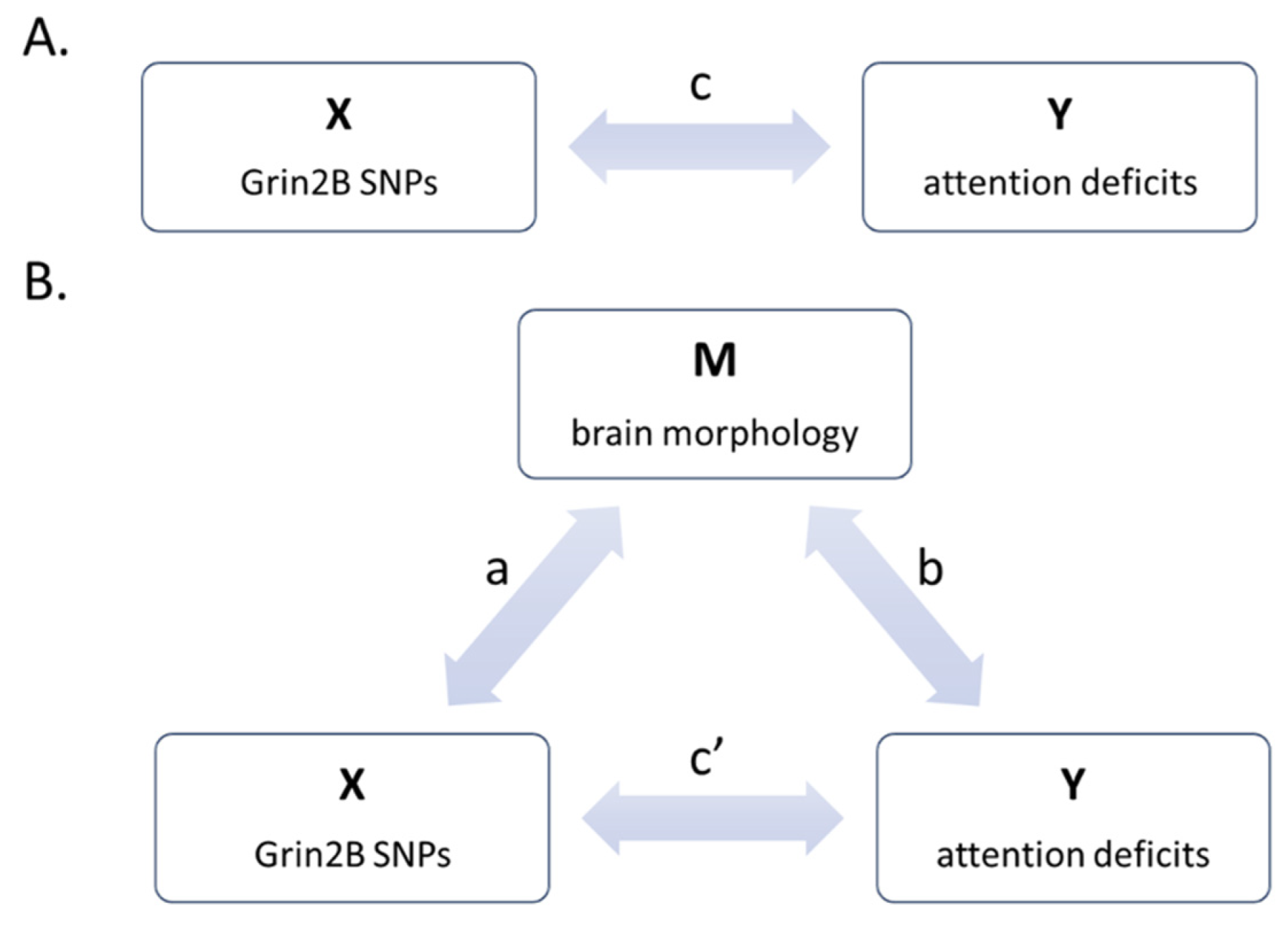
2.7.2. Mediation Analyses

Mediation was assessed using the open-source Bootstrap Regression Analysis of Voxelwise Observations (BRAVO) toolbox (https://sites.google.com/site/bravotoolbox/, downloaded on 5 March 2018) in Matlab. On each triad of selected variables, we designed a simple mediation model, where the GRIN2B SNP was the causal variable X, the CBCL/6-18 AP score the outcome Y, and the ROI morphological parameter the mediator M (Figure 2).
Provided that X significantly accounts for variability in both Y (path c) and M (path a), and M accounts for variability in Y when covarying for X (path b), M is the mediator of the X-Y relationship if the effect of X on Y substantially decreases when M is entered simultaneously with X as a predictor of Y (path c’). Further details on mediation models can be found in [37].
In our study, we used a mediation regression model with age and gender as covariates. The mediation significance was assessed through a permutation procedure with 5000 iterations. Before running the analyses, the values of X, Y, and M were normalized using the Z-score standardization. The strength of model path, for both observed data and bootstrap distributions, was assessed through Ordinary Least Square regression. Confidence intervals and p-values were then estimated using the bias corrected and accelerated formula described in [38]. For all model coefficients (a, b, ab, c, c’), the significance threshold was set to p = 0.05. Multiple comparison corrections were performed if appropriate, based on the number of mediation models applied.

3. Results

3.1. GLM Analyses

3.1.1. Design #1. GRIN2B Effects on Neuroanatomy

The GLM statistics concerning GRIN2B effects on regional brain morphology are reported in Table 3. We detected significant associations between rs5796555-/A marker and regional GMV, and rs2268119A/T and rs2216128T/C markers and regional CSA (p < 0.05, MC corrected). On the contrary, no significant effects of GRIN2B markers on CT emerged.
Specifically, the less frequent allele “A” of marker rs5796555-/A was associated with GMV deficits in left isthmus of cingulate cortex, left precuneus, right caudal and rostral anterior cingulate cortex, right transverse temporal gyrus and bilateral rostral middle frontal gyrus, inferior parietal gyrus, middle temporal cortex and pars orbitalis. Notably, this effect was highly significant (p < 0.01, MC corrected) in regions of the right hemisphere, that is, the caudal and rostral anterior cingulate cortex and inferior parietal gyrus. As inferred from Table 3, the peak T statistics was observed in the caudal portion of the right anterior cingulate cortex.
We also found a negative association between the genotype carrying the less frequent allele “T” of marker rs2268119A/T and CSA in left lateral orbitofrontal cortex (p < 0.05, MC corrected) and right lateral occipital cortex (p < 0.01, MC corrected), and the genotype carrying the minor allele “C” of marker rs2216128G/C and CSA in the right isthmus of the cingulum (p < 0.05, MC corrected). None of the above brain features were affected by gender or age (p > 0.05).

3.1.2. Design #2. GRIN2B Association with Attention/Hyperactivity Problems

The GLM analyses assessing the impact of GRIN2B markers on inattention/hyperactivity revealed a significant positive association between the genotype carrying the minor allele ‘A’ of marker rs5796555-/A and the mean CBCL/6-18 AP score (T(54) = 2.41, p < 0.05). No influences of gender or age on this score emerged (p > 0.05).

3.1.3. Design #3. Neuroanatomy Effects on Attention/Hyperactivity Problems

In view of the results of designs #1 and #2, the neuroanatomy-attention GLM analyses were performed on the only brain morphological parameters influenced by GRIN2B marker rs5796555-/A (Table 3). We found that the CBCL/6-18 AP score was inversely proportional to GMV in the left isthmus of the cingulate cortex (T(54) = 2.67, p < 0.05) and in the right inferior parietal cortex (T(54) = 2.26, p < 0.05), suggesting a possible role of these regions as mediators of the effect of GRIN2B marker rs5796555-/A on inattention/hyperactivity.

3.2. Mediation Analyses

In view of the GLM results, two separate mediation analyses (MA1 and MA2) were performed to investigate the relationship among GRIN2B rs5796555-/A genotype (causal variable X), CBCL/6-18 AP score (outcome Y), and (i) GMV in the left isthmus of the cingulate cortex (mediator variable M1), (ii) GMV in the right inferior parietal cortex (mediator variable M2). No mediation analyses were performed on GRIN2B genotypes or brain features other than those specified due to the absence of the mediation model prerequisites.
The mediation model parameters, 95% confidence intervals (CI), and p-values are reported in Table 4. In line with preliminary GLM results, both mediation analyses confirmed a significant total effect of rs5796555-/A genotype on CBCL/6-18 AP score (c = 0.31, p < 0.05).
As shown in Figure 3, MA1 results confirmed that genotypes carrying the minor allele A of rs5796555-/A were associated with GMV deficits in the left isthmus of the cingulate cortex (a = −0.45, p < 0.001), and in turn that such deficits (while regressing out rs5796555-/A effect) were linked to CBCL/6-18 AP score (b = −0.25, p < 0.05). After inclusion of M1 in the model, the direct effect of rs5796555-/A on CBCL/6-18 AP score was not significant (c’ = 0.20, p = 0.09), whereas its indirect effect through GMV in the left isthmus of cingulate cortex remained significant (ab = 0.11, p < 0.001). Specifically, 35.89% of the total rs5796555-/A effect on attention was mediated by GMV in this region.
On the contrary, MA2 showed that rs5796555-/A genotypes were linked to GMV in the right inferior parietal gyrus (a = −0.48, p < 0.001), but did not confirm a significant influence of the right inferior parietal deficits on attention/hyperactivity problems while controlling for rs5796555-/A contribution (b = −0.18, p = 0.08). The absence of a net effect of M2 on Y ruled out the investigation of any mediated effects of this variable. Of note, since only MA1 analysis was successfully conducted, no multiple comparison corrections were performed on MA1 analysis coefficients.

4. Discussion

In this preliminary work, an innovative genetic-neuroimaging-behavioral approach was adopted to investigate the potential causal effects of GRIN2B markers on developmental attention/hyperactivity problems through neuroanatomy. To this end, we genotyped 6 GRIN2B markers and measured brain morphological parameters and inattention/hyperactivity in a large clinical sample followed from childhood to adolescence.
Our results confirm the presence of a causal chain of relationships among the three variables, showing that GRIN2B rs5796555-/A effects on inattention/hyperactivity over time is significantly mediated by volume in the left isthmus of the cingulate cortex. This unprecedented finding supports the hypothesis that GRIN2B variants affect the structure of key brain regions for executive functioning, ultimately exerting both direct and indirect impact on these developmental problems. This evidence offers new opportunities and translational pathways in the identification and management of attention/hyperactivity deficits in the delicate phase of neurodevelopment.

4.1. GRIN2B Effect on Attention Deficits

To our knowledge, this is the first study on GRIN2B that considered multiple time measures of behavioral attention/hyperactivity problems. The use of the mean CBCL/6-18 AP score across three time points allowed us to smooth the variability due to different manifestations of this complex behavior from childhood to adolescence. In fact, the CBCL/6-18 AP scale assesses both inattentive and hyperactive-impulsive symptoms linked to ADHD, and longitudinal studies suggest that these dimensions may follow separate developmental trajectories and have different manifestations at different ages [3].
In line with previous studies, we found a significant relation between persistent attention deficits and the GRIN2B gene. Specifically, subjects carrying the minor allele ‘A’ of GRIN2B rs5796555-/A were characterized by a higher CBCL/6-18 AP score.
Our findings further confirm the crucial role of GRIN2B in behavioral functions. Given the importance of the Glun2B subunit of NMDA receptor for maturation and plasticity of the central nervous system, it is not surprising that over 60 variants of GRIN2B have been associated with heterogeneous neurodevelopmental and psychiatric disorders [16].
Previous literature reported evidence of the association of GRIN2B gene variants with attention deficits in the general population and patient samples. Of note, [39] found an association between GRIN2B genotypes and CBCL/6-18 AP score in a general population sample aged 6-11. [8] investigated inattention and impulsive symptoms in a sample of ADHD children and found a positive correlation between both symptom classes and nine GRIN2B SNPs. On the same line, a study on attention performance in ADHD patients linked GRIN2B and GRIN2A variants to increased susceptibility to attention problems [12]. Our study confirms and strengthens these findings, showing a role of GRIN2B rs5796555-/A in the genetic risk for inattention/hyperactivity traits that remains stable during development.

4.2. GRIN2B Influence on Brain Structure

Brain imaging features may provide biological endophenotypes for genetic studies on inattention/hyperactivity. Nevertheless, in the growing imaging genetics field, just a few studies explored the link between GRIN2B markers and brain morphology.
For the first time, we assessed the influence of six GRIN2B SNPs on a set of morphological features. The extraction of regional cortical thickness, volume and surface area has offered a unique opportunity to delineate the structural brain correlates of GRIN2B variants with high specificity.
Interestingly, we found selective associations between GRIN2B markers and morphological features. The brain feature that resulted in being most widely influenced by GRIN2B SNPs was regional GMV. On the contrary, the CT feature showed no influence of the GRIN2B markers. The minor allele of marker rs5796555-/A was associated with lower GMV in frontal, parietal and temporal regions. Conversely, the minor allele of marker rs2268119A/T was associated with lower CSA in left frontal and right occipital regions, and rs2216128G/C genotypes carrying the minor allele “C” showed CSA deficits in the right cingulate cortex.
Previous studies already showed an association between glutamatergic genes and regional GMV in children with neurodevelopmental disorders involving, at different levels, attention deficits.
Probably due to different clinical populations and research methods, literature results are mixed. In patients with obsessive compulsive disorder (OCD), [40] reported GRIN2B SNPs to be associated with total thalamus volume. In another pediatric OCD study, a significant association between left orbitofrontal and right anterior cingulate volumes and GRIN2B SNPs emerged [23]. Of note, GRIN2B was linked to left posterior cingulate volume in adolescents with alcohol dependence, one of the disorders most closely related to impulsivity [24]. Since these findings emerged from clinical samples that share only some features with attention deficit syndromes, further investigations are needed to confirm our findings and interpret them in a wider dimensional perspective.
To our knowledge, the only imaging-GRIN2B study that has focused on inattention/hyperactivity is a resting state functional MRI (fMRI) study on ADHD children, which showed GRIN2B influence on regional homogeneity in left superior parietal cortex, being part of the attention circuit and with a role in inhibition [41].
Overall, these results suggest that GRIN2B regulation is not confined to specific brain regions but involves complex brain networks. Indeed, precuneus, cingulate, prefrontal, orbitofrontal, inferior parietal and temporal cortices, which were found to be affected by GRIN2B markers, are included in the default mode network (DMN) [42,43]. The DMN is a spontaneous resting state network that deactivates during task performance, whose activation has been implicated in attention and, specifically, in exteroceptive and interoceptive attentional orientation [44,45,46,47]. The DMN failure to deactivate during tasks might result in attentional intrusions and deficits in performance [48].
Moreover, the posterior and rostral cingulate cortex, prefrontal cortex, and inferior parietal lobule are part of the frontoparietal control network [49,50,51,52], involved in executive control. During tasks demanding direct attention to external information, activity increases in the frontoparietal control network and decreases in DMN [53].
The above evidence supports the hypothesis that GRIN2B effects on brain structure might be interpreted in terms of brain circuitries, especially those that in turn impact on behavioral functions.

4.3. Brain Correlates of Attention/Hyperactivity Problems

In the imaging-behavioral analysis, we deliberately focused on the brain regions that resulted in being affected by GRIN2B rs5796555-/A, which may act as intermediate biological phenotypes in GRIN2B effect on inattention/hyperactivity. The study of the link between other brain features (e.g., regional CSA or CT) and attention/hyperactivity problems went beyond the scope of our study, but could be the subject of future investigations.
Interestingly, we found a negative association between CBCL/6-18 AP score and GMV in the left isthmus of the cingulum and the right inferior parietal cortex. The involvement of the posterior cingulate cortex and inferior parietal lobule in functional networks of attention control may explain the relation between attention problems and structural abnormalities in these areas and further supports the aforementioned network-based perspective.

4.4. From GRIN2B to Behavior through Neuroanatomy

In recent years, imaging genetics aimed to disentangle the pathways from genes to behavior. Instead of directly measuring the association between complex behavioral phenotypes and genetics, brain functionality and anatomy might be used as reliable intermediate phenotype, with a more direct and interpretable relation with genetics [41].
The results of our preliminary GLM analyses support the hypothesis that regional GMV (in left isthmus of cingulate cortex and right inferior parietal gyrus) might mediate GRIN2B effect on inattention/hyperactivity. Hence, two separate mediation analyses were performed to investigate the relationship among GRIN2B rs5796555-/A genotype, mean CBCL/6-18 AP score and, as mediator, GMV in left isthmus of cingulate cortex and right inferior parietal cortex.
The failure to verify the second mediation hypothesis suggests that GMV in the right inferior parietal cortex, besides being regulated by GRIN2B rs5796555-/A, does not shape genetic susceptibility for inattention/hyperactivity.
On the contrary, our results suggest that GMV in the left isthmus of cingulate cortex may play a key role in this mechanism. Indeed, after inclusion of this parameter in the mediation model, the direct effect of GRIN2B rs5796555-/A on CBCL/6-18 AP score became not significant, whereas its indirect effect through GMV in this region emerged to be significant. Specifically, more than 30% of the GRIN2B rs5796555-/A genotype effect on CBCL/6-18 AP score was mediated by GMV in left isthmus of cingulate volume.
Therefore, we believe that this region might play a relevant role in translating GRIN2B variation to the complex attention phenotype. Until now, the isthmus of cingulate cortex, also known as retrosplenial cortex, has received little attention in studies on attention deficits. Nevertheless, a recent research found that the right cingulate isthmus was thinner in children with comorbid developmental coordination disorder (DCD) and ADHD compared to children with DCD alone. Previous fMRI studies reported an involvement of the retrosplenial cortex in spatial attention [54], episodic memory [55] and emotional processing [56]. Overall, this evidence enhances the need for focused research on structural and functional alterations of this region in attention/hyperactivity disorders.

4.5. Limitations

In the discussion of these results, the following limitations should be considered. First, the sample size was limited by difficulties related to the longitudinal study protocol. Specifically, the MRI acquisitions required high level of patient compliance, which was not always achieved due to the young age and clinical characteristics of our sample. The use of a modest sample has limited the statistical power and the reliability of the emerged imaging-genetic-behavioral associations, which need to be reproduced on larger, independent samples.
The integration of genetic, neuroimaging, and psychopathologic information enhanced the possible sources of error. Larger sample size replications are needed in order to minimize the risk of false positive results.
It is worth mentioning that our research protocol included children and adolescents with emotional and behavioral difficulties but not comparison subjects. Despite this limit, the clinical heterogeneity of the sample has enabled its stratification based on attention and hyperactivity problems and the investigation of the underlying genetic and neuroanatomic mechanisms.
Another important point is the lack of environmental factors in the model, which may have affected the findings’ reliability, as gene-environment interactions are implicated in the development of complex psychopathological behaviors [2,57].
Regarding the measurement of attention/hyperactivity problems, we used the CBCL/6-18 “Attention Problems” subscale, fulfilled by the participants’ caregivers. This scale evaluates both inattention and impulsivity from a hetero-referred point of view. In the future, it is desirable to integrate the measurement of these traits with neuropsychological tasks or clinical measures from different raters, and to disentangle the contributions of inattention and hyperactivity problems.
A final, intrinsic limitation of our study concerns the focus on a specific gene of the glutamatergic pathway. This choice was driven by growing evidence from genetic studies, suggesting a role of GRIN2B in attention deficits/hyperactivity traits. Given the increasing interest in polygenic risk factors, future study extensions should additionally consider the effect of other genes implicated in this behavior. We should investigate the interactions between genes, also known as “epistasis” [58], i.e., the extent to which the effect of one gene upon the phenotype is moderated by other genetic variations at a statistical and, possibly, a biological level [13].

5. Conclusions

Using a proof-of-concept imaging genetics mediation design, we explored the relationship between the glutamatergic GRIN2B gene variants and attention deficits and impulsive-hyperactive behaviors by introducing neuroanatomical measures as intermediate factors.
Our findings confirm that brain anatomy, more specifically volumetry, is closely related to GRIN2B variations and can act as an intermediate phenotype between genetics and complex behaviors. The mediation results highlight a possible role of the left isthmus of the cingulum in mediating heritable risk for inattention/hyperactivity linked to GRIN2B variants. Confirmatory longitudinal studies are required to better delineate the genetic, neuronal and environmental mechanisms contributing to developmental risk pathways, with important implications for effective prevention, identification and treatment of early-onset psychiatric disorders.

Author Contributions

M.N.: co-design of the current study, data collection, supervision, analysis and manuscript writing. E.M., M.M. (Maddalena Mauri): integrated data analysis and first manuscript draft writing. M.G., S.P. and C.B.: recruitment, subjects’ evaluation and clinical data collection. R.G. (Roberto Giorda): genetic data analysis. R.G. (Rossano Girometti): neuroimaging data collection. B.T.: neuroimaging data collection and analysis. M.M. (Massimo Molteni): recruitment supervision, manuscript revision. F.F.: recruitment supervision, manuscript revision. P.B.: co-design of the current study, project coordination, analysis supervision and manuscript revision. All authors have read and agreed to the published version of the manuscript.

Funding

This research was funded by the Italian Ministry of Health (RF-2016-02364582 to M.N. and P.B., GR-2018-12367789 to E.M., RC 2018-19 MIMOSA to M.N.).

Institutional Review Board Statement

The study was conducted according to the guidelines of the Declaration of Helsinki, and approved by the Research Ethical Committee of the Scientific Institute IRCCS E. Medea.

Informed Consent Statement

Informed consent was obtained from all subjects involved in the study.

Data Availability Statement

The data presented in this study are available on request from the corresponding author. The data are not publicly available due to privacy and ethical restrictions. Further information and requests for materials and methods should be directed to and will be fulfilled by the corresponding author.

Conflicts of Interest

The authors declare no conflict of interest. The funders had no role in the design of the study; in the collection, analyses, or interpretation of data; in the writing of the manuscript, or in the decision to publish the results.

References

  1. Nobile, M.; Giorda, R.; Marino, C.; Carlet, O.; Pastore, V.; Vanzin, L.; Bellina, M.; Molteni, M.; Battaglia, M. Socioeconomic status mediates the genetic contribution of the dopamine receptor D4 and serotonin transporter linked promoter region repeat polymorphisms to externalization in preadolescence. Dev. Psychopathol. 2007, 19, 1147–1160. [Google Scholar] [CrossRef]
  2. Nobile, M.; Rusconi, M.; Bellina, M.; Marino, C.; Giorda, R.; Carlet, O.; Vanzin, L.; Molteni, M.; Battaglia, M. COMT Val158Met polymorphism and socioeconomic status interact to predict attention deficit/hyperactivity problems in children aged 10–14. Eur. Child Adolesc. Psychiatry 2009, 19, 549–557. [Google Scholar] [CrossRef] [Green Version]
  3. Leopold, D.R.; Christopher, M.E.; Olson, R.K.; Petrill, S.A.; Willcutt, E.G. Invariance of ADHD Symptoms Across Sex and Age: A Latent Analysis of ADHD and Impairment Ratings from Early Childhood into Adolescence. J. Abnorm. Child Psychol. 2018, 47, 21–34. [Google Scholar] [CrossRef] [PubMed]
  4. Kuntsi, J.; Gayan, J.; Stevenson, J. Parents’ and teachers’ ratings of problem behaviours in children: Genetic and contrast effects. Twin Res. 2000, 3, 251–258. [Google Scholar] [PubMed]
  5. Rietveld, M.; Hudziak, J.; Bartels, M.; van Beijsterveldt, T.; Boomsma, D. Heritability of attention problems in children: Longitudinal results from a study of twins, age 3 to 12. J. Child Psychol. Psychiatry 2004, 45, 577–588. [Google Scholar] [CrossRef]
  6. Franke, B.; Faraone, S.; Asherson, P.; Buitelaar, J.; Bau, C.; Ramos-Quiroga, J.A.; Mick, E.; Grevet, E.; Johansson, S.; Havikk, J.; et al. The genetics of attention deficit/hyperactivity disorder in adults, a review. Mol. Psychiatry 2011, 17, 960–987. [Google Scholar] [CrossRef] [Green Version]
  7. Cortese, S. The neurobiology and genetics of Attention-Deficit/Hyperactivity Disorder (ADHD): What every clinician should know. Eur. J. Paediatr. Neurol. 2012, 16, 422–433. [Google Scholar] [CrossRef]
  8. Dorval, K.M.; Wigg, K.G.; Crosbie, J.; Tannock, R.; Kennedy, J.L.; Ickowicz, A.; Pathare, T.; Malone, M.; Schachar, R.; Barr, C.L. Association of the glutamate receptor subunit gene GRIN2B with attention-deficit/hyperactivity disorder. Genes Brain Behav. 2006, 6, 444–452. [Google Scholar] [CrossRef] [Green Version]
  9. Akazawa, C.; Shigemoto, R.; Bessho, Y.; Nakanishi, S.; Mizuno, N. Differential expression of five N-methyl-D-aspartate receptor subunit mRNAs in the cerebellum of developing and adult rats. J. Comp. Neurol. 1994, 347, 150–160. [Google Scholar] [CrossRef]
  10. Gill, I.; Droubi, S.; Giovedi’, S.; Fedder, K.; Bury, L.A.D.; Bosco, F.; Sceniak, M.P.; Benfenati, F.; Sabo, S.L. Presynaptic NMDA receptors: Dynamics and distribution in developing axons in vitro and in vivo. J. Cell Sci. 2015, 128, 768–780. [Google Scholar] [CrossRef] [PubMed] [Green Version]
  11. Monyer, H.; Burnashev, N.; Laurie, D.J.; Sakmann, B.; Seeburg, P.H. Developmental and regional expression in the rat brain and functional properties of four NMDA receptors. Neuron 1994, 12, 529–540. [Google Scholar] [CrossRef]
  12. Kim, J.I.; Kim, J.-W.; Park, S.; Hong, S.-B.; Lee, D.S.; Paek, S.H.; Han, D.H.; Cheong, J.H.; Kim, B.-N. The GRIN2B and GRIN2A Gene Variants Are Associated with Continuous Performance Test Variables in ADHD. J. Atten. Disord. 2016, 24, 1538–1546. [Google Scholar] [CrossRef] [PubMed]
  13. Mascheretti, S.; Facoetti, A.; Giorda, R.; Beri, S.; Riva, V.; Trezzi, V.; Cellino, M.R.; Marino, C. GRIN2B mediates susceptibility to intelligence quotient and cognitive impairments in developmental dyslexia. Psychiatr. Genet. 2015, 25, 9–20. [Google Scholar] [CrossRef] [PubMed]
  14. Endele, S.; Rosenberger, G.; Geider, K.; Popp, B.; Tamer, C.; Stefanova, I.; Milh, M.; Kortüm, F.; Fritsch, A.; Pientka, F.K.; et al. Mutations in GRIN2A and GRIN2B encoding regulatory subunits of NMDA receptors cause variable neurodevelopmental phenotypes. Nat. Genet. 2010, 42, 1021–1026. [Google Scholar] [CrossRef]
  15. Hassan, A.; Heckman, M.G.; Ahlskog, J.; Wszolek, Z.K.; Serie, D.J.; Uitti, R.J.; Van Gerpen, J.A.; Okun, M.S.; Rayaprolu, S.; Ross, O.A. Association of Parkinson disease age of onset with DRD2, DRD3 and GRIN2B polymorphisms. Park. Relat. Disord. 2016, 22, 102–105. [Google Scholar] [CrossRef] [Green Version]
  16. Hu, C.; Chen, W.; Myers, S.J.; Yuan, H.; Traynelis, S.F. Human GRIN2B variants in neurodevelopmental disorders. J. Pharmacol. Sci. 2016, 132, 115–121. [Google Scholar] [CrossRef] [Green Version]
  17. Lorenzi, C.; Delmonte, D.; Pirovano, A.; Marino, E.; Bongiorno, F.; Catalano, M.; Colombo, C.; Bramanti, P.; Smeraldi, E. Searching Susceptibility Loci for Bipolar Disorder: A Sib Pair Study on Chromosome 12. Neuropsychobiology 2010, 61, 10–18. [Google Scholar] [CrossRef]
  18. Seripa, D.; Matera, M.G.; Franceschi, M.; Bizzarro, A.; Paris, F.; Cascavilla, L.; Rinaldi, M.; Panza, F.; Solfrizzi, V.; Daniele, A.; et al. Association Analysis of GRIN2B, Encoding N-Methyl-D-Aspartate Receptor 2B Subunit, and Alzheimer’s Disease. Dement. Geriatr. Cogn. Disord. 2008, 25, 287–292. [Google Scholar] [CrossRef]
  19. Meyer-Lindenberg, A.; Weinberger, D.R. Intermediate phenotypes and genetic mechanisms of psychiatric disorders. Nat. Rev. Neurosci. 2006, 7, 818–827. [Google Scholar] [CrossRef] [PubMed]
  20. Zayed, A.; Robinson, G.E. Understanding the Relationship Between Brain Gene Expression and Social Behavior: Lessons from the Honey Bee. Annu. Rev. Genet. 2012, 46, 591–615. [Google Scholar] [CrossRef] [PubMed] [Green Version]
  21. Kuswanto, C.N.; Sum, M.Y.; Thng, C.R.Z.; Bin Zhang, Y.; Yang, G.L.; Nowinski, W.L.; Sitoh, Y.Y.; Low, C.M.; Sim, K. GRIN2B Gene and Associated Brain Cortical White Matter Changes in Bipolar Disorder: A Preliminary Combined Platform Investigation. BioMed Res. Int. 2013, 2013, 635131. [Google Scholar] [CrossRef] [Green Version]
  22. Stein, J.; Hua, X.; Morra, J.H.; Lee, S.; Hibar, D.P.; Ho, A.J.; Leow, A.D.; Toga, A.W.; Sul, J.H.; Kang, H.M.; et al. Genome-wide analysis reveals novel genes influencing temporal lobe structure with relevance to neurodegeneration in Alzheimer’s disease. NeuroImage 2010, 51, 542–554. [Google Scholar] [CrossRef] [Green Version]
  23. Arnold, P.D.; MacMaster, F.; Richter, M.A.; Hanna, G.; Sicard, T.; Burroughs, E.; Mirza, Y.; Easter, P.C.; Rose, M.; Kennedy, J.L.; et al. Glutamate receptor gene (GRIN2B) associated with reduced anterior cingulate glutamatergic concentration in pediatric obsessive–compulsive disorder. Psychiatry Res. Neuroimaging 2009, 172, 136–139. [Google Scholar] [CrossRef] [Green Version]
  24. Dalvie, S.; Brooks, S.J.; Cardenas, V.; Fein, G.; Ramesar, R.; Stein, D.J. Genetic variation within GRIN2B in adolescents with alcohol use disorder may be associated with larger left posterior cingulate cortex volume. Acta Neuropsychiatr. 2016, 29, 252–258. [Google Scholar] [CrossRef]
  25. Wilens, T.E.; Zulauf, C.A. Attention deficit hyperactivity disorder, and substance use disorders. In Youth Substance Abuse and Co-Occurring Disorders; Kaminer, Y., Arlington, V.A., Eds.; American Psychiatric Publishing: Washington, DC, USA, 2015; pp. 103–129. [Google Scholar]
  26. Norman, L.J.; Carlisi, C.; Lukito, S.; Hart, H.; Mataix-Cols, D.; Radua, J.; Rubia, K. Structural and Functional Brain Abnormalities in Attention-Deficit/Hyperactivity Disorder and Obsessive-Compulsive Disorder. JAMA Psychiatry 2016, 73, 815–825. [Google Scholar] [CrossRef]
  27. Bellina, M.; Brambilla, P.; Garzitto, M.; Negri, G.A.L.; Molteni, M.; Nobile, M. The ability of CBCL DSM-oriented scales to predict DSM-IV diagnoses in a referred sample of children and adolescents. Eur. Child Adolesc. Psychiatry 2012, 22, 235–246. [Google Scholar] [CrossRef]
  28. Bianchi, V.; Brambilla, P.; Garzitto, M.; Colombo, P.; Fornasari, L.; Bellina, M.; Bonivento, C.; Tesei, A.; Piccin, S.; Conte, S.; et al. Latent classes of emotional and behavioural problems in epidemiological and referred samples and their relations to DSM-IV diagnoses. Eur. Child Adolesc. Psychiatry 2016, 26, 549–557. [Google Scholar] [CrossRef] [PubMed] [Green Version]
  29. Kaufman, J.; Birmaher, B.; Brent, D.A.; Ryan, N.D.; Rao, U. K-SADS-PL. J. Am. Acad. Child Adolesc. Psychiatry 2000, 39, 1208. [Google Scholar] [CrossRef]
  30. Achenbach, T.M.; Rescorla, L. Manual for the ASEBA School-Age Forms & Profiles; ASEBA: Burlington, VT, USA, 2001. [Google Scholar]
  31. Barratt, W. The Barratt Simplified Measure of Social Status (BSMSS): Measuring SES; Unpublished manuscript; Indiana State University: Terre Haute, IN, USA, 2006. [Google Scholar]
  32. Wechsler, D. Manual for the Wechsler Intelligence Scale for Children; revised; Psychological Corporation: San Antonio, TX, USA, 1974. [Google Scholar]
  33. Achenbach, T.M.; Verhulst, F.C.; Baron, G.D.; Althaus, M.; Veriiulst, F.C. A Comparison of Syndromes Derived from the Child Behavior Checklist for American and Dutch Boys Aged 6–11 and 12–16. J. Child Psychol. Psychiatry 1987, 28, 437–453. [Google Scholar] [CrossRef]
  34. Verhulst, F.C.; Achenbach, T.M.; Althaus, M.; Akkerhuis, G.W. A Comparison of Syndromes Derived from the Child Behavior Checklist for American and Dutch Girls Aged 6–11 And 12–16. J. Child Psychol. Psychiatry 1988, 29, 879–895. [Google Scholar] [CrossRef]
  35. Fischl, B. FreeSurfer. NeuroImage 2012, 62, 774–781. [Google Scholar] [CrossRef] [Green Version]
  36. Desikan, R.S.; Ségonne, F.; Fischl, B.; Quinn, B.T.; Dickerson, B.C.; Blacker, D.; Buckner, R.L.; Dale, A.M.; Maguire, R.P.; Hyman, B.T.; et al. An automated labeling system for subdividing the human cerebral cortex on MRI scans into gyral based regions of interest. NeuroImage 2006, 31, 968–980. [Google Scholar] [CrossRef]
  37. Preacher, K.J.; Hayes, A.F. Asymptotic and resampling strategies for assessing and comparing indirect effects in multiple mediator models. Behav. Res. Methods 2008, 40, 879–891. [Google Scholar] [CrossRef]
  38. DiCiccio, T.J.; Efron, B. Bootstrap confidence intervals. Stat. Sci. 1996, 11, 189–228. [Google Scholar] [CrossRef]
  39. Riva, V.; Battaglia, M.; Nobile, M.; Cattaneo, F.; Lazazzera, C.; Mascheretti, S.; Giorda, R.; Mérette, C.; Émond, C.; Maziade, M.; et al. GRIN2B predicts attention problems among disadvantaged children. Eur. Child Adolesc. Psychiatry 2014, 24, 827–836. [Google Scholar] [CrossRef]
  40. Wu, K.; Hanna, G.; Easter, P.; Kennedy, J.L.; Rosenberg, D.R.; Arnold, P.D. Glutamate system genes and brain volume alterations in pediatric obsessive-compulsive disorder: A preliminary study. Psychiatry Res. Neuroimaging 2013, 211, 214–220. [Google Scholar] [CrossRef] [Green Version]
  41. Kim, J.I.; Yoo, J.H.; Kim, D.; Jeong, B.; Kim, B.-N. The effects of GRIN2B and DRD4 gene variants on local functional connectivity in attention-deficit/hyperactivity disorder. Brain Imaging Behav. 2017, 12, 247–257. [Google Scholar] [CrossRef]
  42. Greicius, M.D.; Krasnow, B.; Reiss, A.L.; Menon, V. Functional connectivity in the resting brain: A network analysis of the default mode hypothesis. Proc. Natl. Acad. Sci. USA 2003, 100, 253–258. [Google Scholar] [CrossRef] [PubMed] [Green Version]
  43. Ismaylova, E.; Di Sante, J.; Gouin, J.-P.; Pomares, F.; Vitaro, F.; Tremblay, R.E.; Booij, L. Associations Between Daily Mood States and Brain Gray Matter Volume, Resting-State Functional Connectivity and Task-Based Activity in Healthy Adults. Front. Hum. Neurosci. 2018, 12, 168. [Google Scholar] [CrossRef] [Green Version]
  44. Fransson, P. Spontaneous low-frequency BOLD signal fluctuations: An fMRI investigation of the resting-state default mode of brain function hypothesis. Hum. Brain Mapp. 2005, 26, 15–29. [Google Scholar] [CrossRef]
  45. Fransson, P. How default is the default mode of brain function? Further evidence from intrinsic BOLD signal fluctuations. Neuropsychologia 2006, 44, 2836–2845. [Google Scholar] [CrossRef]
  46. Leech, R.; Sharp, D.J. The role of the posterior cingulate cortex in cognition and disease. Brain 2014, 137, 12–32. [Google Scholar] [CrossRef] [Green Version]
  47. Gusnard, D.A.; Akbudak, E.; Shulman, G.L.; Raichle, M.E. Medial prefrontal cortex and self-referential mental activity: Relation to a default mode of brain function. Proc. Natl. Acad. Sci. USA 2001, 98, 4259–4264. [Google Scholar] [CrossRef] [PubMed] [Green Version]
  48. Sonuga-Barke, E.J.; Castellanos, F.X. Spontaneous attentional fluctuations in impaired states and pathological conditions: A neurobiological hypothesis. Neurosci. Biobehav. Rev. 2007, 31, 977–986. [Google Scholar] [CrossRef] [PubMed]
  49. Leech, R.; Kamourieh, S.; Beckmann, C.F.; Sharp, D.J. Fractionating the Default Mode Network: Distinct Contributions of the Ventral and Dorsal Posterior Cingulate Cortex to Cognitive Control. J. Neurosci. 2011, 31, 3217–3224. [Google Scholar] [CrossRef]
  50. Leech, R.; Braga, R.; Sharp, D.J. Echoes of the Brain within the Posterior Cingulate Cortex. J. Neurosci. 2012, 32, 215–222. [Google Scholar] [CrossRef] [Green Version]
  51. Vincent, J.L.; Kahn, I.; Snyder, A.Z.; Raichle, M.E.; Buckner, R.L. Evidence for a Frontoparietal Control System Revealed by Intrinsic Functional Connectivity. J. Neurophysiol. 2008, 100, 3328–3342. [Google Scholar] [CrossRef] [Green Version]
  52. Spreng, R.N. The fallacy of a “task-negative” network. Front. Psychol. 2012, 3, 145. [Google Scholar] [CrossRef] [PubMed] [Green Version]
  53. Kelly, C.; Uddin, L.; Biswal, B.B.; Castellanos, F.X.; Milham, M.P. Competition between functional brain networks mediates behavioral variability. NeuroImage 2008, 39, 527–537. [Google Scholar] [CrossRef]
  54. Mesulam, M.; Nobre, A.C.; Kim, Y.-H.; Parrish, T.; Gitelman, D. Heterogeneity of Cingulate Contributions to Spatial Attention. NeuroImage 2001, 13, 1065–1072. [Google Scholar] [CrossRef]
  55. Wagner, A.D.; Shannon, B.J.; Kahn, I.; Buckner, R.L. Parietal lobe contributions to episodic memory retrieval. Trends Cogn. Sci. 2005, 9, 445–453. [Google Scholar] [CrossRef] [PubMed]
  56. Maddock, R. The retrosplenial cortex and emotion: New insights from functional neuroimaging of the human brain. Trends Neurosci. 1999, 22, 310–316. [Google Scholar] [CrossRef]
  57. Nobile, M.; Rusconi, M.; Bellina, M.; Marino, C.; Giorda, R.; Carlet, O.; Vanzin, L.; Molteni, M.; Battaglia, M. The influence of family structure, the TPH2 G-703T and the 5-HTTLPR serotonergic genes upon affective problems in children aged 10–14 years. J. Child Psychol. Psychiatry 2009, 50, 317–325. [Google Scholar] [CrossRef]
  58. Plomin, R. Child Development and Molecular Genetics: 14 Years Later. Child Dev. 2012, 84, 104–120. [Google Scholar] [CrossRef]
Figure 1. GRIN2B linkage disequilibrium. Haploview plot showing pairwise linkage disequilibrium (r2 values) for 6 SNPs of GRIN2B based on the sample’s genotypes.
Genes 12 01193 g001
Figure 2. Mediation model design. (A) Model of direct effect of X on Y. (B) Mediation model, where X has both direct and indirect (through M) effects on Y.
Genes 12 01193 g002
Figure 3. Mediation analysis 1 (MA1) results. (A) Model of direct effect of X on Y. (B) Mediation model, where X has both direct and indirect (through M) effects on Y. Significant pathways are highlighted with *. CBCL/6-18 AP: Child Behavior Checklist Attention Problems score.
Genes 12 01193 g003
Table 1. Sociodemographic, clinical and cognitive characteristics of the sample.
Males (n = 42)Females (n = 16)Total (n = 58)
Wave 0
Age (years) 18.55 ± 2.519.38 ± 2.078.78 ± 2.43
SES 149.05 ± 18.5936.25 ± 21.1845.52 ± 20.17
CBCL Attention Problems 163.10 ± 9.4066.56 ± 10.3764.05 ± 9.80
FSIQ 1108.81 ± 16.39109.38 ± 13.93108.96 ± 15.65
K-SADS-PL DIAGNOSIS 2
ADHDN = 9 (21.4%)N = 4 (25.0%)N = 13 (22.4%)
Any behavioral disorder N = 10 (23.8%)N = 0 (0.0%)N = 10 (17.2%)
Any mood disorder N = 12 (28.6%)N = 6 (37.5%)N = 18 (31.0%)
Any anxiety disorder N = 19 (45.2%)N = 9 (56.3%)N = 28 (48.3%)
Other diagnoses N = 7 (16.7%)N = 5 (31.3%)N = 12 (20.7%)
No current diagnosisN = 2 (4.8%)N = 1 (6.3%)N = 3 (5.2%)
Comorbidities1 diagnosis: N = 25 (59.5%)1 diagnosis: N = 8 (50.0%)1 diagnosis: N = 33 (56.9%)
2 diagnoses: N = 13 (31.0%)2 diagnoses: N = 5 (31.3%)2 diagnoses: N = 18 (31.0%)
3 diagnoses: N = 2 (4.8%)3 diagnoses: N = 2 (12.5%)3 diagnoses: N = 4 (6.9%)
Wave 1
Age (years) 114.12 ± 2.0915.58 ± 2.2414.52 ± 2.23
CBCL Attention Problems159.48 ± 7.8662.06 ± 7.2460.19 ± 7.78
Wave 2
Age (years) 115.80 ± 2.3017.14 ± 2.4316.17 ± 2.41
CBCL Attention Problems 157.83 ± 6.8859.00 ± 4.9858.16 ± 6.43
1 Mean ± standard deviation; 2 N (%). SES: socioeconomic status; FSIQ: full-scale intelligence quotient; K-SADS-PL: Kiddie schedule for affective disorders and schizophrenia for school-age children-present and lifetime version; ADHD: attention deficits/hyperactivity disorder. CBCL: Child Behavior Checklist.
Table 2. GRIN2B allele frequencies and Hardy-Weinberg equilibrium’s p-values.
GRIN2B SNPAlleleFrequency 1Hardy-Weinberg Equilibrium
rs5796555-0.710.201
A0.29
rs1012586G0.660.744
C0.34
rs2268119A0.730.213
T0.27
rs2216128A0.740.146
G0.26
rs11609779C0.840.546
T0.16
rs2192973G0.780.115
A0.22
SNP: single nucleotide polymorphism. 1 Fraction of the total. - is marker of the allele.
Table 3. General Linear Model results of GRIN2B markers effect on brain morphological parameters.
GRIN2B SNPAlleleFS FeatureBrain RegionT 1ppcorr 2
rs5796555“-/A” and “A/A”VolumeLeft inferior parietal3.72<0.001<0.05
Left isthmus cingulate3.62<0.001<0.05
Left middle temporal3.90<0.001<0.05
Left pars orbitalis3.72<0.001<0.05
Left precuneus3.78<0.001<0.05
Left rostral middle frontal3.59<0.001<0.05
Right caudal ACC4.32<0.0001<0.01
Right inferior parietal4.14<0.001<0.01
Right middle temporal3.430.001<0.05
Right pars orbitalis3.72<0.001<0.05
Right rostral ACC3.95<0.001<0.01
Right rostral middle frontal3.49<0.001<0.05
Right transverse temporal3.400.001<0.05
rs2268119“A/T” and “T/T”AreaLeft lateral orbitofrontal3.400.001<0.05
Right lateral occipital3.98<0.001<0.01
rs2216128“G/C” and “C/C”Right isthmus cingulate3.420.001<0.05
SNP: single nucleotide polymorphism; FS: FreeSurfer software; ACC: anterior cingulate cortex. ROI: region of interest. 1 general linear model design: ROI parameter ~ 1 + Gender + Age + GRIN2B SNP. Observations were 58, error degrees of freedom were 54. ROI volumes were normalized using total intracranial volume. ROI surface areas were normalized using total cortical surface area. 2 Level of significance after multiple comparison correction: p < 0.0016.
Table 4. Summary of mediation results.
Mediation Analysis 1Mediation Analysis 2
ParameterValue95% CIpParameterValue95% CIp
a–0.45[–0.26 0.26]<0.001a0.48[–0.26 0.26]<0.001
b–0.25[–0.27 0.26]0.037b−0.18[–0.25 0.29]0.08
c0.31[–0.27 0.26]0.011c0.31[–0.27 0.26]0.011
ab0.11[–0.04 0.05]<0.001abn.e.n.e.n.e.
c’0.20[–0.27 0.27]0.09c’n.e.n.e.n.e.
Causal variable X: GRIN2B marker rs5796555-/A. Outcome variable Y: CBCL/6-18 attention problem score. Mediator variable MA1: left isthmus of cingulate cortex volume. MA2: right inferior parietal cortex. CI: confidence interval. n.e: not evaluated.
Publisher’s Note: MDPI stays neutral with regard to jurisdictional claims in published maps and institutional affiliations.

