Article

Chiral Recognition of Azo-Schiff Base Ligands, Their Cu(II) Complexes, and Their Docking to Laccase as Mediators

Department of Chemistry, Faculty of Science, Tokyo University of Science, 1-3 Kagurazaka, Shinjuku-ku, Tokyo 162-8601, Japan
*
Author to whom correspondence should be addressed.
Submission received: 25 April 2019 / Revised: 10 May 2019 / Accepted: 12 May 2019 / Published: 14 May 2019
(This article belongs to the Special Issue Symmetric and Asymmetric Metal Complexes)

Abstract

:
Chiral crystals were discovered due to spontaneous resolution when enantiomers of 4-phenyldiazenyl-2-[(R or S)-(1-phenylethyl)-iminomethyl]phenol and its racemic mixture were prepared. Using two ligands per molecule, optically active R,R and S,S enantiomers and meso R,S diastereomer of Cu(II) complexes were prepared. Strong chiral recognition was expected for them. Laccase has attracted attention as a catalyst that reduces oxygen to water in a cathode of biofuel cells, which can be effectively mediated by metal complexes. Furthermore, azobenzene can align perpendicularly to the polarization direction of irradiating linearly polarized ultraviolet light (Weigert effect) as well as to the conventional cis-trans photoisomerization accompanying the shift of redox potential. Thus, we also studied the photo-induced control of cis-trans forms and the alignment of these Cu(II) complexes as a mediator to fit laccase appropriately. We discuss photo-induced control on not only electronically but also sterically-favored redox conditions. The meso(R,S)-form of the Cu(II) complex in cis-form was found to be the best at increasing the current of cyclic voltammetry (CV) among the three R,R and S,S enantiomers and the R,S diastereomer of the Cu(II) complexes.

Graphical Abstract

1. Introduction

Laccase has attracted attention as a catalyst for the four-electron oxygen reduction reaction which is used for the electrodes of biofuel cells [1,2]. Proteins of this type containing functional copper atoms are referred to as members of the multicopper oxidase family [3]. Among them, laccase is used as a catalytic enzyme on the cathode side of biofuel cells. Biofuel cells have the advantage of being able to generate electricity at room temperature and under low environmental loads [4,5]. The four copper atoms, which are the active centers of the laccase, are divided into three types, T1, T2 and T3, respectively, and T2 copper and two T3 copper atoms form triple nuclear clusters, which catalyze the reduction of oxygen (O2) to water (H2O) [6]. Substrates are oxidized at the T1 copper site and electrons are transferred to the cluster site, followed by four-electron reduction. However, there are also problems related to the instability of the enzyme and small electric power compared to other fuel cells. Herein, we explore some ways to solve this problem of low power [7].
In this case, a redox substance called a mediator is generally used for electron transfer between the electrode and laccase [2]. In order to facilitate electron transfer between the electrode and the oxygen reductase in the biofuel cell cathode, our laboratory studied the use of Schiff base metal complexes as mediators [8,9,10,11]. Mediators are required to be able to fit to the hydrophobic pocket sites on the laccase surface to supply electrons to the electron accepting T1 site inside the laccase molecule. We have searched for methods to introduce not only central metal ions (Cu, Mn), but also redox groups of anthraquinone [8,9] and polarization orientation of azobenzene [10,11], to organic ligands. However, whether photoisomerization and photo-induced orientation of the azobenzene moiety is effective for increasing current, and what the appropriate way to fit a complex to laccase is, are still unknown.
Herein, we designed new enantiomers and diastereomers of Cu(II) complexes as mediators (Figure 1) incorporating 1-phenylethylamine derivative ligands. The purpose was to investigate favorable docking with the hydrophobic pocket site of the laccase surface using chiral molecular recognition. In addition, cis-trans forms produced by photoisomerization of three enantiomer and diastereomer Cu(II) mediators by chirality were compared by means of computational simulations. It was found that chiral molecular recognition resulting from spontaneous resolution could be observed for the racemic organic ligand.

2. Materials and Methods

2.1. General Procedures

Chemicals of the highest commercial grade available (solvents from Kanto Chemical (Japan), organic compounds from Tokyo Chemical Industry (Japan), and metal sources from Wako (Japan)) were used as received without further purification. 4-Phenyldiazenyl-2-[(1-phenylethyl)-iminomethyl]phenol and its single crystals were prepared according to methods detailed in the literature [12] and checked with X-ray crystallography, proton nuclear magnetic resonance (1H-NMR), and infrared (IR) spectroscopy (not shown).

2.2. Preparation of Complexes

To an orange methanol solution of 4-phenyldiazenyl-2-[(1-phenylethyl)-iminomethyl]phenol of appropriate chirality, Cu(II) acetate hydrate (0.2012 g, 1.00 mmol) was added, and this was stirred at 313 K for 2 h to give rise to a dark brown solution as the resulting complex. This crude compound was filtered and recrystallized from chloroform or diethyl ether, and dried in a desiccator for several days.
1: Yield: 0.5861 g (81.37%). Anal. found: C, 69.19; H, 4.70; N,11.59%. Calcd for C84H74Cu2N12O5: C, 69.17; H, 5.11; N, 11.52%. IR (KBr): 1617 cm−1 (C=N). UV–vis (diffuse reflectance): 550 nm. UV–vis (0.01 mM acetone): 260 (π–π *), 400 (n–π *). Circular dichroism (CD) (0.01 mM acetone): 260 (positive, π–π *), 400 (negative, n–π *), 640 (positive, d–d) nm.
2: Yield: 0.4763 g (66.12%). Anal. found: C, 68.69; H, 5.05; N,11.55%. Calcd for C42H38CuN6O3: C, 68.32; H, 5.19; N, 11.38%. IR (KBr): 1616 cm−1 (C = N). UV–vis (diffuse reflectance): 550 nm. UV–vis (0.01 mM acetone): 260 (π–π *), 400 (MLCT). CD (0.01 mM acetone): 260 (negative, π–π *), 400 (positive, MLCT), 640 (negative, d–d) nm.
3: Yield: 0.4505 g (62.54%). Anal. found: C, 69.29; H, 4.97; N,11.82%. Calcd for C84H74Cu2N12O5: C, 69.17; H, 5.11; N, 11.52%. IR (KBr): 1616 cm−1 (C=N). UV–vis (diffuse reflectance): 550 nm. UV–vis (0.01 mM acetone): 260 (π–π *), 400 (MLCT) nm.
The hybrid materials of the complexes and laccase were treated according to procedures described in the literature [11,13]; namely, 13 were prepared by mixing 0.5 mL of acetone solution (0.0019 g in 5 mL acetone) and laccase (0.0152 g in 2 mL Tris buffer) with 13. Cast films of the complex and laccase were prepared and measured using polarized UV–vis spectra before and after polarized UV light irradiation. Cyclic voltammetry (CV) was measured using a Nafion-coated glassy carbon electrode before and after UV light irradiation.

2.3. Physical Measurements

IR spectra were recorded on an FT-IR 4200 spectrophotometer (JASCO, Tokyo, Japan) in the range of 4000–400 cm−1 at 298 K. (Polarized) electronic (UV–vis) spectra were measured on a V-650 spectrophotometer (JASCO, Tokyo, Japan) equipped with a polarizer in the range of 800–220 nm at 298 K. CD spectra were measured on a J-725 spectropolarimeter (JASCO, Tokyo, Japan) in the range of 800–200 nm at 298 K. Fluorescence spectra were measured on an FP-6200 spectrophotometer (JASCO, Tokyo, Japan) in the range from 720 to 220 nm at 298 K. Electrochemical CV was carried out on an SEC2000-UV/VIS and ALS2323 system (BAS, Tokyo, Japan) with Ag/AgCl electrodes in the range of −0.50 to 0.80 V vs. Ag/Ag+. Photo-illumination was carried out using a lamp (1.0 mW/cm2) made by Hayashi Tokei (Tokyo, Japan) with optical filters (UV λ = 200–400 nm) led to a sample using optical fibers and polarization through optical filters.

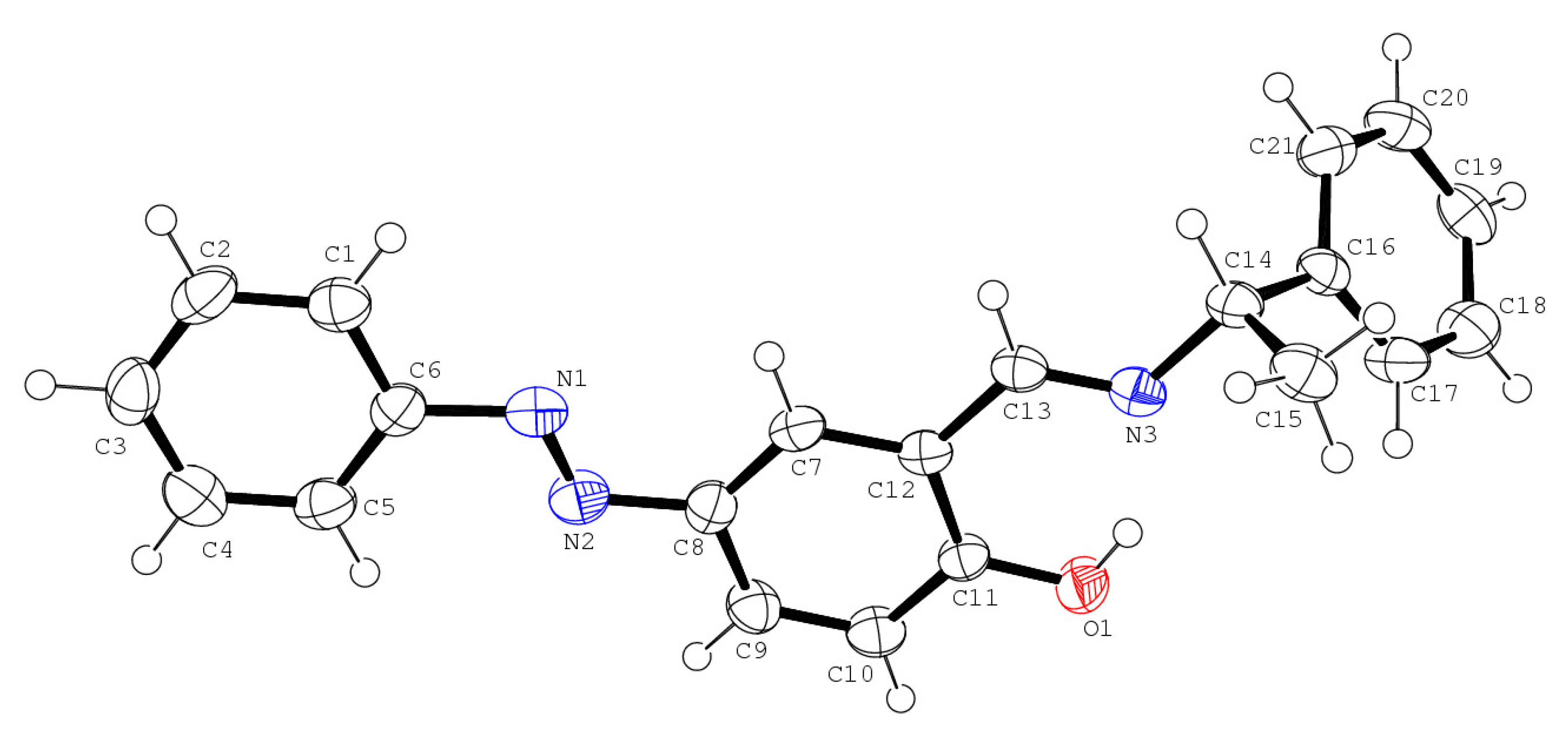
2.4. X-Ray Crystallography

Orange prismatic crystals of 4-phenyldiazenyl-2-[(1-phenylethyl)-iminomethyl]phenol from S-1-phenylethylamine (S-form crystallized in C2 with a = 22.506 (3) Å, b = 5.9736 (9) Å, c = 13.435 (2) Å, β = 106.461 (2)°, V = 1732.2 (4) Å3) or racemic 1-phenylethylamine (the picked-up crystal was R-form crystallized in C2 with a = 22.460 (7) Å, b = 5.9618 (19) Å, c = 13.478 (4) Å, β = 106.301 (4)°, V = 1732.2 (9) Å3) were glued to the top of a glass fiber rod. We coated the rod with a thin layer of epoxy resin. Intensity diffraction data were collected on a Bruker APEX2 CCD diffractometer (Bruker, Billerica, MA, USA) with graphite-monochromated Mo–K radiation (λ = 0.7107 Å). Data analysis was carried out with a SAINT program package (Bruker, Billerica, MA, USA). The structures were solved by direct methods with a SHELXS-97 program [14], expanded by Fourier techniques, and refined by the full-matrix least-squares methods based on F2 using the SHELXL-97 program [14]. An empirical absorption correction was applied by the SADABS program (Bruker, Billerica, MA, USA). All non-hydrogen atoms were readily located and refined using anisotropic thermal displacement parameters. All hydrogen atoms were located at geometrically calculated positions and they were refined using riding models. Only selected crystallographic data were reported herein, because they were merely re-determination consequently.

2.5. Computational Methods

Calculations of all complexes were performed using the Gaussian 09W software Revision D.02 (Gaussian, Inc., Wallingford, CT, USA) [15]. All geometries were optimized by using the B3LYP level of theory and SDD as a basis set. We also performed frequency calculations on optimized geometry using the same level of theory and basis set.
A complex and protein docking simulation was carried out using GOLD suite calculation software (ver. 5.5.0) (Cambridge, UK) [16]. From the Protein Data Bank [17], we obtained 1GYC single crystal structure data of laccase from Trametes versicolor and used this to calculate how the complex behaves in the vicinity of the hydrophobic pocket at coordinates close to the T1 site.

3. Results and Discussion

3.1. Ligands Exhibiting Spontaneous Resolution

The chiral photochromic Schiff base compound 4-phenyldiazenyl-2-[(1-phenylethyl)-iminomethyl]phenol (C21H19N3O) was synthesized from {racmic-1-phenylethylamine} or {(S)-1-phenylethylamine} and the salicylaldehyde of an azobenzene derivative. The molecule corresponds to the S-enantiomer of the previously reported {(R)-1-phenylethylamine} and the C=N and N–C bond distances are 1.278(2) and 1.475(2) Å, respectively (Figure 2). There are two important intramolecular interactions in the crystal. The diazenyl group adopts a cis form with an N–N distance of 1.243(2) Å. On the other hand, racemic-1-phenylethylamine yields the R-enantiomer [12] (re-determination of reported structure) crystals by spontaneous resolution, where the C=N and N–C bond distances are 1.272(3) and 1.477(3) Å, respectively. An intramolecular O–H…N hydrogen bond occurs. The diazenyl group adopts a trans form with an N=N distance of 1.237(3) Å. As shown in Figure 3, characteristic intermolecular interactions, such hydrogen bonds and CH–pi interactions, were observed in the crystal structures.
Optical isomers of chiral or racemic 1-phenylethylamine derivatives may be some of the basic units to obtain enantiomers or diastereomers of crystalline compounds [18], though it is sometimes difficult to compare all R, S, and racemic derivatives. Indeed, spontaneous resolution is one of the well-known phenomena associated with chirality, even for catalytic compounds [19]. We aimed to obtain diastereomers or racemic crystals of previously reported chiral derivatives, and we employed the racemic precursor for this. Of course, structural features of the R-form from racemic amine were trivial because they were similar to previously reported ones prepared from pure R-form of amine.

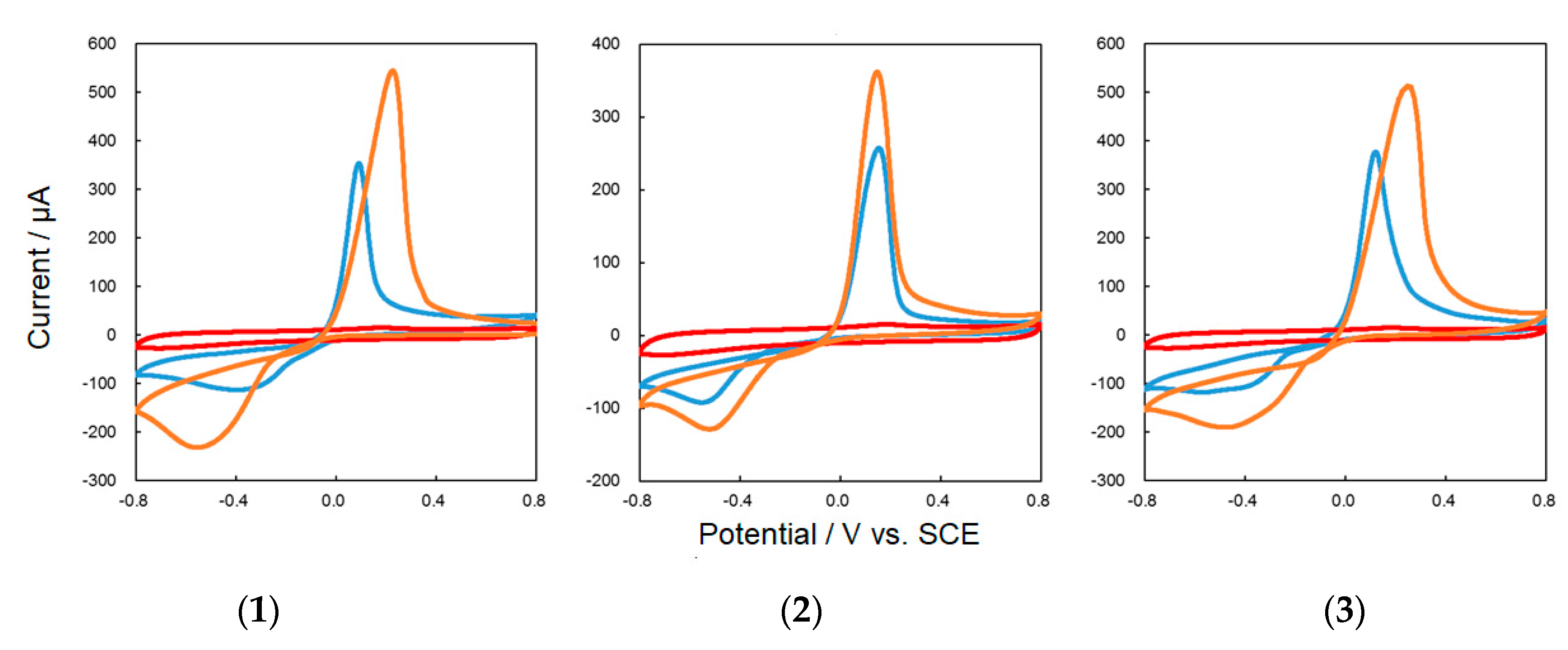
3.2. Complexes of Enantiomers and Diastremers

Crystals 13 were prepared by using 4-phenyldiazenyl-2-[(R)-(1-phenylethyl)-iminomethyl]phenol, 4-phenyldiazenyl-2-[(S)-(1-phenylethyl)-iminomethyl]phenol and racemic 4-phenyldiazenyl-2-[(1-phenylethyl)-iminomethyl]phenol (prepared from racemic 1-phenylethylamine) and confirmed by means of CD spectra. That 1 and 2 are symmetric, and 3 is absent, suggests formation of the meso complex from racemic 4-phenyldiazenyl-2-[(1-phenylethyl)-iminomethyl]phenol in solution). Unfortunately, single crystals of all 13 could not be obtained; hence, a structural discussion was carried out using density functional theory (DFT) optimized structures, which was confirmed by a qualitatively reasonable comparison of simulated and experimental UV–vis and CD spectra (not shown) of the related compounds [20]. Cis-trans photoisomerization of the azo-moiety was also confirmed for 13 by alternate irradiation of UV and visible light for 3 min [21].
Changes in the electrochemical properties of 13 in an isotropic media by linearly polarized UV light were examined by CV as follows. The working electrode was a glassy carbon electrode, the reference electrode was an Ag/AgCl electrode, and the counter electrode was a platinum electrode. The buffer solution was a 200 mM acetate buffer solution at pH 5.0, and the scan rate was measured at 0.05 V/s. Complexes 13 (5 mg) were dissolved in 2 mL of acetone to prepare solutions 13, and solutions 13 (40 μL) were applied to the electrode. To prevent the electrode material from eluting into the buffer solution, 20 μL of Nafion® was added after drying. The electrode material was irradiated with UV light and linearly polarized UV light for 3 min, and the CV spectra were measured under an oxygen atmosphere. Potential shifts from about −0.4 to −0.2 V and about −0.5 to −0.3 V were also observed for 1 and 2, respectively, similar to that of 3 (Figure 4), because of cis-trans photoisomerization of the azo-moiety. This feature suggested that 13 can act as mediators for laccase as cis-form but not as trans-form.

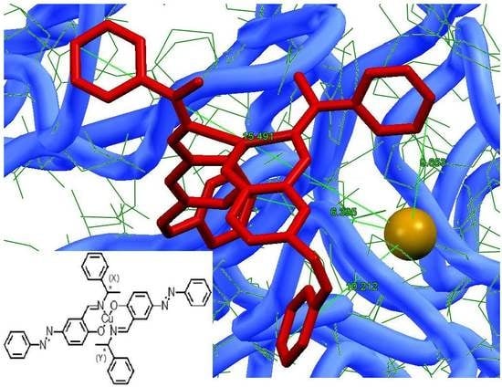
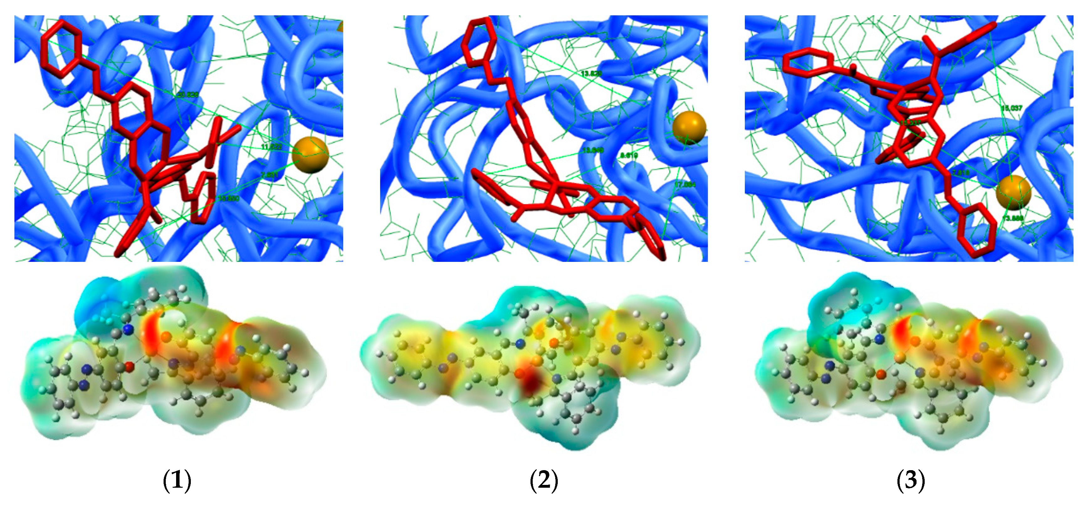
3.3. Hybrid Composites of Complexes in Laccase

The polarized UV–vis spectra measurement results showed that induced optical anisotropy was increased in systems 13 in laccase (anisotropic media). Hybrid materials 13 were prepared by mixing 0.5 mL of acetone solution with 13 (0.0019 g in 5 mL acetone) and laccase (0.0152 g in 2 mL Tris buffer) dropwise onto the PMMA film and drying at room temperature for 2 days. Docking of 13 in laccase was also confirmed with the quenching of the fluorescence intensity of laccase (λem = 350 nm, λex = 270 nm). For the evaluation of the Weigert effect, we employed two parameters (R and S) to determine the degree of photo-induced optical anisotropy [22,23]:
R = A 0 A 90 ;
S = A 0 A 90 A 0 + A 90 ,
where A90 and A0 denote the absorbance measured perpendicular and parallel to the direction of electric vector of irradiation polarized light with the polarizer. For both polarized UV–vis spectra, complete isotropic systems of S = 0 and R = 1 and both S and R parameters were changed as the dichroism by alignment increased. As for the intense π–π* bands around 320 nm, the S and R parameters were 0.8530 and −0.0515 for 1 and laccase, 0.8559 and −0.0545 for 2 and laccase, and 0.8403 and −0.0562 for 3 (Figure 5) and laccase, respectively. The assignments of the most intense peaks were HOMO-3 to LUMO+1 (HOMO-2 to LUMO), HOMO to LUMO+2 (HOMO-2 to LUMO+1), and HOMO-8 to LUMO (HOMO-1 to LUMO+2) for the trans-forms (cis-forms) of 13, respectively. Therefore, it is confirmed that the azobenzene derivative chiral Schiff base Cu(II) complex is photo-aligned in laccase. A control test for pure laccase cast films afforded R = 0.9871 and S = −0.0043 without radiation damage by UV light.
As a preliminary result of the CV measurement in the hybrid system of each complex and laccase (not shown), it was confirmed that the oxygen reduction current value increased compared with the simple substance of trans-form complexes (showing similar behavior to Figure 6) and laccase (whose currents at −0.7 V and +0.2 V were increased under an oxygen rather than a nitrogen atmosphere) alone. Thus, all complexes fully fulfilled their functions as mediators for laccase under oxygen [11].
Besides the addition of the complexes as mediators, and as a result of CV measurements before and after UV light irradiation, the reduction potential was possibly positively shifted due to cis-trans photoisomerization as well as molecular orientation. To resolve both effects, we compared natural (non-polarized) UV light and linearly polarized UV light irradiation (Figure 7). For all complexes, natural UV light irradiation resulted in a positive shift in the reduction potential, which could be attributed to photoisomerization of complexes to cis-form. On the other hand, linearly polarized UV light irradiation resulted in not only a potential shift but also an increased current for oxygen reduction. In detail, a slight increase in current was observed for 1 and 2 in laccase, while a considerable increase in current was observed for 3 in laccase. Thus, it is considered that the oxygen reduction reaction of laccase was further promoted by the molecular orientation of the appropriate stereochemical molecules [9,10].
Since laccase supplies electrons from the T1 copper site and oxygen is reduced by T2/T3 copper sites, it is desirable that the ligand complex intervenes at the electrode–T1 copper site and functions as an electron mediator. Therefore, a calculation was done to join the coordinates (x, y, z) = (13, 25, 38) such that they entered the hydrophobic region pocket including the T1 copper site. As for the trans-forms of 13, docking scores by GOLD were 55.6926, 54.1818, and 58.6724 with the distance of the Cu(II) of 13 to the T1 site being 9.913, 10.448, and 10.918 Å, respectively (Figure 8). On the other hand, assuming UV light irradiation of the cis-forms of 13, docking scores by GOLD were 44.2980, 42.4267, and 65.4168 with distances of the Cu(II) of 13 to the T1 site of 9.990, 10.030, and 10.9898 Å, respectively (Figure 9).
Low docking scores were obtained for the cis-forms of enantiomers 1 and 2 in laccase, which suggested enlarged molecular orientation distances between the Cu(II) in complexes and the T1 site without keeping suitable docking features. However, for cis-forms of diastereomer 3 in laccase, suitable docking features were kept after photoisomerization and molecular orientation, which resulted in an increase in the current values of CV.

4. Conclusions

In summary, as a ligand, only racemic 4-phenyldiazenyl-2-[(1-phenylethyl)-iminomethyl]phenol, exhibited spontaneous resolution. A characteristic hydrogen bond that could affect chiral molecular recognition was identified in the crystals of spontaneous resolution. Using this ligand, enantiomer or diastereomer Cu(II) complexes were obtained and were effective for the chiral molecular recognition of laccase, as were their cis-trans stereoisomers. The cis-form of 3 exhibited the best docking score, which corresponded to the shortest Cu(II)–T1 side distance and increased the current of CV for 3 under an oxygen atmosphere. Additionally, 3 showed a change in redox potential due to the molecular orientation accompanying cis-trans photoisomerization, which was separately confirmed for the first time by using polarized and natural UV light. Thus, it can be expected that 3 exhibits docking advantages for the oxygen reduction reaction of laccase by proving optical orientation control, thereby affecting the improvement of electron transfer toward laccase. In the application to cathode material as an enzyme type biofuel cell, this concept of photo-tunable mediators may be a promising new strategy for molecular design. Further study of this method and the analogous materials is currently in progress.

Author Contributions

Conceptualization, T.A.; Experiments, F.K., J.-Y.K.; X-ray crystallography, S.Y. (Shiomi Yagi); Computation, S.Y. (Shu Yamzaki); Writing, F.K., T.H., T.A.

Funding

This research received no external funding.

Conflicts of Interest

The authors declare no conflict of interest.

References

  1. Mano, N.; de Poulpiquet, A. O2 Reduction in Enzymatic Biofuel Cells. Chem. Rev. 2018, 118, 2392–2468. [Google Scholar] [CrossRef] [PubMed]
  2. Goff, A.; Holzinger, M.; Cosnier, S. Recent progress in oxygen-reducing laccase biocathodes for enzymatic biofuel cells. Cell. Mol. Life Sci. 2015, 72, 941–952. [Google Scholar] [CrossRef] [PubMed]
  3. Quintanar, L.; Stoj, C.; Taylor, A.B.; Hart, P.J.; Kosman, D.J.; Solomon, E.I. Shall We Dance? How A Multicopper Oxidase Chooses Its Electron Transfer Partner. Chem. Rev. 2007, 40, 445–452. [Google Scholar]
  4. Moehlenbrock, M.J.; Minteer, S.D. Extended lifetime biofuel cells. Chem. Soc. Rev. 2008, 37, 1188–1196. [Google Scholar] [CrossRef] [PubMed]
  5. Sakai, H.; MIta, H.; Sugiyama, T.; Tokita, Y.; Shirai, S.; Kano, K. Construction of a Multi-stacked Sheet-type Enzymatic Biofuel Cell. Electrochemistry 2014, 82, 156–161. [Google Scholar] [CrossRef] [Green Version]
  6. Solomon, E.I.; Szilagyi, R.K.; DeBeer George, S.; Basumallick, L. Electronic structures of metal sites in proteins and models: Contributions to function in blue copper proteins. Chem. Rev. 2004, 104, 419–458. [Google Scholar] [CrossRef] [PubMed]
  7. Kurosawa, Y.; Tsuda, E.; Takase, M.; Yoshida, N.; Takeuchi, Y.; Mitsumoto, Y.; Akitsu, T. Spectroscopic and Electrochemical Studies on Metalloprotein (Laccase) and Cu(II) Complex Mediators As Model Systems for Biofuel Cell Cathodes. In Threonine: Food Sources, Functions and Health Benefits; Nova Science Publishers, Inc.: Hauppauge, NY, USA, 2015; Chapter 4; pp. 73–86. [Google Scholar]
  8. Takeuchi, Y.; Akitsu, T. Anthraquinone Derivative Chiral Schiff Base Copper(II) Complexes for Enzyme Type Bio-Fuel Cell Mediators. J. Electr. Eng. 2016, 4, 189–195. [Google Scholar] [CrossRef]
  9. Takeuchi, Y.; Sunaga, N.; Akitsu, T. Anthraquinone and L-amino Acid Derivatives Schiff Base Cu(II) Complexes as a Mediator between Cathode of Biofuel Cell and Oxygen-reducing Laccase. Trend Green Chem. 2017, 3, 3. [Google Scholar] [CrossRef]
  10. Mitsumoto, Y.; Sunaga, N.; Akitsu, T. Polarized light induced molecular orientation in laccase for chiral azosalen Mn(II), Co(II), Ni(II), Cu(II), Zn(II) mediators toward application for biofuel cell. SciFed J. Chem. Res. 2017, 1, 1. [Google Scholar]
  11. Sano, A.; Yagi, S.; Haraguchi, T.; Akitsu, T. Synthesis of Mn (II) and, Cu (II) complexes including azobenzene and its application to mediators of laccase for biofuel cells. J. Indian Chem. Soc. 2018, 95, 487–494. [Google Scholar]
  12. Aritake, Y.; Watanabe, Y.; Akitsu, T. Chiral photochromic Schiff base: 4-phenylazo-2-[[(R)-(1-phenylethyl)imino]methyl]phenol. Acta Crystallogr. E 2010, 66, o749. [Google Scholar] [CrossRef] [PubMed]
  13. Kominato, C.; Akitsu, T. Photoinduced molecular orientation of catalytic-like chiral azo-Schiff base complexes in PMMA or laccase matrices. Lett. Appl. NanoBioSci. 2015, 2, 264–270. [Google Scholar]
  14. Sheldrick, G.M. A short history of SHELX. Acta Crystallogr. Sect. A 2008, 64, 112–122. [Google Scholar] [CrossRef] [PubMed]
  15. Frisch, M.J.; Trucks, G.W.; Schlegel, H.B.; Scuseria, G.E.; Robb, M.A.; Cheeseman, J.R.; Scalmani, G.; Barone, V.; Mennucci, B.; Petersson, G.A.; et al. Gaussian 09, Revision D.01; Gaussian, Inc.: Wallingford, CT, USA, 2009. [Google Scholar]
  16. CCDC CSD. Available online: https://www.ccdc.cam.ac.uk/solutions/csd-discovery/components/Gold/ (accessed on 24 April 2019).
  17. PDB. Available online: http://www.rcsb.org/ (accessed on 24 April 2019).
  18. Akitsu, T.; Komorita, S.; Tamura, H. Diastereomers of copper(II) complexes exhibiting difference in coordination geometry by R- or S-1-phenylethylamine lignads in the solid state and structural conversion of crystals into solutions. Inorg. Chim. Acta 2003, 348, 25–32. [Google Scholar] [CrossRef]
  19. Xia, Z.; Jing, X.; He, C.; Wang, X.; Duan, C. Coordinative Alignment of Chiral Molecules to Control over the Chirality Transfer in Spontaneous Resolution and Asymmetric Catalysis. Sci. Rep. 2017, 7, 15418. [Google Scholar] [CrossRef] [PubMed] [Green Version]
  20. Aritake, Y.; Takanashi, T.; Yamazaki, A.; Akitsu, T. Polarized spectroscopy and hybrid materials of chiral Schiff base Ni(II), Cu(II), Zn(II) complexes with included or separated azo-groups. Polyhedron 2011, 30, 886–894. [Google Scholar] [CrossRef]
  21. Akitsu, T. Photofunctional supramolecular solution systems of chiral Schiff base nickel(II), copper(II), and zinc(II) complexes and photochromic azobenzenes. Polyhedron 2007, 26, 2527–2535. [Google Scholar] [CrossRef]
  22. Yamazaki, A.; Akitsu, T. Polarized spectroscopy and polarized UV light-induced molecular orientation of chiral diphenyl Schiff base Ni(II) and Cu(II) complexes and azobenzene in a PMMA film. RSC Adv. 2012, 2, 2975–2980. [Google Scholar] [CrossRef]
  23. Ito, M.; Akitsu, T.; Palafox, M.A. Theoretical interpretation of polarized light-induced supramolecular orientation on the basis of normal mode analysis of azobenzene as hybrid materials in PMMA with chiral Schiff base Ni(II), Cu(II), and Zn(II) complexes. J. Appl. Solut. Chem. Model. 2016, 5, 30–47. [Google Scholar]
Figure 1. Structures of Cu(II) complexes (X = R, Y = R for 1; X = S, Y = S for 2; X = R, Y = S for 3).
Symmetry 11 00666 g001
Figure 2. Molecular structures of 4-phenyldiazenyl-2-[(S)-(1-phenylethyl)-iminomethyl]phenol (newly analyzed enantiomer crystal structure).
Symmetry 11 00666 g002
Figure 3. Crystal structures of 4-phenyldiazenyl-2-[(R)-(1-phenylethyl)-iminomethyl]phenol prepared from racemic 1-phenylethylamine showing characteristic intermolecular interactions.
Symmetry 11 00666 g003
Figure 4. CV for 3 before (blue; trans-form) and after (orange; cis-form) linearly polarized UV light irradiation for 3 min.
Symmetry 11 00666 g004
Figure 5. (Left) Polarized UV–vis spectra of 3 and laccase, and (right) angular dependence of absorbance of these spectra at 320 nm (π–π*) before (blue; random trans-form) and after (orange; orientated cis-form) linearly polarized UV light irradiation for 3 min, as determined by the polarizer.
Symmetry 11 00666 g005
Figure 6. CV for 13 (blue; trans-form), laccase (red), and a hybrid system of 13 in laccase (orange) under an oxygen atmosphere.
Symmetry 11 00666 g006
Figure 7. CV for 3 and laccase before (blue; trans-form) and after (orange; cis-form) (above) exposure to natural UV light (below) and linearly polarized UV light irradiation for 3 min.
Symmetry 11 00666 g007
Figure 8. (Above) Docking simulation of the trans-forms of 13 in laccase. (Below) DFT optimized structures with charge distributions of the trans-forms of 13 with dipole moments of 8.3449, 4.9374, and 8.4552 D, respectively.
Symmetry 11 00666 g008
Figure 9. (Above) Docking simulation of the cis-forms of 13 in laccase. (Below) DFT optimized structures with the charge distributions of the cis-forms of 13 with dipole moments of 8.4361, 4.3523, and 11.7205 D, respectively.
Symmetry 11 00666 g009

