Article

Effect of Carbon Sources in Carotenoid Production from Haloarcula sp. M1, Halolamina sp. M3 and Halorubrum sp. M5, Halophilic Archaea Isolated from Sonora Saltern, Mexico

by
Ana Sofía Vázquez-Madrigal
1,
Alejandra Barbachano-Torres
1,
Melchor Arellano-Plaza
1,
Manuel Reinhart Kirchmayr
1,
Ilaria Finore
2,
Annarita Poli
2,
Barbara Nicolaus
2,
Susana De la Torre Zavala
3 and
Rosa María Camacho-Ruiz
1,*
1
Biotecnología Industrial, Centro de Investigación y Asistencia en Tecnología y Diseño del Estado de Jalisco, Guadalajara A.C. 44270, Mexico
2
Istituto di Chimica Biomolecolare, Consiglio Nazionale delle Ricerche, Via Campi Flegrei 34, 80078 Pozzuoli, Italy
3
Facultad de Ciencias Biológicas, Instituto de Biotecnología, Universidad Autónoma de Nuevo León, Av. Pedro de Alba S/N Ciudad Universitaria, San Nicolás de los Garza C.P. 66455, Mexico
*
Author to whom correspondence should be addressed.
Submission received: 21 April 2021 / Revised: 29 April 2021 / Accepted: 11 May 2021 / Published: 20 May 2021

Abstract

:
The isolation and molecular and chemo-taxonomic identification of seventeen halophilic archaea from the Santa Bárbara saltern, Sonora, México, were performed. Eight strains were selected based on pigmentation. Molecular identification revealed that the strains belonged to the Haloarcula, Halolamina and Halorubrum genera. Neutral lipids (quinones) were identified in all strains. Glycolipid S-DGD was found only in Halolamina sp. strain M3; polar phospholipids 2,3-O-phytanyl-sn-glycerol-1-phosphoryl-3-sn-glycerol (PG), 2,3-di-O-phytanyl-sn-glycero-1-phospho-3′-sn-glycerol-1′-methyl phosphate (PGP-Me) and sodium salt 1-(3-sn-phosphatidyl)-rac-glycerol were found in all the strains; and one unidentified glyco-phospholipid in strains M1, M3 and M4. Strains M1, M3 and M5 were selected for further studies based on carotenoid production. The effect of glucose and succinic and glutamic acid on carotenoid production was assessed. In particular, carotenoid production and growth significantly improved in the presence of glucose in strains Haloarcula sp. M1 and Halorubrum sp. M5 but not in Halolamina sp. M3. Glutamic and succinic acid had no effect on carotenoid production, and even was negative for Halorubrum sp. M5. Growth was increased by glutamic and succinic acid on Haloarcula sp. M1 but not in the other strains. This work describes for first time the presence of halophilic archaea in the Santa Bárbara saltern and highlights the differences in the effect of carbon sources on the growth and carotenoid production of haloarchaea.

1. Introduction

Halophilic archaea are mostly aerobic, Gram-negative, chemoorganotrophic microorganisms that grow from 1M NaCl to the point of saturation in a temperature range from 4 to 68 °C [1,2]. They produce carotenoid pigments synthesized through the mevalonate pathway in order to tolerate such extreme conditions [3]. Carotenoids are important bioactive compounds with antioxidant and free radical scavenging properties [4]. They have a great economic impact, especially in the market that seeks to replace synthetic pigments with those of natural origin. Bacterioruberin, the main carotenoid pigment in halophilic archaea, is a 50-carbon structure with 13 double bonds, involved in the protection of oxidative stress, solar radiation [5] and influences the fluidity of cell membrane [6]. Bacterioruberin is not exclusive to the archaea domain, as some bacteria (Rubrobacter radiotolerans, Kocuria rosea) have been reported as bacterioruberin producers [5]. In particular, this pigment has a great industrial potential due to its high antioxidant capacity, which is considerably greater that β-carotene, astaxanthin and other carotenoids [7,8]. In addition, bacterioruberin already has been tested in animal cells, exhibiting no toxicity in human monocytes and improving the viability in ram sperm [8].
Haloarchaea are the main microorganisms present in hypersaline environments. These habitats contain at least three times the sea salt concentration [9]. The Santa Bárbara saltern (Figure 1) is a salt-producing area in a non-protected zone, classified as a thalassohaline environment (NaCl as the main salt), salted wetland, with a permanent connection to the sea located in the south coast of Sonora, México. Santa Bárbara houses a great diversity of species of halophilic plants, birds and mammals [10], and so far, the evidence of microorganisms in Sonora State include halophilic bacteria [11] and methanogenic archaea [12]; however, until this work, the halophilic archaea phenotype was still unknown in this area.
Archaea species populate extreme environments and their challenging identification and characterization make them organisms in need of constant elucidation. The discovery of new halophilic species and the use of different identification tools provokes a continuously changing classification and promotes the emergence of new taxonomic groups. For example, the Halorubrum genus, the largest in the class Halobacteria, has been rearranged several times since it was proposed in 1995 [13].
It has been shown that some carbon sources, such as carbohydrates, amino acids and other compounds, could stimulate microbial growth; nevertheless, information on how these carbon sources affect carotenoid production is scarce. The complexity of the substrate and the metabolic profile of the organism are important factors, as the latter has a high variability in archaea species, sometimes even in species within the same genera [14].
Although complete metabolism and the origin of metabolic routes in haloarchaea are still unknown, it is clear that they have acquired genes from bacterial pathways such as mevalonate through horizontal gene transfer [15]. The origin and understanding of this pathway have not been fully decrypted due to the erratic genetic pattern that codes for the enzymes of this route [16]; the carotenoid pigments in these organisms are synthetized by this pathway, which starts with acetyl-CoA, and starting from this point, it should be verify which compounds could stimulate pigmentation. Some halophilic archaea, such as Halobacterium, contain enzymes that allow some amino acids, such as glutamic acid, to be directly incorporated into the tricarboxylic acid cycle (TCA) and obtain greater availability of acetyl-CoA that could be used for the mevalonate pathway [17]. Succinic acid also has been used to improve carotenoid production in the yeast Xantophyllomyces dendrohous, entering directly into the TCA cycle and increasing the activity of the enzymes [18].
Another way to obtain acetyl-CoA in species of archaea capable of utilizing carbohydrates is from glucose. Glucose is converted to pyruvate in four steps via the Entner–Doudoroff (ED) pathway, and a carboxyl group is removed and coenzyme A is attached to form acetyl-CoA [19]. Gochnauer et al. used various concentrations of glucose, stimulating the growth and the pigmentation process in Halobacterium, and verified that this monosaccharide acted optimally at 2% [20,21].
Thus, the aims of this work are to provide information about the halophilic microorganisms that inhabit Santa Bárbara saltern, search for carotenoid producers, perform their chemotaxonomic and molecular identification and to assess the effect of glucose, succinic and glutamic acid on the production of carotenoid pigments in some of the isolated species.

2. Materials and Methods

2.1. Organisms and Culture Media

The microorganisms used in this study were isolated from saline mud and salt crust samples collected in November 2017. Samples were collected from 10 different points up to 20 cm deep within an 8.2 km2 area of the Santa Bárbara saltern, located in the Municipality of Huatabampo, Sonora, México. Soil temperatures in Santa Bárbara range from 35 to 36 °C during the day and has a soil pH from 7.7 to 8.6. A map of the sampling sites is showed in Figure 1. The environmental samples were stored in refrigerated sterile plastic bags until their analysis.
The samples (1 g) were diluted in 9 mL of 20% sterile saline solution and used to inoculate a solid ATCC2185 medium, pH 8, containing (g/L) MgSO4 × 7H2O 20, sodium citrate 3, KCl 2, tryptone 5, yeast extract 3, NaCl 233.3, agar 10, and trace elements solution 100 µL. Trace element solution was prepared containing (g/L) ZnSO4 × H2O 1.32 g, MgSO4 × H2O 0.34 g, Fe (NH4)2SO4 × 6H2O 0.82 g, and CuSO4 × 5H2O 0.14 g. This culture medium is generally employed for the growth of halophilic archaea [22]. After 1 week of incubation at 37 °C in an aerobic incubation stove with no artificial light, the presence of colonies with a different morphology was revealed and they were purified by restreaking on the same solid medium. The isolates were stained with methyl violet for microscopic observations and cryopreserved at −80 °C in 75% glycerol. All reagents were purchased from Sigma-Aldrich, St. Louis, MO, USA.

2.2. DNA Extraction and Phylogenetic Analysis

For the DNA analysis, the cells of all isolates were collected after 4 days of incubation at a temperature of 37 °C via centrifugation at 5000× g for 15 min.
The cells were subjected to total genomic DNA extraction and purification by using the Gen EluteTM Plant Genomic DNA Miniprep Kit Protocol (Sigma-Aldrich, USA) according to manufacturer’s specifications, followed by a PCR of the complete 16S rRNA gene using forward D30 (5′-ATTCCGGTTGATCCTGC-3′) and reverse D56 (5′GYTACCTTGTTACGACTT-3′) primers [23] and the Invitrogen Platinum Taq DNA Polymerase (ThermoFisher Scientific, Waltham, MA, USA) protocol. The following conditions were used: initial denaturation at 94 °C for 3 min, followed by 30 cycles of 94 °C for 45 s, 53.5 °C for 45 s and 72 °C for 2 min, and a final extension at 72 °C for 10 min. The strains were cloned using a CloneJet PCR Cloning Kit (ThermoFisher Scientific, MA, USA) into E. coli calcium competent cells. Consensus sequences were generated with the sequenced fragments using software CLC Workbench 8. A BLASTn was performed with sequences from the GenBank 16S database (Bacteria and archaea) [24].
Reference 16S rRNA gene sequences of the closest haloarchaea were selected from the NCBI-GenBank. Sequences were aligned with ClustalW [25] and trimmed using MEGA V 7.0 [26] obtaining a 453 bp sequence alignment. The optimal substitution model was determined with MEGA for the sequence alignment. A calculated T92+1 model [27] was used to reconstruct a phylogenetic tree with a Maximum Likelihood (ML) algorithm in MEGA V 7.0, with 1000 bootstraps.

2.3. Chemo-Taxonomic Identification

The lipid extraction was carried out according to Finore et al. [28] using 2.5 g of freeze-dried cells harvested at the stationary growth phase. Quinones were extracted from freeze-dried cells with n-hexane and were purified by TLC on silica gel (0.25 mm; F254, Merck) and eluted with n-hexane/ethylacetate (96:4, v/v). The purified UV-bands from TLC were then analysed by LC/MS on a reverse-phase RP-18 Lichrospher column eluted with n-hexane/ethylacetate (99:1, v/v) with a flow rate of 1.0 mL min−1 and identified by electrospray ionization (ESI)-MS and 1H-NMR spectrometry. NMR spectra were acquired on a Bruker DPX-300 operating at 300 MHz, using a dual probe. The residual cellular pellet, after n-hexane extraction of freeze-dried cells, was subjected to extraction with CHCl3/CH3OH/H2O (65:25:4, by vol.) for polar lipids recovery. The polar lipid extract was analysed by TLC on silica gel (0.25 mm, F254, Merck) eluted in the first dimension with CHCl3/CH3OH/H2O (65:25:4, by vol.) and in the second dimension with CHCl3/CH3OH/acetic acid/H2O (8:12:15:4, by vol.). All polar lipids were detected by spraying the plates with 0.1% (w/v) Ce(SO4)2 in 1 M H2SO4 or with 3% (w/v) methanolic solution of molybdophosphoric acid followed by heating at 100 °C for 5 min. Phospholipids and aminolipids were detected by spraying TLC plates with the Dittmer-Lester and ninhydrin reagents, respectively, and glycolipids were visualized with α-naphtol [29].

2.4. Carotenoid Production

Flasks of 250 mL volume containing 80 mL of culture medium were inoculated with all isolates and incubated at 37 °C, 200 rpm, pH 8. Aliquots were taken every 24 h for 6 days to measure biomass and carotenoid production. Biomass was quantified using a correlation between the optical density (OD620) and dry weight: biomass g/L = 0.688 × OD620. The pigment extraction was performed using a modified Naziri method [30] that consisted firstly of obtaining the cellular pellets by centrifugation at 4000 rpm, 4 °C for 1 h, and frozen at −20 °C. Then, a solution of acetone/methanol (7:3, v/v) was added to the cellular pellets and the cell membranes were disrupted by vortexing and sonicating. Afterwards, the mixtures were centrifuged at 6000 rpm, 4 °C for 30 min, and the supernatants were collected. Finally, the solvent was evaporated using a SpeedVac concentrator (ThermoFisher Scientific, MA, USA).
The carotenoid quantification was calculated with the Lambert–Beer law as Britton describes it [31], reading absorbance at λ490 nm and using 2660 as the molar attenuation coefficient of bacterioruberin in methanol. The most carotenoid-producing strain of each genus was selected for further studies.
To identify the main pigment, the carotenoid extracts were suspended in acetone and a spectrophotometric scan from λ400 to λ600 nm was carried out in a Biotek Eon Microplate Spectrophotometer (Agilent, Santa Clara, CA, USA).
Experiments were carried out in triplicate.

2.5. Optimization of Carotenoid Production: Effect of Carbon Sources

The most carotenoid-producing strain of each genus was selected for deeper investigation; in particular, the microbial response to carbon source presence was evaluated. For this purpose, three different carbon sources, namely, glucose (10 and 25 g/L), succinic acid (3.5 g/L) or glutamic acid (3 g/L), were added at a time to 40 mL of the culture medium ATTCC2185 at the optimal conditions of each isolate. After regular interval times, growth was monitored for 120 h by measuring the OD620 for biomass calculation according to the equation described above and by extracting the carotenoids as reported. Experiments were carried out in triplicate.

2.6. Statistical Analysis

Statistical analysis was performed using a one-way analysis of variance (ANOVA) to test the significance of the model (S1). Significance was determined at p < 0.05.

3. Results

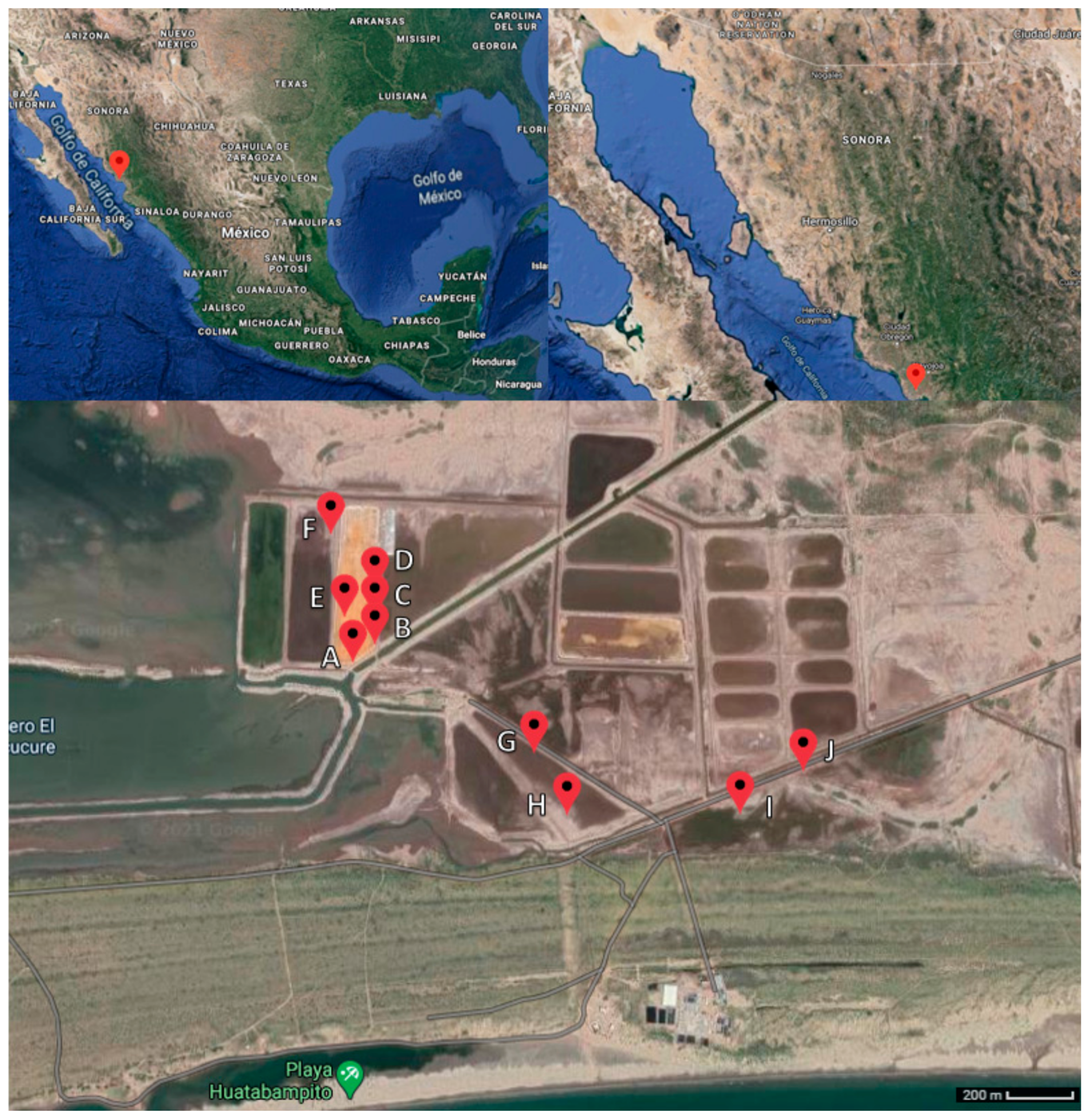
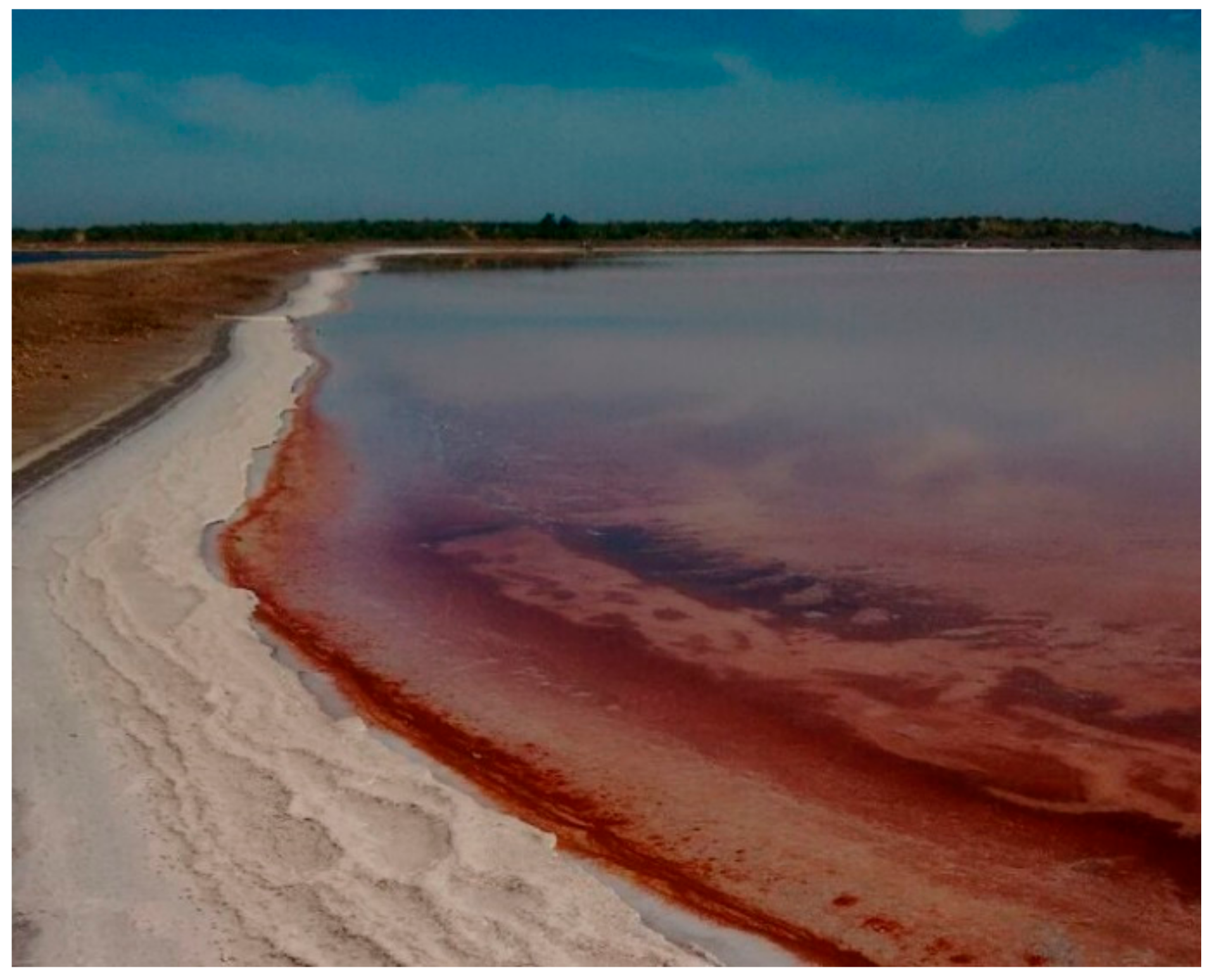
3.1. Sampling and Isolation

Santa Bárbara saltern (Figure 2) is a salt-producing area in a non-protected zone located in the Municipality of Huatabampo, Sonora, México, at 10 m above sea level. It belongs to the physiographic province of the pacific coastal plain. This region is a flat relief next to the ocean with flooded areas parallel to the coast covered by alluvial sediments from the Sierra Madre Occidental [32]. The weather is dry with an average temperature of 23.4 °C, ranging from 0 °C in winter to 49 °C in summer. The annual precipitation is less than 300 mm; the fauna is composed of a variety of endemic birds, reptiles, amphibians, felines and turtles; and the vegetation is mainly xerophytic and halophilic plants [33,34].
Seventeen strains were obtained from the sample isolation and eight of them, the most pigmented, were selected for further investigation. All isolates (Figure 3) are pleomorphic, Gram-negative and orange-red colored.

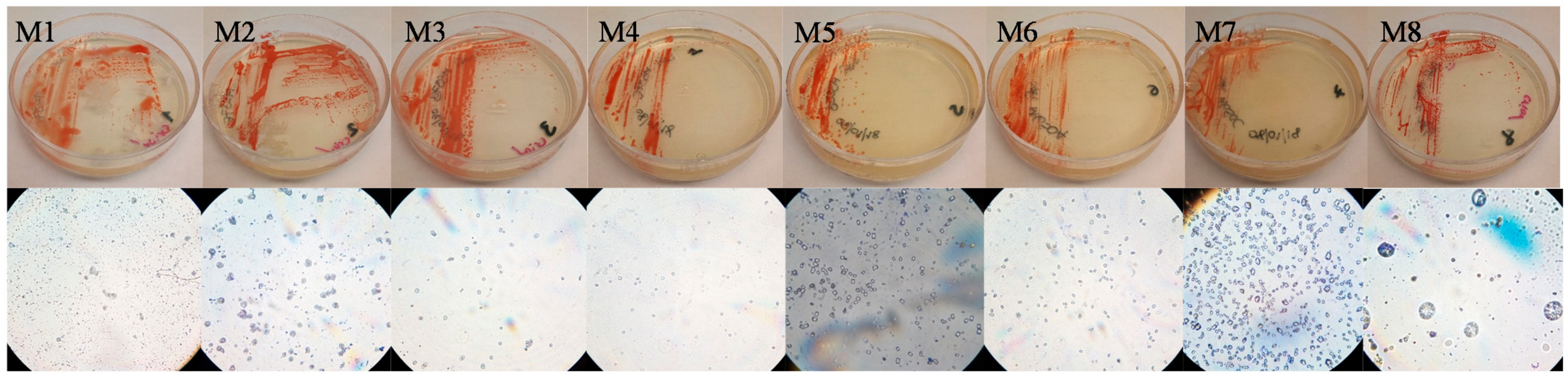
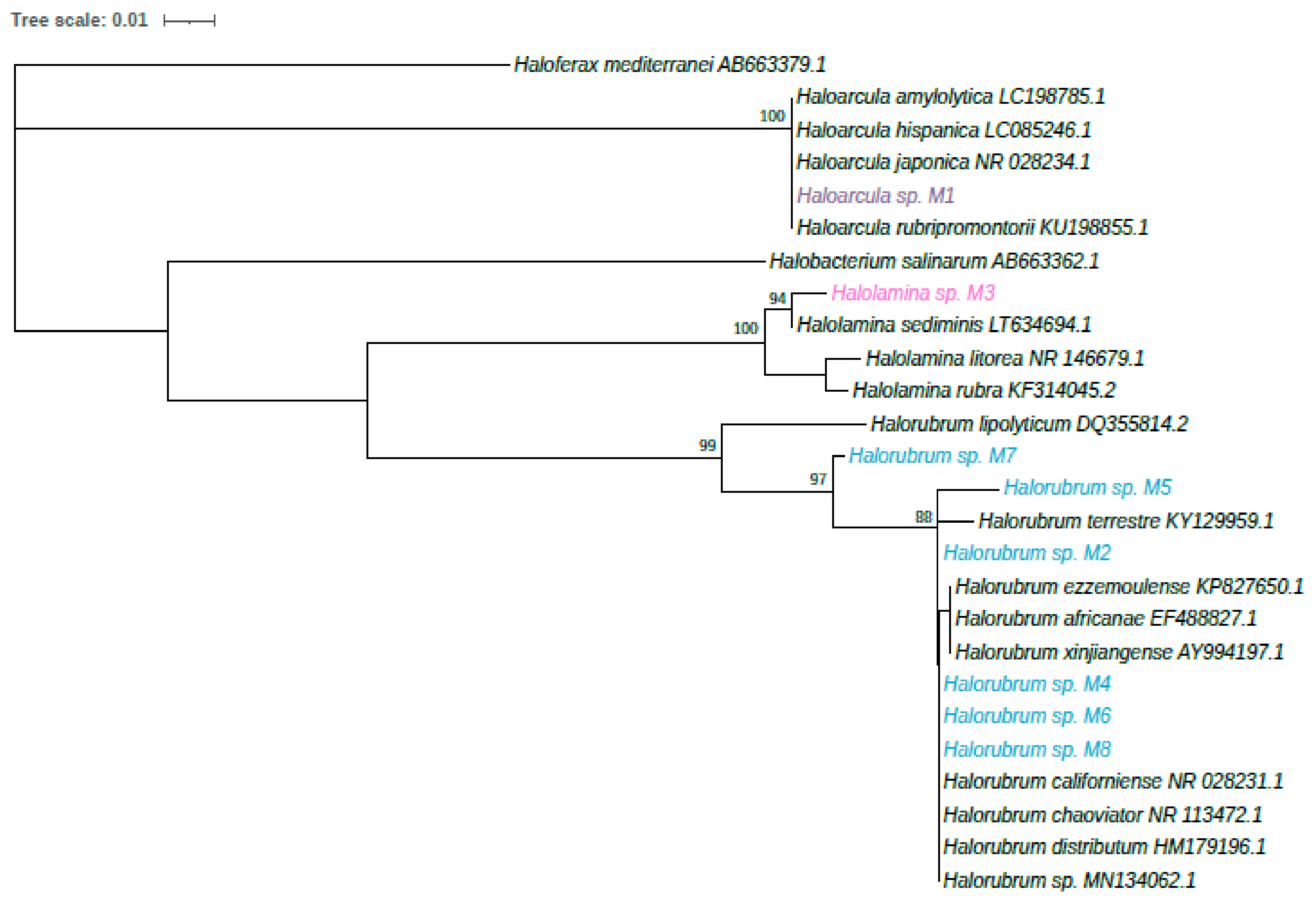
3.2. Identification of Halophilic Archaea Strains

The closest relative of each strain in the database, as found with BLAST, is showed in Table 1. To achieve an unambiguous taxonomic identification of the isolates, a phylogeny was reconstructed using 16S rRNA genes (Figure 4). All isolates belong to three genera Haloarcula, Halorubrum and Halolamina, and will be further identified with the strain code.
Neutral lipid identification (quinones) by LC–mass spectrometry (Table 2) showed menaquinone with eight isoprenoid units (MK-8; 717 g/mol molecular mass) for all strains except strain M5; in addition, strains M2, M3, M4, M6 and M7 exhibited dihydromenaquinones-8 (MK-8 (H2); 719 g/mol molecular mass). Finally, strain M5 showed methylmenaquinone-8 (MMK-8; 733 g/mol molecular mass).
The polar lipid profile of the strains comprised derivatives of the polar phospholipids (PG), in which the hydrocarbon isoprenoid structure is composed by two chains containing 20 carbons (diether C20-C20 moiety) linked by an ether linkage to the glycerol structure. Phosphatidylglycerol phosphate methyl ester (PGP-Me), sodium salt 1-(3-sn-phosphatidyl)-rac-glycerol (PI) and unknown glycolipids (GP) were found in all isolated strains; in addition, derivatives of unidentified glyco-phospholipid were found in strains M1, M3 and M4 and, finally, mannose-6-sulfate(1-2)-glucose glycerol diether (S-DGD) was described for strain M3. Two unidentified aminolipids were detected in all isolated strains.

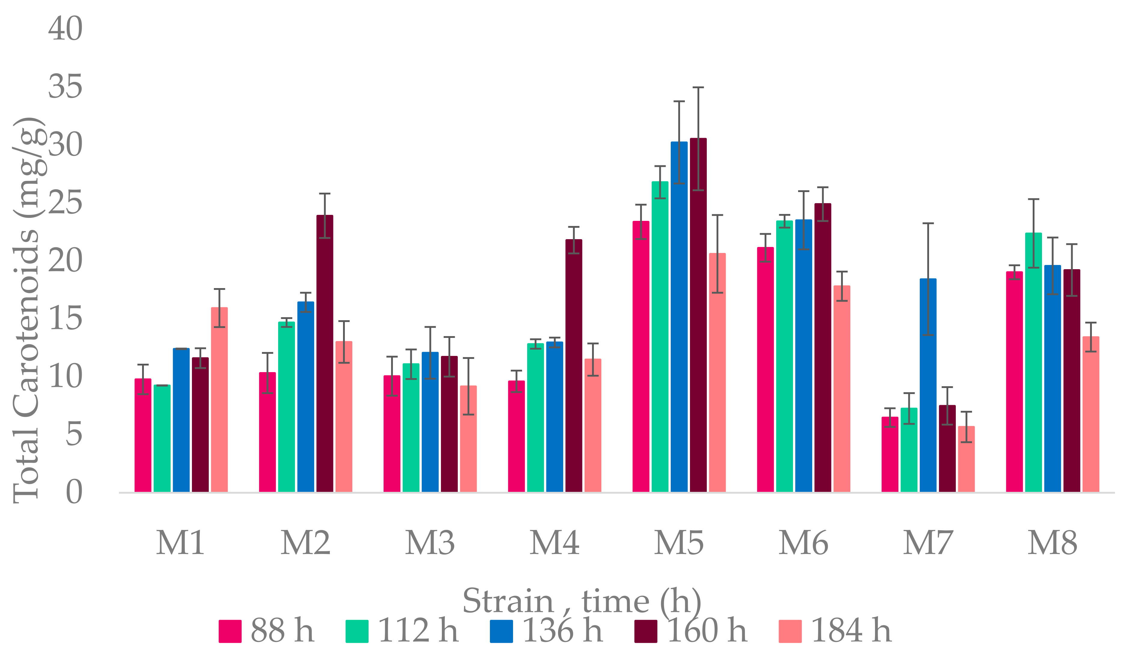
3.3. Carotenoid Producers

All strains were carotenoid producers; the highest carotenoid production started from 136–160 h, at the stationary phase. A graph of the yield of carotenoids per gram of biomass produced by each strain is found in Figure 5.
The highest yield was achieved by the Halorubrum genus, especially in Halorubrum sp. M5 at 160 h with 30.55 mg/g of biomass.

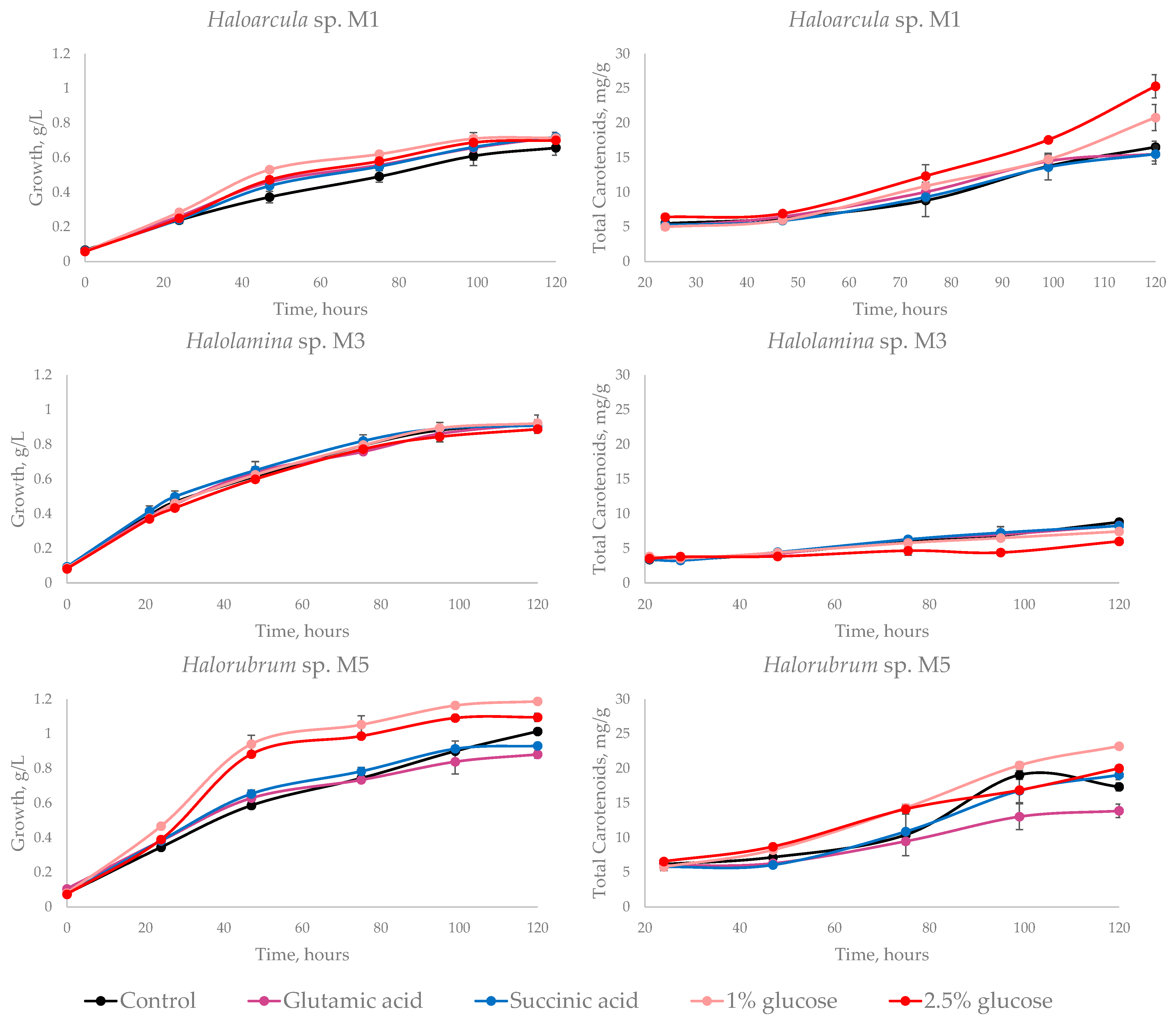
3.3.1. Carbon Source Influence in Carotenoid Production

Statistical analysis showed, in strain Haloarcula sp. M1, significant differences in growth when an extra carbon source was added to the medium with respect to the control medium (p-value = 0.001), but no differences were observed between the carbon source used; whereas, pigment production improved with both glucose concentrations employed, especially at 2.5% (p-value = 0.0000004). Halolamina sp M3 showed no differences comparing to control for growth (p-value = 0.322), and pigmentation was negatively affected when glucose was added to the control medium (p-value = 0.00000003). Glucose also stimulated growth (p-value = 0.0000000000004) and pigmentation (p-value = 0.00000002) in Halorubrum sp. M5, 1% glucose being the carbon source that improved the most pigment production, reaching 23.21 mg/g of biomass at 120 h. In Halorubrum sp. M5, pigment production was affected negatively by glutamic acid. The biomass and carotenoid production of the three selected strains are shown in Figure 6. Detailed statistical analysis is provided in Supplementary Material Figure S1.

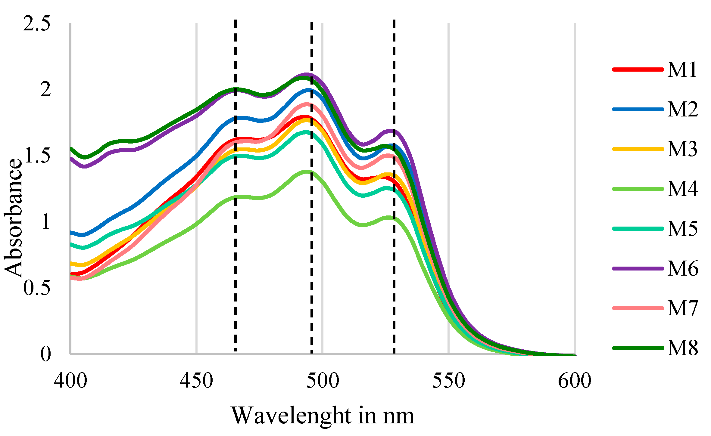
3.3.2. Pigments Identification

The eight strains were able to produce carotenoids as revealed from spectrophotometric investigation. The spectrophotometric scans showed the three characteristic peaks of bacterioruberin in acetone (λ468, 498 and 532 nm) according to Britton’s carotenoid manual [31], which confirms the presence of bacterioruberin as the main pigment in all of the strains. The scan is shown in Figure 7. Although bacterioruberin is the main pigment, there may be other carotenoids in which its spectrum is hidden behind that of bacterioruberin, as chromatographic characterization is necessary to detect all pigments.

4. Discussion

Pigment-producing haloarchaea inhabit high salinity environments worldwide [39]. In this work, we successfully isolated eight haloarchaeal strains from the Santa Bárbara saltern in northwest México, which produce pigments of biotechnological potential. These isolates belong to three genera (Haloarcula, Halolamina and Halorubrum) previously found to be pigmented strains [40,41,42]; however, to our knowledge, this is the first report of an isolate of the genus Halolamina being evaluated on pigment production potential. Unlike other members of the Halobacteriaceae family, Halolamina has been recently isolated and recognized as a novel genus [43]. As observed in Figure 2, phylogenetic reconstruction shows that each isolate might represent different species; nevertheless, a genome-based taxonomy is currently directing research on uncultivated species [44,45] as culturing and sequencing techniques keep expanding the tree of life in the Archaea domain [46].
Menaquinone-8 (MK-8) is commonly found in halophilic archaea; its weight is 717 Da and 719 Da in its hydrogenated form [47]. Several studies report MK-8 in Haloarcula [48,49,50] and in Halorubrum [51,52], but so far, we have not found any reports of menaquinones in Halolamina. The weight 733 Da found in strain Halorubrum sp. M5 corresponds to methyl-menaquinone-8 (MMK-8) also found in Natronobacterium gregoryi [53].
The glycolipid S-DGD could be associated with gas vesicles [54]; it was reported by Cui when proposing the genus Halolamina [43] and it is described in other Halolamina species [41]. In this work, it was found only in Halolamina sp. M3. In the Haloarcula genus it has not been reported yet. PG and PGP-Me were present in all strains; this lipid is related with cytochrome C oxidase and alkalophilic archaea lack this phospholipid [55].
The strains have the capacity to grow at various values of NaCl, pH and all temperature; this may be due to the ever-changing ecosystem from which the archaea were isolated, where temperature and water retention varies depending on the season [10]. This represents a great advantage for industrial culturing, since it reduces incubation and cooling costs.
The maximum carotenoid yield of 30.55 mg/g obtained in Halorubrum sp. M5 is similar than those obtained in optimized conditions by de la Vega [42] in the same genus, species Halorubrum SH1. This strain was selected to investigate the effect of carbon source on pigment production due to its high pigmentation, whereas Haloarcula sp. M1 and Halolamina sp. M3 were chosen for being the only ones of their genus in our study.
Most of the studies regarding haloarchaea pigment production are focused on culture conditions, such as the salt concentration, agitation, light or oxygen content [5,40,42]. However, the research related to the effect of carbon sources in pigment production by haloarchaea is scarce, although glucose and glycerol have been studied on Halobacterium [5,21]. While succinic and glutamic acid had been reported as carbon sources for some archaea, their effect on pigment production has not been elucidated. Regarding the archaeal metabolism, not all haloarchaea function with the same metabolism, as the utilization of metabolic pathways depends not entirely on the genetics but also on ecology.
Glutamic acid assimilation in halophilic archaea has not been fully elucidated. Halobacterium contain enzymes that allow glutamic acid to be directly incorporated into the tricarboxylic acid cycle (TCA) and obtain acetyl-CoA [17]; it can be metabolized by Halobacterium salinarum and Haloarcula marismortui [56,57]. However, other authors report that Haloarcula and Halorubrum are incapable to grow in glutamic acid [14], but Haloarcula metabolizes glutamic acid when it is used as the sole carbon source [58]. Our results indicate that addition of glutamic acid in the control medium did not increase the growth on Halolamina sp. M3, and a negative effect on growth was observed for Halorubrum sp. M5 whereas a slight increment on growth was achieved for Haloarcula sp. M1. Besides, pigment production was not affected in Haloarcula sp. M1 and Halolamina sp. M3 but negatively affected Halorubrum sp. M5. It could mean that glutamic acid affects growth unless it is the only carbon source available; that the ability to metabolize it varies in species within the same genus; and that it does not have a positive effect on pigment production.
The addition of succinic acid in the control medium was proposed on the basis that halophilic archaea contain enzymes that allow amino acids to be directly incorporated into the tricarboxylic acid cycle [17]. The effect of succinic acid in carotenoid production in yeasts is remarkable, as reported for Xantophyllomyces dendrorhous [18]. The effect of succinic acid on the growth of the strains studied was similar to those observed with glutamic acid; however, succinic acid did not increase the pigment production of the three strains studied, although halophilic archaea and yeasts possess similar pathways and enzymes. Ultimately, they are phylogenetically further apart, and more studies about the impact of amino acids on archaeal pigment production are needed.
Glucose metabolism is complex and variable in halophiles; it is degraded via different variants of the ED pathway within them [57]. In the most known way, glucose is converted to pyruvate via ED, a carboxyl group is removed, and coenzyme A is attached to form acetyl-CoA [19]. The effect of glucose on growth and pigmentation was evaluated in Halobacterium by Goshnauer, obtaining inhibition of pigmentation when 4% glucose was used as carbon source, and little effect on growth but considerable improvement on pigmentation with 2% of glucose [21]. Our results suggest that the effect of glucose on pigment production depends on the species. In Haloarcula sp. M1 and Halorubrum sp. M5, pigment production improved when glucose was added, but in Halolamina sp. M3 pigment production decreased. The effect of glucose on growth was evident in Halorubrum sp. M5 and Haloarcula sp. M1 but not in Halolamina sp. M3. Glucose concentration has different effects, depending on the strain. In the case of Haloarcula sp. M1, 2.5% glucose increased pigment production, where as in Halorubrum spp. pigment production increased with 1% glucose. Finally pigment production can be improved using single carbon sources as glucose, but not for all halophilic archaea. A deeper understanding of pigments metabolism in haloarchaea is needed, as well as research on pigment production, combining carbon sources with growth parameters such as oxygen, light or salt concentration.

5. Conclusions

This work represents first evidence of halophilic archaea in the Santa Barbara saltern, exhibiting at least eight species distributed across three different genera of halophilic archaea, namely, Haloarcula, Halolamina and Halorubrum, with the possibility that Halolamina sp. strain M3 could be a new species. All eight strains are carotenoid producers with bacterioruberin as the main pigment. There is a relationship between growth and pigment production, although the latter can also be influenced by stress factors and components in the culture media. The addition of glucose to the ATCC2185 media increases considerably the production of carotenoids and growth in Haloarcula sp. M1 and Halorubrum sp. M5 but not in Halolamina sp. M3. This paper highlights the diversity of carbohydrate metabolism in haloarchaea and their effect on carotenoid production.

Supplementary Materials

The following are available online at https://0-www-mdpi-com.brum.beds.ac.uk/article/10.3390/microorganisms9051096/s1, Figure S1: Detailed statistical analysis of carotenoid effect on pigment production at 120 h of culture.

Author Contributions

Conceptualization, R.M.C.-R.; data curation, A.S.V.-M.; formal analysis, I.F., S.D.l.T.Z. and R.M.C.-R.; funding acquisition, R.M.C.-R.; investigation, A.S.V.-M.; methodology, A.B.-T., M.A.-P., M.R.K., I.F., A.P., S.D.l.T.Z. and R.M.C.-R.; resources, A.P. and B.N.; supervision, A.B.-T., M.A.-P., M.R.K., I.F., A.P. and S.D.l.T.Z.; validation, B.N.; writing—original draft, A.S.V.-M.; writing—review and editing, M.R.K., I.F., A.P., S.D.l.T.Z. and R.M.C.-R. All authors have read and agreed to the published version of the manuscript.

Funding

This research was funded by CIATEJ (Centro de Investigación y Desarrollo en Tecnología y Diseño del Estado de Jalisco).

Institutional Review Board Statement

Not applicable.

Informed Consent Statement

Not applicable.

Data Availability Statement

The data presented in this study are available on request from the corresponding author. The data are not publicly available due to data are in process of publishing in the CIATEJ public repository as part of Sofia Vazquez thesis.

Acknowledgments

The authors thank Alessandro Esposito of the NMR Service of Institute of Biomolecular Chemistry of CNR (Pozzuoli, Italy). We also thank Raul Balam Martínez of CIATEJ for the sampling.

Conflicts of Interest

The authors declare no conflict of interest.

References

  1. Stackebrandt, P.D.F.E.; Sanderson, K.; Volkman, K.; Cameron, D.E. Halobacterium lacusprofundi sp. Nov., a Halophilic Bacterium Isolated from Deep Lake, Antartica. Syst. Appl. Microbiol. 1988, 11, 20–27. [Google Scholar]
  2. Cayol, J.; Olivier, B.; Patel, B.K.C.; Prensier, G.; Guezennec, J.; Garcia, J. Isolation and Characterization of Halothermothrix orenii gen. sp nov, a Halophilic, Thermophilic, Fermentative Strictly Anaerobic Bacterium. Int. J. Syst. Evol. Microbiol. 1994, 44, 534–540. [Google Scholar] [CrossRef] [Green Version]
  3. Yang, Y.; Yatsunami, R.; Ando, A.; Miyoko, N.; Fukui, T.; Takaichi, S.; Nakamura, S. Complete biosynthetic pathway of the C50 carotenoid bacterioruberin from lycopene in the extremely halophilic archaeon Haloarcula japonica. J. Bacteriol. 2015, 197, 1614–1623. [Google Scholar] [CrossRef] [Green Version]
  4. Mandelli, F.; Miranda, V.S.; Rodrigues, E.; Mercadante, A.Z. Identification of carotenoids with high antioxidant capacity produced by extremophile microorganisms. World J. Microbiol. Biotechnol. 2012, 28, 1781–1790. [Google Scholar] [CrossRef] [PubMed]
  5. Calegari-Santos, R.; Diogo, R.A.; Fontana, J.D.; Bonfim, T.M.B. Carotenoid Production by Halophilic Archaea Under Different Culture Conditions. Curr. Microbiol. 2016, 72, 641–651. [Google Scholar] [CrossRef] [PubMed]
  6. Souza, S.E.D.; Altekar, W.; D’Souza, S.F. Adaptive response of hfx mdi to low NaCl. Arch. Microbiol. 1997, 168, 68–71. [Google Scholar] [CrossRef]
  7. Yatsunami, R.; Ando, A.; Yang, Y.; Takaichi, S.; Kohno, M.; Matsumura, Y.; Ikeda, H.; Fukui, T.; Nakasone, K.; Fujita, N.; et al. Identification of carotenoids from extremely halophilic archaeon Haloarcula Japonica. Front. Microbiol. 2014, 5, 100. [Google Scholar] [CrossRef]
  8. Zalazar, L.; Pagola, P.; Miró, M.V.; Churio, M.S.; Cerletti, M.; Martínez, C.; Iniesta-Cuerda, M.; Soler, A.J.; Cesari, A.; De Castro, R. Bacterioruberin extracts from a genetically modified hyperpigmented Haloferax volcanii strain: Antioxidant activity and bioactive properties on sperm cells. J. Appl. Microbiol. 2019, 126, 796–810. [Google Scholar] [CrossRef] [PubMed]
  9. McGenity, T.J.; Oren, A. Hypersaline Environments. In Life at Extremes: Environments, Organisms and Strategies for Survival; Bell, M.E., Ed.; Cabi: Cambridge, MA, USA, 2016; pp. 402–437. [Google Scholar]
  10. Villa-Andrade, M.F. Humedales de Yavaros–Moroncarit. Ficha Informativa de los Humedales de Ramsar (FIR); Comisión de Ecología y Desarrollo Sustentable del Estado de Sonora (CEDES): Hermosillo, Mexico, 2011; Volume 2009–2012. [Google Scholar]
  11. Coronado, J.C.; De los Santos, S.; Prado, L.A.; Buenrostro, J.J.; Vásquez-Murrieta, M.S.; Estrada, M.I.; Cira, L.A. Isolation of moderately halphilic bacteria in saline environments of Sonora State searching for proteolylic hydrolases. Open Agric. 2018, 3, 207–213. [Google Scholar]
  12. Vigneron, A.; L’Haridon, S.; Godfroy, A.; Roussel, E.; Cragg, B.; Parkes, R.; Toffin, L. Evidence of active methanogen communities in shallow sediments of the Sonora margin cold seeps. Appl. Environ. Microbiol. 2015, 81, 3451–3459. [Google Scholar] [CrossRef] [Green Version]
  13. McGenity, T.J.; Grant, W.D. Transfer of Halobacterium saccharovorum, Halobacterium sodomense, Halobacterium trapanicum NRC 34021 and Halobacterium lacusprofundi to the Genus Halorubrum gen. nov., as Halorubrum saccharovorum comb. nov., Halorubrum sodomense comb. nov., Halorubrum. Syst. Appl. Microbiol. 1995, 18, 237–243. [Google Scholar] [CrossRef]
  14. Sabet, S.; Diallo, L.; Hays, L.; Jung, W.; Dillon, J.G. Characterization of halophiles isolated from solar salterns in Baja California, Mexico. Extremophiles 2009, 13, 643–656. [Google Scholar] [CrossRef]
  15. Peck, R.; Graham, S.; Gregory, A. Species widely distributed in halophilic archaea exhibit opsin-mediated inhibition of bacterioruberin biosynthesis. J. Bacteriol. 2019, 201, 1–14. [Google Scholar] [CrossRef] [Green Version]
  16. Giani, M.; Miralles-Robledillo, J.M.; Peiró, G.; Pire, C.; Martínez-Espinoza, R.M. Deciphering Pathaways for carotenogenesis in haloarchaea. Molecules 2020, 25, 1197. [Google Scholar] [CrossRef] [Green Version]
  17. Ng, W.; Mahairas, G.; Pan, M.; Lasky, S.; Thorsson, V.; Swartzell, S.; Hall, J.; Dahl, T.; Welti, R.; Goo, Y.; et al. Genome sequence of Halobacterium species NRC-1. Proc. Natl. Acad. Sci. USA 2000, 97, 12176–12181. [Google Scholar] [CrossRef] [PubMed] [Green Version]
  18. Martinez-Moya, P.; Niehaus, K.; Alcaíno, J.; Baeza, M.; Cifuentes, V. Proteomic and metabolomic analysis of the carotenogenic yeast Xantophyllomyces dendrorhous using different carbon sources. BMC Genom. 2015, 16, 1–18. [Google Scholar] [CrossRef] [PubMed] [Green Version]
  19. Bräsen, C.; Esser, D.; Rauch, B.; Siebers, B. Carbohydrate Metabolism in Archaea: Current Insights into Unusual Enzymes and Pathaways and Their Regulation. Microbiol. Mol. Biol. Rev. 2014, 78, 89–175. [Google Scholar] [CrossRef] [PubMed] [Green Version]
  20. Gochnauer, M.B.; Kushner, D.J. Growth and nutrition of extremely halophilic bacteria. Can. J. Microbiol 1969, 15, 1157–1165. [Google Scholar] [CrossRef]
  21. Gochnauer, M.B.; Kushwaha, S.C.; Kates, M.; Kushner, D.J. Nutritional control of pigment and isoprenoid compound formation in extremely halophilic bacteria. Archiv Für Mikrobiol. 1972, 84, 339–349. [Google Scholar] [CrossRef]
  22. Camacho, R.M.; Mateos-Diaz, J.C.; Diaz-Montaño, D.M.; González-Reynoso, O.; Córdova, J. Carboxyl ester hydrolases production and growth of a halophilic archaeon, Halobacterium sp. NRC-1. Extremophiles 2009, 14, 99–106. [Google Scholar] [CrossRef]
  23. Arahal, D.R.; Dewhirst, F.E.; Paster, B.J. Phylogenetic Analyses of Some Extremely Halophilic Archaea Isolated from Dead Sea Water, Determined on the Basis of Their 16S rRNA Secuences. Appl. Environ. Microbiol. 1996, 62, 3779–3786. [Google Scholar] [CrossRef] [PubMed] [Green Version]
  24. Altschul, S.F.; Gish, W.; Miller, W.; Myers, E.W.; Lipman, D.J. Basic local alignment search tool. J. Mol. Biol. 1990, 215, 403–410. [Google Scholar] [CrossRef]
  25. Higgins, G. CLUSTAL V: Multiple alignment of DNA and protein sequences. Methods 634 Mol. Biol. 1994, 25, 307–318. [Google Scholar]
  26. Kumar, S.; Stecher, G.; Tamura, K. MEGA7: Molecular Evolutionary Genetics Analysis 636 Version 7.0 for Bigger Datasets. Mol. Biol. Evol. 2016, 33, 1870–1874. [Google Scholar] [CrossRef] [Green Version]
  27. Tamura, K. Estimation of the number of nucleotide substitutions when there are strong transition-transversion and G+C content bases. Mol. Biol. Evol. 1992, 9, 678–687. [Google Scholar]
  28. Finore, I.; Gioello, A.; Leone, L.; Orlando, P.; Romano, I.; Nicolaus, B.; Poli, A. Aeribacillus composti sp. nov. a thermophilic bacillus isolated from olive mill pomace compost. Int. J. Syst. Evol. Microbiol. 2017, 67, 4830–4835. [Google Scholar] [CrossRef]
  29. Finore, I.; Orlando, P.; Di Donato, P.; Leone, L.; Nicolaus, B.; Poli, A. Nesterenkonia aurantiaca sp. Nov., an alkaliphilic actinobacterium isolated from Antarctica. Int. J. Syst. Evol. Microbiol. 2016, 66, 1554–1560. [Google Scholar] [CrossRef]
  30. Naziri, D.; Hamidi, M.; Hassanzadeh, S.; Tarhriz, V.; Zanjani, B.M.; Nazemyieh, H.; Hejazi, M.A.; Hejazi, M.S. Analysis of Carotenoid Production by Halorubrum sp. TBZ126; an Extremely Halophilic Archeon from Urmia Lake. Adv. Pharm. Bull. 2014, 4, 61–67. [Google Scholar] [PubMed] [Green Version]
  31. Britton, G.; Liaaen-Jensen, S.; Pfander, H. Carotenoids Handbook; Birkhauser Verlag: Basel, Switzerland, 2012; pp. 456–460. [Google Scholar]
  32. Instituto Nacional de Ecología y Cambio Climático. Available online: http://www2.inecc.gob.mx/publicaciones2/libros/421/cap2.html#:~:text=Se%20distribuyen%20principalmente%20en%20cuatro,la%20Sierra%20Madre%20del%20Sur (accessed on 26 January 2021).
  33. Ortiz, E. Características Edafológicas, Fisiográficas, Climáticas e Hidrográficas de México; Instituto Nacional de estadística y geografía INEGI: Aguascalientes, Mexico, 2008. [Google Scholar]
  34. Llanura Costera del Pacífico: Hidrología, Clima, Flora, Fauna. Available online: https://www.lifeder.com/llanura-costera-pacifico/ (accessed on 26 January 2021).
  35. Viver, T.; Cifuentes, A.; Díaz, S.; Rodríguez-Valdecantos, G.; González, B.; Antón, J.; Rosselló-Mora, R. Diversity of extremely halophilic cultivable prokaryotes in Mediterranean, Altlantic and Pacific solar salterns: Evidence that unexplored sites constitute sources of cultivable novelty. Syst. Appl. Microbiol. 2015, 38, 266–275. [Google Scholar] [CrossRef] [PubMed] [Green Version]
  36. Feng, Y.; Louyakis, A.; Makkay, A.; Guerrero, R.; Papke, T.; Gogarten, P. Complete Genome Sequence of Halorubrum ezzemoulense Strain Fb21. Microbiol. Resour. Announc. 2019, 8, e00096-19. [Google Scholar] [CrossRef] [PubMed] [Green Version]
  37. GenBank NCBI. Available online: https://0-www-ncbi-nlm-nih-gov.brum.beds.ac.uk/nuccore/HM031393.1 (accessed on 11 May 2021).
  38. Atanasova, N.; Roine, E.; Oren, A.; Bamford, D.; Oksanen, H. Global network of specific virus-host interactions in hypersaline environments. Environ. Microbiol 2012, 14, 426–440. [Google Scholar] [CrossRef] [PubMed]
  39. Oren, A. The Microbiology of Red Brines. In Advances in Applied Microbiology, 1st ed.; Jackman, A.M., Johnson, H., Eds.; Elsevier: London, UK, 2020; Volume 113, pp. 57–110. [Google Scholar]
  40. Sahli, K.; Gomri, M.; Esclapez, J.; Gómez-Villegas, P.; Ghennai, O.; Bonete, M.; León, R.; Kharroub, K. Bioprospecting and characterization of pigmented halophilic archaeal strains from Algerian hypersaline environments with analysis of carotenoids produced by Halorubrum sp. BS2. J. Basic Microbiol. 2020, 60, 624–638. [Google Scholar] [CrossRef] [PubMed]
  41. Zhang, W.; Huo, Y.Y.; Zhang, X.Q. Halolamina salifodinae sp. nov. and Halolamina salina sp. nov. two extremely halophilic archaea isolated from a salt mine. Int. J. Syst. Evol. Microbiol. 2013, 63, 4380–4385. [Google Scholar] [PubMed]
  42. De la Vega, M.; Sayago, A.; Ariza, J.; Barneto, A.G.; León, R. Characterization of a bacterioruberin-producing Haloarchaea isolated from the marshlands of the Odiel river in the southwest of Spain. Biotechnol. Prog. 2016, 32, 592–600. [Google Scholar] [CrossRef]
  43. Cui, H.; Gao, X.; Yang, X.; Xu, X. Halolamina pelagica gen. nov., sp. nov., a new member of the family Halobacteriaceae. Int. J. Syst. Evol. Microbiol. 2011, 61, 1617–1621. [Google Scholar] [CrossRef]
  44. Parks, D.H.; Chuvochina, M.; Chaumeil, P.A.; Rinke, C.; Mussig, A.J.; Hugenholtz, P. A complete domain-to-species taxonomy for Bacteria and Archaea. Nat. Biotechnol. 2020, 38, 1079–1086. [Google Scholar] [CrossRef]
  45. Tang, L. Taxonomy of Bacteria and Archaea. Nat. Methods 2020, 17, 562. [Google Scholar] [CrossRef] [PubMed]
  46. Spang, A.; Caceres, E.; Ettema, T. Genomic exploration of the diversity, ecology, and evolution of the archaeal domain of life. Science 2017, 357, eaaf3883. [Google Scholar] [CrossRef] [PubMed] [Green Version]
  47. Chen, Y.; Mu, Q.; Hu, K.; Chen, M.; Yang, J.; Chen, J.; Sun, Z. Characterization of MK 8 (H2) from Rhodococcus sp. B7740 and its potential antiglycation capacity measurements. Mar. Drugs 2018, 16, 391. [Google Scholar] [CrossRef] [PubMed] [Green Version]
  48. Barreteau, H.; Vandervennet, M.; Guèdon, L.; Point, V.; Canaan, S.; Rebuffat, S.; Peduzzi, J.; Carrè-Mlouka, A. Haloarcula sebkhae sp. nov., an extremely halophilic archaeon from Algerian hypersaline environment. Int. J. Syst. Evol. Microbiol. 2019, 69, 732–738. [Google Scholar] [CrossRef]
  49. Namwong, S.; Tanasupawat, S.; Kudo, T.; Itoh, T. Haloarcula salaria sp. Nov. and Haloarcula tradensis sp. nov., isolated from salt in Thai fish sauce. Int. J. Syst. Evol. Microbiol. 2011, 61, 231–236. [Google Scholar] [CrossRef] [Green Version]
  50. Yang, Y.; Cui, H.L.; Zhou, P.J.; Liu, S.J. Haloarcula amylolytica sp. nov., an extremely halophilic archaeon isolated from Aibi Salt Lake in Xin-Jiang, China. Int. J. Syst. Evol. Microbiol. 2007, 57, 103–106. [Google Scholar] [CrossRef] [PubMed]
  51. Mori, K.; Nurcahyanto, D.; Kawasaki, H.; Lisdiyanti, P.; Suzuki, K.I. Halobium palmae gen. nov., sp. nov., an extremely halophilic archaeon isolated from a solar saltern. Int. J. Syst. Evol. Microbiol. 2016, 66, 3799–3804. [Google Scholar] [CrossRef] [PubMed] [Green Version]
  52. Yim, K.; Cha, I.; Lee, H.; Song, H.; Kim, K.; Lee, S.; Nam, Y.; Hyun, D.; Bae, J.; Rhee, S.; et al. Halorubrum halophilum sp. nov., an extremely halophilic archaeon isolated from a salt-fermented seafood. Anton Leeuw 2014, 105, 603–612. [Google Scholar] [CrossRef] [PubMed]
  53. Collins, M.; Tindall, B. Ocurrence of menaquinones and some novel methylated menaquinones in the alkaliphilic, extremely halophilic archaebacterium Natronobacterium gregoryi. FEMS Microbiol. Lett. 1987, 43, 307–312. [Google Scholar] [CrossRef]
  54. Oren, A. Halophilic Microorganisms and Their Environments; Kluwer Academic Publishers: Norwell, MA, USA; Hebrew University of Jerusalem: Jerusalem, Israel, 2003; Volume 5, pp. 91–92. [Google Scholar]
  55. Oberwinkler, T. Metabolic and Genomic Annotations in Halophilic Archaea. Ph.D. Thesis, Fakultät für Chemie und Pharmazie der Ludwig-Maximilians-Universität München, München, Germany, 2011. [Google Scholar]
  56. Flores, N.; Hoyos, S.; Venegas, M.; Galetovic, A.; Zúñiga, L.; Fábrega, F.; Paredes, B.; Salazar-Ardilles, C.; Vilo, C.; Ascaso, C.; et al. Haloterrigena sp. strain SGH1, a bacterioruberin-rich, perchlorate-tolerant halophilic archaeon isolated from halite microbial communities, Atacama Desert, Chile. Front. Microbiol. 2020, 11, 1–18. [Google Scholar] [CrossRef]
  57. Falb, M.; Müller, K.; Königsmaier, L.; Oberwinkler, T.; Horn, P.; Von Gronau, S.; Oesterhelt, D. Metabolism of halophilic archaea. Extremophiles 2008, 12, 177–196. [Google Scholar] [CrossRef] [PubMed] [Green Version]
  58. Torreblanca, M.; Rodriguez-Valera, F.; Juez, G.; Ventosa, A.; Kamekura, M.; Kates, M. Classification of Non-alkaliphilic Halobacteria Based on Numerical Taxonomy and Polar Lipid Composition, and Description of Haloarcula gen. nov. and Haloferax gen. nov. Syst. Appl. Microbiol. 1986, 8, 89–99. [Google Scholar] [CrossRef]
Figure 1. Maps of Huatabampo municipality in México, in the Sonora State, and of the Santa Bárbara saltern, showing the 10 sampling points (A: N26°42.421′W109°38.510′; B: N26°42.444′W109°38.475′; C: N26°42.469′W109°38.468′; D: N26°42.512′W109°38.469′; E: N26°42.498′W109°38.530′; F: N26°42.619′W109°38.554′; G: N26°42.269′W109°38.176′; H: N26°42.175′W109°38.119′; I: N26°42.167′W109°37.800′; J: N26°42.240′W109°37.677′). Satellite photo from Google maps.
Microorganisms 09 01096 g001
Figure 2. Santa Bárbara saltern, Huatabampo, Sonora.
Microorganisms 09 01096 g002
Figure 3. Petri and microscope pictures of the eight isolates.
Microorganisms 09 01096 g003
Figure 4. Maximum likelihood phylogenetic tree showing the relationship between the isolated strains and related taxa. Reconstruction was based on partial 16S rRNA sequences with the calculated T92+1 evolutionary model. Bootstrap values, indicated at nodes harboring the studied strains, were obtained from 1000 bootstrap replicates and are reported as percentages. Haloferax mediterranei was used as the outgroup. The closest reference strains are shown in black.
Microorganisms 09 01096 g004
Figure 5. Yield of pigments produced per gram of dry weight biomass. On the X axis, the time in hours and the strain code; on the Y axis, the total yield of carotenoids expressed in mg/g.
Microorganisms 09 01096 g005
Figure 6. Effect of carbon source on growth and carotenoid production; the standard deviation at each point is shown.
Microorganisms 09 01096 g006
Figure 7. Spectrophotometric scans of the pigment extracts from all isolated strains.
Microorganisms 09 01096 g007
Table 1. Closest related strains of isolates based on 16S rRNA gene sequence similarity.
Strains CodeClosest Strains in GenBank DatabaseAccession NumbersPercentage of SimilarityReference
M1
CA_13B53
Haloarcula sp. M1
Haloarcula salaria
MW567147
LN649977.1
100%
99.4%
From this study
[35]
M2
Fb21
Halorubrum sp. M2
Halorubrum ezzemoulense
MW567148
CP034940.1
100%
99.3%
From this study
[36]
M3
UAH-SP14
Halolamina sp. M3
Haloarchaeon UA-SP14
MW567149
HM031393.1
100%
99.46%
From this study
[37]
M4
SD683
Halorubrum sp. M4
Halorubrum sp. SD683
MW567151
LT578362.2
100%
99.85%
From this study
[38]
M5
Fb21
Halorubrum sp. M5
Halorubrum ezzemoulense
MW567150
CP034940.1
100%
93.3%
From this study
[36]
M6
Fb21
Halorubrum sp. M6
Halorubrum ezzemoulense
MW567152
CP034940.1
100%
99.85%
From this study
[36]
M7
Fb21
Halorubrum sp. M7
Halorubrum ezzemoulense
MW56567153
CP034940.1
100%
100%
From this study
[36]
M8
E302-1
Halorubrum sp. M8
Halorubrum sp. E302-1
MW567154
JN196504.1
100%
99.17%
From this study
[38]
Table 2. The eight strains and their lipid content, MK-8 = menaquinone-8; UK = unknown lipid; GP = unidentified glycol-phospholipid; PG = polar phospholipids; PGP-Me = methylated polar phospholipids; PI-Na = phosphatidylinositol sodium salt; S-DGD = mannose-6-sulfate(1-2)-glucose glycerol diether.
Strain IDQuinoneGlycolipidsPhospholipids
Haloarcula sp. M1MK-82 UK, 1 GPPG, PGP-Me, PI-Na, 3 UK
Halorubrum sp. M2MK-8, MK-8(H2)1 UKPG, PGP-Me, PI-Na, 3 UK
Halolamina sp. M3MK-8, MK-8(H2)S-DGD, 1 UK, 1 GPPG, PGP-Me, PI-Na, 2 UK
Halorubrum sp. M4MK-8, MK-8(H2)2 UK, 1 GPPG, PGP-Me, PI-Na, 3 UK
Halorubrum sp. M5MMK-81 UKPG, PGP-Me, PI-Na, 3 UK
Halorubrum sp. M6MK-8, MK-8(H2)2 UKPG, PGP-Me, PI-Na, 3 UK
Halorubrum sp. M7MK-8, MK-8(H2)1 UKPG, PGP-Me, PI-Na, 3 UK
Halorubrum sp. M8MK-81 UKPG, PGP-Me, PI-Na, 3 UK
Publisher’s Note: MDPI stays neutral with regard to jurisdictional claims in published maps and institutional affiliations.

