Article

Signaling Pathways Associated with Macrophage-Activating Polysaccharide Isolated from Korea Red Ginseng

1
College of Korean Medicine, Gachon University, Seongnam 13120, Korea
2
Department of Life Science, College of Bio-Nano Technology, Gachon University, Seongnam 13120, Korea
*
Authors to whom correspondence should be addressed.
Submission received: 12 June 2021 / Revised: 26 July 2021 / Accepted: 29 July 2021 / Published: 31 July 2021
(This article belongs to the Special Issue Polysaccharides: From Extraction to Applications)

Abstract

:
Background and Objectives: Korean red ginseng (KRG) is known as an immune-enhancing health food and has been approved by the Korea Food and Drug Administration. We analyzed the immune-enhancing activity of KRG and its polysaccharide (KRG-P) using RAW264.7 murine macrophage cells. Materials and Methods: The protein and mRNA expression levels of IL-6 and TNF-α were measured using ELISA and qRT-PCR, respectively. Nitric oxide levels were measured using the Griess reagent. The phosphorylation and total protein levels of ERK, p38, JNK, p65, and GAPDH were determined by immunoblot assay. Results: The polysaccharide (KRG-P), but not KRG, produced nitric oxide, interleukin 6 (IL-6), and tumor necrosis factor-α (TNF-α) in RAW264.7 cells. KRG-P increased nitric oxide synthase 2 (NOS2), IL-6, and TNF-α expression in RAW264.7 cells. KRG-P also increased phosphorylation of MAPKs (mitogen-activate protein kinases) including ERK, p38, JNK, and NF-κB (nuclear factor kappa-light-chain-enhancer of activated B cells) in a concentration-dependent manner in RAW264.7 cells. Conclusions: The polysaccharide KRG-P is the active component responsible for the immune-enhancing activity of Korean red ginseng and may modulate the systemic immune system in vivo.

1. Introduction

Panax ginseng C.A. Meyer (P. ginseng) is a perennial herb that has been used as a traditional Chinese medicine in Asian countries including Korea, China, and Japan for thousands of years. P. ginseng has been shown to have anti-inflammatory [1], anti-oxidant [2], anti-cancer [3,4], anti-stress [5], cognitive function enhancement [6], anti-fatigue [7], and anti-wrinkle [8,9] activities. It is composed of ginsenosides, phenolic compounds, polysaccharides, peptides, polyacetylenes, fatty acids, and other ingredients [10].
In previous studies, red ginseng, which results from the heat processing of white ginseng, exhibits beneficial effects in the human body from the chaining of ginsenosides [11,12]. It is known that during heat processing, the natural ginsenosides, Rg1, Re, Rb1, Rc, and Rd gradually decrease, whereas the content of effective active substances, such as ginsenoside Rg3, Rg2, and Rh1, increases [13,14].
In addition to ginsenosides, the active ingredients in ginseng include ginseng polysaccharides. Presently, a significant amount of research has been done on ginsenosides, however, few studies have been conducted on the polysaccharides in red ginseng. Recently, pharmacological activity of plant polysaccharides has been reported including anti-oxidant [15,16], anti-cancer [17,18], and immunity regulatory [19] effects. Thus, research on polysaccharides has grown significantly in recent years. Previous reports showed that Korean red ginseng (KRG) polysaccharides regulate the immune system by effectively reducing cytotoxicity caused by ginsenosides and promoting anti-inflammatory activity [20,21], regulating intestinal homeostasis [22], macrophage activation [23], and preventing influenza [24]. Thus, red ginseng polysaccharides play an important role in regulating the immune system.
The innate immune system represents the first defense mechanism against invading foreign pathogens or harmful substances. In addition to the physical defense of the skin, innate immunity also consists of immune cells such as the complement system, phagocytes, and NK cells [25]. Of these, macrophages in phagocytes can non-specifically recognize foreign bodies and bind to surface receptors to activate phagocytosis and produce nitric oxide (NO) and reactive oxygen species to kill foreign bodies [24,26]. In addition, cytokines (i.e., IL-6, TNF-α, and IL-1) secreted by activated macrophages induce and regulate the inflammatory response and transmit signals to the acquired immune system consisting of T lymphocytes. Shin [23] reported that plant polysaccharides or glycoproteins can activate macrophages by recognizing and binding to receptors, such as toll-like receptor 4 (TLR4), cluster of differentiation 14 (CD14), complement receptor 3 (CR3), scavenger receptor, dectin-1, or the mannose receptor. This triggers a cascade of intracellular signaling molecules in the activated macrophages, which are transmitted to the nucleus through the MAPK (ERK, JNK p38) and NF-κB signaling pathways, thereby upregulating gene transcription and secreting inflammatory cytokines (i.e., IL-6, TNF-α, IL-1, and IFN-γ) and NO [27].
Although previous papers have shown that red ginseng has various effects, the specific active ingredients in KRG have not been fully elucidated [1,2,3,4,5,6,7,8,9,10]. To the best of our knowledge, ginsenoside is one of the active ingredients responsible for the pharmacological effects of ginseng; however, not all the biological activities of ginseng are attributable to ginsenosides. Therefore, in this study, we isolated the polysaccharide component (KRG-P) from KRG extracts and analyzed its chemical properties and evaluated its immune-enhancing effects and mechanism.

2. Materials and Methods

2.1. Materials

The RAW264.7 macrophage cell line was purchased from the Korean Cell Line Bank (KCL, Seoul, Korea). Dulbecco’s modified Eagle’s medium (DMEM) and penicillin/streptomycin were purchased from Welgene (Daegu, Korea), and fetal bovine serum (FBS) was obtained from the American Type Culture Collection (ATTC, Manassas, Virginia, USA). For the cytotoxicity assay, we purchased LPS from Sigma Aldrich (Burlington, MI, USA), the EZ-Cytox analysis kit from DoGEN (Seoul, Korea), and ELISA kits (IL-6 and TNF-α) from BD Biosciences (Flanklin Lakes, NJ, USA). Radioimmunoprecipitation assay (RIPA) buffer and polyvinylidene difluoride (PVDF) membranes were obtained from Millipore (Burlington, MA, USA) and the protease inhibitor cocktail and Enhanced Chemiluminescence (ECL) solution were purchased from Roche Diagnostics Corporation (SIN, USA) and Thermo Fisher Scientific (Waltham, MI, USA), respectively. Antibodies against p65 (C-20), p38 (C-20), ERK1 (C-16), JNK (FL), and β-actin (I-19) were purchased from Santa Cruz Biotechnology (Santa Cruz, CA, USA). Antibodies against phospho-NF-κB p65 (Ser-536) (93H1), phospho-p44/42 MAPK ERK1/2 (Thr202/Tyr204) (20G11), phospho-SAPK/JNK (Thr-183/Tyr-185) (81E11), phospho-p38 MAPK (Thr-180/Tyr-182) (D3F9) were purchased from Cell Signaling Technology, Inc. (Danvers, MA, USA). Ultrapure LPS from Escherichia coli (0111:B4) was obtained from Invitrogen (San Diego, CA, USA). Anti-rabbit IgG horse radish peroxidase-conjugated antibody was purchased from Santa Cruz Biotechnology Inc. (Santa Cruz, CA, USA). For real-time reverse transcription PCR, we purchased the RNeasy mini kit from Qiagen (Hilden, Germany) and the SYBR Green PCR Master Mix from Applied Biosystems (Waltham, MA, USA).

2.2. Extraction and Purification of KRG-P

KRG extract, which has been recognized by the Ministry of Food and Drug Safety (formerly known as the Korea Food and Drug Administration) as a healthy functional food was provided by the Korea Ginseng Corporation (Seoul, Korea). The Korean red ginseng polysaccharide (KRG-P) was extracted and purified as described previously [22]. Briefly, to collect the polysaccharides fraction from KRG, KRG was diluted using distilled water and then precipitated by the addition of four volumes of ethanol. Following this, the precipitate was collected by centrifugation and low molecular weight substances were removed using dialysis with Slide-A-Lyzer 20K dialysis cassettes (molecular cutoff: 20 KDa). The polysaccharide fraction (KRG-P) was then collected after lyophilization.

2.3. Cell Culture

RAW264.7 macrophages cells were cultured in DMEM media containing 100 U/mL penicillin, 100 µg/mL streptomycin, and 10% FBS in a 37 °C incubator containing 5% CO2. The RAW264.7 macrophage cells were sub-cultured every two days.

2.4. Cytotoxicity Assay

RAW264.7 macrophage cells were seeded into 96-well flat-bottomed microplates overnight at a density of 1 × 10⁵ cells/well. The RAW264.7 macrophage cells were then treated with KRG or KRG-P (final concentration: 7.8, 15.6, 31.2, 62.5, 125, and 250 µg/mL) for 24 h and the control (CTL) group was treated with DMEM medium. The cell viability was determined by adding 10 µL of EZ-Cytox solution to each well, incubating at 37 °C for 1 h, and the absorbance at 450 nm was measured with a microplate reader. The percent cell viability was expressed as a percentage compared with the CTL group.

2.5. Cytokine and NO Production

RAW264.7 macrophage cells were seeded into 96-well flat-bottomed microplates overnight at a density of 1 × 10⁵ cells/well. The RAW264.7 macrophage cells were treated with KRG or KRG-P (final concentration: 7.8, 15.6, 31.2, 62.5, 125, and 250 µg/mL) for 24 h, the control group was treated with DMEM medium, and the positive group was treated with LPS (final concentration: 500 ng/mL). The supernatants were then collected and used to measure the production of IL-6, TNF-α, and NO. IL-6 and TNF-α were measured according to the manufacturer’s instructions using sandwich ELISA kits from eBioscience (San Diego, CA, USA). The production of nitrite oxide was measured with the Griess reagent by mixing the same volume of supernatant for 10 min and measuring the absorbance at 550 nm. The production of NO was calculated using a standard curve established with sodium nitrite.

2.6. Immunoblot Analysis

RAW264.7 macrophage cells were seeded into a 6-well plate overnight at a density of 2 × 10⁶ cells/well. The next day, the cells were treated with KRG-P (5, 10, 25, and 50 µg/mL), the control group was treated with DMEM medium, and the positive group was treated with LPS (500 ng/mL) for 30 min. The plates were then washed twice with Dulbecco’s phosphate-buffered saline (Welgene, Gyeongsangbuk-do, Korea) and the cells were harvested. The cells were then lysed with RIPA buffer, which contained 1 mM sodium orthovanadate, 1 mM dithiothreitol, 10 mM β-glycerophosphate, 10 mM phenylmethanesulfonylfluoride (PMSF), and a cocktail of proteinase inhibitors on ice. The cell lysates were separated on 10% SDS-PAGE gels and transferred to PVDF membranes. The membranes were blocked in TBS-T buffer (137 mM sodium chloride, 20 mM Tris, and 0.1% Tween-20) containing 5% skim milk for 2 h. After blocking, the membranes were washed three times with TBS-T buffer and incubated with primary antibodies overnight at 4 °C. The membranes were then washed three times and incubated with anti-rabbit IgG conjugated to horseradish peroxidase (HRP) for 90 min at room temperature. The membranes were washed, and the proteins were visualized with ECL solution using the Fusion Solo System (Vilber, Lourmat, Paris, France). The resulting images were analyzed with ImageJ software.

2.7. Real-Time PCR

RAW264.7 macrophage cells were seeded into 6-well plates at a density of 2 × 10⁶ cells/well. The cells were then treated with KRG-P (10, 25, and 50 µg/mL) for 6 h, the control group was treated with DMEM medium, and the positive group was treated with LPS (500 ng/mL). Total RNA was extracted from the cells using the RNeasy mini kit and cDNA was synthesized using AccuPower® RocketScript™ Cycle RT PreMix (Bioneer, Daejeon, Korea). All quantitative real-time PCR reactions were performed with Power SYBR Green PCR Master Mix (ThermoScientific, Waltham, MI, USA) using the Quant Studio 3 real-time PCR system (Applied Biosystems, Waltham, MA, USA). The following forward and reverse primers were used for amplification: β-Actin forward primer: ATCACTATTGGCAACGAGCG-3, β-actin reverse primer: TCAGCAATGCCTGGGTACAT; IL-6 forward primer: GAGGATACCACTCCCAACAGACC, IL-6 reverse primer: AAGTGCATCATCGTTGTTCATACA; TNF-α forward primer: GCCTCTTCTCATTCCTGCTTG, TNF-α reverse primer: CTGATGAGAGGGAGGCCATT; NOS2 forward primer: GAGAACAGACC, NOS2 reverse primer: AATGTTCTACA. All of the experiments included a non-template control group, which was treated with DEPC water instead of cDNA to eliminate the possibility of reagent contamination.

2.8. Statistical Analysis

The results were expressed as the mean ± standard deviation (SD) of triplicate experiments. The results were statistically analyzed with Mann–Whitney using Prism 8 (GraphPad Software, San Diego, CA, USA).

3. Results

3.1. Chemical Properties of Polysaccharide (KRG-P) Isolated from KRG

Our previous study demonstrated that KRG-P primarily consisted of neutral sugars (74.3%), uronic acid (24.6%), and trace amounts of proteins [22]. Further, analysis of monosaccharides revealed that glucose is the major sugar component (60.5%), which may be related to the starch-like polysaccharide, though its structure warrants further analysis. KRG-P contained large amounts of galacturonic acid, galactose, and arabinose, which are probably derived from the RG-I structure of the pectin polysaccharides of ginseng extracts. The monosaccharide composition of KRG-P was consistent with that of RG-I as reported by Lee et al. and included galacturonic acid, galactose, and arabinose [28].

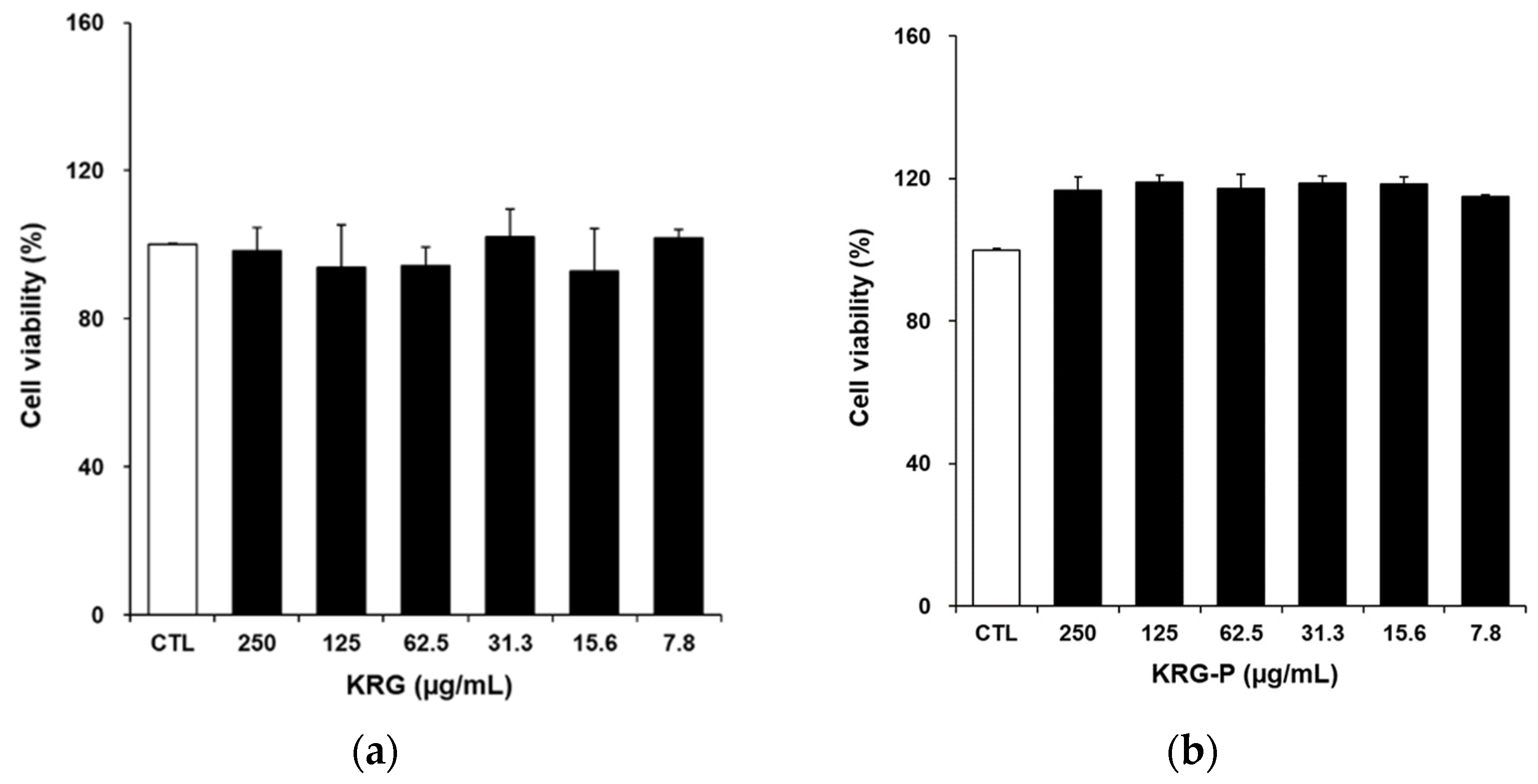
3.2. Cytotoxicity Assay of KRG and KRG-P

Plant polysaccharides not only have anti-cancer, immune regulatory, and other functions, but also exhibit low toxicity and safety. To determine the cytotoxicity of KRG and KRG-P, RAW264.7 macrophage cells were treated with KRG or KRG-P at a concentration of 7.8–200 μg/mL for 24 h. The results are shown in Figure 1. KRG was non-toxic to RAW264.7 cells. KRG-P was also non-toxic and showed a proliferative effect on RAW264.7 cells, which was not statistically significant. These results are consistent with the low toxicity of polysaccharides mentioned earlier.

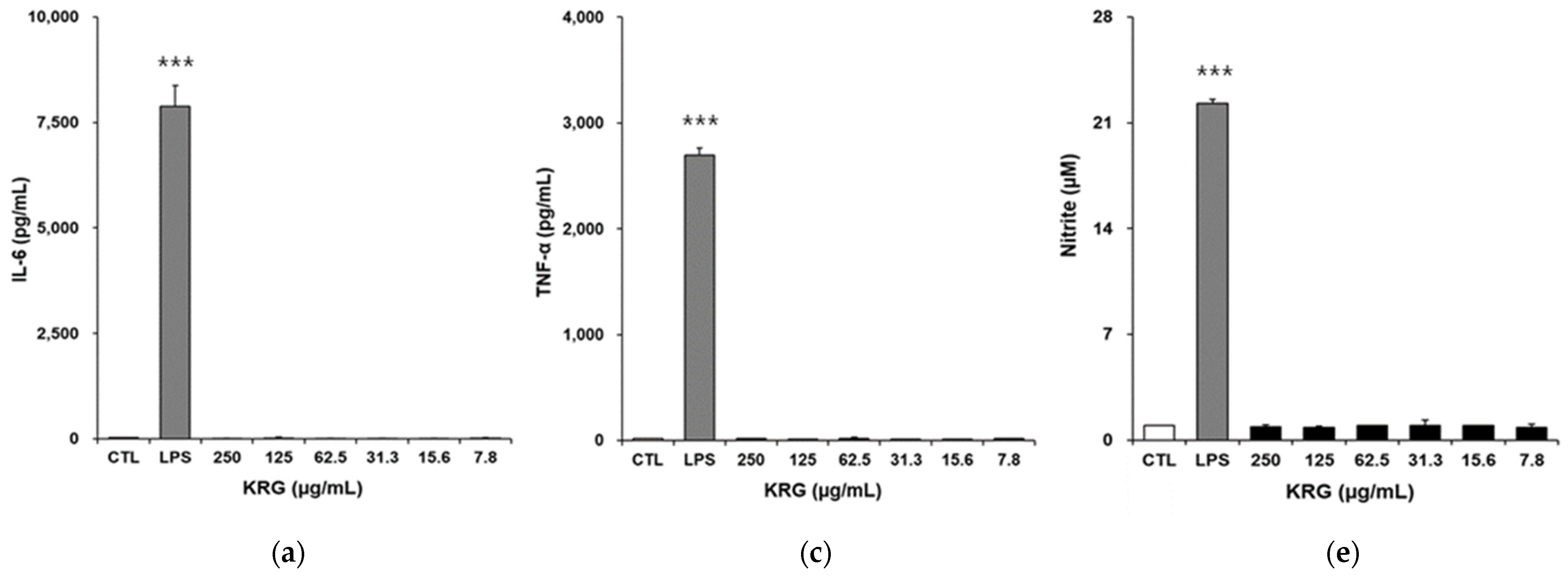
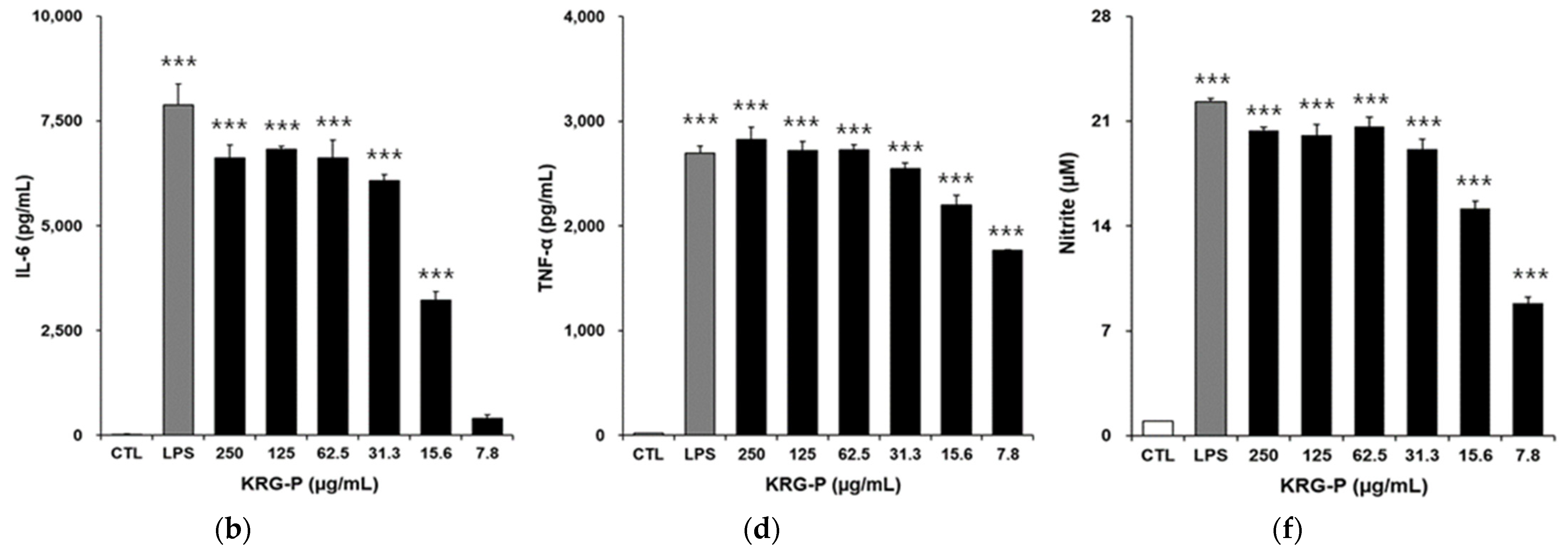
3.3. Effects of KRG and KRG-P on IL-6, TNF-α, and NO Production in RAW264.7 Cells

Following activation of macrophages, they not only secrete NO, but also proinflammatory cytokines (IL-1, IL-6, and TNF-α) [29]. To study KRG or KRG-P activated macrophages, RAW264.7 cells were treated with KRG or KRG-P at a concentration of 7.8~200 μg/mL for 24 h and the production of cytokines and NO was determined. As shown in Figure 2, IL-6, TNF-α, and NO were not produced by KRG treatment of RAW264.7 macrophage cells. However, the production of IL-6 (2b), TNF-α (2d), and NO (2f) was significantly enhanced by KRG-P treatment in a concentration-dependent manner in RAW264.7 cells. Although KRG was not toxic to RAW264.7 cells (Figure 1a), it did not induce the production of IL-6, TNF-α, or NO, which indicates that KRG cannot activate RAW264.7 cells. In contrast, KRG-P was not only non-toxic to RAW264.7 cells (Figure 1b), but also induced the production of IL-6 (2b), TNF-α (2d), and NO (2f) in a concentration-dependent manner. This suggests that KRG-P is able to stimulate the innate immune system.

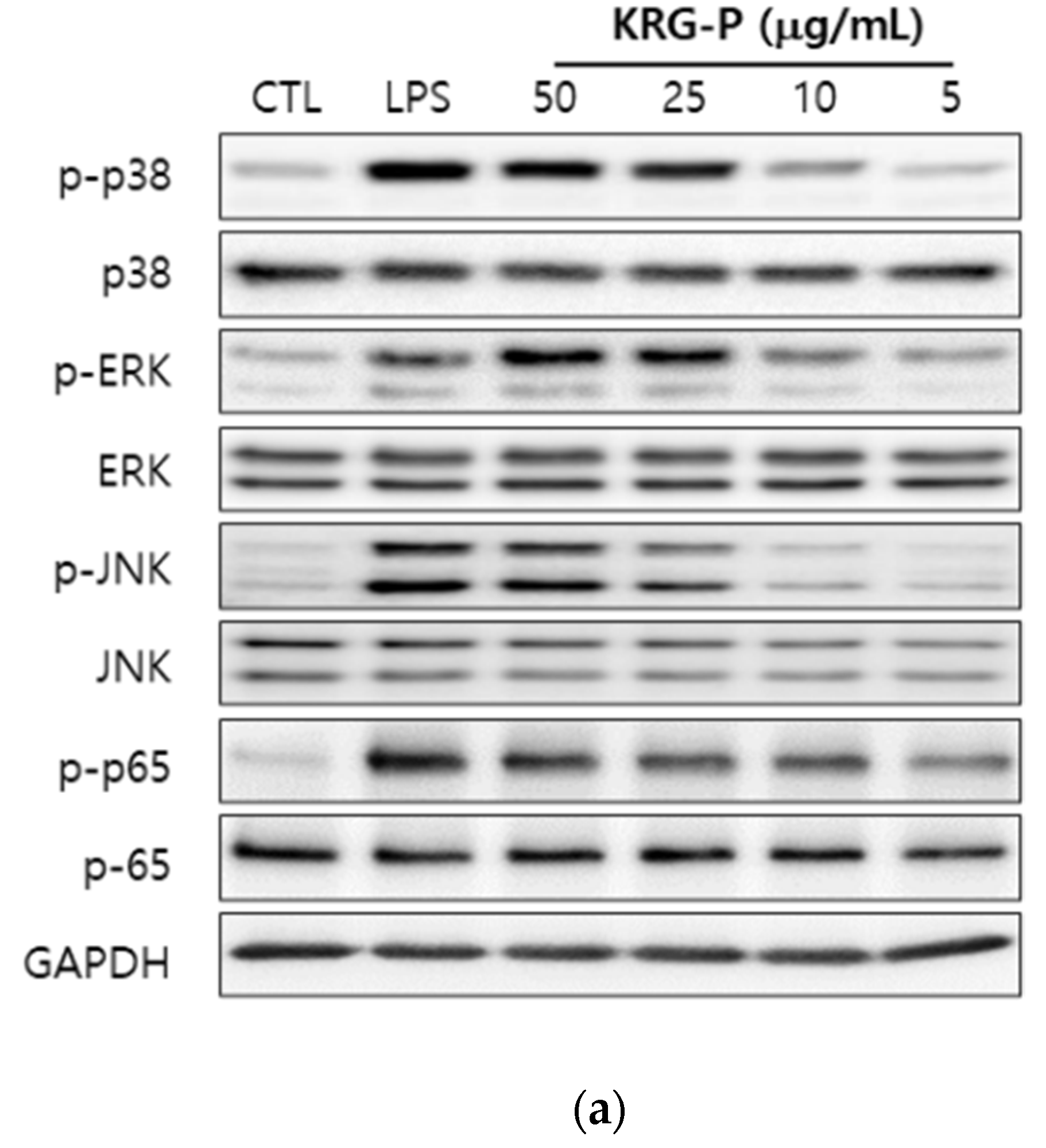
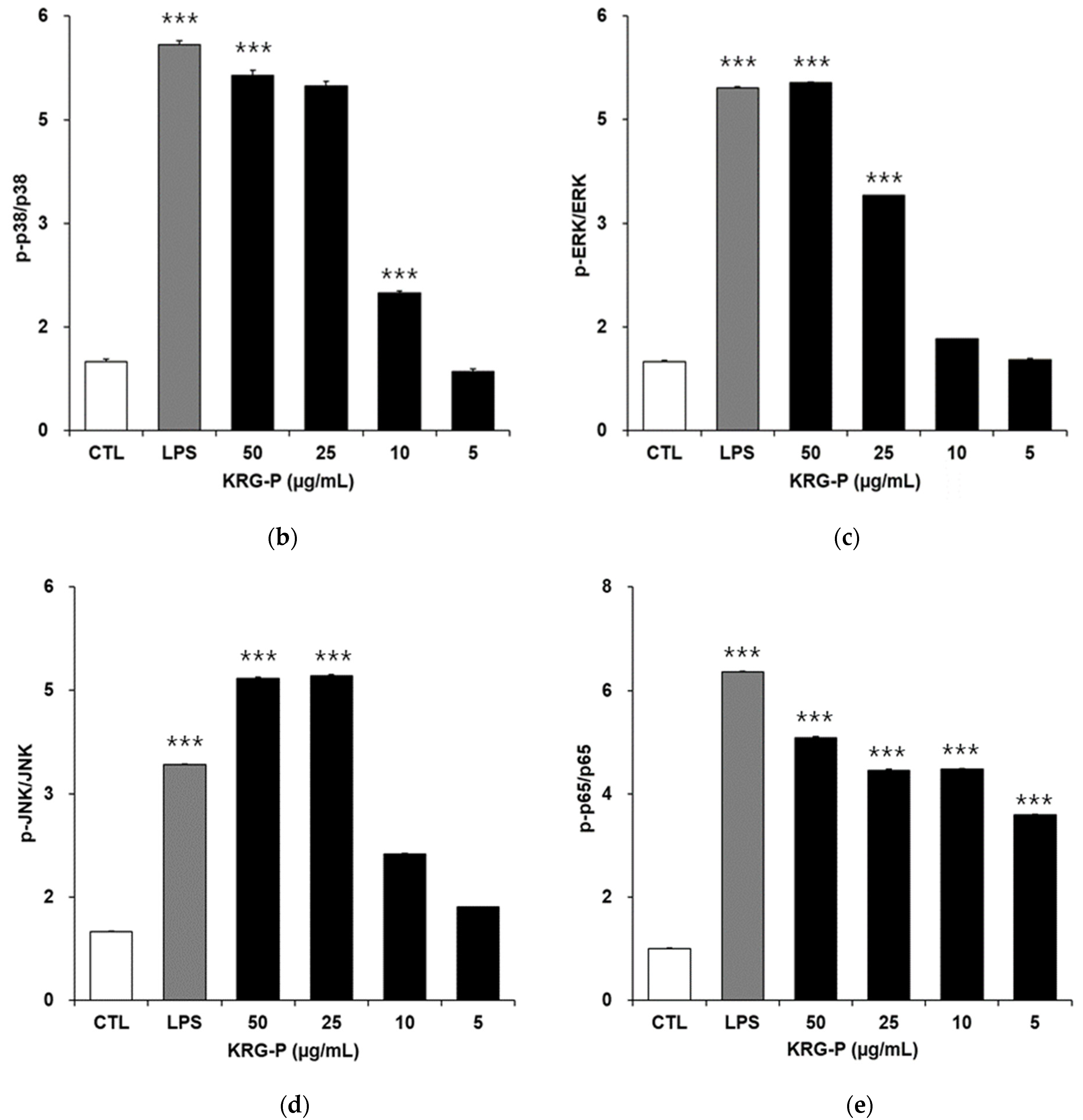
3.4. Effects of KRG-P on MAPKs and NF-κB Phosphorylation in RAW264.7 Cells

Shin [23] reported that macrophages are activated by plant polysaccharides through MAPK and NF-κB phosphorylation and ultimately regulate gene transcription. Rao et al. [30] also reported that RAW264.7 cells treated with LPS can activate MAPK pathways to produce NO, IL-6, and TNF-α. To study the mechanism of macrophage activation by KRG-P, RAW264.7 cells were treated with KRG-P at concentrations of 5~50 μg/mL for 30 min. As shown in Figure 3, p38, ERK, and JNK were significantly phosphorylated by KRG-P treatment compared with the control group in a concentration-dependent manner. The p65 subunit of NF-κB was also phosphorylated by KRG-P treatment in a concentration-dependent manner compared with the control group. The levels of the total p38, JNK, ERK, and p65 protein were not affected. These results indicate that MAPK and NF-kB pathways are significantly phosphorylated by KRG-P in a concentration-dependent fashion (Figure 3).

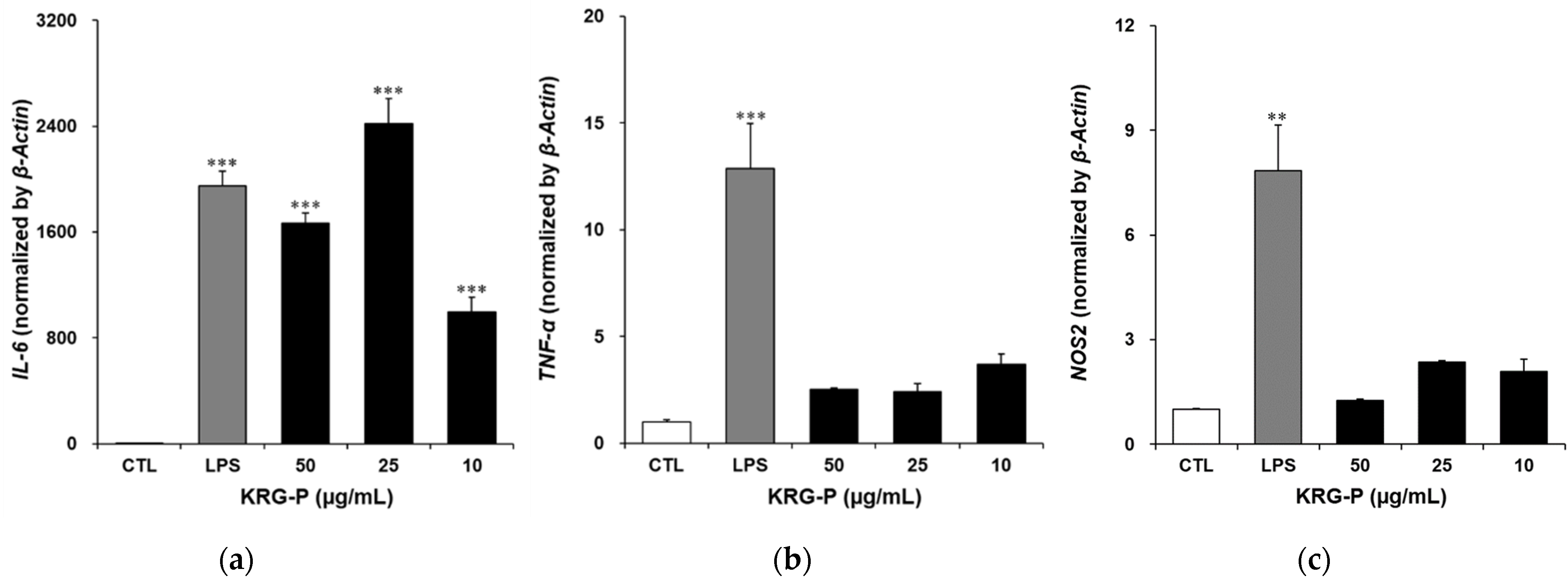
3.5. Effects of KRG-P on IL-6, TNF-α, and NOS2 Gene Expression in RAW264.7 Cells

Macrophages activate the expression of the IL-6, TNF-α, and NOS2 genes through the MAPK and NF-κB pathways, thereby releasing cytokines and NO. The NO produced by iNOS encoded by the nitric oxide synthase 2 (NOS2) gene, which can kill bacteria and viruses, plays an important role in the immune response. To determine the effect of cell signaling pathways activated by KRG-P on expressed genes, we evaluated its effect on the expression of IL-6, TNF-α, and NOS2 in RAW264.7 cells. RAW264.7 cells were treated with KRG or KRG-P at concentrations of 10–50 μg/mL for 6 h. RNA was evaluated and the results are shown in Figure 4. The expression of the IL-6 gene was significantly increased by KRG-P compared with the control group and the expression of TNF-α and NOS2 were also increased by KRG-P. The results indicate that the expression of IL-6, TNF-α, and INOS were enhanced by KRG-P in RAW264.7 cells (Figure 4). Although the gene expression results did not show a concomitant concentration-dependent increase with the production of cytokines following KRG-P treatment for 6 h, it still showed an increased trend compared with that of the control group, which may be related to the time of KRG-P treatment. Overall, these results demonstrate the production of NO and cytokines (IL-6 and TNF-α) by KRG-P exposure.

4. Discussion

Studies on polysaccharides isolated from natural products with pharmacological activities, such as suppression of cancer metastasis through NK cells and macrophages, have been reported [17,18]. These polysaccharides are known to have low toxicity and side effects in animal experiments, suggesting a high possibility of their development as health functional food materials or pharmaceuticals in the future. However, detailed structural analysis of polysaccharides and studies investigating detailed mechanisms of their pharmacological activity are warranted. Recently, Lee et al. reported that acidic polysaccharide (RGP-AP-I) purified from KRG mainly consists of rhamnose (9.5%), galacturonic acid (18.4%), galactose (30.4%), and arabinose (35.0%), which showed macrophage stimulatory effects such as the production of IL-6, IL-12, and TNF-α in RAW264.7 cells [27]. Further, Lee et al. analyzed glycosyl linkages of RGP-AP-I, which comprised 21 different glycosyl linkages and possessed the rhamnogalacturonan-I structure. In addition, Kim et al. purified a polysaccharide (RG-CW-EZ-CP) from KRG using α-amylase and amyloglucosidase, which mainly comprised arabinose (15.94%), glucose (5.06%), galactose (13.97%), and galacturonic acid (59.81) and increased NO, IL-6, and IL-12 production mediated by MAPK (ERK, p38, and JNK) phosphorylation in RAW264.7 cells [31]. Our previous study already reported that KRG-P comprised glucose (60.5%), galactose (11.0%), arabinose (6.8%), rhamnose (1.9%), and galacturonic acid (19.7%) [22]. In this study, we demonstrated that KRG-P activates macrophages by increasing the production of cytokines, NO, and mRNAs of IL-6 and TNF-α, which is in turn mediated by the phosphorylation of p38, ERK, JNK, and NF-κB p65. Based on these results, it is thought that the similarity in the composition of monosaccharides of the polysaccharide may result in similar pharmacological activity.

5. Conclusions

This study revealed that the polysaccharide fraction (KRG-P) isolated from KRG water extracts can enhance innate immune response (NO, IL-6, and TNF-α production) and results in the phosphorylation of intermediates of the intercellular signal pathways (p38, ERK, JNK, and NF-κB p65) in RAW264.7 cells. However, chemical structure of KRG-P remains unclear and warrants further analysis such as detailed glycosyl linkages analysis data of purified KRG-P after gel permeation chromatography. Additionally, it is necessary to investigate the relationship between structure and activity of polysaccharide. In this study, we demonstrated that KRG-P polysaccharide, not KRG was identified as a potentially useful immunostimulant and a promising therapeutic candidate for further development.

Author Contributions

Conceptualization, M.-S.S.; methodology, J.G.; formal analysis, S.L. and J.-H.L.; investigation, J.G.; writing—original draft preparation, J.G.; writing—review and editing, M.-S.S.; project administration, M.-S.S.; funding acquisition, K.-S.K. All authors have read and agreed to the published version of the manuscript.

Funding

This study was supported by the 2019 grant from the Korean Society of Ginseng, funded by the Korean Ginseng Corporation.

Conflicts of Interest

The authors declare no conflict of interest.

References

  1. Baek, K.S.; Hong, Y.D.; Kim, Y.; Sung, N.Y.; Yang, S.; Lee, K.M.; Park, J.Y.; Park, J.S.; Rho, H.S.; Shin, S.S.; et al. Anti-inflammatory activity of AP-SF, a ginsenoside-enriched fraction, from Korean ginseng. J. Ginseng Res. 2015, 39, 155–161. [Google Scholar] [CrossRef] [PubMed] [Green Version]
  2. Han, B.H.; Park, M.H.; Woo, L.K.; Woo, W.S.; Han, Y.N. Studies on the antioxidant components of Korean ginseng. In Proceedings of the Ginseng Society Conference 1978, Seoul, Korea, 7–11 September 1978; pp. 13–17. [Google Scholar]
  3. Wong, A.S.; Che, C.M.; Leung, K.W. Recent advances in ginseng as cancer therapeutics: A functional and mechanistic overview. Nat. Prod. Rep. 2015, 32, 256–272. [Google Scholar] [CrossRef]
  4. Shibata, S. Chemistry and cancer preventing activities of ginseng saponins and some related triterpenoid compounds. J. Korean Med. Sci. 2001, 16, S28–S37. [Google Scholar] [CrossRef] [PubMed] [Green Version]
  5. Lee, S.H.; Jung, B.H.; Kim, S.Y.; Lee, E.H.; Chung, B.C. The antistress effect of ginseng total saponin and ginsenoside Rg3 and Rb1 evaluated by brain polyamine level under immobilization stress. Pharmacol. Res. 2006, 54, 46–49. [Google Scholar] [CrossRef]
  6. Kennedy, D.O.; Scholey, A.B. Ginseng: Potential for the enhancement of cognitive performance and mood. Pharmacol. Biochem. Behav. 2003, 75, 687–700. [Google Scholar] [CrossRef]
  7. Wang, J.; Li, S.; Fan, Y.; Chen, Y.; Liu, D.; Cheng, H.; Gao, X.; Zhou, Y. Anti-fatigue activity of the water-soluble polysaccharides isolated from panax ginseng CA Meyer. J. Ethnopharmacol. 2010, 130, 421–423. [Google Scholar] [CrossRef] [PubMed]
  8. Kim, M.J.; Kwon, R.H.; Jang, M.W.; Ha, B.J. Antioxidant and anti-wrinkle effects of steamed three ginseng extracts. J. Soc. Cosmet. Sci. Korea 2012, 38, 155–162. [Google Scholar]
  9. Lee, J.H.; Cho, S.H.; Yun, M.Y.; An, S.; Jang, H.H.; Lee, S.N.; Song, G.Y. Anti-wrinkle effect of rare ginsenosides, produced from ginsenoside Rd. Asian J. Beauty Cosmetol. 2015, 13, 909–916. [Google Scholar]
  10. Attele, A.S.; Wu, J.A.; Yuan, C.S. Ginseng pharmacology: Multiple constituents and multiple actions. Biochem. Pharmacol. 1999, 58, 1685–1693. [Google Scholar] [CrossRef]
  11. Christensen, L.P. Ginsenosides: Chemistry, biosynthesis, analysis, and potential health effects. Adv. Food Nutr. Res. 2009, 55, 1–99. [Google Scholar]
  12. Kim, C.S.; Choi, K.J.; Kim, S.C.; Ko, S.Y.; Sung, H.S.; Lee, Y.G. Controls of the hydrolysis of ginseng saponins by neutralization of organic acids in red ginseng extract preparations. J. Ginseng Res. 1998, 22, 205–210. [Google Scholar]
  13. Xu, X.F.; Gao, Y.; Xu, S.Y.; Liu, H.; Xue, X.; Zhang, Y.; Zhang, H.; Liu, M.N.; Xiong, H.; Lin, R.C.; et al. Remarkable impact of steam temperature on ginsenosides transformation from fresh ginseng to red ginseng. J. Ginseng Res. 2018, 42, 277–287. [Google Scholar] [CrossRef]
  14. In, G.; Ahn, N.G.; Bae, B.S.; Lee, M.W.; Park, H.W.; Jang, K.H.; Cho, B.G.; Han, C.K.; Park, C.K.; Kwak, Y.S. In situ analysis of chemical components induced by steaming between fresh ginseng, steamed ginseng, and red ginseng. J. Ginseng Res. 2017, 41, 361–369. [Google Scholar] [CrossRef] [Green Version]
  15. Thetsrimuang, C.; Khammuang, S.; Chiablaem, K.; Srisomsap, C.; Sarnthima, R. Antioxidant properties and cytotoxicity of crude polysaccharides from Lentinus polychrous Lév. Food Chem. 2011, 128, 634–639. [Google Scholar] [CrossRef]
  16. Schepetkin, I.A.; Quinn, M.T. Botanical polysaccharides: Macrophage immunomodulation and therapeutic potential. Int. Immunopharmacol. 2006, 6, 317–333. [Google Scholar] [CrossRef] [PubMed]
  17. Lee, E.H.; Park, H.R.; Shin, M.S.; Cho, S.Y.; Choi, H.J.; Shin, K.S. Antitumor metastasis activity of pectic polysaccharide purified from the peels of Korean Citrus Hallabong. Carbohydr. Polym. 2014, 111, 72–79. [Google Scholar] [CrossRef] [PubMed]
  18. Shin, M.S.; Hwang, S.H.; Yoon, T.J.; Kim, S.H.; Shin, K.S. Polysaccharides from ginseng leaves inhibit tumor metastasis via macrophage and NK cell activation. Int. J. Biol. Macromol. 2017, 103, 1327–1333. [Google Scholar] [CrossRef] [PubMed]
  19. Ruijun, W.; Shi, W.; Yijun, X.; Mengwuliji, T.; Lijuan, Z.; Yumin, W. Antitumor effects and immune regulation activities of a purified polysaccharide extracted from Juglan regia. Int. J. Biol. Macromol. 2015, 72, 771–775. [Google Scholar] [CrossRef] [PubMed]
  20. Kim, H.; Cho, S.M.; Kim, W.J.; Hong, K.B.; Suh, H.J.; Yu, K.W. Red ginseng polysaccharide alleviates cytotoxicity and promotes anti-inflammatory activity of ginsenosides. Food Sci. Technol. 2021, 41, 1–6. [Google Scholar]
  21. Uthaisangsook, S.; Day, N.K.; Bahna, S.L.; Good, R.A.; Haraguchi, S. Innate immunity and its role against infections. Ann. Allergy Asthma Immunol. 2002, 88, 253–264. [Google Scholar] [CrossRef]
  22. Park, D.H.; Han, B.; Shin, M.S.; Hwang, G.S. Enhanced intestinal immune response in mice after oral administration of Korea red ginseng-derived polysaccharide. Polymers 2020, 12, 2186. [Google Scholar] [CrossRef]
  23. Shin, K.S. Immunostimulating plant polysaccharides: Macrophage immunomodulation and its possible mechanism. Food Sci. Ind. 2012, 45, 12–22. [Google Scholar]
  24. West, A.P.; Brodsky, I.E.; Rahner, C.; Woo, D.K.; Erdjument-Bromage, H.; Tempst, P.; Walsh, M.C.; Choi, Y.; Shadel, G.S.; Ghosh, S. TLR signalling augments macrophage bactericidal activity through mitochondrial ROS. Nature 2011, 472, 476–480. [Google Scholar] [CrossRef] [PubMed] [Green Version]
  25. Yoo, D.G.; Kim, M.C.; Park, M.K.; Park, K.M.; Quan, F.S.; Song, J.M.; Wee, J.J.; Wang, B.Z.; Cho, Y.K.; Compans, R.W.; et al. Protective effect of ginseng polysaccharides on influenza viral infection. PLoS ONE 2012, 7, e33678. [Google Scholar] [CrossRef] [PubMed]
  26. MacMicking, J.; Xie, Q.W.; Nathan, C. Nitric oxide and macrophage function. Annu. Rev. Immunol. 1997, 15, 323–350. [Google Scholar] [CrossRef]
  27. Lee, S.J.; In, G.; Han, S.T.; Lee, M.H.; Lee, J.W.; Shin, K.S. Structural characteristics of a red ginseng acidic polysaccharide rhamnogalacturonan I with immunostimulating activity from red ginseng. J. Ginseng Res. 2020, 44, 570–579. [Google Scholar] [CrossRef] [PubMed]
  28. Stimpel, M.; Proksch, A.; Wagner, H.; Lohmann-Matthes, M.L. Macrophage activation and induction of macrophage cytotoxicity by purified polysaccharide fractions from the plant Echinacea purpurea. Infect. Immun. 1984, 46, 845–849. [Google Scholar] [CrossRef] [Green Version]
  29. Byeon, S.E.; Lee, J.; Kim, J.H.; Yang, W.S.; Kwak, Y.S.; Kim, S.Y.; Choung, E.S.; Rhee, M.H.; Cho, J.Y. Molecular mechanism of macrophage activation by red ginseng acidic polysaccharide from Korean red ginseng. Mediat. Inflamm. 2012, 2012, 732860. [Google Scholar] [CrossRef]
  30. Rao, K.M.K. MAP kinase activation in macrophages. J. Leukoc. Biol. 2001, 69, 3–10. [Google Scholar] [PubMed]
  31. Kim, H.; Kim, H.W.; Yu, K.W.; Suh, H.J. Polysacchrides fractionated from enzyme digests of Korean red ginseng water extracts enhanced the immunostimulatory activity. Int. J. Biol. Macromol. 2019, 121, 913–920. [Google Scholar] [CrossRef]
Figure 1. Cytotoxic effect of KRG (a) and KRG-P (b) isolated from Korean red ginseng on RAW264.7 macrophage cells in vitro. RAW264.7 macrophage cells (1.0 × 10⁵ cells/well) were treated with KRG or KRG-P using a series of concentrations (7.8~250 μg/mL) in 96-well flat-bottomed microplates for 24 h. Cytotoxicity was determined using the EZ-Cytox based colorimetric assay. CTL; control. Data are presented as the means ± SD of three independent experiments.
Applsci 11 07111 g001
Figure 2. Effect of KRG and KRG-P on the production of IL-6 (a,b), TNF-α (c,d), and NO (e,f) in RAW264.7 macrophage cells in vitro. RAW264.7 macrophage cells (1.0 × 10⁵ cells/well) were treated with KRG and KRG-P using a series of concentrations (7.8~250 μg/mL) or LPS (500 ng/mL) in 96-well flat-bottomed microplates for 24 h. The supernatants were collected for the measurement of cytokines including IL-6 (a,b), TNF-α (c,d), and NO (e,f). CTL; control, LPS; lipopolysaccharide. Data are presented as the means ± SD of three independent experiments. *** p < 0.001 vs. the control group.
Applsci 11 07111 g002aApplsci 11 07111 g002b
Figure 3. Activation of MAPK and NF-κB pathways by treatment with KRG-P in RAW264.7 macrophage cells. (a) RAW264.7 cells (3.0 × 10⁶ cells/well) were treated with KRG-P at a series of concentrations (5~50 μg/mL) or with LPS (500 ng/mL) in 6-well flat-bottom plate for 30 min. CTL; control, LPS; lipopolysaccharide. Western blots show the expression of the representative proteins. The bar graphs respectively show density ratios of p-p38 to total p38 (b), pERK to total ERK (c), pJNK to total JNK (d) and p-p65 to total p65 (e) as analyzed by ImageJ Software. Data are presented as the means ± SD. *** p < 0.001 vs. the control group.
Applsci 11 07111 g003aApplsci 11 07111 g003b
Figure 4. Effect of KRG-P of mRNA expression of IL-6 (a), TNF-α (b) and NOS2 (c) in RAW264.7 macrophage cells using quantitative real-time PCR. RAW264.7 cells (3.0 × 10⁶ cells/well) were treated with KRG-P using a series of concentrations (10~50 μg/mL) or with LPS (500 ng/mL) in 6-well flat-bottom plates for 6 h. CTL; control, LPS; lipopolysaccharide. Data are presented as the means ± SD of three independent experiments. ** p < 0.01 or *** p < 0.001 vs. the control group.
Applsci 11 07111 g004
Publisher’s Note: MDPI stays neutral with regard to jurisdictional claims in published maps and institutional affiliations.

