Article

Chromatographic Characterization and In Vitro Bioactivity Evaluation of Lactobacillus helveticus Hydrolysates upon Fermentation of Different Substrates

by
Federica Ianni
1,*,
Alessandra Anna Altomare
2,
Beniamino T. Cenci-Goga
3,*,
Francesca Blasi
1,
Luca Grispoldi
3,
Luca Regazzoni
2 and
Lina Cossignani
1,4
1
Food Science and Nutrition Section, Department of Pharmaceutical Sciences, University of Perugia, 06126 Perugia, Italy
2
Department of Pharmaceutical Sciences, University of Milan, Via Mangiagalli 25, 20133 Milano, Italy
3
Department of Veterinary Medicine, University of Perugia, Via San Costanzo 4, 06126 Perugia, Italy
4
Center for Perinatal and Reproductive Medicine, University of Perugia, Santa Maria della Misericordia University Hospital, Sant’Andrea delle Fratte, 06132 Perugia, Italy
*
Authors to whom correspondence should be addressed.
Submission received: 30 November 2020 / Revised: 11 January 2021 / Accepted: 13 January 2021 / Published: 16 January 2021
(This article belongs to the Special Issue Bioactive Peptides from Foods)

Abstract

:

Featured Application

Exploration of Lactobacillus helveticus strains as source of bioactive peptides for potential applications as functional foods as well as for potential uses in nutraceuticals.

Abstract

Among various food sources, milk proteins remain the major vector for functional peptides endowed with several biological activities. Particularly, the proteolytic activity of lactic acid bacteria during milk fermentation has been one of the most followed strategies to produce bioactive peptides. In the present study, the exploration of the activity of several starter cultures, at different fermentation times, was firstly investigated by reversed phase-high performance liquid chromatography. Among the tested strains, Lactobacillus helveticus showed a higher proteolytic activity and it was submitted to further investigations by changing the fermentation substrate (skim milk, brain heart infusion, peptone water) as well as the extraction strategy (trichloroacetic acid vs. glass beads). The chromatographic analyses and the in vitro antioxidant and antihypertensive assays highlighted considerable differences for L. helveticus hydrolysates from different substrates, while a negligible impact by the two extraction protocols emerged. Furthermore, nano-high pressure liquid chromatography coupled with a high resolution mass spectrometry analyzer allowed the preliminary discrimination of fractions from fermented skim milk, likely responsible for the found activity. The obtained results suggest the possibility of varying the fermentation parameters in order to maximize the functional effects of the bioactive peptides.

Graphical Abstract

1. Introduction

In the last decades, the role of proteins in food has gained increasing acknowledgement. Beyond their purely nutritional role, bioactive peptides (BPs) encrypted in dietary protein sequences are emerging as an important tool for the treatment of various diseases [1,2]. The release of the active form of BPs from the sequence of the parent protein could mainly follow three processes, namely, (i) the hydrolysis by gastrointestinal digestive enzymes, (ii) the hydrolysis by proteolytic microorganisms (during fermentation), or (iii) the action of proteolytic enzymes derived from microorganisms or plants [3]. Once split from their precursors, BPs contribute to the modulation of different body functions exerting, inter alia, antioxidant, digestive, immunomodulatory, hypotensive, antithrombotic, antibacterial, and opiate-like properties. Such potential health benefits justify the emerging employment of BPs and protein hydrolysates as valuable sources for physiological functioning and human well-being promotion [1,2,3].
Although potential BPs have been identified from various food proteins (of either animal or plant origin) [1,4] and their intake could be regularly associated with the current diet, milk proteins remain the major source of functional BPs endowed with a wide range of biological activities. The occurrence of biofunctional peptides with high nutritional value has been also widely reported in fermented milk products such as yogurt, cheese, sour milk, or kefir, and some of them have been shown to contain health beneficial properties [2,5]. This feature accounts for the growing interest by food scientists in exploiting protein hydrolysates and bioactive peptides from milk and dairy products for applications as functional foods as well as for potential uses in nutraceuticals [2,5].
The proteolytic activity of lactic acid bacteria (LAB) during milk fermentation has been, and still is, among the most pursued strategies to produce functional peptides. In this scenario, the research of novel tailored strategies to exploit dairy fermented foods as sources of health peptides is widely increasing. As reported in literature [5,6,7,8], BPs generated during milk fermentation mostly range from 2 to 20 amino acids and some of them are known to display multi-functional properties. BPs have been mainly screened for their capacity to exhibit specific biological properties, with particular attention paid to the antioxidant [9,10], angiotensin-converting enzyme-inhibitory (ACEi) [11,12,13], and antimicrobial [14,15] actions among others. Although numerous approaches dealing with the production of BPs from a variety of sources are described in literature [16,17], often misleading and incomplete evidences still drive the rational selection and fermentation conditions of specific strains for the generation of active components. Moreover, the lack of systematic approaches, allowing overcoming of challenging peptide extractions and purifications, represents one of the major limitations in BP production.
The present study was aimed at being a preliminary exploration of the proteolytic activity of several starter cultures, sampled at different fermentation times and in the presence of diverse substrates, in producing protein hydrolysates. The fermentation activity was monitored by reversed phase-high performance liquid chromatography (RP-HPLC) in order to get a comprehensive characterization of the obtained peptide pool and select the best parameters to maximize the production of BPs. Moreover, the in vitro antioxidant and antihypertensive activities of protein hydrolysates were evaluated as pilot discriminant before following steps addressed to the purification and characterization of the final BP products. In this frame, nano-high pressure liquid chromatography coupled with high resolution mass spectrometry analyzer (nano-HPLC-HRMS) allowed the preliminary identification of fractions from fermented skim milk likely responsible for the above-mentioned bioactive properties (an overview of the anaytical approaches applied in the present study is given in the Graphical Abstract).

2. Materials and Methods

2.1. Chemicals and Reagents

HPLC-grade and MS-grade acetonitrile (ACN), trifluoroacetic acid (TFA), formic acid (FA), trichloroacetic acid (TCA), ethanol (EtOH), urea, TRIS-HCl, diammonium 2,2′-azino-bis(3-ethylbenzothiazoline-6-sulphonate) (ABTS), 2,4,6-tris(2-pyridyl)-s-triazine (TPTZ), 6-hydroxy-2,5,7,8-tetramethyl-2-carboxylic acid (Trolox), ferric chloride (FeCl3), sodium acetate buffer, potassium persulfate, sodium borate, sodium chloride (NaCl), hippuryl-L-histidyl-L-leucine (HHL), hippuric acid, glass beads, and rabbit lung powder containing ACE (0.1 U) were purchased from Sigma Aldrich (Milan, Italy). Water for HPLC analysis was purified with a Milli-Q Plus185 system from Millipore (Milford, MA, USA). All the employed mobile phases were mixed and preliminarily degassed by sonication for 10 min before use.

2.2. HPLC-UV Conditions

The HPLC study was performed on a Thermo Separation low-pressure quaternary gradient pump system (Spectra system Series, Thermo Scientific, Waltham, MA, USA) supplied with a GT-154 vacuum degasser (Shimadzu, Kyoto, Japan). The system was equipped with a SPD-10A UV-Vis detector (Shimadzu, Kyoto, Japan) and a Rheodyne 7725i injector (Rheodyne Inc., Cotati, CA, USA) with a 20 µL stainless steel loop. Data management and acquisition was made by means of Clarity Lite chromatography software. UV detection was carried out at 210 nm. The analytical columns were: Robusta RP18 (250 × 4.6 mm i.d., 5 µm, 100 Å pore size from Sepachrom, Milan, Itay), Grace Smart RP 18 (250 × 4.6 mm i.d., 5 µm, 120 Å pore size from Sepachrom, Milan, Itay), Kinetex EVO C18 (250 × 4.6 mm i.d., 5 µm, 100 Å pore size from Phenomenex, Milan, Itay), Gemini NX (250 × 4.6 mm i.d., 5 µm, 110 Å pore size from Phenomenex, Milan, Itay). In the optimized conditions, column temperature was fixed at 40 °C through a Grace (Sedriano, Italy) heater/chiller (Model 7956R) thermostat. The final gradient program, at a flow rate of 0.8 mL/min, was the following: eluent A (0.1% (v) TFA in water), eluent B (0.1% (v) TFA in acetonitrile, ACN); 0–45 min, linear gradient from 100% to 75% A; 45–60 min, linear gradient to 60% A. At the end of each run, a column cleaning step (10 min) with 100% B was added to remove more hydrophobic compounds before column re-equilibration with 100% A (30 min).

2.3. Bacterial Strain Selection and Propagation

Several strains, which showed growth in the range 10–37 °C, both in aerobic and anaerobic conditions without gas production from dextrose, were tested for acidifying activity in skim milk (SM, BD Difco, 232,100). Then, based on the instantaneous acidification rate and its maximum value [18,19,20,21,22], a selection of ten strains was used for the production of BPs.
The strains tested in this work were: Lactococcus lactis ssp. lactis (internal reference (ref. 79, field strain), Lactococcus lactis ssp. lactis (ref. 81, field strain), Lactobacillus casei ssp. casei (ref. 80, filed strain), Lactobacillus acidophilus ATCC 4356 (ref. 618), Lactobacillus acidophilus LA 14 (ref. 1004), Lactobacillus acidophilus (ref. 80/2, field strain), Lactobacillus acidophilus (ref. 80/3, field strain), Lactobacillus helveticus (ref. LH, field strain), Enterococcus faecium UBEF-41 (ref. 1003), and Saccharomyces cerevisiae var boulardii MTCC-5375 (ref. 1005). The morphological, biochemical and physiological characterization, the growth curves at several temperatures, including refrigeration conditions, the acidifying activity and their ability to improve palatability of certain food, along with safety considerations, have been reported by the authors in previous papers [18,19,20,21,22]. Before the test, freeze-dried strains of the starter cultures were grown aerobically in nutrient broth (NB, Oxoid CM0001, Basingstoke, UK) at 37 °C for 24 h. Each strain was then sub-cultured in skim milk (BD Difco, Franklin Lakes, NJ, USA, 232,100) at 37 °C for 24 h. The total viable cell (TVC) count (on nutrient agar, NA, Oxoid CM0003, incubated at 37 °C in aerobiosis for 24 h) after 24 h of incubation (Sanyo MIR-153 incubator, Moriguchi-City Osaka, Japan) was approximately 1 × 10⁹ cfu/mL. For tests, the strains were inoculated into skim milk (BD Difco) to get an initial concentration of approximately 1 × 10⁷ cfu/g, which mimics the initial starter concentration in dairy products fabrication. The fermentation activity was monitored at different time points (Table S1, Supplementary Material).

2.4. Evaluation of Different Substrates and Comparison of Two Extraction Protocols for Milk Fermented with L. helveticus

L. helveticus (Ref. LH) was inoculated in three different culture media, skim milk (SM, BD Difco), brain heart infusion (BHI, Oxoid) and peptone water (PW, Oxoid) and fermentation was carried out for 12 h and 110 h at 37 °C in aerobiosis. The conditions of bacterial growth and inoculation described in Section 2.3 were applied when the three substrates (SM, BHI, and PW) were used as culture media.
Two extraction protocols were used for peptide recovery within the fermented products, as described below:
(i) an aliquot of fermented substrate (1.0 mL) was treated with an aqueous TCA solution (1.0% w/v, 500 µL), vortexed (ZX3 VELP Scientifica, Usmate, Italy) for 1 min and centrifuged (ALC 4239R high speed refrigerated centrifuge, International PBI, Milan, Italy) at 10,000× g for 10 min at 4 °C. For each sample the supernatant was separated from the precipitated fraction and stored at −20 °C until further investigations;
(ii) an aliquot of fermented substrate (1.0 mL) was treated with glass beads (100 mg) and an aqueous solution of urea/TRIS-HCl (urea 2.0 M + Tris-HCl 50 mM, pH 7.4, 500 µL). The mixture was sonicated for 10 min at 25 °C, and then centrifuged at 10,000× g for 10 min at 20 °C. For each sample the supernatant was separated from the precipitated fraction and stored at −20 °C until further investigations.

2.5. Determination of the ACE Inhibitory Activity

The ACE inhibitory (ACEi) activity of each hydrolysate (from SM, BHI, and PW) was measured spectrophotometrically (PerkinElmer, Inc.; Waltham, MA, USA) using the slightly modified method of Muguerza et al. [12]. A buffer solution of sodium borate (100 mM) containing NaCl (300 mM) was prepared and the pH value was adjusted to 8.3. An aliquot of HHL solution (5 mM in buffer, 100 µL) was mixed with each hydrolysate (40 µL) and the mixture was submitted to pre-incubation at 37 °C for 2 min (distilled water was used for the control sample). The reaction was initiated by addition of an aqueous ACE solution (0.1 U/mL, 20 µL) and carried out at 37 °C for 30 min. Finally, HCl (1.0 N, 150 µL) was added to stop the enzymatic reaction. The released hippuric acid was extracted with ethyl acetate (1.0 mL), after a vigorous stirring for 20 s and centrifugation at 1500× g for 10 min. An aliquot of ethyl acetate extract (150 µL) was dried under vacuum (BUCHI Rotavapor R-114/B-480 waterbath, Cornaredo, Italy), re-dissolved in water (50 µL), and analyzed via HPLC by using a Robusta RP18 column to determine the hippuric acid formed during the enzymatic reaction. The following experimental conditions were used: mobile phase, 0.1% (v) TFA in water/0.1% (v) TFA in ACN-80/20 (v/v); eluent flow rate, 1.0 mL/min; column temperature, 30 °C; and wavelength of detection, 228 nm. The hippuric acid quantification was performed by relying upon a calibration curve built up by using standard solutions with concentration values in the range 0.001–0.1 mg/mL (y = 509.55x + 0.1626, R2 = 0.9999). The % inhibitory activity was calculated according Equation (1) as follows:
%   Inhibitory   activity =   ( Cc - Cs ) ( Cc - Cb ) × 100
where Cc is the hippuric acid concentration in the presence of ACE without hydrolysate (control, 100% ACE activity); Cs is the hippuric acid concentration in the presence of both ACE and hydrolysate; and Cb is the concentration in the presence of hydrolysate without ACE (Blank, 0% ACE activity).
HPLC analyses were performed in duplicate.

2.6. Determination of the Radical Scavenging Capacity by the ABTS Method

The ABTS radical-scavenging assay was measured according to previously reported methods [23,24]. The ABTS+• radical cation was generated by reaction of ABTS with potassium persulfate and incubating the mixture in the dark at room temperature for 12 h. Then reagent obtained was diluted with EtOH until its absorbance at 734 nm was 0.70 (±0.02). An aliquot of ABTS+•/EtOH solution was added to the hydrolysate (60 µL of supernatant) and the mixture was kept in the dark for 30 min. Analyses were performed in duplicate for each hydrolysate and the radical scavenging capacity was expressed as µmol Trolox equivalents/mL (µmol TE/mL).

2.7. Determination of the Antioxidant Capacity by the Ferric Reducing Antioxidant Power (FRAP) Assay

The reducing capacity of the peptide hydrolysates was determined according to previously reported methods [24,25] with minor modifications. The FRAP reagent was prepared by mixing TPTZ solution with FeCl3 solution and acetate buffer, and then adding it to the hydrolysates (50 µL of supernatant). The absorbance was determined at 593 nm after incubation of the reaction mixture in the dark at room temperature for 4 min. Analyses were performed in duplicate for each hydrolysate and the antioxidant capacity was expressed as µmol TE/mL.

2.8. Nano HPLC-HRMS Analysis

To further purify the peptide mixtures, a protein precipitation step has been performed by adding to 100 µL of each sample (SM-substrate, SM-TCA, SM-beads) 5 volumes (500 µL) of glacial acetone; after 30 s of sample vortexing they were incubated at −20 °C for 1 h, centrifuged (Eppendorf 5804R refrigerated centrifuge with A-4-44 rotor, Eppendorf Srl, Milan, Italy) for 20 min at 14,500 g, and the supernatant containing the peptide mixture was transferred in a new Eppendorf tube. The remaining pellets were washed by adding additional 500 µL of glacial acetone and another centrifugation step was performed together with the recovery of the supernatant. The two aliquots of supernatant were dried in a vacuum concentrator (Vacuum Concentrator RVC 2–18 CD plus, Martin Christ, Osterode am Harz, Germany) and the new pellets obtained were resuspended in 30 µL of 5% ACN, 0.1% FA. Peptide mixtures were extracted by using two C18 Ziptips with a binding capacity of 5 µg each (10 µg) and eluted in 80% ACN, 0.1% FA. The hydrolysates were further dried in a vacuum concentrator and finally dissolved in an appropriate volume (30 µL) of mobile phase (0.1% TFA) for mass spectrometry (MS) analysis.
Peptide mixtures were analyzed in duplicate by nano-LC-HRMS as described by Degani et al. with few modifications in the multi-step separative gradient [26]. Briefly, for each sample, 5 µL of solubilized peptides in 0.1% TFA were injected onto the Acclaim PepMap C18 column (75 µm × 15 cm, 100 Å pore size), protected by a pre-column (Acclaim PepMap, 100 µm × 2 cm, 100 Å pore size), both from Thermo Scientific (Milan, Italy). After the sample loading, peptide separation was performed by the nanoflow pump with a linear gradient of buffer B (0.1% FA in ACN) from 1% to 40% (70 min), followed by a further 5 min of linear gradient from 40% to 95% (buffer B); then 5 min at 95% of buffer B served to rinse the column before the re-equilibration to initial conditions (7 min at 99% of buffer A, 0.1% FA in H2O). The nano-chromatographic system was connected to an LTQ-Orbitrap XL mass spectrometer equipped by a Thermo Scientific (Milan, Italy) dynamic Nanospray ion source, and operated in data-dependent acquisition mode (DDA) to acquire selected full MS spectra, and MS/MS spectra. Xcalibur software (version 2.0.7, Thermo Scientific Inc., Milan, Italy) was used to control the mass spectrometer. A Pierce LTQ ESI Positive Ion Calibration Solution was used for positive calibration of Thermo Scientific orbitrap instrument.

2.9. Statistical Analysis

The comparison between L. acidophilus (LA 14) and L. helveticus (LH) activity upon 72 h fermentation is based on the area values of reference peaks (measured in mV·s). The mean area values ± standard deviation (SD) (n = 3) is reported. The statistical analysis was performed by an unpaired Student’s t-test in GraphPad Prism Version 6.0 h.

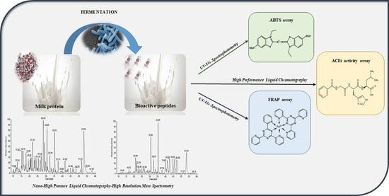
3. Results and Discussion

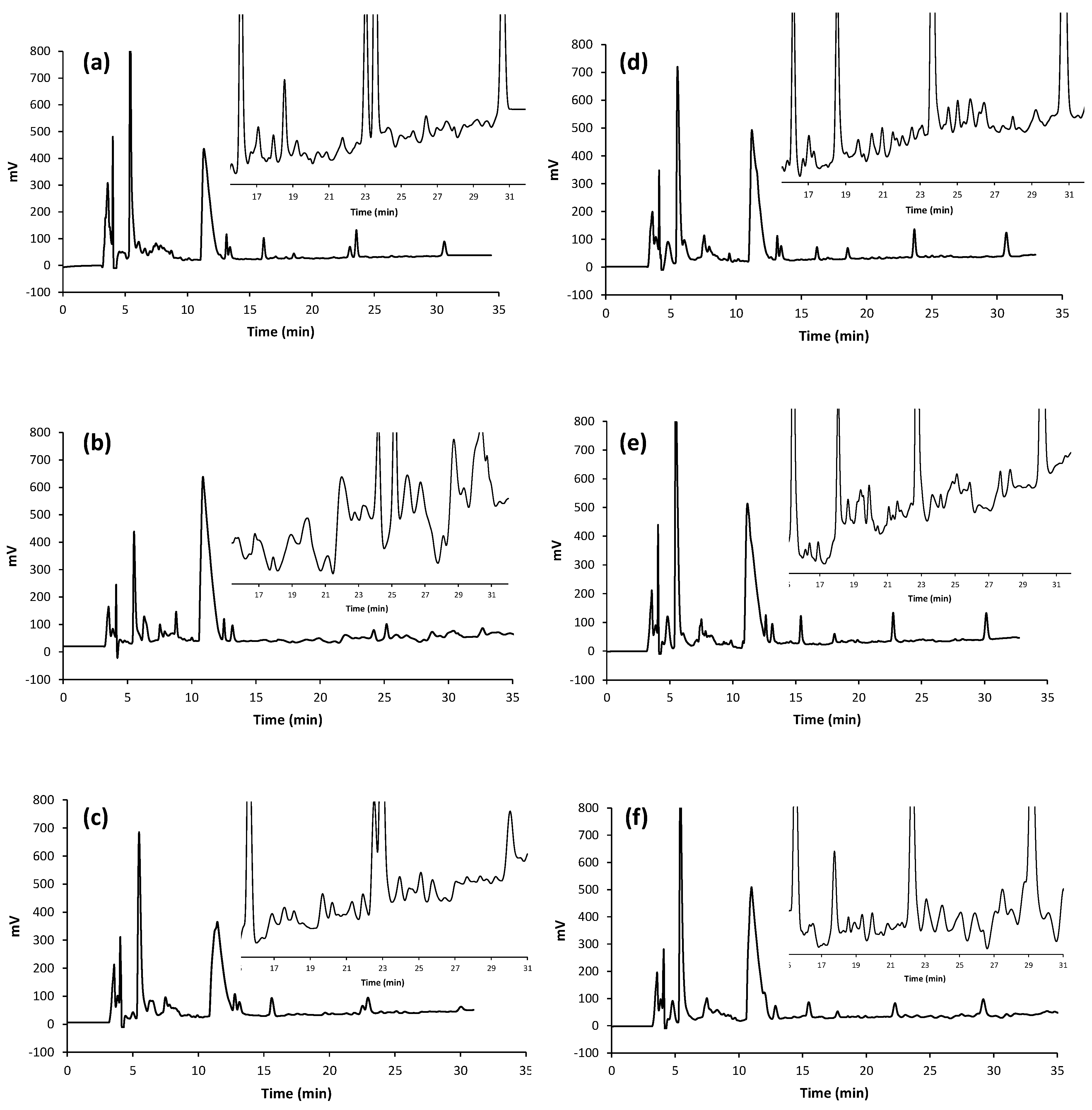
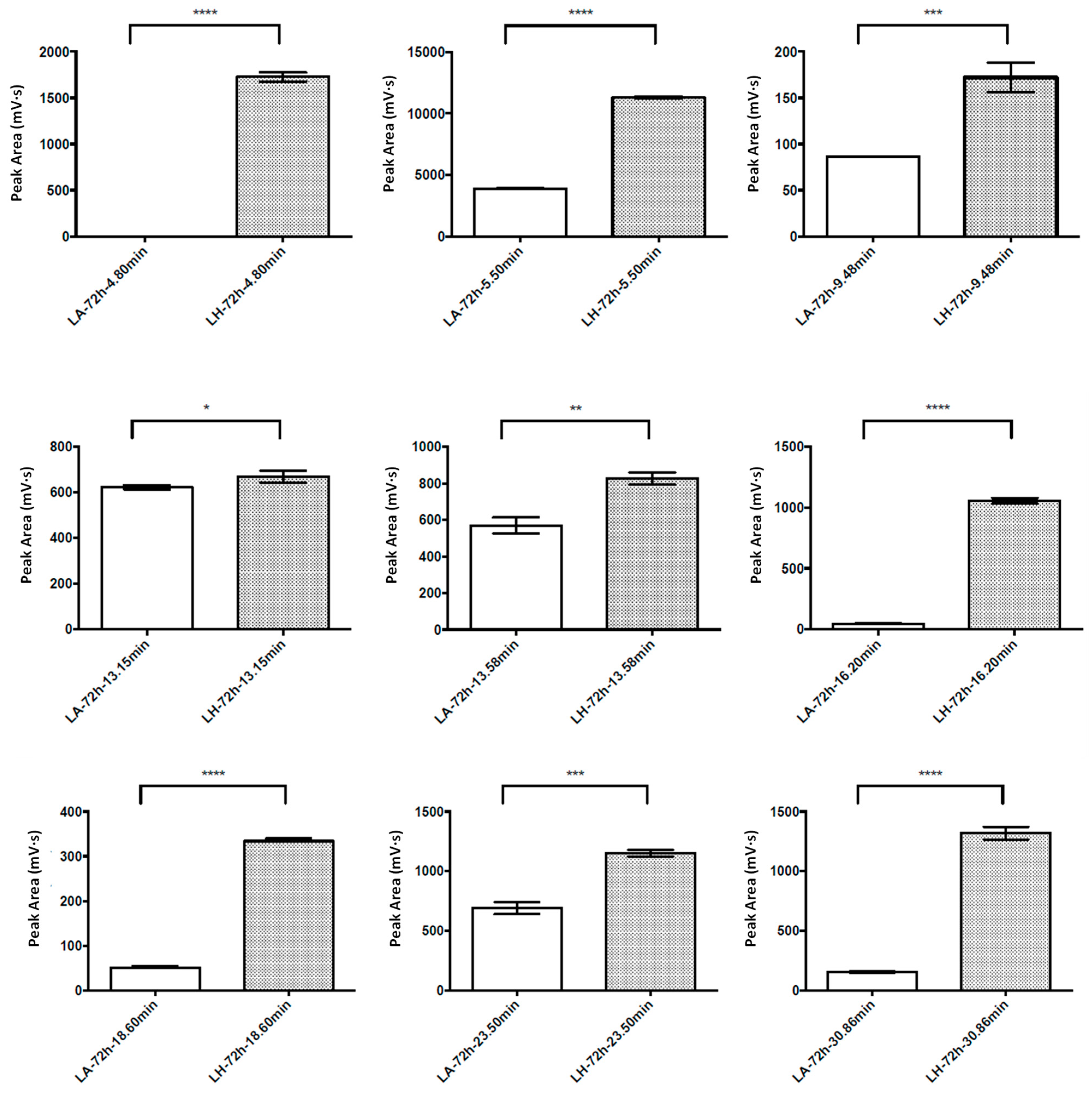
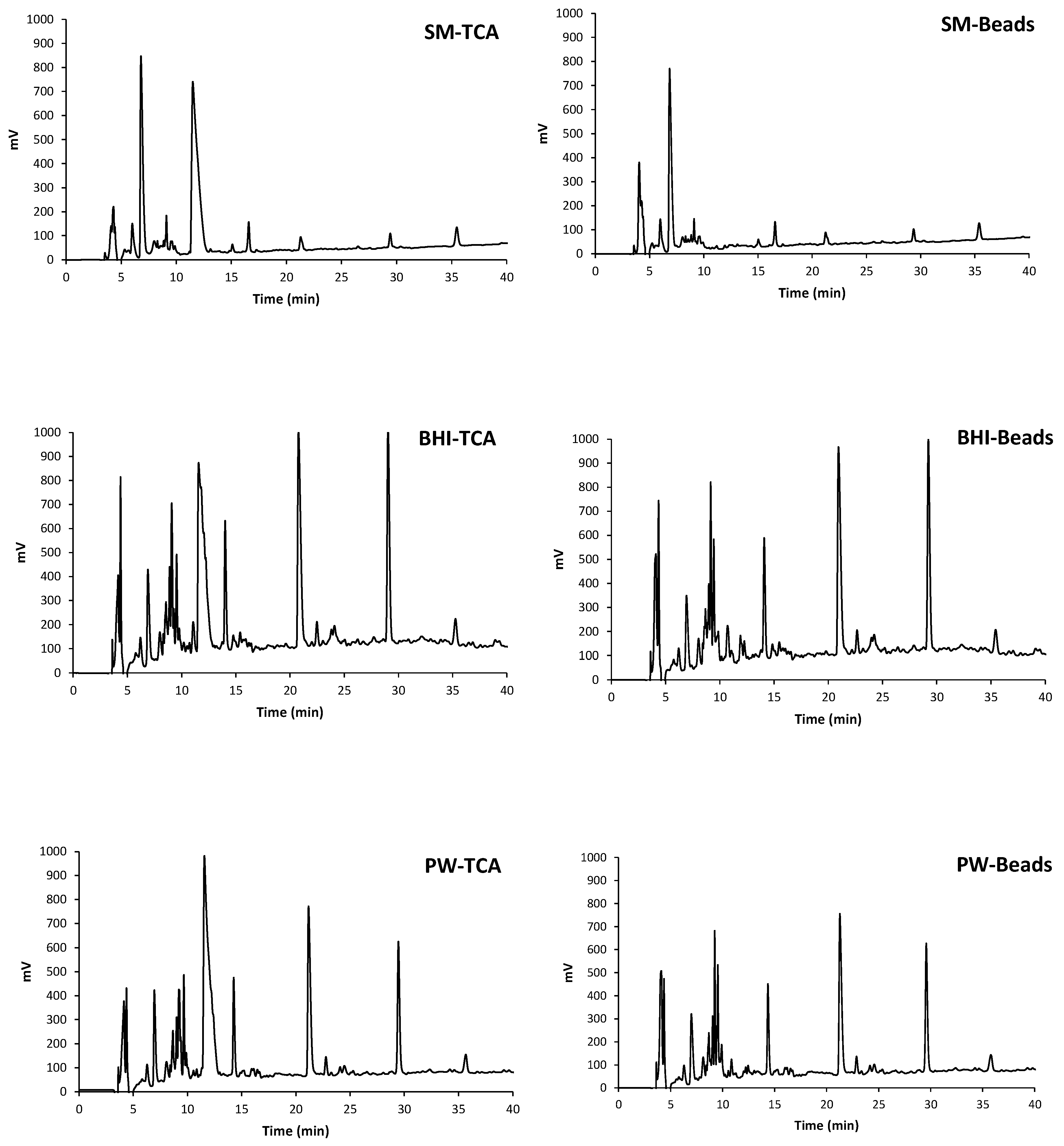
3.1. Evaluation of the Fermentation Activity by RP-HPLC

A selection of ten starter cultures (see Section 2.3 for details), isolated from meat and dairy products and identified in previous works [18,19,20,21,22], was tested for their ability to grow in aerobic and anaerobic conditions. The selected strains were inoculated in reconstituted SM and screened via RP-HPLC to assess their proteolytic activity. The preliminary outcomes highlighted a higher activity, chromatographically evaluated, of L. acidophilus LA 14 (ref. 1004) and L. helveticus (ref. LH, field strain) for the production of peptide components in fermented milk. Therefore, such selected microorganisms were submitted to more detailed investigations by monitoring their activity at different times (between 12 h and 400 h upon fermentation).
Based on data reported by other authors [27,28], in order to get the separation of the major number of peaks belonging to the desirable peptide fraction, several chromatographic experimental conditions were properly selected. Accordingly, the gradient profile, along with column temperature and flow rate, was progressively adapted by relying upon the heuristic “trial-and-error” method [29]. The preliminary optimization step was carried out by using a common reversed phase stationary phase. The addition of 0.1% (v) TFA as lipophilic ion-pairing additive to both components of the mobile phase (water and ACN) revealed to be an optimal compromise to guarantee the reproducibility of chromatographic runs [30,31,32,33,34]. Additionally, the simultaneous optimization of both temperature (from 25 to 40 °C) and gradient steepness demonstrated being a powerful tool to control band spacing and separation of the complex mixtures under investigation [32,33,34]. The optimal identified experimental conditions were screened on several RP columns [29,35] (See Section 2.2 for details). The Gemini NX column proved to be the best performing one in terms of separation and resolution factor and, therefore, was selected to carry out the chromatographic analyses applied to the real samples.
The scanning of microorganism activity and successive selection were made by following the chromatographic trend of some reference peaks during the gradient elution. In Figure 1, the chromatographic profiles obtained from SM fermented with an L. acidophilus strain and L. helveticus, in the timeframe 12–400 h, are shown.
Based on these preliminary results, and in line with data reported by other authors [13,36], L. helveticus showed a higher likelihood, and more constant over the time, proteolytic activity than L. acidophilus. Figure 2 shows the comparison between the two strains’ activities (upon 72 h fermentation), based on area values of reference peaks. Moreover, a comparison of antioxidant and ACEi activities between the two LAB strains, which confirms the higher properties of hydrolysate from L. helveticus (upon 12 h fermentation), is exemplarily reported in Table 1.
Detailed mean area values ± SD (n = 3) are reported in Table S2 (Supplementary Material).
Therefore, L. helveticus stood out as a producer of BPs in fermented milk, further investigations were deepened to explore its use as potential candidate for BPs production. With the aim of maximizing L. helveticus activity, the results obtained from fermented SM were compared with those produced by inoculating BHI and PW as substrates (Figure 3). In this phase, two extraction strategies were appraised: the more “conventional” extraction by centrifugation in the presence of TCA at 4 °C [37], and the extraction by using glass beads [38]. Comparing these two extraction methods, it was possible to determine the potential cell membrane disruption for the recovery of intracellular bioactive compounds. The results highlighted no significant differences between the two methods. Indeed, the chromatographic profiles obtained on cell lysates produced by mechanical glass bead disruption were found to be similar to those achieved by treatment with TCA. This would suggest that the majority of BPs are in the culture medium and any other intracellular component did not influence their concentration.
The elution profiles shown in Figure 3 highlighted substantial differences in terms of proteolytic activity by changing the substrate. On the contrary, a negligible impact was recorded by comparing the profiles relative to the two extractive protocols (TCA vs. beads) for each substrate. It can be observed that hydrolysates from BHI and PW showed similar and richer chromatographic profiles with respect to hydrolysate from SM.

3.2. Antioxidant and ACE-Inhibitory Activity of Peptide Hydrolysates

Several food-derived peptides have been found to exhibit antioxidant properties generally without noticeable side effects and with high activity and easy absorption [39,40]. Several properties such as structure, hydrophobicity, amino acids composition and their specific positioning and configuration in the peptide sequence, seem to significantly influence the antioxidant activity of peptides [41,42]. Furthermore, hydrolysate concentration and molecular weight were also found to affect antioxidant properties [43].
In the present study, the antioxidant activity of the hydrolysates from L. helveticus fermented substrates was evaluated and compared by relying upon the free radical scavenging activity (measured through the ABTS assay) and the reducing capacity (measured through the FRAP assay). As shown in Table 2, the hydrolysates obtained by using BHI as starting fermentation substrates showed the highest values of antioxidant activity determined by FRAP and ABTS assays. Instead, a controversial behavior was observed for the hydrolysates from SM and PW substrates; indeed, while in the former case the lowest antioxidant activity was measured by ABTS, in the latter the lowest FRAP activity was obtained.
Concerning the ACEi activity, L. helveticus strains have been designated among the preferential sources to achieve a hypotensive effect, thanks to the excellent microbial proteolytic system involved in conversion of milk proteins into oligopeptides [16,43]. Accordingly, we tested the ACEi activity of L. helveticus hydrolysates produced in the presence of the three selected substrates. The results highlighted an in vitro activity close or over 50%, comparable with data reported by other authors [12,13,43]. The higher ACEi activity in the presence of PW with respect to the SM (Table 2) could be related to the higher peptide content of the starting substrate which, in turn, supports or integrates the efficiency of the proteolytic system. As shown in Table 2, the lowest ACEi activity was obtained for BHI hydrolysates and, outstandingly, was inversely correlated to the antioxidant activity (both in terms of ABTS and FRAP assay). A similar trend was recorded between ACEi and FRAP assay when PW was the starting substrate; in fact the highest ACEi activity matched the lowest FRAP value.

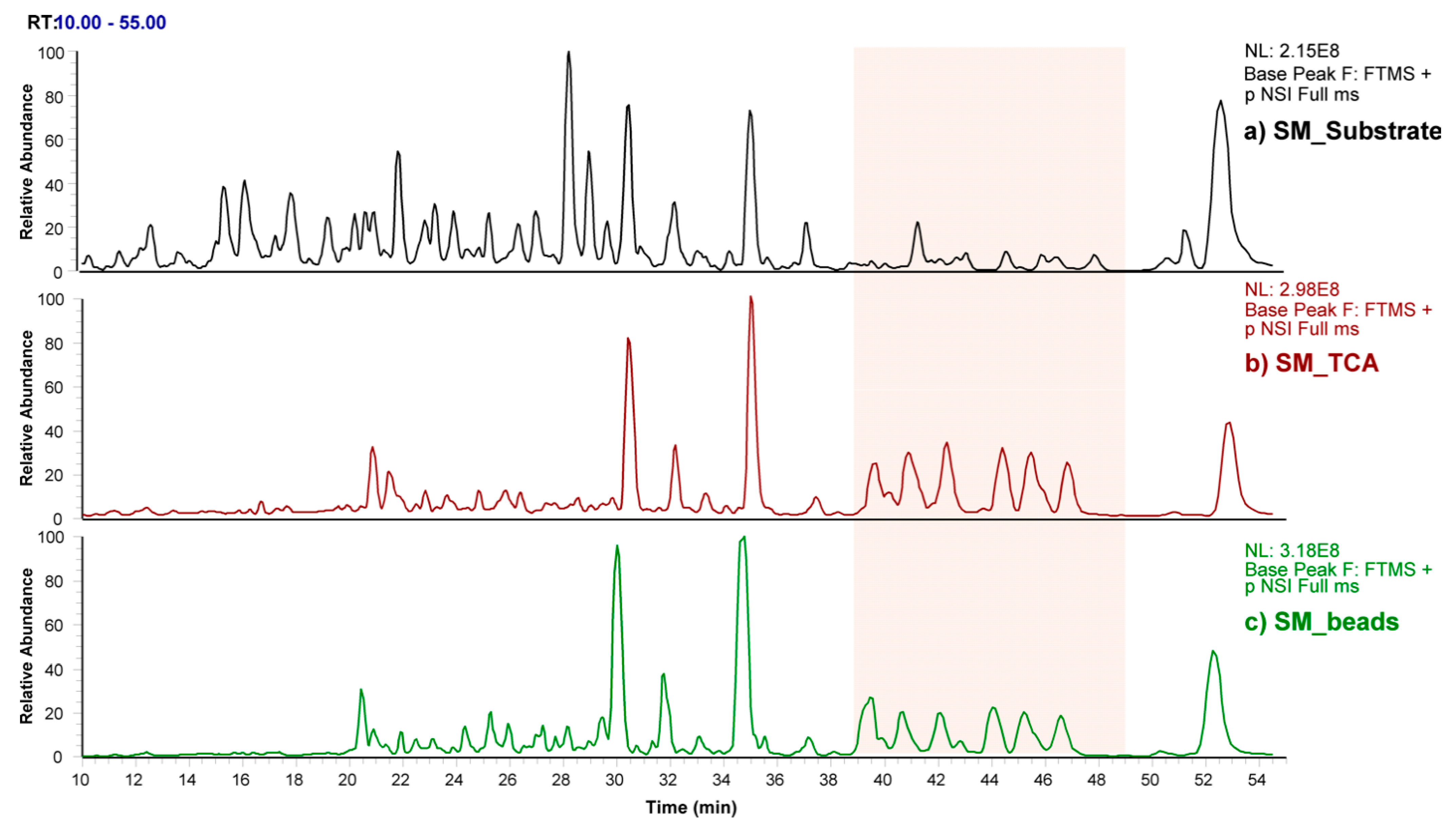
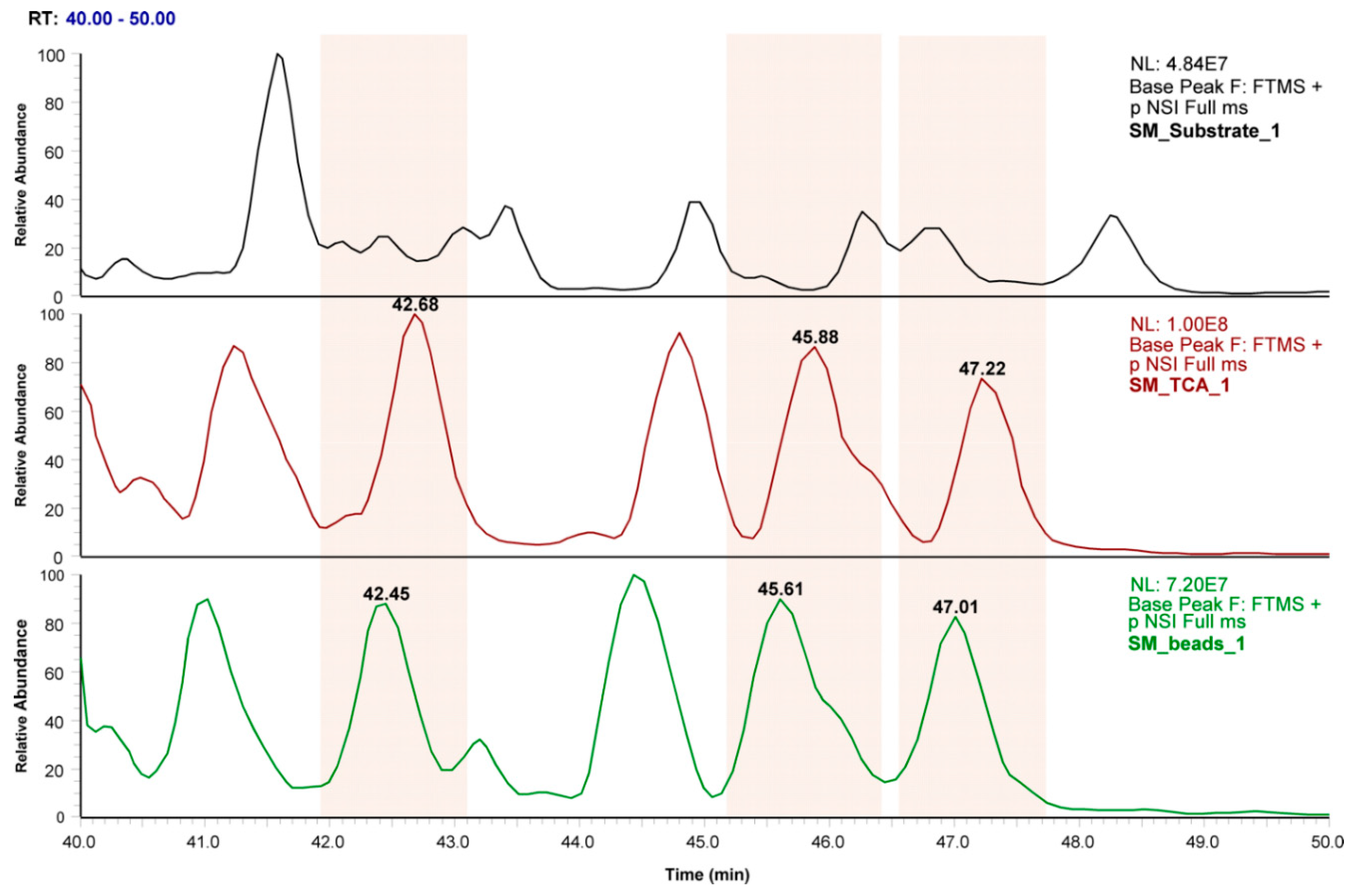
3.3. Profiling of SM Samples by Mass Spectrometry

To highlight any difference between the produced hydrolysates (by treatment in TCA or glass beads) and the starting substrate (not submitted to fermentation, and extracted by TCA analogously to the fermented samples), a nano-HPLC-HRMS analysis was carried out. This part of the study, preliminarily addressing profiling of SM samples, allowed distinguishing of those fractions likely responsible for either the above antioxidant, ACEi, or both, activities.
Figure 4 compares the MS profiles (total ion current—TIC) of the three samples analyzed by HRMS (SM-substrate, SM-TCA, and SM-beads) zooming into the separative gradient interval characterized by the presence of the main peaks (10–55 min). Overall, the MS profiles showed some overlapping peaks flanked by some other peaks uniquely present in the fermented samples (SM-TCA and SM-beads) with respect to the SM-substrate. Therefore, the preliminary identification of fractions in SM was focused on the TIC interval characterized by an appreciable diversity in terms of newly formed species (Figure 5) due to the sample treatment with L. helveticus (40–50 min).
From the zoomed-in window (40–50 min) shown in Figure 5, three major peaks in the profile of the two fermented samples with respect to the substrate were highlighted. By in-depth investigation of the new species eluting under those peaks, the newly formed species were defined on the basis of their m/z and shown in Figures S1–S3 (Supplementary Material), where the full MS spectra of each peak, with RT 42.57 ± 0.16 min, 45.75 ± 0.19, and 47.12 ± 0.15 min, respectively, are reported for the three samples.

4. Conclusions

In the present work, the proteolytic activity of several starter cultures, isolated from meat and dairy products, sampled at different fermentation times and grown in different substrates, was evaluated. The fine optimization of the RP-HPLC method allowed screening of the proteolytic activity by following the chromatographic profile of selected peaks during the gradient elution. Of the investigated strains, L. helveticus showed a higher likelihood, and more constant over the time, proteolytic activity and was, therefore, selected for further investigations. The results of the chromatographic analyses, coupled with in vitro antioxidant (ABTS and FRAP) and antihypertensive (ACE inhibitory) assays, evidenced a remarkable dependence of L. helveticus activity on the starting substrate. This aspect could be gainfully exploited when a specific activity needs to be privileged to functionalize a food with pro-health peptides. Moreover, a nano-HPLC-HRMS system was implemented to discriminate those fractions which, differently from the non-fermented substrates, would be potentially responsible for the found activity.
The preliminary results obtained in this study underline the necessity of considering a multiplicity of factors to exploit the potential bioactivity associated with peptides produced upon fermentation. Evidently, innovative separation and purification processes are mandatory to isolate the desirable peptide fractions following an initial screening, being aware, at the same time, that a significant variation in terms of bioactivities can be expected by the use of purified peptides over the non-purified hydrolysates. All these aspects become particularly crucial when large-scale production of healthful food or food ingredients is intended. In this context, more in-depth investigations addressing peptide sequence identification would enable the molecular characterization of such bioactive components. This, in turn, would also result in being helpful to reach a better knowledge of specific targets.

Supplementary Materials

The following are available online at https://0-www-mdpi-com.brum.beds.ac.uk/2076-3417/11/2/811/s1, Table S1: Time sampling scheme for strain fermentation activity evaluation, Table S2: Comparison between L. acidophilus (LA 14) and L. helveticus (LH) activity upon 72 h fermentation, reported as mean area values of reference peaks (measured in mV·s) ± standard deviation (SD), n = 3 for each selected peak., Figure S1: Full MS spectrum of the three samples a) SM-Substrate, b) SM-TCA, c) SM-Beads, related to the RT including the peak at 42.57 ± 0.16 min. The m/z of the newly formed peptides are highlighted in red., Figure S2: Full MS spectrum of the three samples a) SM-Substrate, b) SM-TCA, c) SM-Beads, related to the RT including the peak at at 45.75 ± 0.19 min. The m/z of the newly formed peptides are highlighted in red. Figure S3: Full MS spectrum of the three samples a) SM-Substrate, b) SM-TCA, c) SM-Beads, related to the RT including the peak at at 47.12 ± 0.15 min. The m/z of the newly formed peptides are highlighted in red.

Author Contributions

Conceptualization, L.C. and B.T.C.-G.; investigation, F.I., A.A.A., and L.G.; writing—original draft preparation, F.I.; data curation, L.R. and F.B.; writing—review and editing, F.I., F.B., and L.C.; supervision, L.C. All authors have read and agreed to the published version of the manuscript.

Funding

This research received no external funding.

Institutional Review Board Statement

Not applicable.

Informed Consent Statement

Not applicable.

Data Availability Statement

Data contained within the article or supplementary material are here presented for the first time.

Conflicts of Interest

The authors declare no conflict of interest.

References

  1. Beltrán-Barrientos, L.M.; Hernández-Mendoza, A.; Torres-Llanez, M.J.; González-Córdova, A.F.; Vallejo-Córdoba, B. Invited review: Fermented milk as antihypertensive functional food. J. Dairy Sci. 2015, 99, 4099–4110. [Google Scholar] [CrossRef] [PubMed] [Green Version]
  2. Prasanna, P.H.P.; Grandison, A.S.D.; Charalampopoulos, D. Bifidobacteria in milk products: An overview of physiological and biochemical properties, exopolysaccharide production, selection criteria of milk products and health benefits. Food Res. Int. 2014, 55, 247–262. [Google Scholar] [CrossRef]
  3. Chakrabarti, S.; Guha, S.; Majumder, K. Food-Derived Bioactive Peptides in Human Health: Challenges and Opportunities. Nutrients 2018, 10, 1738. [Google Scholar] [CrossRef] [PubMed] [Green Version]
  4. Montesano, D.; Gallo, M.; Blasi, F.; Cossignani, L. Biopeptides from vegetable proteins: New scientific evidences. Curr. Opin. Food Sci. 2020, 31, 31–37. [Google Scholar] [CrossRef]
  5. Rai, A.K.; Sanjukta, S.; Jeyaram, K. Production of angiotensin I converting enzyme inhibitory (ACE-I) peptides during milk fermentation and their role in reducing hypertension. Crit. Rev. Food Sci. Nutr. 2017, 57, 2789–2800. [Google Scholar] [CrossRef]
  6. Saito, T.; Nakamura, H.; Kitazawa, H.; Kawai, Y.; Itoh, T. Isolation and structural analysis of antihypertensive peptides that exist naturally in Gouda cheese. J. Dairy Sci. 2000, 83, 1434–1440. [Google Scholar] [CrossRef]
  7. Schanbacher, F.L.; Talhouk, R.S.; Murray, F.A.; Gherman, L.I.; Willet, L.B. Milk borne bioactive peptides. Int. Dairy J. 1998, 8, 393–403. [Google Scholar] [CrossRef]
  8. Hafeez, Z.; Cakir-Kiefer, C.; Roux, E.; Perrin, C.; Miclo, L.; Dary-Mourot, A. Strategies of producing bioactive peptides from milk proteins to functionalize fermented milk products. Food Res. Int. 2014, 63, 71–80. [Google Scholar] [CrossRef]
  9. Power, O.; Jakeman, P.; FitzGerald, R.J. Antioxidative peptides: Enzymatic production, in vitro and in vivo antioxidant activity and potential applications of milk-derived antioxidative peptides. Amino Acids 2013, 44, 797–820. [Google Scholar] [CrossRef]
  10. Rana, S.; Bajaj, R.; Mann, B. Characterization of antimicrobial and antioxidative peptides synthesized by L. rhamnosus C6 fermentation of milk. Int. J. Pept. Res. Ther. 2018, 24, 309–321. [Google Scholar] [CrossRef]
  11. Pihlanto, A.; Virtanen, T.; Korhonen, H. Angiotensin I converting enzyme (ACE) inhibitory activity and antihypertensive effect of fermented milk. Int. Dairy J. 2010, 20, 3–10. [Google Scholar] [CrossRef]
  12. Muguerza, B.; Ramos, M.; Sanchez, E.; Manso, M.A.; Miguel, M.; Aleixandre, A.; Delgado, M.A.; Recio, I. Antihypertensive activity of milk fermented by Enterococcus faecalis strains isolated from raw milk. Int. Dairy Res. 2006, 16, 61–69. [Google Scholar] [CrossRef]
  13. Wang, J.; Li, C.; Xue, J.; Yang, J.; Zhang, Q.; Zhang, H.; Chen, Y. Fermentation characteristics and angiotensin I-converting enzyme-inhibitory activity of Lactobacillus helveticus isolate H9 in cow milk, soy milk, and mare milk. J. Dairy Sci. 2015, 98, 3655–3664. [Google Scholar] [CrossRef] [PubMed] [Green Version]
  14. Khan, M.U.; Pirzadeh, M.; Förster, C.Y.; Shityakov, S.; Shariati, M.A. Role of milk-derived antibacterial peptides in modern food biotechnology: Their synthesis, applications and future perspectives. Biomolecules 2018, 8, 110. [Google Scholar] [CrossRef] [PubMed] [Green Version]
  15. Davoodi, S.H.; Shahbazi, R.; Esmaeili, S.; Sohrabvandi, S.; Mortazavian, A.; Jazayeri, S.; Taslimi, A. Health-Related Aspects of Milk Proteins. Iran. J. Pharm. Res. 2016, 15, 573–591. [Google Scholar] [PubMed]
  16. Tagliazucchi, D.; Martini, S.; Solieri, L. Bioprospecting for bioactive peptide production by lactic acid bacteria isolated from fermented dairy food. Fermentation 2019, 5, 96. [Google Scholar] [CrossRef] [Green Version]
  17. Mohanty, D.; Jena, R.; Choudhury, P.K.; Pattnaik, R.; Mohapatra, S.; Saini, M.R. Milk derived antimicrobial bioactive peptides: A review. Int. J. Food Prop. 2016, 19, 837–846. [Google Scholar] [CrossRef]
  18. Cenci-Goga, B.T.; Karama, M.; Sechi, P.; Iulietto, M.F.; Grispoldi, L.; Selvaggini, R.; Ceccarelli, M.; Barbera, S. Fate of selected pathogens in spiked «SALAME NOSTRANO» produced without added nitrates following the application of NONIT™ technology. Meat Sci. 2018, 139, 247–254. [Google Scholar] [CrossRef]
  19. Cenci-Goga, B.T.; Karama, M.; Sechi, P.; Iulietto, M.F.; Novelli, S.; Selvaggini, R.; Barbera, S. Effect of a novel starter culture and specific ripening conditions on microbiological characteristics of nitrate-free dry-cured pork sausages. Ital. J. Anim. Sci. 2016, 15, 358–374. [Google Scholar] [CrossRef]
  20. Cenci-Goga, B.T.; Karama, M.; Sechi, P.; Iulietto, M.F.; Novelli, S.; Selvaggini, R.; Mattei, S. Growth inhibition of selected microorganisms by an association of dairy starter cultures and probiotics. Ital. J. Anim. Sci. 2015, 14, 246–250. [Google Scholar] [CrossRef] [Green Version]
  21. Cenci-Goga, B.T.; Rossitto, P.V.; Sechi, P.; Parmegiani, S.; Cambiotti, V.; Cullor, J.S. Effect of selected dairy starter cultures on microbiological, chemical and sensory characteristics of swine and venison (Damadama) nitrite-free dry-cured sausages. Meat Sci. 2012, 90, 599–606. [Google Scholar] [CrossRef] [PubMed]
  22. Clementi, F.; Cenci Goga, B.T.; Trabalza Marinucci, M.; Di Antonio, E. Use of selected starter cultures in the production of farm manufactured goat cheese from thermized milk. Ital. J. Food Sci. 1998, 10, 41–56. [Google Scholar]
  23. Urbani, E.; Blasi, F.; Simonetti, M.S.; Chiesi, C.; Cossignani, L. Investigation on secondary metabolite content and antioxidant activity of commercial saffron powder. Eur. Food Res. Technol. 2016, 242, 987–993. [Google Scholar] [CrossRef]
  24. Rocchetti, G.; Pagnossa, J.P.; Blasi, F.; Cossignani, L.; HilsdorfPiccoli, R.; Zengin, G.; Montesano, D.; Cocconcelli, P.S.; Lucini, L. Phenolic profiling and in vitro bioactivity of Moringa oleifera leaves as affected by different extraction solvents. Food Res. Int. 2020, 127, 108712. [Google Scholar] [CrossRef] [PubMed]
  25. Rocchetti, G.; Blasi, F.; Montesano, D.; Ghisoni, S.; Marcotullio, M.C.; Sabatini, S.; Cossignani, L.; Lucini, L. Impact of conventional/non-conventional extraction methods on the untargeted phenolic profile of Moringa oleifera leaves. Food Res. Int. 2019, 115, 319–327. [Google Scholar] [CrossRef] [PubMed]
  26. Degani, G.; Altomare, A.; Digiovanni, S.; Arosio, B.; Fritz, G.; Raucci, A.; Aldini, G.; Popolo, L. Prothrombin is a binding partner of the human receptor of advanced glycation end products. J. Biol. Chem. 2020, 295, 12498–12511. [Google Scholar] [CrossRef]
  27. Chen, G.W.; Tsai, J.S.; Pan, B.S. Purification of angiotensin I-converting enzyme inhibitory peptides and antihypertensive effect of milk produced by protease-facilitated lactic fermentation. Int. Dairy J. 2007, 17, 641–647. [Google Scholar] [CrossRef]
  28. Hernandez-Ledesma, B.; Amigo, L.; Ramos, M.; Recio, I. Application of high-performance liquid chromatography-tandem mass spectrometry to the identification of biologically active peptides produced by milk fermentation and simulated gastrointestinal digestion. J. Chromatogr. A 2004, 1049, 107–114. [Google Scholar] [CrossRef]
  29. Ianni, F.; Sardella, R.; Lisanti, A.; Gioiello, A.; Cenci Goga, B.T.; Lindner, W.; Natalini, B. Achiral-chiral two-dimensional chromatography of free amino acids in milk: A promising tool for detecting different levels of mastitis in cows. J. Pharm. Biomed. Anal. 2005, 116, 40–46. [Google Scholar] [CrossRef]
  30. Mant, C.T.; Hodges, R.S. HPLC of Biological Macromolecules; Gooding, K.M., Regnier, F.E., Eds.; Marcel Dekker: New York, NY, USA, 2002; pp. 433–511. [Google Scholar]
  31. Chen, Y.; Mehok, A.R.; Mant, C.T.; Hodges, R.S. Optimum concentration of trifluoroacetic acid for reversed-phase liquid chromatography of peptides revisited. J. Chromatogr. A 2004, 1043, 9–18. [Google Scholar] [CrossRef]
  32. Dolan, J.W.; Snyder, L.R.; Blanc, T.; Van Heukelem, L. Selectivity differences for C18 and C8 reversed-phase columns as a function of temperature and gradient steepness: I. Optimizing selectivity and resolution. J. Chromatogr. A 2000, 897, 37–50. [Google Scholar] [CrossRef]
  33. Hancock, W.S.; Chloupek, R.C.; Kirkland, J.J.; Snyder, L.R. Temperature as a variable in reversed-phase high-performance liquid chromatographic separations of peptide and protein samples: I. Optimizing the separation of a growth hormone tryptic digest. J. Chromatogr. A 1994, 686, 31–43. [Google Scholar] [CrossRef]
  34. Chloupek, R.C.; Hancock, W.S.; Marchylo, B.A.; Kirkland, J.J.; Boyes, B.E.; Snyder, L.R. Temperature as a variable in reversed-phase high-performance liquid chromatographic separations of peptide and protein samples. II. Selectivity effects observed in the separation of several peptide and protein mixtures. J. Chromatogr. A 1994, 686, 45–59. [Google Scholar] [CrossRef]
  35. Sardella, R.; Lisanti, A.; Marinozzi, M.; Ianni, F.; Natalini, B.; Blanch, G.P.; Ruiz del Castillo, M.L. Combined monodimensional chromatographic approaches to monitor the presence of D-amino acids in cheese. Food Control 2013, 34, 478–487. [Google Scholar] [CrossRef] [Green Version]
  36. Chen, Y.; Liu, W.; Xue, J.; Yang, J.; Chen, X.; Shao, Y.; Kwok, L.; Bilige, M.; Mang, L.; Zhang, H. Angiotensin-converting enzyme inhibitory activity of Lactobacillus helveticus strains from traditional fermented dairy foods and antihypertensive effect of fermented milk of strain H9. J. Dairy Sci. 2014, 97, 6680–6692. [Google Scholar] [CrossRef] [Green Version]
  37. Solieri, L.; Rutella, G.S.; Tagliazucchi, D. Impact of non-starter lactobacilli on release of peptides with angiotensin-converting enzyme inhibitory and antioxidant activities during bovine milk fermentation. Food Microbiol. 2015, 51, 108–116. [Google Scholar] [CrossRef] [Green Version]
  38. Pan, D.; Luo, Y.; Tanokura, M. Antihypertensive peptides from skimmed milk hydrolysate digested by cell-free extract of Lactobacillus helveticus JCM1004. Food Chem. 2005, 91, 123–129. [Google Scholar] [CrossRef]
  39. Li, B.; Chen, F.; Wang, X.; Ji, B.; Wu, Y. Isolation and identification of antioxidant peptides from porcine collagen hydrolysate by consecutive chromatography and electrospray ionization-mass spectrometry. Food Chem. 2007, 102, 1135–1143. [Google Scholar] [CrossRef]
  40. Sarmadi, B.H.; Ismail, A. Antioxidative peptides from food proteins: A review. Peptides 2010, 31, 1949–1956. [Google Scholar] [CrossRef]
  41. Chen, H.M.; Muramoto, K.; Yamauchi, F.; Nokihara, K. Antioxidant activity of designed peptides based on the antioxidative peptide derived from digests of a soybean peptide. J. Agric. Food Chem. 1996, 44, 2619–2623. [Google Scholar] [CrossRef]
  42. Chen, H.M.; Muramoto, K.; Yamauchi, F. Structural analysis of antioxidative peptides from soybean β-conglycinin. J. Agric. Food Chem. 1995, 43, 574–578. [Google Scholar] [CrossRef]
  43. Chen, Y.; Li, C.; Xue, J.; Kwok, L.Y.; Yang, J.; Zhang, H.; Menghe, B. Characterization of angiotensin-converting enzyme inhibitory activity of fermented milk produced by Lactobacillus helveticus. J. Dairy Sci. 2015, 98, 5113–5124. [Google Scholar] [CrossRef] [PubMed]
Figure 1. Chromatographic profiles obtained from SM substrate fermented with (ac) L. acidophilus strain (strain LA 14) and (df) L. helveticus (LH field strain), at time points of 12 h, 72 h, and 400 h, respectively. The hydrolysates were extracted by TCA at 4 °C (see Section 2.4 for details). An enlarged portion of each chromatogram in the time-window 15–30 min is reported to evidence the richness of the chromatographic profile relatively to minor intensity detected peaks. The interference peak detected at 12 min corresponds to the TCA additive.
Applsci 11 00811 g001
Figure 2. Graphical representation of the comparison between L. acidophilus (LA 14) and L. helveticus (LH) activity upon 72 h fermentation, based on area values of reference peaks (measured in mV·s). Data are the mean ± standard deviation (SD) of three replicates. Data were analyzed by a Student’s unpaired t-test. **** p < 0.0001, *** p < 0.001, ** p < 0.01, * p < 0.05.
Applsci 11 00811 g002
Figure 3. Chromatographic analysis of L. helveticus hydrolysates obtained with the three substrates (SM, BHI, and PW) and extracted by TCA at 4 °C or by using glass beads. The interference peak detected at 12 min corresponds to the TCA additive.
Applsci 11 00811 g003
Figure 4. FTMS (Fourier Transform Mass Spectrometry)-Nanospray Ionization (NSI) spectrum: TIC (total ion current) zoomed into the separative gradient interval 10–55 min of SM-substrate, SM-TCA, and SM-beads samples acquired in data-dependent acquisition (DDA) mode. Light red square highlights the interval characterized by the greater diversity in terms of newly formed species due to the sample treatment.
Applsci 11 00811 g004
Figure 5. TIC (total ion current) zoomed-in the separative gradient interval 40–50 min of SM-Substrate, SM-TCA, and SM-Beads samples acquired in DDA mode. The light red squares highlight the peaks of interest.
Applsci 11 00811 g005
Table 1. Comparison of the antioxidant (by ABTS and FRAP assays) and ACEi activities of hydrolysates after 12 h fermentation with L. acidophilus and L. helveticus. Values are expressed as mean values ± standard deviation (SD), n = 2.
StrainABTS (µmol TE/mL)FRAP (µmol TE/mL)ACEi Activity%
L. acidophilus (LA 14)12.07 ± 0.0023.14 ± 0.0036.81 ± 2.62
L. helveticus (LH)42.07 ± 0.0030.06 ± 0.0074.37 ± 3.82
Table 2. Summary of antioxidant (by ABTS and FRAP assays) and ACEi activities of hydrolysates. Values are expressed as mean values ± standard deviation (SD), n = 2.
HydrolysateABTS (µmol TE/mL)FRAP (µmol TE/mL)ACEi Activity% *
SM-TCA17.00 ± 0.0023.02 ± 0.0081.77 ± 1.37
BHI-TCA245.40 ± 0.0029.55 ± 0.0053.65 ± 1.82
PW-TCA55.67 ± 0.009.58 ± 0.0087.30 ± 1.59
SM-Beads26.27 ± 0.0020.84 ± 0.0070.12 ± 3.81
BHI-Beads261.40 ± 0.0025.12 ± 0.0049.59 ± 0.49
PW-Beads64.87 ± 0.0010.53 ± 0.0097.75 ± 1.87
* Measured by reversed phase-high performance liquid chromatography (RP-HPLC).
Publisher’s Note: MDPI stays neutral with regard to jurisdictional claims in published maps and institutional affiliations.

