Article

Antimicrobial Cocktail Combining Specific Peptide Extracts from Native Probiotic Bacteria Hamper Adulteration of Ready-to-Eat Mango Wedges

Biofood and Nutraceutics Research and Development Group, Faculty of Engineering in Agricultural and Environmental Sciences, Technical University of the North, Av. 17 de Julio s-21, Barrio El Olivo, 100150 Ibarra, Ecuador
*
Author to whom correspondence should be addressed.
Submission received: 2 December 2020 / Revised: 13 December 2020 / Accepted: 16 December 2020 / Published: 4 March 2021
(This article belongs to the Special Issue Bioactive Peptides from Foods)

Abstract

:
Consumption of ready-to-eat chopped fruits sold in the streets is a concern, as such activities are outside the regulation and protection in most developing countries. Ready-to-eat mangos are commonly sold as wedges in plastic cups at ambient temperature by mobile vendors in Ecuador, thus they are prone to contamination by bacteria, which poses a safety issue of concern. This work aimed to evaluate the effect of several antimicrobial cocktails consisting of previously designed specific peptide extract combinations from two probiotic bacteria Lactobacillus plantarum UTNCys5-4 and Lactococcus lactis subsp. lactis UTNGt28, along with nisin, a commercial food additive, on mango wedges artificially inoculated with a logarithmic phase culture of a five-strain bacterial mixture (FSBM). Preliminary bacteriological analysis of mango wedges purchased from mobile vendors showed the presence of multiple antibiotic-resistant isolates such E. coli spp., Enterobacter spp., Shigella spp., Salmonella spp., along with yeasts and molds, indicating non-compliance with the food safety standards. The results revealed that two antimicrobial cocktails, T2 and T5, containing cell-free supernatant based (CFS) and precipitated peptides (PP) based cocktails from UTNCys5-4 and UTNGt28 strains applied at dose 1:3 (v/v), were the most efficient combinations that inhibited the colonization of total bacterial counts with 56.03% and 55.61% in mango wedges stored with refrigeration. The reduction of total E. coli counts was 64.93%, while Salmonella and Shigella counts were reduced by 98.09% and 97.93%, respectively, when mango wedges were treated with T5-cocktail. The commercial nisin inhibited total Salmonella spp. counts by 40.13%, while E. coli spp. and Shigella spp. diminished by 28.20% and 37.22%, respectively. Moreover, we showed that T5 but not T7 (nisin) damaged the target cell integrity, thereby eventually inhibiting their growth and reproduction. The selected antimicrobial cocktails exerted a bacteriolytic effect by killing the FSBM simultaneously in a fruit matrix and preventing their accumulation in mango wedges. Furthermore, there is a possibility of using peptide combinatorial treatments to combat drug-resistant bacteria in ready-to-eat fruits.

1. Introduction

Ecuador, a megabiodiverse country, is an ideal place for tropical fruit lovers, where you can find myriad varieties of fruits with unique flavors. Due to the high climatic variation and tropical temperature, the fruits are commercialized throughout the year.
Mango (Mangifera indica) is one of the most important tropical fruits in the world, as ascorbic acid, carotenoids, polyphenols, and antioxidants are present in the edible part of the fruit [1]. Mango production in Ecuador dates back dozens of years as a highly desirable seasonal fruit, with an exquisite sweet taste, especially the traditional variety. Mango is considered a noble plant since it does not require watering [2]. Ecuador is the second mango exporting country to Europe, after Brazil; unfortunately, there is insufficient support for sustainable growth production by the agricultural sector as the exportation was not expanded to other countries except Germany, Spain and, the United Kingdom [3]. Usually, at the local level, mango fruits are sold in the market at different ripening stages, from green, green red to yellow. The change in maturation is expressed when the color of the peel changes from green to yellow, in addition, the pulp color is the best indicator of the physiological maturity of the mango [4]. Due to the environmental conditions (temperate climate over the year) in Ecuador, with inappropriate storage, these fruits are susceptible to contamination by spoilage or pathogenic microorganisms. To reduce damage due to early deterioration, mobile vendors are selling the fruits as ready-to-eat wedges in plastic containers in the street, near parks, or public transportation. This approach is very widespread in Ecuador. These foods are low-priced, but the risk of infection is elevated, as mobile sale is not regulated. In the capital of Quito, more than 30,000 mobile vendors were registered, eight sellers per 1000 habitants. A recent report presented by the Ministry of Health indicated that 47% of 4000 food samples analyzed in 2018 did not meet food quality standards [5]. Due to the poor handling and lack of control at the point of sale by the authorities, the likelihood of fruit contamination is elevated. Tropical fruits that are commercially available and sold as cut fruits may be contaminated at harvest and the proliferation of microorganisms can occur until they reach their sale point [6]. In cut form, tropical fruits are highly perishable with a very short shelf life and physiological deterioration. A salmonellosis outbreak due to the consumption of mangos imported from Peru and Brazil was registered in 2001 in the USA, and the epidemiological investigation indicated that the water used in the hydrothermal treatment was responsible for the contamination [7]. The detection of Salmonella spp. and E. coli in fresh-cut melons, cucumber, lettuce, tomatoes, and Red Delicious apples raises concerns about the safety of fruit consumption and consumer health [8,9,10,11,12,13]. However, it is important to optimize the sale condition to avoid fruit deterioration along with expanding vendor training on fruit manipulation, storage, and good production practices. The use of antimicrobial peptides secreted from animals, plants, or bacteria was proposed as a viable alternative for food preservation [14,15]. Among these, lactic acid bacteria (LAB) are considered the most outstanding microorganisms in the process of fermentation, which produce organic acids, other metabolites, and antimicrobial proteins known as bacteriocins [16]. Considered as safe antimicrobial agents by the United States Food and Drug Administration (USFDA), bacteriocins from LAB are excellent candidates for the control of pathogenic microorganisms present in the food industry [17,18]. The use of edible films and bacteriocin coatings does not affect the organoleptic characteristics of fruits and vegetables [19]. Additionally, their use in the preservation of cut vegetables or fruits was suggested as a promising natural approach [19]. The use of carboxymethyl cellulose (CMC) edible coatings with bacteriocin from Bacillus methylotrophicus BM47 showed a positive influence on antioxidant activity, and improvement in commercial appearance and shelf life [20].
Recently, we investigated the antimicrobial capacity of several peptide extracts from LAB strains isolated from wild tropical fruits of the Amazonian region of Ecuador and selected several candidates with high potential for use in natural food preservation [21,22]. An edible coating solution was developed to protect fresh tomatoes and Ananas comosus fruits from contamination. However, several combinations in the form of cell-free supernatant (CFS) or precipitated peptides (PP) were selected [22] as promising competitors to be tested and validated ex vitro in different raw materials. As the inhibitory effect depends on the food matrix composition, the efficiency of the peptide or peptide combinations, the dose and form of application, and the presence of one or more pathogenic bacteria in a sample at the same time, the effectiveness of each antimicrobial should be evaluated separately. In this study, we compared the inhibitory effect of seven antimicrobial cocktails consisting of a peptide extract mixture from two native LAB strains applied as CFS-cocktail and PP-cocktail on ready-to-eat mango wedges and selected an optimal inhibitory composition that diminishes the artificially inoculated five-strain target bacteria simultaneously. The native LAB strains produce two-peptide bacteriocins, plantaricin W produced by L. plantarum UTNCys5-4, and lacticin 3147, lactococcin M, and lactococcin A produced by L. lactis subsp. lactis UTNGt28) [23]. The results were compared with nisin, a commercial food additive. Considering the health problems associated with the consumption of ready-to-eat fruits, these antimicrobial cocktails might represent a solution from a safety viewpoint and could reduce the bacterial population that may be present in the fruits during manipulation.

2. Materials and Methods

2.1. Microbiological Diagnostic Quality of Mango Fruit and Fresh-Cut Wedges Purchased from the Local Market

The microbiological quality was carried out in accordance with the Ecuadorian Regulations [24,25]. Mango fruits were purchased from a local open low-cost market (two venders, V1 and V2), while the ready-to-eat wedges were purchased from mobile vendors located near the central park (V3) and central bus station (V4). Briefly, 25 g of mango peel collected independently from three fruits at different stages of ripening (2, 3, and 4) [26], 25 g of remaining pulp after peeling, and 25 g of wedges (three independent plastic fruit cups containing mango wedges only and mango wedges mixed with red melon) were inoculated in pre-enrichment in buffered peptone water (0.1%), homogenized and incubated for 4 h at 37 °C; decimal dilutions made with sterile water were inoculated on 3M Petrifilm Aerobic (3M Science Applied to Life, Detroit, MI, USA) to determine the total aerobic microbial population (37 °C, 48 h). To detect and differentiate the presumptive presence of Salmonella spp. and Shigella spp., aliquots (100 μL) were plated on SS (Shigella-Salmonella SS agar, Difco, Detroit, MI, USA), incubated for 48 h at 37–40 °C. The presence of Salmonella was confirmed as previously described [27] and for confirmation, the agglutination test with Salmonella latex (Oxoid Limited, Wade Road, Basingstoke, Hampshire, UK) was performed [28]. Independent experiment aliquots (100 μL) were placed on Chromocult Coliform agar (Merck, Kenilworth, NJ, USA) to determine the total coliforms and E. coli spp. and eosin methylene blue (Difco, Detroit, MI, USA) to detect the presence of Enterobacter spp., along with E. coli spp. In addition, 3M Petrifilm yeasts and Molds (3M Science Applied to Life, Detroit, MI, USA) for the enumeration of yeasts and molds (incubation at 25–28 °C for 7 days) were used. The microbial counts were expressed as CFU/g.

2.2. Antibiotic Susceptibility Testing

A total of 240 random colonies from the detected bioindicator isolates in mango peel (50 colonies) and remaining pulp (30 colonies) and wedges (160 colonies) were picked up and used for antibiotic susceptibility test using commercial discs such as: Amoxicillin (AMX: 25 μg), Ampicillin (AM: 10 μg) Gentamicin (CN: 10 μg), Kanamycin (K: 30 μg), Tetracycline (TE: 30 μg), and Cefuroxime (CXM: 30 μg). For the disk diffusion assay, we used the concentrations recommended by the Scientific Committee on Animal Nutrition (discs provided by Merck, USA). For quality control of the disks, Escherichia coli ATCC25922 was used. The microbiological breakpoints reported by the FEEDAP standards were used to categorize the isolates as susceptible, intermediary, or resistant [29]. The % of resistance was determined as the number of total bacteria resistant/number of total isolates.

2.3. Lactic Acid Bacterial Strains Growth Conditions, and Preparation of Cell-Free Supernatant (CFS) and Precipitated Peptides (PP)

CFS and PP from L. plantarum UTNCys5-4 (GenBank accession No. KY041686.1) and L. lactis subsp. lactis UTNGt28 (GenBank accession No. MG675576.1) were obtained as described [21]. Briefly, the LAB strains grown in MRS (de Man Ragosa and Sharpe) broth at 37 °C for 24 h were used to extract CFS by centrifugation at 13,000× g for 30 min (4 °C), followed by filtration using 0.22 µm porosity syringe filter (#STF020025H, Chemlab Group, Washington, USA). To obtain partially purified peptides (PP), ammonium sulfate was added at 80% saturation to the CFS extract followed by overnight incubation with refrigeration without stirring and centrifuged at 8000× g for 30 min and 4 °C. The PPs were recovered in 25 mM ammonium acetate (pH 6.5), desalted by using a midi dialysis kit (cat # PURD10005-1KT, Sigma-Aldrich Co. LLC, Saint Louis, MO, USA), was pre-equilibrated with phosphate buffer (pH 7.0), and stored at (-) 20 °C before use in antimicrobial assays. Titer, estimated as AU/mL (defined as the highest dilution that inhibited the growth of the indicator strain), was determined as described [21]. The CFS and PP with a determined concentration (ranged from 800 to 12,800 AU/mL) were added independently into broth tubes containing the indicator bacteria, incubated for 24 h at 37 °C followed by plate agar to determine the minimum concentration that inhibits 50% of the target bacteria. The titer was estimated at 6400 AU/mL for CFS and 9600 AU/mL for PP toward each indicator tested.

2.4. Composition of Five-Strain Bacterial Mixture (FSBM)

The indicator strains used were: E. coli UTNEc1 (lab collection, isolated from fresh cheese), E. coli ATCC25922, Salmonella enterica subsp. enterica ATCC51741, Salmonella UTNSm2 (lab collection, isolated from cooked chicken), and Shigella sonnei ATCC25931, were grown in Luria Bertani (Difco, Detroit, MI, USA) and nutrient broth (NB) culture media (Difco, Detroit, MI, USA). An overnight cell culture (1 mL) of each indicator bacteria was transferred to 100 mL of LB and NB culture media and incubated for 24 h at 37–40 °C. Then, the cells were recovered by centrifugation at 6000 rpm for 5 min. The cells with a density of 1.0 (OD605) were washed one time with distilled water, resuspended in sterile peptone water (0.01%), and mixed to obtain the FSBM for further inoculation of mango wedges.

2.5. Establishment of Antimicrobial Cocktails Composition

The CFS and PP previously prepared as described in Section 2.3 were used to prepare the CFS-, and PP-cocktails as follows: (1) T1: 1:1 (v/v) consisting of (UTNCys5-4 + UTNGt28)CFS (6400 AU/mL); (2) T2: 1:3 (v/v) (UTNCys5-4 + UTNGt28)CFS; (3) T3: 3:1 (v/v) (UTNCys5-4 + UTNGt28)CFS; (4) T4: 1:1 (v/v) consisting of (UTNCys5-4 + UTNGt28)PP (9600 AU/mL); (5) T5: 1:3 (v/v) of (UTNCys5-4 + UTNGt28)PP; (6) T6: 3:1 (v/v) of (UTNCys5-4 + UTNGt28)PP; (7) T7: nisin from Lactococcus lactis (2.5% balance sodium chloride, cat # N5764, Sigma-Aldrich Co. LLC, Saint Louis, MO, USA) at the final concentration of 50 μg/mL diluted in distilled water.

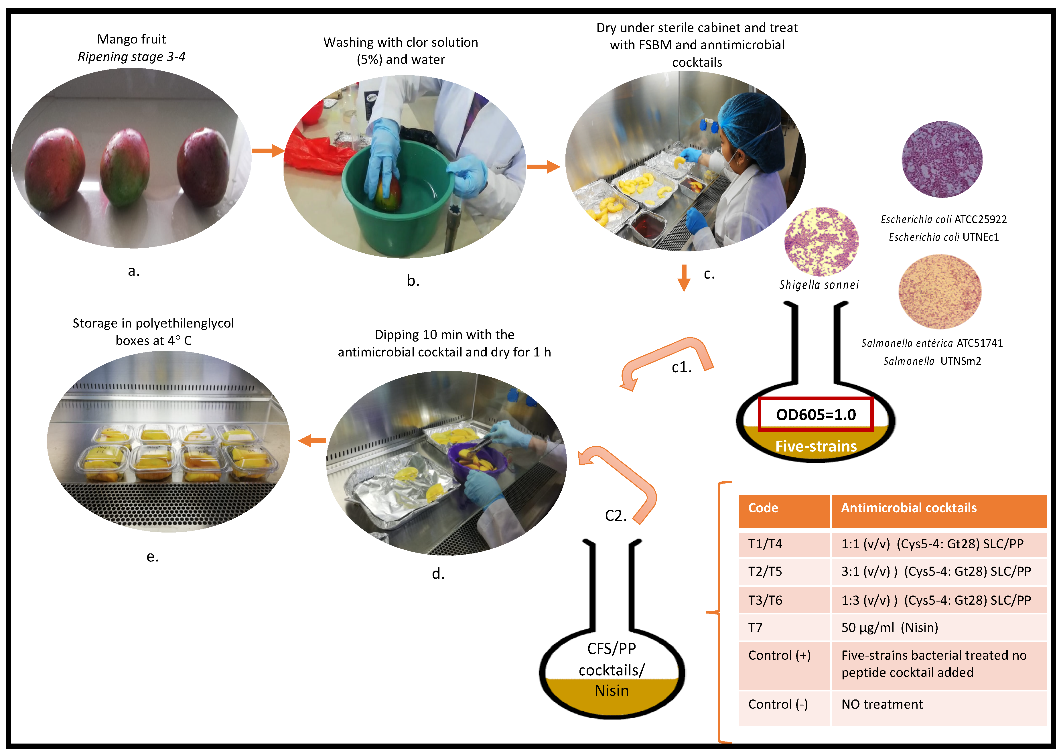
2.6. Inoculation of Fresh-Cut Mango Wedges with FSBM

Mango fruits at stage 3–4 of ripening were purchased from a local distributor, washed with 5% bleach solution for 10 min then twice with distillate water, and left to dry under the laminar flow cabinet. The fruit peel was removed with a sterile knife, and the pulp was manually cut into several wedges about 1 cm thick. To assure that the fruit pulp was free of contamination, the slices were washed for 3 min with 0.3% commercial antibacterial solution (for fruits and vegetables) and twice with distillate water and left to dry under the safety cabinet for 25 min. Mango wedges (5–6 pieces × 10 g × tray × each treatment) were submerged in 400 mL of the FSBM inoculum (1 × 10⁸ CFU/mL) and rotated by stirring with a glove-covered hand for 10 min to ensure that bacteria infiltrate the fruit slices, followed by air drying for 1 h in a biosafety cabinet (Figure 1). Control samples (+) were plunged only in FSBM solution, while control (-) samples were not plunged in the FSBM solution (they were not antimicrobial cocktails treated). The experiment was repeated three times using mango fruits purchased independently from the same distributor.

2.7. Treatment of Mango Wedges with Antimicrobial Cocktails

The antimicrobial cocktails described in Section 2.5 (400 mL) were used to immerse the fruit slices, manually rotating each slice for 10 min to assure complete coverage and contact of the surface with the solution, and then left to dry for 1 h in the biosafety cabinet (Figure 1). The fruit slices were packed in food trays covered with plastic food film and then stored for 5 days with refrigeration at 4 °C. Mango slices inoculated with FSBM, but not antimicrobial cocktails added, were used as control (+) and mango slices without any treatment as control (-).

2.8. Determination of the Total Cell Counts in Mango Wedges during Storage

To enumerate the microbial colonization during storage in mango wedges treated with antimicrobial cocktails, 25 g of mango slices were placed in peptone water (0.1%) for 4 h at 37 °C; serial dilution (1:10) with saline solution (NaCl 8.6 g/L) was used to determine total cell counts by plating on nutrient agar after incubation at 37 °C for 48 h. Independently, decimal dilutions (0.1 mL) were plated in triplicate on Chromocult Coliform agar (Merck, USA) to detect and enumerate E. coli and SS (Salmonella/Shigella SS agar, Difco, Detroit, MI, USA) agar to discriminate and enumerate Salmonella and Shigella cells. The results were expressed in CFU/g and daily determined during storage; at day 5, the percentage of the total cell counts reduction as well as with determination of reduction % of Shigella/Salmonella/E. coli was calculated by the equation: (R%) = [(A − B)/A × 100%]; where R is the percentage of bacterial reduction and B and A are the surviving bacterial cells (CFU/g) of the antimicrobial cocktail treated and control plates, respectively.

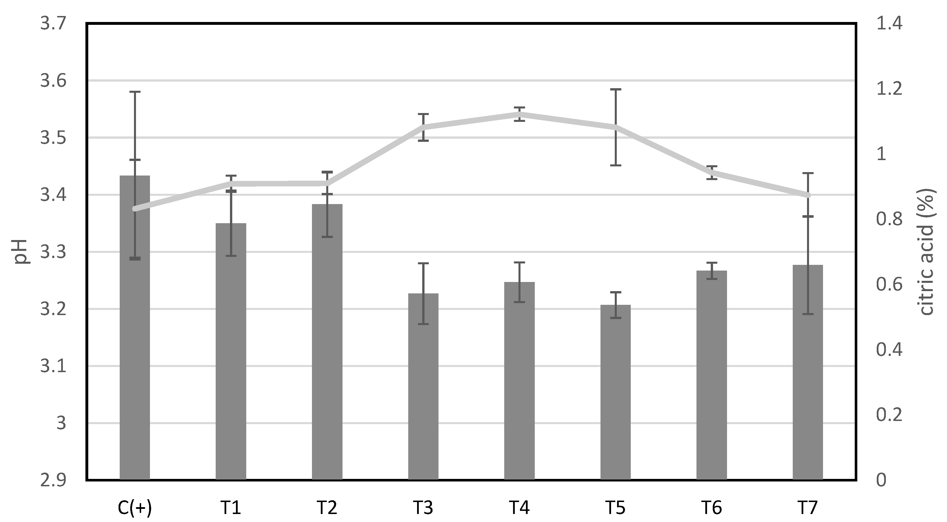
2.9. Monitoring the pH and Acidity of Mango Wedges during Storage

The pH was monitored daily during fruit storage using a pH meter (Seven Compact S210, Mettler Toledo LCC, Columbus, OH, USA). Total acidity, expressed as a percent of citric acid, was determined by titrating with 0.01 M NaOH to pH 8.2 as described [21].

2.10. Cytoplasmic Membrane Permeabilization

The cytoplasmic membrane permeabilization of E. coli ATCC25922, S. enterica subsp. enterica ATCC51741, and S. sonnei ATCC25931 by T5, T6 (9600 AU/mL final concentration), and T7 (50 μg/mL) cocktails were investigated using ONPG (o-nitro-phenyl-l-d-galactoside, no. N1127, Sigma-Aldrich Co. LLC, Saint Louis, MO, USA) as described [30]. In brief, the T5, T6, and T7 cocktails were added to each bacterial suspension independently at optical density 0.6, incubated 5 min at 30 °C, and then ONPG with a final concentration of 30 mM was added to each cell suspension. The hydrolysis of ONPG to o-nitrophenol (ONP) was monitored at 415nm at 120 min of incubation. To distinguish between cytoplasmic enzyme release and peptide uptake in the cells, β-galactosidase release was measured from the supernatant [30].

2.11. Cell Membrane Integrity Assay

The cell membrane integrity was assessed as previously described [28]. In brief, independent bacterial suspensions of E. coli ATCC25922, S. enterica ATCC51741, and S. sonnei ATCC25931 were grown overnight in appropriate broth culture media, harvested by centrifugation, and washed twice with 1 × PBS (phosphate-buffered saline, pH 7.5). The bacterial cells were treated independently with T5-, T6-, and T7-cocktails and incubated overnight at 30 °C. As a control, one flask for each target was maintained and the peptide cocktail was replaced with 1 × PBS. Cell cultures were centrifuged, the supernatant filtered and precipitated with isopropanol and ammonium acetate (3M), washed with 75% ethanol, followed by electrophoresis in 1% agarose gel with ethidium bromide, in 1 × TBE (Tris-borate EDTA, pH 8.0) migration buffer (Sigma-Aldrich Co. LLC, Saint Louis, MO, USA) to detect the presence of DNA/RNA molecules.

2.12. Statistical Analysis

All experiments were performed in triplicate. The results were reported as mean ± standard deviation. The normal distribution of the data was employed with the Shapiro-Wilk test (RStudio Version 1.2.1335, RStudio, Inc., Boston, MA, USA, 2019). The effect of the antimicrobial cocktails on target cell viability during storage was evaluated using ANOVA with a split-plot experimental design. Then, Tukey multiple range test and LSD (Least Significant Difference with Bonferroni correction) were applied to determine significant differences between the means (SPSS 13.0, Inc., Chicago, IL, USA).

3. Results

3.1. Mango Wedges Sold by Mobile Vendors Hold Antibiotic-Resistant Microorganisms

The microbiological quality of mango fruits was evaluated after purchasing. The results indicated the presence of several indicator bacteria and their amount was linked to the fruit ripening stage and purchasing origin (V1, V2). The highest number of aerobe counts was detected in fruits of stage 4 of ripening (Table 1). In the remaining pulp (after skin removal), the amount of the total aerobes decreased to 30–66% in the fruits from V1, thus accomplished the minimum accepted counts [22,23]. Unsatisfactory results were obtained in the case of fruits from V2 as the total aerobe counts were above accepted limited in both peel and remaining pulp (Table 1). The indicators, E. coli spp. and Enterobacter spp., were detected in both mango peel and pulp (Table 1), while Salmonella spp. and Shigella spp. were detected only in mango peels of fruits purchased from V2. Neither Salmonella nor Shigella was found in remaining mango pulp from V1 nor V2. While yeasts were detected in almost all samples, the molds were detected in the peel of fruits from V2 (Table 1). In mango wedges, the total coliforms varied from 1.30 × 10⁵ to 1.68 × 10⁶ CFU/g, while yeasts and molds varied from 3.03 × 10² to 6.30 × 10³ CFU/g (Table 2). By means of the standard bacteriological analysis, the total coliform bioindicators, E. coli spp. (varied from 7.00 × 10² to 1.74 × 10⁴ E+04 CFU/g), and Enterobacter spp. (varied from 1.00 × 10² to 1.16 × 10⁶ CFU/g) were predominant in all samples, while Shigella cells were detected in the mango wedges purchased from V4. No Salmonella colonies were detected in the mango wedges purchased from V3, while 2.00 × 101 CFU was detected in the mango wedges (25 g) from V4 (Table 2). The antibiotic susceptibility analysis indicated that the isolates from mango peel (50 randomly selected isolates) and remaining pulp (30 randomly selected isolates) were highly resistant to all antibiotics tested except tetracycline (Table S1). The results indicated that all selected isolates from mango wedges were resistant to at least one or two compounds of three different classes of antibiotics (cephalosporins, beta-lactamases, aminoglycosides, and penicillin-like antibiotics) (Table 3). All selected Enterobacter spp. and E. coli spp. isolates exhibited high resistance to ampicillin, gentamycin, amoxicillin, and cefuroxime (Table 3). The selected Shigella isolates were resistant to ampicillin and gentamycin, and 66.67% resistant to kanamycin. Salmonella isolates were resistant to all antibiotics tested except kanamycin, gentamycin, and tetracycline.

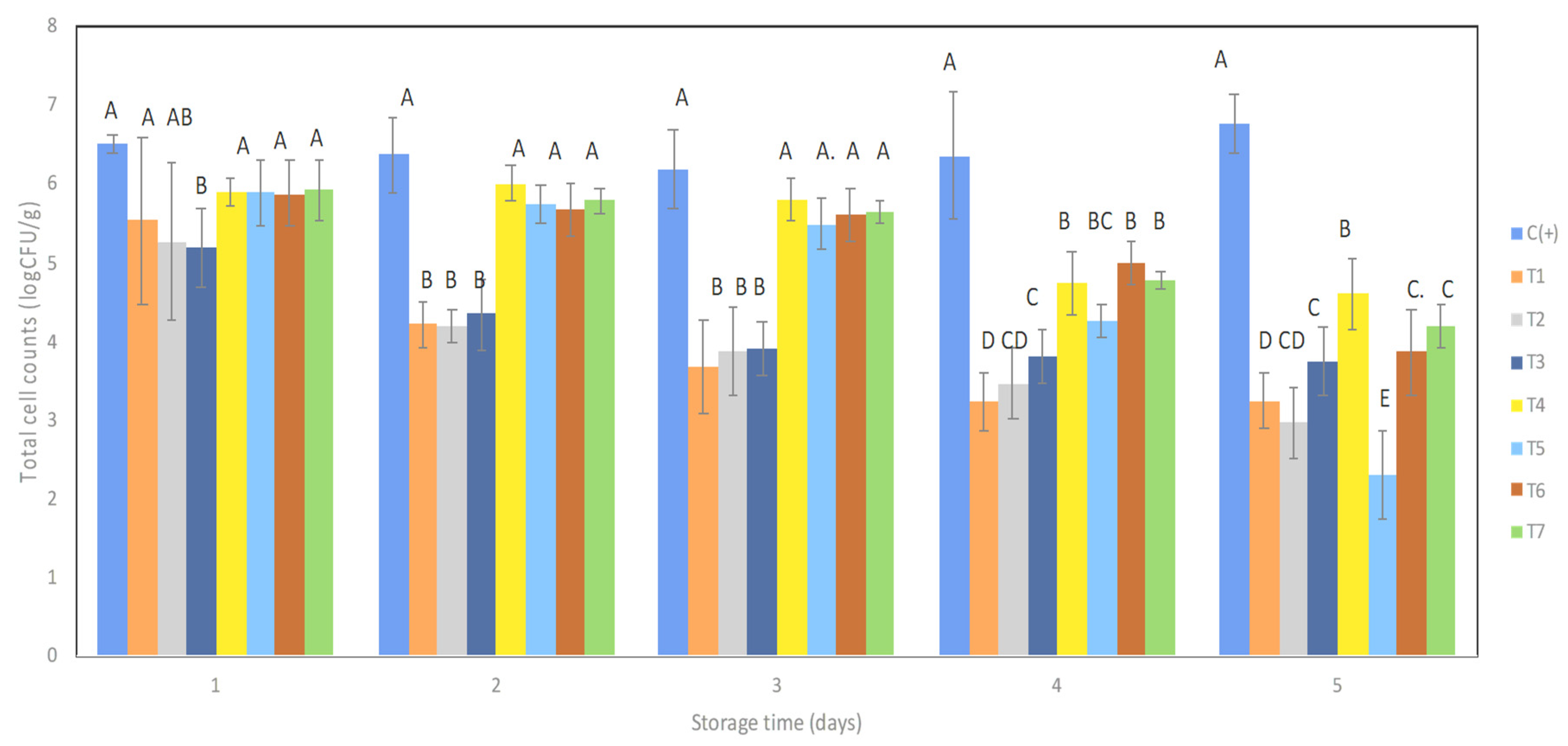
3.2. Antimicrobial Cocktails Diminished the Total FSBM Viability in Mango Wedges during Storage with Refrigeration

A significant reduction (p > 0.05) in the total cell counts was registered in mango wedges from day 2 of storage upon treatment with CFS-cocktails compared with the untreated control slices where a slight increase of the total cell counts was detected. A moderate reduction in cell counts was recorded with PP-cocktails as well as commercial nisin on day 2, while on day 4 the reduction was significant (p > 0.05) (Figure 2). Based on LSD analysis (group versus group contrast), at the last day of storage (day 5), all antimicrobial cocktails showed a significant reduction (p > 0.05) in cell counts compared with the control indicating their efficiency to simultaneously diminish FSBM in mango wedges. The maximum cell reduction of 56.03% was registered for T2-cocktail, followed by T5 (55.61% reduction), T1 (52.10% reduction), and T6, T7, and T4 with 42.88%, 37.85%, and 31.93% reduction, respectively. We suggest that the efficiency of CFS cocktail to decrease the overall cell counts might be related to the presence of both acids and peptides that work synergistically as a hurdle to block colonization by undesirable microorganisms (Table 4). By overloading the dose of UTNGt28, a significant difference (p > 0.05) in the overall cell counts reduction was observed at day 5 of storage for T2 and T5-cocktail, indicating that the dose of Gt28 might enhance the global killing event.

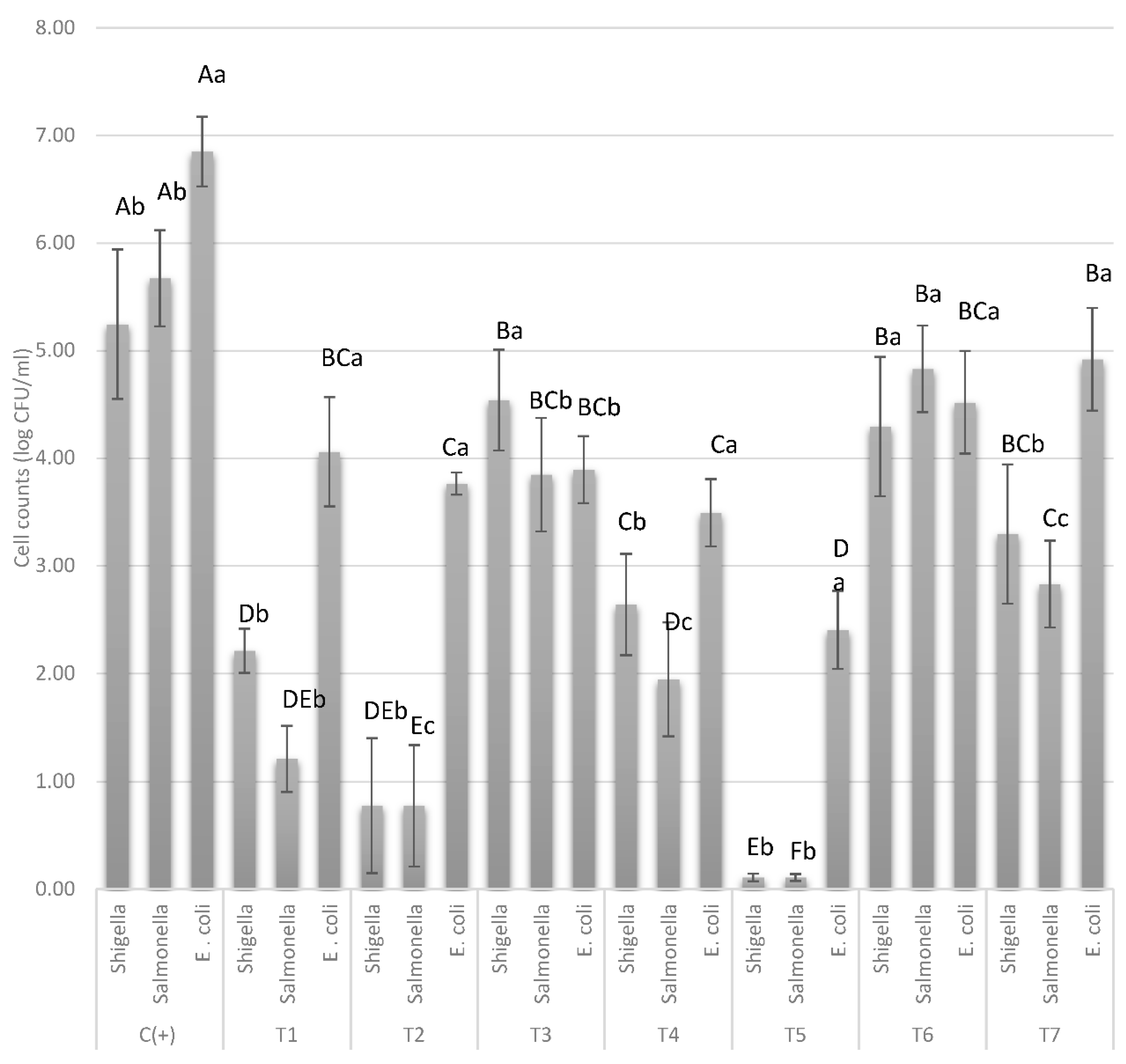
3.3. The Antimicrobial Cocktails T2 and T5 Inhibit Salmonella/Shigella and to Some Extent E. coli Growth in Mango Wedges

The effect of antimicrobial cocktails was analyzed individually for Salmonella (2 strain mix), Shigella (1 strain), and E. coli (2 strain mix) in selective media at day 5 of storage. In the control samples, the cell counts showed about the same amount at the end of storage indicating the adaptation of the microorganisms to the mango surface (Figure 3). When compared to the individual strain and its counterpart control, a significant reduction (p > 0.05) was registered for all antimicrobial treatments. The most significant reduction (p > 0.05) was detected for Salmonella and Shigella for treated mango with T2 and T5 combinations. For the untreated mango, Salmonella yielded 5.67 log CFU/g and Shigella yielded 5.24 log CFU/g, while the application of T2 and T5 resulted in a significant decrease of the cell counts (p > 0.05), Salmonella yielded 0.77 log CFU/g and Shigella, 0.1 log CFU/g, respectively, which corresponded to the incorporation of three times UTNGt28 peptide extract. This result suggested that by increasing the dose of UTNGt28, the cell death of Salmonella/Shigella was enhanced. A moderate reduction was observed with T1, T3, T4, T6, and T7. A decrease in E. coli cell counts was observed for all treatments when compared with the counterpart untreated control, with T5 being the most effective treatment (Figure 3). Overall results indicated that Salmonella and Shigella were more susceptible (98% and 97% reduction, respectively) than E. coli (64.93%) to the mixture when using the T5-cocktail (Table 4). Nonetheless, E. coli adapted well to the mango matrix as an increase in cell counts was registered from 6.0 to 6.85 log CFU/g at the end of storage, while Shigella / Salmonella cell counts were maintained stable during storage. The pH of mango slices was about 3.30 at the time of purchase and the acidity was 0.858 expressed in % of citric acid. By day 3 of storage, a progressive decrease with 0.1–0.15 units of pH induced a little decrease in the total cell viability, while from day 4, the cell viability was maintained lower in the T2, T5, and T7 treatments despite increase or decrease of pH (Figure S1). On the last day of storage, a little increase of pH was detected in the control (+) samples and T2 treatment. The samples treated with T5 showed the lowest pH value (3.2), which corresponded to the lowest Salmonella/Shigella counts in mango wedges. The cocktails T3, T4, T6, and T7 showed very similar values, indicating that in mango wedges the acidity might influence to a lesser extent the survival of the target bacteria (Figure 4).

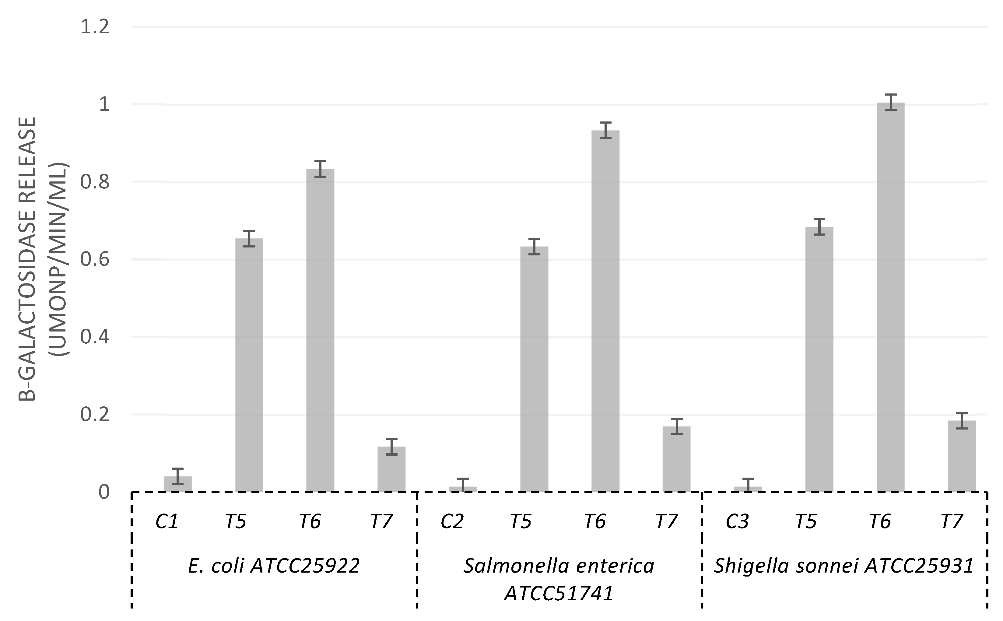
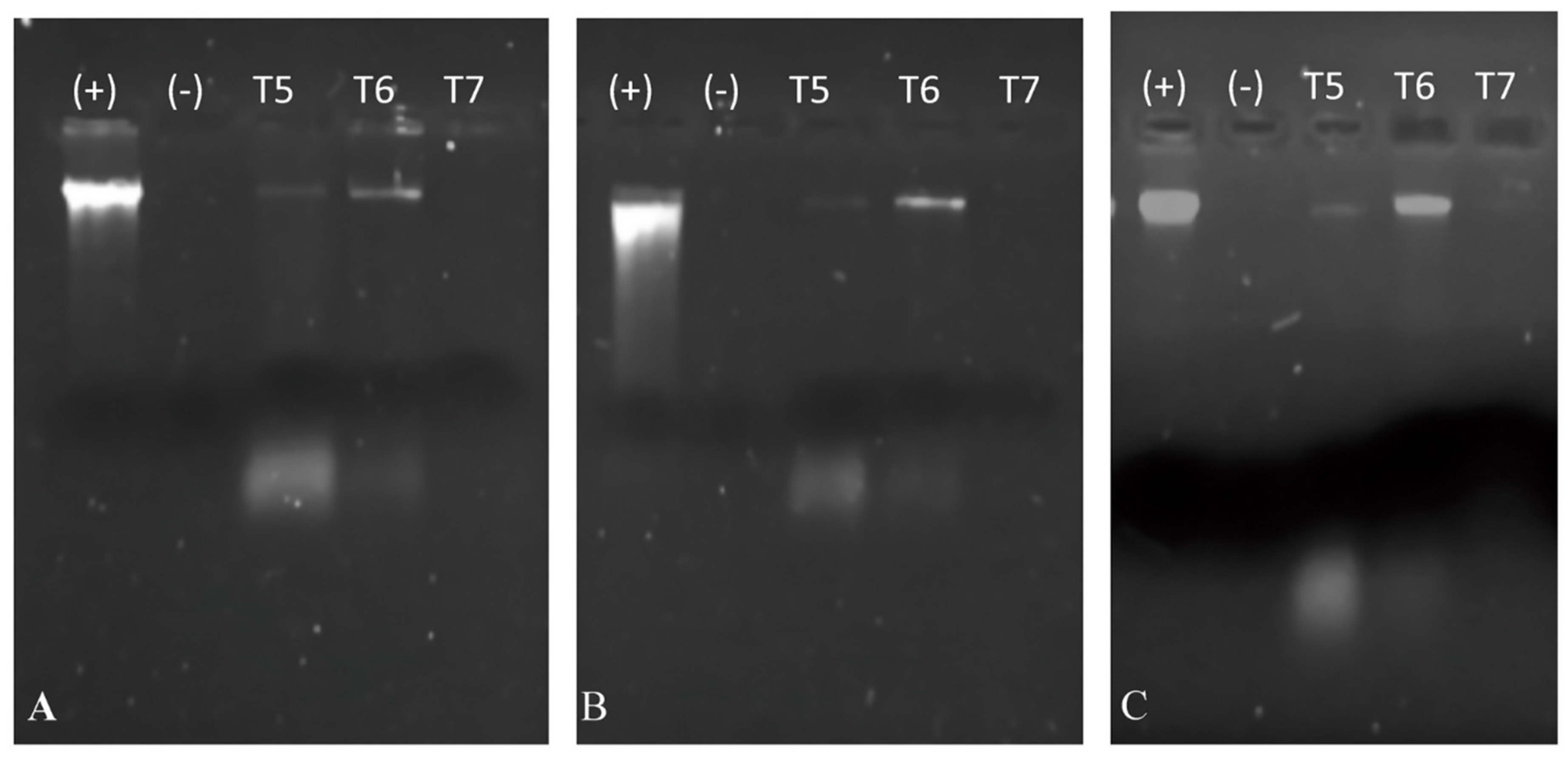
3.4. Cytoplasmic Membrane Permeation and Alteration of Cell Membrane Integrity

To evaluate the effect of antimicrobials on the target membrane cell permeation, the leakage of cytoplasmic content from target cells upon interaction with T5, T6, and T7 cocktails was monitored as a function of cytoplasmic β-galactosidase release, with bacteria grown in lactose-containing medium. T6-cocktail was included in this experiment showed about the same total cell count reduction as T7-cocktail (nisin). The results indicated that the antimicrobial cocktails caused a considerable release of the enzyme into the medium at 120 min of incubation (Figure 5). Less membrane permeability was observed in the case of T7, while no β-galactosidase activity was detected in the culture medium of bacterial cells without antimicrobial cocktail treatment. The release of DNA/RNA molecules was examined by gel electrophoresis as shown in Figure 6A–C. The presence of both DNA/RNA bands was observed when treated with the T5-, T6-cocktail for all strains, indicating that the target membrane integrity was compromised. Neither DNA nor RNA molecules were detected when cells were treated with the T7-cocktail (nisin).

4. Discussion

The perception of consumers regarding the microbial hazards of street foods is in general driven by their level of education, income, knowledge of food safety, age, and gender [31]. Due to the lack of official data on the volume of trade involved and the deficiency of food quality, there is an increased risk of disease within the population.
In Ecuador, mobile vendors target high human traffic areas, including parks, schools, highway traffic light-stops, and bus stations; thus, these sectors are fraught with unhealthy activities with serious concerns over the safety of the practitioners, especially the health of the consumers, particularly infants and tourists who buy these products. There is a considerable risk of food cross-contamination as the vendors neglect basic hygiene practices at the preparation and vending sites. Thus, searching for environmentally safe practices to protect the fruits from contamination and prolonging their shelf life must be considered.
The use of bacteriocins from lactic acid bacteria is an important tool to reduce pathogens or spoilage from bacterial growth [32]. Nisin, a food-grade bacteriocin (E234) produced by Lactococcus lactis, remains the only additive used in the food industry [33,34], recognized to be safe by the Joint Food and Agriculture Organization/World Health Organization (FAO/WHO) Expert Committee on Food Additives [35,36]. The antimicrobial activity of nisin against Listeria monocytogenes and Staphylococcus aureus was demonstrated early [37], but its efficacy against Gram-negative bacteria increases when combined with chelating agents like EDTA (ethylenediaminetetraacetic acid) that destabilize the outer membrane of the pathogenic bacteria [38,39].
During the past decade, outbreaks of human infection due to the consumption of vegetables and fruits has increased [40]. For example, the presence of Salmonella in mangos was associated with inappropriate manipulation [7]. If traditional salmonellosis has been linked to the consumption of meat products, the illness registered due to the consumption of raw fruits like mango has already been disclosed [41]. Furthermore, the presence of E. coli, Salmonella spp., and L. monocytogenes was detected on the surface of cut fruits and vegetables causing serious damage to human health [42].
In this study, the bacteriological analysis of mango peel, remaining pulp, and wedges purchased from the local market indicated the presence of several bioindicators, Enterobacter spp., Salmonella spp., Shigella spp., and E. coli spp. (Table 1; Table 2). The total aerobes counts were superior in ready-to-eat mango wedges than in the peel or remaining pulp, indicating that the hygiene practices of the vendors along with the inappropriate storage might explain the presence of harmful microbes. For safety reasons and according to international food standards [27], the presence of Salmonella is not acceptable. The high concentration of Salmonella spp. counts detected on mango peels in stage 4 of ripening might be explained by different factors such as natural maturity of the mango fruit after post-harvesting, possible mechanical damage, physiological disorders non-detectable at the moment of harvesting, along with the humidity and inappropriate storage conditions that might increase the contamination by several commensal microorganisms including Salmonella. The Salmonella positive mango fruits and wedges samples were not compliant with the safety criteria and their marketing should not be permitted, albeit the impact on consumer health cannot be estimated. Nonetheless, mango is a perishable fruit by nature and must be stored and manipulated accordingly to minimize microbial contamination. Thus, the presence of microorganisms in ready-to-eat mango wedges might be related to the vendors’ poor handling and storage. Once cut, the fruit slices are good substrates for microorganism growth, due to their organic composition and high-water activity, thus are highly susceptible to contamination. Therefore, a sanitation step or treatment with antimicrobials is mandatory. Similar studies conducted on street food in Brazil [43] and South Africa [44] showed high levels of bioindicators (coliforms and E. coli), suggesting that food handlers increase contamination. Based on the antibiotic results of the selected isolates found in the mango wedges, we conclude that E. coli spp. and Enterobacter spp. isolates were highly resistant to all antibiotics tested (Table 3). The presence of antibiotic-resistant isolates was found in both mango fruit peel and remaining pulp indicating the risk of ingesting a high number of resistant bacteria unless prophylactic disinfection and appropriate storage conditions are considered.
Contamination with antibiotic-resistant strains is of concern as food can act as a vector for the transfer of antimicrobial resistance genes to humans [45,46]. Applying a combination of different peptide doses of two native lactic bacterial species, which efficiently inhibit various microorganisms simultaneously in ready-to-eat fruits was recently demonstrated in pineapple [22]. As the fruit matrix is a complex structure, the peptide combinations must be individually tested for their efficacy to inhibit microorganisms that invade the fruits during storage. The peptide dose, pH, and acidity of the fruit matrix simultaneously might contribute to the inhibitory activity by shifting the peptide through the target cell wall [47]. However, as a proof of concept to verify the feasibility of using edible peptide extract cocktails as antimicrobials in cut fruits, we extended the work to mango wedges. In the present study, all designed antimicrobial cocktails showed the capacity to diminish concurrently five-strain bacterial mixture in mango wedges (Figure 2; Figure 3). Nonetheless, three antimicrobial combinations (T1, T2, and T5) showed a greater bactericidal performance than nisin (T7).
A previous study indicated that a combination of nisin from Lactococcus lactis with curvaticin 13 from Lactobacillus curvatus strain SB13 had a greater bactericidal effect against Listeria monocytogenes ATCC15313 than the use of a single bacteriocin [48] in vitro. The peptides from both lactic acid bacteria strains used in this study were previously detected as potential antimicrobials [27]. The inhibitory efficacy of T1 formulation, for example, was already demonstrated in fresh-cut pineapple [22], but its effectiveness was greater in mango, expanding its application in acidic fruits. Although the difference in the overall cell reduction between T2 and T5 was not significant (Table 4), we suggest that from the commercial perspective, the use of CFS-, rather than PP-cocktail, might have advantages as no extra purification step is needed and the cost of production decreases. Early research indicated that the use of cell-free supernatant from lactic acid bacteria might be a solution for biopreservation [49]. Another advantage of using these peptide extracts from lactic bacteria isolated from acidic fruits is that both bacterial strains originate from wild tropical fruits.
A recent study reports the successful utilization of Lactobacillus rhamnosus GG as a preservative agent for maintaining the quality of fresh-cut bell pepper, preventing its microbial colonization [19]. In our study, a significant reduction of Salmonella and Shigella was detected when mango wedges were treated with T5-cocktail (Figure 3). This might be related to the overloading of the UTNGt28 peptide extract in the samples. Recently, we demonstrated that UTNGt28 peptide extract induced lysis and the formation of ghost cells as a secondary killing event in Salmonella [30]. Similar to our previous study in pineapple [22], in mango wedges, the acidity (pH 3.3) does not influence the target cell survival during storage (Figure 4). For the control wedges, a slight increase of pH from 3.3 to 3.43 was observed, while a decrease with 0.1–0.2 units was detected in all antimicrobial cocktail treatments. Considering that the pH of PP-cocktail and nisin at the time of application was 6.80, it seems that the acidity of the fruit increased their solubility, however, they might rapidly bind the target membrane and induce killing. pH is known to be a factor that increases peptide solubility [50]. The outer membrane of Gram-negative bacterial cells constitutes a semi-permeable barrier that is responsible for preventing antimicrobials from reaching their cytoplasmic membrane and eventually inducing membrane disruption [51]. A recent study indicated that killing occurs only when bacterial cell membranes are completely saturated with peptides [52]. In this study, T5 and T6 permeated the cytoplasmic membrane while T7 showed minor effects (Figure 5). This indicates that nisin did not permeate the membrane to the extent required for membrane disruption as it did not cause leakage of cell contents from the target cells (Figure 6). T5- and T6-cocktails alter the cell membrane integrity demonstrated by the detection of DNA/RNA molecules (Figure 6). The result was in agreement with our previous findings showing that the extract containing more than one peptide from L. plantarum UTNGt2 showed a bactericidal mode of action toward Salmonella while nisin does not have any effect on the membrane integrity [22,28]. Nonetheless, the mechanism of action of peptide cocktails against Gram-negative bacteria is different from nisin, as nisin needs an extra permeabilization agent that perturbs the outer membrane of target bacteria and sensitizes Gram-negative bacteria to several antibiotics [53].

5. Conclusions

In conclusion, this study drew attention to the contamination of tropical ready-to-eat mango wedges with antibiotic-resistant bacteria that could be acquired during manipulation by street vendors. The results proved the antimicrobial effect of peptide-, or cell-free supernatant cocktails against a five-strain bacterial mixture that might colonize the fruit at once, simultaneously. Although a complete survival blockage might not be possible, the peptide treatment and lower temperature of storage can ensure a significant reduction in the dynamic multiplication of different microorganisms. On the basis of the results obtained, the antimicrobial cocktails from native UTNCys5–4 and UTNGt28 strains confirmed our hypothesis that a combination of different peptide extracts might be effective in diminishing bacterial growth in fresh cut fruits and could further reduce the risk of food-borne illness by contamination with Salmonella spp., Shigella spp., and E. coli spp. The production of bacteriocins and their incorporation in fruit wedges as cell-free extracts or precipitated peptides could be a technically effective method to increase the protection and shelf-life of fresh fruits but its commercial viability needs to be further addressed.

Supplementary Materials

The following are available online at https://0-www-mdpi-com.brum.beds.ac.uk/2076-3417/11/5/2246/s1, Table S1: The antibiotic susceptibility (%) of isolates randomly selected from mango peel and pulp. Figure S1. The pH and total cell viability changes during storage.

Author Contributions

Conceptualization, G.N.T.; methodology, G.N.T.; software, D.O.; validation, D.O.; formal analysis, G.N.T. and D.O.; investigation, G.N.T. and D.O. resources, G.N.T.; writing—original draft preparation, G.N.T.; writing—review and editing, G.N.T.; visualization, G.N.T.; supervision, G.N.T.; project administration, G.N.T.; funding acquisition, G.N.T. All authors have read and agreed to the published version of the manuscript.

Funding

This work was supported by the Centre of Research (CUICYT) of the Technical University of the North, Grant No: 2929/ 2019.

Acknowledgments

The authors would like to thank C. Ortega for technical support and D. Brown for helpful comments, suggestions, and the correction of the manuscript.

Conflicts of Interest

The authors declare no conflict of interest.

References

  1. Bernal, A.C.; Maldonado, M.E.; Urango, L.A.; Franco, M.C.; Rojano, B.A. Sugar mango (Mangifera indica), variety from Colombia: Antioxidant, nutritional and sensorial characteristics. Rev. Chil. Nutr. 2014, 41. [Google Scholar] [CrossRef] [Green Version]
  2. Guererro, G.H. La produccion del mango ecuatoriano. Perspectiva 2018. Available online: https://perspectiva.ide.edu.ec/investiga/2018/06/26/la-produccion-del-mango-ecuatoriano/ (accessed on 3 June 2020).
  3. Arequipa, X.B. Diseño de un plan de marketing para la exportación del mango ecuatoriano hacia el mercado español. Bibdigital 2010. Available online: http://bibdigital.epn.edu.ec/handle/15000/1612 (accessed on 8 June 2020).
  4. Jadán Solís, K.P.; Najarro Quintero, R. Análisis de las políticas públicas agrícolas y la seguridad alimentaria de la provincia de Los Ríos. Rev. Cientif. Ecocien. 2019, 6, 1–16. [Google Scholar] [CrossRef]
  5. Ministerio De Salud Pública. MSP lidera la primera campaña de capacitación en manipulación de alimentos. Quito: Ministerio de Salud Pública. 2014. Available online: http://www.salud.gob.ec/msp-lidera-la-primera-campana-de-capacitacion-en-manipulacion-de-alimentos/ (accessed on 10 October 2020).
  6. Nazuka, E.; Inatsu, Y.; Kayasaki, S.; Miyamaru, M. The investigation of bacterial contamination in fresh-cut fruits, and the behavior of Escherichia coli O157: H7 and Salmonella enteritidis on fruit cubes. Jap. J. Food Microbio. 2004, 21, 269–274. [Google Scholar] [CrossRef] [Green Version]
  7. Branquinho Bordini, M.E.; Asturiano Ristori, C.; Jakabi, M.; Scala Gelli, D. Incidence, internalization and behavior of Salmonella in mangoes, var. Tommy Atkins. Food Control. 2007, 18, 1002–1007. [Google Scholar] [CrossRef]
  8. Alegre, I.; Abadias, M.; Anguera, M.; Oliveira, M.; Viñas, I. Factors affecting growth of foodborne pathogens on minimally processed apples. Food Microbiol. 2010, 27, 70–76. [Google Scholar] [CrossRef]
  9. CDC (Center for Disease Control and Prevention). Outbreak of Salmonella Poona Infections Linked to Imported Cucumbers (Final Update). 2016. Available online: https://www.cdc.gov/salmonella/poona-09-15/index.html (accessed on 11 October 2020).
  10. CDC (Center for Disease Control and Prevention). Outbreak of Listeriosis Linked to Whole Cantaloupes from Jensen Farms, Colorando (Final Update). 2012. Available online: https://www.cdc.gov/listeria/outbreaks/cantaloupes-jensen-farms/index.html (accessed on 6 October 2020).
  11. CDC (Center for Disease Control and Prevention). Outbreak of Salmonella Infections Linked to Pre-Cut Melons. 2019. Available online: https://www.cdc.gov/salmonella/carrau-04-19/index.html (accessed on 11 October 2020).
  12. CDC (Center for Disease Control and Prevention). Outbreak of Salmonella Infections Linked to Cut Fruit. 2020. Available online: https://www.cdc.gov/salmonella/javiana-12-19/index.html (accessed on 11 October 2020).
  13. CDC (Center for Disease Control and Prevention). Outbreak of Salmonella Infections Linked to Gavi Brand Whole, Fresh Papayas. 2020. Available online: https://www.cdc.gov/salmonella/uganda-06-19/index.html (accessed on 11 October 2020).
  14. Rojas-Graü, M.A.; Soliva-Fortuny, R.; Martín-Belloso, O. Edible coatings to incorporate active ingredients to fresh-cut fruits: A review. Trends Food Sci. Technol. 2009, 20, 438–447. [Google Scholar] [CrossRef]
  15. Oliveira, M.; Abadias, M.; Colás-Medà, P.; Usall, J.; Viñas, I. Biopreservative methods to control the growth of foodborne pathogens on fresh-cut lettuce. Int. J. Food Microbiol. 2015, 214, 4–11. [Google Scholar] [CrossRef]
  16. Rai, M.; Pandit, R.; Gaikwad, S.; Kövics, G. Antimicrobial peptides as natural bio-preservative to enhance the shelf-life of food. J. Food Sci. Technol. 2016, 53, 3381–3394. [Google Scholar] [CrossRef] [PubMed] [Green Version]
  17. López-Meza, J.E.; Ochoa-Zarzosa, A.; Barboza-Corona, J.E.; Bideshi, D.K. Antimicrobial peptides: Current and potential applications in biomedical therapies. Biomed Res. Int. 2015, 367243. [Google Scholar] [CrossRef] [PubMed]
  18. Upendra, R.S.; Khandelwal, P.; Jana, K.; Ajay Kumar, N.; Gayathri Devi, M.; Stephaney, M.L. Bacteriocin production from indigenous strains of lactic acid bacteria isolated from selected fermented food sources. Int. J. Pharma. Res. Health Sci. 2016, 4, 982–990. Available online: www.pharmahealthsciences.net (accessed on 20 October 2020).
  19. Saravanakumar, K.; Sathiyaseelan, A.; Mariadoss, A.V.A.; Chelliah, R.; Hu, X.; Oh, D.H.; Wang, M.H. Lactobacillus rhamnosusGG and Biochemical Agents Enrich the Shelf Life of Fresh-Cut Bell Pepper (Capsicum annuum L. var. grossum (L.) Sendt). Foods 2020, 9, 1252. [Google Scholar] [CrossRef] [PubMed]
  20. Tumbarski, Y.; Nikolova, R.; Petkova, N.; Ivanov, I.; Lante, A. Biopreservation of fresh strawberries by carboxymethyl cellulose edible coatings enriched with a bacteriocin from Bacillus methylotrophicus BM47. Food Technol. Biotechnol. 2019, 57, 230–237. [Google Scholar] [CrossRef] [PubMed] [Green Version]
  21. Tenea, G.N.; Pozo Delgado, T. Antimicrobial peptides from Lactobacillus plantarum UTNGt2 prevent harmful bacteria growth on fresh tomatoes. J. Microbiol. Biotechnol. 2019, 29, 1553–1560. [Google Scholar] [CrossRef] [PubMed]
  22. Tenea, G.N.; Olmedo, D.; Ortega, C. Peptide-based formulation from lactic acid bacteria Impairs the pathogen growth in Ananas comosus (Pineapple). Coatings 2020, 10, 457. [Google Scholar] [CrossRef]
  23. Tenea, G.N.; Hurtado, P.; Ortega, C. Inhibitory effect of substances produced by native Lactococcus lactis strains of tropical fruits towards food pathogens. Prev. Nutr. Food Sci. 2018, 23, 260–268. [Google Scholar] [CrossRef] [PubMed]
  24. NTE INEN 1529-15:2009. Control Microbiológico de los Alimentos. Toma, Envío y Preparación de Muestras Para el Análisis Microbiológico. Instituto Ecuatoriano de Normalización (INEN), 14. Available online: https://archive.org/stream/ec.nte.1529.15.1996/ec.nte.1529.15.1996_djvu.txt (accessed on 10 July 2020).
  25. NTE INEN 1529-8: 2015.Norma. Técnica. Ecuatoriana. Control Microbiológico de los Alimentos. Detección y Recuento de Escherichia coli Presuntiva por la Técnica del Número más Probable. pp. 1–13. Available online: https://archive.org/stream/ec.nte.1529.8.1990#mode/2up (accessed on 10 July 2020).
  26. García Martínez, R.; López Jiménez, A.; Saucedo Veloz, C.; Salazar-García, S.; Suárez Espinosa, J. Maduración y calidad de frutos de mango ‘Kent’ con tres niveles de fertilización. Rev. Mex. Cienc. Agric. 2015, 6, 665–678. Available online: http://www.scielo.org.mx/scielo.php?script=sci_arttext&pid=S2007-09342015000400001&lng=es&tlng=es (accessed on 13 October 2020).
  27. ISO, 6579: 2002 (E). Microbiology-General Guidance on Methods for the Detection of Salmonella, 4th ed.; International Organization for Standardization: Geneve, Switzerland, 2002; Available online: https://www.iso.org/standard/29315.html (accessed on 10 July 2020).
  28. Tenea, G.N.; Guana, J.M. Inhibitory substances produced by native Lactobacillus plantarum UTNUTNCys5-4 control microbial population growth in meat. J. Food Qual. 2019, a9516981. [Google Scholar] [CrossRef] [Green Version]
  29. European Food Safety authority (EFSA). Guidance on the assessment of bacterial susceptibility to antimicrobials of human and veterinary importance. EFSA J. 2012, 10, 2740. [Google Scholar] [CrossRef]
  30. Tenea, G.N. Peptide extracts from native lactic acid bacteria generate ghost cells and spheroplasts upon interaction with salmonella enterica, as promising food antimicrobials. BioMed Res. Int. 2020, 6152356. [Google Scholar] [CrossRef]
  31. Alimi, B.A.; Workneh, T.S. Consumer awareness and willingness to pay for safety of street foods in developing countries: A review. Int. J. Consum. Stud. 2016, 40, 242–248. [Google Scholar] [CrossRef]
  32. Agriopoulou, S.; Stamatelopoulou, E.; Sachadyn-Król, M.; Varzakas, T. Lactic acid bacteria as antibacterial agents to extend the shelf life of fresh and minimally processed fruits and vegetables: Quality and safety aspects. Microorganisms 2020, 8, 952. [Google Scholar] [CrossRef]
  33. Hansen, J.N.; Sandine, W.E. Nisin as a model food preservative. Crit. Rev. Food Sci. Nutr. 1994, 34, 69–93. [Google Scholar] [CrossRef]
  34. Artes-Hernandez, F.; Martinez-Hernandez, G.R.; Aguayo, E.; Gomes, P.A.; Artes, F. Fresh-Cut Fruit and Vegetables: Emerging Eco-friendly Techniques for Sanitation and Preserving Safety. In Postharvest Handling; Kahramanoglu, I., Ed.; IntechOpen: London, UK, 2017. [Google Scholar] [CrossRef] [Green Version]
  35. FAO (Food and Agricultural Organization of the United States). Probiotics in Food: Health and Nutritional Properties and Guidelines for Evaluation; FAO: Rome, Italy, 2016; Available online: http://www.fao.org/3/a-a0512e.pdf (accessed on 15 July 2020).
  36. Favaro, L.; Penna, A.L.B.; Todorov, S.D. Bacteriocinogenic LAB from cheeses–application in biopreservation? Trends Food Sci. Technol. 2015, 41, 37–48. [Google Scholar] [CrossRef]
  37. Arqués, J.L.; Rodríguez, E.; Nuñez, M.; Medina, M. Combined effect of reuterin and lactic acid bacteria bacteriocins on the inactivation of food-borne pathogens in milk. Food Control. 2011, 22, 457–461. [Google Scholar] [CrossRef]
  38. Bari, M.L.; Ukuku, D.O.; Kawasaki, T.; Inatsu, Y.; Isshiki, K.; Kawamoto, S. Combined efficacy of nisin and pediocin with sodium lactate, citric acid, phytic acid, and potassium sorbate and EDTA in reducing the Listeria monocytogenes population of inoculated fresh-cut produce. J. Food Prot. 2005, 68, 1381–1387. [Google Scholar] [CrossRef] [PubMed]
  39. Okuku, D.O.; Fett, W.F. Effect of nisin in combination with EDTA, sodium lactate, and potassium sorbate for reducing Salmonella on whole and fresh-cut cantaloupe. J. Food Prot. 2004, 67, 2143–2150. [Google Scholar] [CrossRef]
  40. Ragazzo-Sanchez, J.A.; Sanchez-Prado, L.; Gutiérrez-Martínez, P.; Luna-Solano, G.; Gomez-Gil, B.; Calderon-Santoyo, M. Inhibition of Salmonella spp. isolated from mango using bacteriocin-like produced by lactobacilli, CyTA. J. Food. 2009, 7, 181–187. [Google Scholar] [CrossRef] [Green Version]
  41. Penteado, A.L.; Eblen, B.S.; Miller, A.J. Evidence of Salmonella internalization into fresh mangos during simulated postharvest insect disinfestations procedures. J. Food Prot. 2004, 67, 181–184. [Google Scholar] [CrossRef]
  42. Qadri, O.S.; Yousuf, B.; Srivastava, A.K. Fresh-cut fruits and vegetables: Critical factors influencing microbiology and novel approaches to prevent microbial risks—A review. Cogent Food Agric. 2015, 1, 1121606. [Google Scholar] [CrossRef]
  43. Bezerra, A.C.D.; Reis, R.B.; Bastos, D.H.M. Microbiological quality of hamburgers sold in the streets of Cuiabá–MT, Brazil and vendor hygiene- awareness. Food Sci. Technol. 2010, 30, 520–524. [Google Scholar] [CrossRef] [Green Version]
  44. Lues, J.F.R.; Rasephei, M.R.; Venter, P.; Theron, M.M. Assessing food safety and associated food handling practices in street food vending. Int. J. Environm. Health Res. 2006, 16, 319–328. [Google Scholar] [CrossRef] [PubMed]
  45. Vincenti, S.; Raponi, M.; Sezzatini, R.; Giubbini, G.; Laurenti, P. Enterobacteriaceae antibiotic resistance in ready-to-eat foods collected from hospital and community canteens: Analysis of Prevalence. J. Food Prot. 2018, 81, 424–429. [Google Scholar] [CrossRef] [PubMed]
  46. Meade, E.; Slattery, M.A.; Garvey, M. Bacteriocins, Potent Antimicrobial Peptides and the Fight against Multi Drug Resistant Species: Resistance Is Futile? Antibiotics 2020, 9, 32. [Google Scholar] [CrossRef] [PubMed] [Green Version]
  47. Khaneghah, A.M.; Hashemi, S.M.B.; Limbo, S. Antimicrobial agents and packaging systems in antimicrobial active food packaging: An overview of approaches and interactions. Food Bioprod. Process. 2018, 111, 1–19. [Google Scholar] [CrossRef]
  48. Bouttefroy, A.; Millière, J.B. Nisin-curvaticin 13 combinations for avoiding the regrowth of bacteriocin resistant cells of Listeria monocytogenes ATCC 15313. Int. J. Food Microbiol. 2000, 62, 65–75. [Google Scholar] [CrossRef] [PubMed]
  49. Arena, M.P.; Silvain, A.; Normanno, G.; Grieco, F.; Drider, D.; Spano, G.; Fiocco, D. Use of Lactobacillus plantarum Strains as a Bio-Control Strategy against Food-Borne Pathogenic Microorganisms. Front. Microbiol. 2016, 7, 464. [Google Scholar] [CrossRef] [Green Version]
  50. Silva, C.; Silva, S.; Ribeiro, S.C. Application of Bacteriocins and Protective Cultures in Dairy Food Preservation. Front. Microbiol. 2018, 9, 594. [Google Scholar] [CrossRef]
  51. Gravel, J.; Paradis-Bleau, C.; Schmitzer, A.R. Adaptation of a bacterial membrane permeabilization assay for quantitative evaluation of benzalkonium chloride as a membrane-disrupting agent. Medchemcomm. 2017, 8, 1408–1413. [Google Scholar] [CrossRef]
  52. Helander, I.M.; Mattila-Sandholm, T. Permeability barrier of the gram-negative bacterial outer membrane with special reference to nisin. Inter. J. Food Microbiol. 2000, 60, 153–161. [Google Scholar] [CrossRef]
  53. Heesterbeek, D.A.C.; Martin, N.I.; Velthuizen, A.; Duijst, M.; Ruyken, M.; Wubbolts, R.; Rooijakkers, S.H.M.; Bardoel, B.W. Complement-dependent outer membrane perturbation sensitizes Gram-negative bacteria to Gram-positive specific antibiotics. Sci. Rep. 2019, 9, 3074. [Google Scholar] [CrossRef] [Green Version]
Figure 1. Treatment of mango wedges with antimicrobial cocktails. (a) mango fruits at stage 3-4 of ripening; (b) washing and disinfection; (c) submersion of wedges with the five-strain bacterial mixture (c1) and dipping with antimicrobial cocktails (c2): (d) drying; (e) storage in polyethyleneglycol boxes.
Applsci 11 02246 g001
Figure 2. Changes in the total cell counts of the target FSBM detected in mango wedges treated with the antimicrobial cocktails during storage. The results are expressed in log (CFU/g). Legend: Control (+): mango wedges + FSBM; T1: (Cys5-4+ Gt28) CFS: 1:1 (v/v); T2: (Cys5-4+ Gt28) CFS: 1:3 (v/v); T3: (Cys5-4+ Gt28) CFS: 3:1 (v/v); T4: (Cys5-4+ Gt28) PP: 1:1 (v/v); T5: (Cys5-4+ Gt28) PP: 1:3 (v/v); T6: (Cys5-4+ Gt28) PP: 3:1 (v/v); T7: Nisin (50 μg/mL); FSBM-five-strain bacterial mixture; CFS-cell free supernatant; PP: partial precipitated peptides. The bars represent the mean ± SD of three independent repetition. The capital letters represent the significant decrease (p < 0.05) in cell counts vs control (+) at different time points (days).
Applsci 11 02246 g002
Figure 3. Viability of Shigella, Salmonella, and E. coli in mango wedges at day 5 of storage with refrigeration. The results are expressed in log CFU/g. Legend: Control (+): mango wedges + FSBM; T1: (Cys5-4+ Gt28)CFS: 1:1 (v/v); T2: (Cys5-4+ Gt28) CFS: 1:3 (v/v); T3: (Cys5-4+ Gt28)CFS: 3:1 (v/v); T4: (Cys5-4+ Gt28)PP: 1:1 (v/v); T5: (Cys5-4+ Gt28)PP: 1:3 (v/v); T6: (Cys5-4+ Gt28)PP: 3:1 (v/v); T7: Nisin (50 μg/mL); FSBM-five-strain bacterial mixture; CFS-cell-free supernatant; PP: partial precipitated peptides. The bars represent the mean ± SD of three independent repetition. The capital letters represent the significant decrease (p < 0.05) of each target bacterial cells vs control (+). Small letters represent the difference between targets exposed at different treatments. The values with different subscripts (a, b, c) indicate a significant difference (p < 0.05).
Applsci 11 02246 g003
Figure 4. pH and acidity determined at day 5 of storage with refrigeration in mango wedges treated with different antimicrobial cocktails. Legend: Control (+): mango wedges + FSBM; T1: (Cys5-4+ Gt28)CFS: 1:1 (v/v); T2: (Cys5-4+ Gt28) CFS: 1:3 (v/v); T3: (Cys5-4+ Gt28)CFS: 3:1 (v/v); T4: (Cys5-4+ Gt28)PP: 1:1 (v/v); T5: (Cys5-4+ Gt28)PP: 1:3 (v/v); T6: (Cys5-4+ Gt28)PP: 3:1 (v/v); T7: Nisin (50 μg/mL); FSBM-five-strain bacterial mixture; CFS-cell free supernatant; PP: partial precipitated peptides. The bars represent the mean ± SD of three independent repetition.
Applsci 11 02246 g004
Figure 5. The membrane permeation of target E.coli ATCC25922, Salmonella enterica subsp. enterica ATCC51741, and Shigella sonnei ATCC25931. Bacteria (after 120 min incubation) were removed by centrifugation and enzyme release was assayed in the cell-free supernatant. Legend: C1, C2, C3: untreated cells; T5: (Cys5-4+ Gt28) PP: 1:3 (v/v); T6: (Cys5-4+ Gt28) PP: 3:1 (v/v); T7: nisin (Sigma-Aldrich, USA). Results are representative of three independent experiments each made in triplicate. The release of o-nitrophenol (ONP) per minute per mL and calculated as described: [A415 × 1000/sample volume (μL)]/reaction time (min) × 4.86; where A415 was the absorbance at 415 nm and 4.86 was the coefficient of extinction (mM−1 cm−1) of ONP, respectively.
Applsci 11 02246 g005
Figure 6. Leakage of DNA/RNA molecules from: upon the treatment with the antimicrobial cocktails. Legend: (+) genomic DNA of E. coli ATCC25922 (A), Salmonella enterica ATCC51741 (B), Shigella sonnei ATCC25931 (C) (-) negative control (no antimicrobial tested); T5: (Cys5-4+ Gt28)PP: 1:3 (v/v); T6: (Cys5-4+ Gt28)PP: 3:1 (v/v); T7: nisin (Sigma-Aldrich, USA).
Applsci 11 02246 g006
Table 1. Indicator microorganisms counts detected in mango fruits at different stages of ripening expressed as CFU/g.
Mango Fruit SamplesIndicator Microorganisms Counts (CFU/g)
Total AerobesE. coli spp.Salmonella spp. *Shigella spp.Enterobacter spp.YeastsMolds
PE2V11.00 × 1033.00 × 102NDND4.00 × 1021.40 × 103ND
PE3V11.30 × 1026.00 × 102NDND1.25 × 1033.80 × 101ND
PE4V11.80 × 1033.00 × 102NDND1.43 × 1044.00 × 102ND
PL2V11.00 × 101NDNDNDNDNDND
PL3V12.97 × 1012.15 × 101NDND3.00 × 1017.00 ×100ND
PL4V12.40 × 1013.00 × 102NDND4.00 × 1014.00 × 101ND
PE2V27.70 × 1037.10 × 1033.25 × 1031.25 × 1032.15 × 1032.30 × 1033.00 × 100
PE3V23.08 × 1042.40 × 1035.80 × 1032.25 × 1031.70 × 1045.40 × 1032.00 × 102
PE4V21.93 × 1041.65 × 1044.55 × 1031.80 × 1031.14 × 1043.90 × 1032.00 × 102
PL2V26.00 × 1025.00 × 1027.50 × 102 4.00 × 1024.00 × 1021.00 × 102ND
PL3V22.40 × 1033.20 × 1039.00 × 1025.00 × 1028.75 × 1031.40 × 102ND
PL4V28.10 × 1034.64 × 1031.20 × 1021.70 × 1031.10 × 1031.30 × 102ND
* for Salmonella spp. presence from 25 g of mango peel/ pulp. Legend: PE2, PE3, PE4: Peel at ripening stage 2, 3, and 4; PL2, PL3, PL4: remaining pulp from fruit at ripening stage 2, 3, and 4. V1, V2: vendor 1 and 2; ND: Not detected.
Table 2. Bacteriological analysis of ready-to-eat mango wedges (expressed as CFU/g).
Mango Wedges Sample SourceIndicator Microorganisms Counts (CFU/g)
Total AerobesE. coli. spp.Yeasts/MoldsShigella spp.Salmonella spp. *Enterobacter spp.
V31.30 × 1057.00 × 1026.30 × 103/NDNDND1.00 × 102
V41.68 × 1061.74 × 1043.03 × 102/ND2.55 × 1032.00 × 1011.16 × 106
* for Salmonella spp. the presence from 25 g of mango wedges. Legend: V3: wedges from vendor 3; V3: wedges from vendor 4; ND: not detected.
Table 3. Antimicrobial resistance (%) of isolates selected from mango ready-to-eat wedges.
Breakpoints (CLSI, 2017) R/I/S (mm)Antimicrobials/Antibiotic Class% Resistance
V3V4
Enterobacter spp. (n = 30)E. coli spp. (n = 30)Enterobacter spp. (n = 30)E. coli spp. (n = 30)Shigella spp./Salmonella spp. (n = 20/20)
≤13/14–17/≥18Kanamycin 30/Aminoglycosides66.6766.6710066.6766.67/25
≤12/13–14/≥15Gentamycin 10/Aminoglycosides100100100100100/25
≤13/14–16/≥17Ampicillin 10/β-lactam-Lactamase inhibitor combinations10010010010033.33/100
≤13/14–17/≥18Amoxicillin 25/Penicillin like antibiotics100100100100100/100
≤11/12–14/≥15Tetracycline 30/Tetracycline66.6766.6766.6733.3333.33/0
≤16/17–19/≥20Cefuroxime 30/Cephalosporins66.6733.3310010033.33/100
% was calculated as no. total bacteria resistant/no. total isolates, R-resistant; I-intermediate resistance; S-susceptible; V3, V4: vendor 3, vendor 4.
Table 4. Antibacterial cocktails activity against Shigella/Salmonella/E. coli at day 5 with storage at 4 °C determined in mango wedges. Percentages of reductions were calculated relative to control (+).
Antimicrobial Cocktails% of Total Cell Counts Reduction% of Shigella Counts Reduction% of Salmonella Counts Reduction% of E. coli Counts Reduction
T152.1057.8978.7040.77
T256.0385.2386.3545.08
T344.5113.4632.2043.19
T431.9349.6965.7049.03
T555.6197.9398.0964.93
T642.8818.14 14.8734.05
T737.8537.22 40.1328.21
Legend: T1: (Cys5-4+ Gt28)CFS: 1:1 (v/v); T2: (Cys5-4+ Gt28) CFS: 1:3 (v/v); T3: (Cys5-4+ Gt28)CFS: 3:1 (v/v); T4: (Cys5-4+ Gt28)PP: 1:1 (v/v); T5: (Cys5-4+ Gt28)PP: 1:3 (v/v); T6: (Cys5-4+ Gt28)PP: 3:1 (v/v); T7: Nisin (50 μg/mL); CFS-cell free supernatant; PP: partial precipitated peptides; Control (+) wedges were plunged in FSBM solution only. FSBM: five-strain bacterial mixture.
Publisher’s Note: MDPI stays neutral with regard to jurisdictional claims in published maps and institutional affiliations.

