
Salvia Species as Nutraceuticals: Focus on Antioxidant, Antidiabetic and Anti-Obesity Properties


1
LAQV-REQUIMTE, Department of Chemistry, University of Aveiro, 3810-193 Aveiro, Portugal
2
Public Health Laboratory of Bragança, Local Health Unit, Rua Eng. Adelino Amaro da Costa, 5300-146 Bragança, Portugal
3
Centro de Investigação de Montanha (CIMO), Instituto Politécnico de Bragança, Campus de Santa Apolónia, 5300-253 Bragança, Portugal
*
Authors to whom correspondence should be addressed.
Appl. Sci. 2021, 11(20), 9365; https://0-doi-org.brum.beds.ac.uk/10.3390/app11209365
Submission received: 6 September 2021
/
Revised: 2 October 2021
/
Accepted: 4 October 2021
/
Published: 9 October 2021
(This article belongs to the Special Issue Functional Foods and Natural Products: Bioactive Compounds and Beneficial Effects on Health)
Abstract
:Salvia plants belong to the Lamiaceae family and are recognized as being strongly aromatic, being widely used for different purposes in culinary or traditional medicine. These plants are well recognized as being rich in phenolic acids, flavonoids and terpenic compounds, which exhibit health-beneficial activities, protecting against oxidative and inflammatory-related diseases, such as diabetes and obesity. Because of these properties, phytochemicals from Salvia species have been investigated as health promoting agents, for application in distinct fields. However, the growing demand for natural products with possible uses and applications in industry requires scientific validation studies. This review consists of a compilation of relevant studies with an emphasis on the antioxidant, antidiabetic and anti-obesity properties of phenolic-rich extracts from Salvia plants.
1. Introduction
There is a clear trend in consumer preference towards natural food ingredients and additives that are perceived to be healthy [1]. It is well known that diet supplementation with plant-based products rich in phytochemicals, like phenolic compounds [2,3], can provide a positive influence on human health—reducing the risk of cancer, diabetes and inflammatory diseases, and preventing neurodegenerative and cardiovascular system disorders, among others [4,5,6]. In this context, knowledge of the bioactive potential of unexplored plant secondary metabolites may grant the development of new plant-based functional foods, nutraceuticals and drugs [6,7].
Salvia is the largest genus in the Lamiaceae family of plants, with over 900 species [8]. The name Salvia has its origin in the Latin word salvare, that means “healer”, in reference to the curative properties of these plants, which have been used since ancient times as a medicinal herb [9,10]. The plants of this genus are distributed worldwide, although they are particularly prevalent in tropical and temperate regions of Europe around the Mediterranean area, in South-East Asia and in Central and South America [8,11]. They are mostly aromatic and perennial shrubs, with flowers of distinct colors (e.g., violet, blue, lilac or pale-blue), size and structure—such as their staminal lever, the length and form of the corolla tube and their floral nectary morphology. In general, stems are long, reaching between 50 to 100 cm, with 3 to 5 branches, containing opposite, simple, ovate and petiolate leaves [12,13,14].
Sage plants have been used for centuries in culinary, cosmetic and fragrance industries, and in traditional medicine [9,10,15]. The latter application includes their use in a wide range of ailments, including digestive, respiratory, renal, hepatic, neurological, cardiac, blood circulation and metabolic, among others [16,17,18,19]. In fact, many authors confirmed the potential bioactive properties of Salvia constituents, such as those in essential oils [20,21,22,23]. More recently, polar extracts rich in phenolic compounds have also emerged as promising bioactive agents, enabling them to be applied in different areas [15,24,25,26,27].
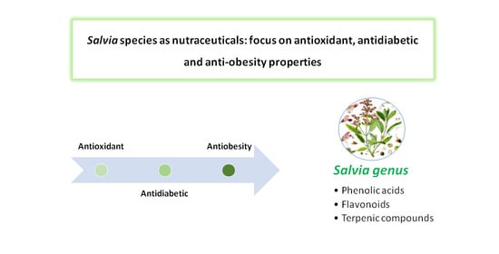
This review highlights the health benefits of several Salvia species as potential sources of phenolic compounds that can combat oxidative stress and related diseases, namely diabetes and obesity. The summary of relevant studies focusing on these issues is shown in the four tables of this manuscript which also include the available information regarding the main phenolic constituents.
1.1. Antioxidant Activity
Oxidative stress results from an imbalance condition between generation and elimination of reactive oxygen species (ROS), including superoxide (O2●−), hydroxyl radicals (●OH) and hydrogen peroxide (H2O2), together with reactive nitrogen species (RNS) such as nitric oxide (NO●) and peroxynitrite (ONOO−), which cause severe damage to cellular components such as DNA, proteins and lipids—contributing to the pathogenesis of many diseases [3,28,29]. In turn, enzymatic ((e.g. catalase (CAT), superoxide dismutase (SOD), glutathione peroxidase (GPx) and glutathione-S-transferase (GST)) and non-enzymatic ((e.g. vitamins A, C and E, reduced glutathione (GSH), β-carotene)) antioxidants counteract oxidative stress [3,29,30,31]. In this regard, a growing body of evidence suggests that diet supplementation with antioxidant compounds, through a daily consumption of plant-based products, can provide an important tool to counteract the adverse effects of oxidative stress and related diseases [2,3,32,33]. Having this in mind, the gathering of scientific data to corroborate the traditional usage of these plants is a key step in promoting their industrial application [2,6,34,35,36].
1.1.1. Antioxidant Activity of Salvia Extracts—Chemical Models
As for plants in general, the antioxidant potential of sage extracts has been evaluated as a first approach through in vitro methods (Table 1), including the capacity to scavenge stable free radicals such as DPPH● (2.2-diphenyl-1-picrylhydrazyl), ABTS● (2.2´-azino-bis-3-ethylbenzthiazoline-6-sulphonic acid), hydroxyl (●OH), superoxide (O2●−) and nitric oxide (NO●), and also by the ability to reduce metal ions from their higher to their lower oxidation state by the reducing power method (RP) and ferric reducing antioxidant power assay (FRAP). Additional assays include the oxygen radical absorbance capacity (ORAC), for assessing radical chain-breaking ability through the inhibition of peroxyl radical (RO2●)–induced oxidation, and the trolox equivalence antioxidant capacity (TEAC), as well as the thiobarbituric acid reactive substances (TBARS) and β-carotene bleaching assays, to estimate the lipid peroxidation inhibitory capacity [24,37,38].
Among sage plants, S. officinalis is the most widespread worldwide and also the most studied regarding antioxidant capacity [24,39]. Due to their high antioxidant capacity, S. officinalis polar phenolic extracts, usually alcoholic or hydroalcoholic extracts, are also frequently used as a reference to assess antioxidant properties of other less investigated plants [40,41]. The DPPH EC50 values (half maximal effective concentration, equivalent to the sample concentration that provides 50% of antioxidant activity) of distinct extracts from this species were established in the range of 1.98–10.1 μg/mL [41,42,43], being close to that of the flavonol quercetin (2.79 μg/mL) [44]. The antioxidant activity of S. officinalis phenolic-rich extracts has also been extensively assessed by the ABTS● scavenging assay (EC50 = 4.79–50.8 μg/mL) [43,44], through evaluation of the ability to scavenge superoxide (EC50 = 5.3–10.1 μg/mL) [42] and hydroxyl radicals (EC50 = 1.36 μg/mL) [43], and also by the ORAC assay (0.4–1.8 mmol TE/100 mL) [45]. Moreover, methanolic extracts and decoctions of S. officinalis were shown to exhibit very low EC50 values, as estimated by ß-carotene bleaching (EC50 = 6.6 and 50.9 μg/mL, respectively) and TBARS assays (EC50 = 2.1 and 10.4 μg/mL, respectively) [46]. More recently, Soxhlet extraction of S. officinalis provided samples with high antiradical activity, with respective DPPH● and ABTS● EC50 values of 116.5 and 88.2 μg/mL for aqueous extract, and 124.7 and 99.3 μg/mL for the hydroethanolic extract. Additionally, the authors showed that extracts obtained with a Soxhlet or by traditional methods presented a similar antioxidant activity against RO2● (EC50 values of 0.52 and 0.61 μg/mL for hydroethanolic and aqueous soxhlet extraction versus 0.52 and 0.69 μg/mL for hydroethanolic maceration and infusion traditional extraction, respectively) [47].
As for other Salvia species, methanolic extracts of Salvia from Tunisia origin (Salvia aegyptiaca, Salvia argentea and Salvia verbenaca), were reported to scavenge DPPH● (EC50 = 21–77 μg/mL) and ABTS● (141–318 μM TE/mg DW), as well as to reduce the ferric form (81–164 mM Fe(II)/mg DW) [41]. Moreover, the DPPH EC50 values of methanol:chloroform (1:1) extracts of 14 Salvia plants from South Africa, which in general contained rosmarinic acid as their major phenolic constituent, were shown to vary between 1.6 µg/mL (Salvia schlechteri) and 74.2 µg/mL (Salvia garipensis) [48]. In the same line, polar extracts of Salvia amplexicaulis [49] and Salvia ringens [50], both rich in kaempferol glycosides (298 to 636 mg/g extract and 120 to 465 mg/g extract, respectively), were suggested as good antioxidant agents, as estimated by DPPH●, ABTS● and FRAP methods. Moreover, a methanolic extract of Salvia syriaca, with considerable amounts of rutin and rosmarinic acid, displayed a high DPPH● scavenging activity, with the same magnitude of the standard solution 3.5-di-tert-butyl-4-hydroxytoluene (BHT; EC50 values of 70 and 66 μg/mL, respectively) [51]. In addition, phenolic-rich extracts from different populations of Salvia fruticosa [52] and aqueous and methanolic extracts of Salvia cadmica from Turkey [53], were demonstrated to scavenge DPPH● and ABTS●, and to reduce ferric ion as well. In these studies, rosmarinic acid was detected as the main compound, ranging from 20.6 to 41.9 mg/g extract [53].
In good agreement with these results, other Salvia polar extracts, such as S. amplexicaulis [49], Salvia halophila [54], Salvia virgata, Salvia persica, Salvia cereal and Salvia reuterana [40], were shown to inhibit β-carotene bleaching, in some cases with equal capacity to those of the standard compounds. Similarly, the potential of Salvia species to inhibit lipid peroxidation, as estimated by TBARS assay, was reported for an ethyl acetate extract of S. halophila, which exerted an inhibitory ability similar to the commercial antioxidant BHT [54]. Moreover, among five solvent extracts from the aerial parts of Salvia pachyphylla, the ethyl acetate extract was the most active based on DPPH● scavenging assay (EC50 = 0.28 mg/ml) [55]. In addition, Asadi et al [56] reported the scavenging ability of six Salvia species methanolic extracts as varying between 1 to 487.1 mg ascorbic acid equivalent /g dried extract, in the order: Salvia hydrangea, Salvia macilenta > Salvia multicalis, Salvia sclarea > Salvia xanthocheila and Salvia lachnocalyx [56]. Moreover, Kostic et al. [57] reported good antioxidant ability of ethanolic extracts of S. sclarea, as estimated through DPPH● (EC50 = 27.8 μg/mL) and inhibition of ß-carotene (EC50 = 19.1 μg/mL) methods. Likewise, methanolic extracts of S. syriaca and Salvia nemorosa collected in Iran were shown to possess a scavenging ability towards DPPH● similar to that of BHT [51,58]. The authors found a good correlation between total phenolic compounds (TPC) and flavonoids (TFC), and their antioxidant activities, highlighting their richness in rosmarinic acid as well as quercetin and its glycosides. Other methanolic extracts of a collection of Salvia species, including Salvia eremophila, Salvia santolinifolia, S. nemorosa and Salvia atropatana, were demonstrated to be good DPPH● scavengers as well [16], and those of S. sclarea and S. nemorosa were even claimed to be 1.4–2.2 times lower than those of BHT [40]. The high antioxidant potential of the two latter extracts was also attested to by other chemical in vitro tests, namely ORAC and ABTS [59].
Although less studied, aqueous extracts obtained from Salvia plants have also been reported to be promising antioxidant agents. In this regard, an S. officinalis decoction extract, rich in quinic acid, protocatechuic acid, gallic acid and salvianolic acid, was shown to inhibit β-carotene bleaching with IC50 values 2.7-fold higher than BHT [60]. In another study, our group demonstrated that the decoctions of Salvia africana, Salvia officinalis ‘Icterina’, Salvia mexicana [26] and Salvia elegans [27], had DPPH● EC50 values in the range of 6.6 ± 0.7 and 10.7 ± 2.1 μg/mL, with a potency of about half or the same order of ascorbic acid. Likewise, decoctions of S. africana, S. officinalis ‘Icterina’ and S. mexicana were shown to hamper lipid peroxidation, as measured by TBARS assay, in the same magnitude of trolox [26]. In addition, a decoction of Salvia apiana was also emphasized for its capacity to prevent lipid peroxidation events [25]. Moreover, decoctions of S. elegans, S. officinalis and Salvia greggii were demonstrated to have a high capacity to scavenge NO● [27].
Rosmarinic acid and other caffeic acid derivatives were frequently reported as being the most abundant phenolic compounds in S. africana, S. apiana and S. elegans decoctions, while glycosidic flavones were particularly abundant in S. officinalis and S. officinalis ‘Icterina’ decoctions [25,26,27]. In our study [25], we also concluded that the high antioxidant potential of S. apiana decoction might be partially associated with its high levels of phenolic compounds, particularly of rosmanol, a derivative of sageone, and hydroxycarnosic acid [25]. Moreover, a significant correlation between ORAC and rosmarinic acid (R = 0.68, p = 0.00355), was previously reported for 16 samples of commercial sage tea infusions that were claimed to have a high content of phenolic compounds [45].
The antioxidant capacity of phenolic compounds isolated from sage plants have in fact been underlined (Table 1). Rosmarinic acid was previously isolated from the methanolic extracts of four Salvia species leaves (S. officinalis, Salvia glutinosa, Salvia aethiopis and S. sclarea) and identified as a dominant radical scavenger towards the DPPH● in the HPLC–DPPH system [61]. More recently, Pavic et al. [62] evidenced the high DPPH● scavenging capacity of carnosol and carnosic acid, both extracted from S. officinalis by supercritical CO2 extraction. In addition, a study performed by Etsassala et al. [63] focused on the antioxidant potential of new and known abietane diterpene compounds isolated from methanolic extracts of Salvia africana-lutea collected in South Africa. In particular, the authors described a strong activity of 19-acetoxy-12-methoxycarnosic acid and clinopodiolides A on ORAC (1.5- and 1.7-fold that of the reference antioxidant epigallocatechingallate, respectively), and a high antioxidant potential of 3b-acetoxy-7a-methoxyrosmanol and clinopodiolides A on FRAP (about 3.3-fold that of the control). The same group of researchers [64] demonstrated that rosmanol, carnosol and 7-methoxyrosmanol exerted excellent scavenging abilities for peroxyl radicals, as estimated by ORAC assay (six times higher than those of the reference commercial compound), while carnosol and rosmanol also exerted high antioxidant capacity in FRAP, and in 4,7-dimethylapigenin and rosmanol with TEAC.

Table 1.
Antioxidant properties of phenolic rich extracts of distinct Salvia species (in vitro experiments).
Table 1.
Antioxidant properties of phenolic rich extracts of distinct Salvia species (in vitro experiments).
| Salvia Species | Origin | Solvent Extraction/Phenolics (mg/g Extract) | Results of Screen Assay | Ref. |
|---|---|---|---|---|
| S. aegyptiaca | Tunisia | MeOH/RA (46.3–41.7), methyl-Carn (9.6–41.4), Api-7-Glc (5.9–8.7), Lut-7-Glc (5.6–7.2) | DPPH (EC50, µg/mL): 21.1–22.6; FRAP (mM Fe(II)/mg): 149–164; ABTS (μM TE/mg dry plant): 312–318 | [41] |
| S. africana | Portugal | H2O/RA (77), Lut-O-Glr (18.7), YA-iso (30.8) | DPPH (EC50, μg/mL): 6.6 (H2O), 6.7 (AA); RP (EC50, μg/mL): 21.2 (H2O), 16.1 (BHA); β-carot bleach (EC50 μg/mL): 128.6 (H2O), 41.7 (Trolox); TBARS (EC50 μg/mL): 21.0 (H2O), 23.0 (Trolox) | [26] |
| S. africana-lutea | South Africa | MeOH/19-acy-12-mxyCA, 3β-acy-7-mxyRos, 19-acy-7-mxyRos, 19-acy-12-mxy Car, clipd A, clipd B (ND) | ORAC (µmol TE/g): 2588 (19-acy-12-mxyCA), 2234 (3β-acy-7-mxyRos), 735 (19-acy-7-mxyRos), 560 (19-acy-12-mxyCar), 2357 (clipd A), 1502 (clipd B), 3977 (EGCG); TEAC (µmole TE/g): 694 (19-acy-12-mxyCA), 636 (3β-acy-7-mxyRos), 124 (19-acy-7-mxyRos), 440 (19-acy-12-mxyCar), 862 (clipd A), 724 (clipd B), 4146 (EGCG); FRAP (µM acy/g): 1217 (19-acy-12-mxyCA), 2201 (3β-acy-7-mxyRos), 1440 (19-acy-7-mxyRos), 1257 (19-acy-12-mxyCar), 2263 (clipd A), 1480 (clipd B), 7525 (EGCG) | [63] |
| S. apiana | Portugal | H2O/Ros (192.4), Sage-deriv (174.1), RA (69.7), HycarAc (56.8) | DPPH (EC50, μg/mL): 13.3 (H2O), 6.7 (AA); RP (EC50, μg/mL): 55.0 (H2O), 16.1 (BHA); β-carot bleach (EC50 μg/mL): 41.2 (H2O), 41.7 (Trolox); TBARS (EC50 μg/mL): 2.79 (H2O), 23.0 (Trolox) | [25] |
| S. amplexicaulis | Lithuania | EtOH/H2O/CO2 EtOH/TPC, 97.4; H2O/TPC, 5.9, mg GAE/g | ORAC (µM TE/g DW): 4735 (EtOH), 676 (H2O), 1914 (CO2); ABTS (µM TE/g DW): 1177 (EtOH), 79.5 (H2O) | [59] |
| S. amplexicaulis | Macedonia | DCM/Hyper (54), Coumarin (29), EA/Hyper (55), Coumarin (30), H2O/RA (67), Kam-3-O-acetil Glc-7-O-rhm (410), EtOH/Lut- 5-O-Glc (138), Kam-3-O-acetil Glc-7-O-Rhm (636), MeOH/Lut- 5-O-Glc (238), Kam-3-O-acetil Glc-7-O-Rhm (298) | DPPH (EC50, μg/mL): 27.8 (EtOH), 15.8 (H2O), 15.1 (MeOH), 196.5 (EA), 593.2 (DCM), 5.1 (AA); ABTS (mg AAE/g): 2.8 (EtOH), 2.8 (H2O), 2.4 (MeOH), 0.7 (EA), 0.4 (DCM), 2.8 (BHT); FRAP (μmol Fe(II)/g): 979 (EtOH), 1372 (H2O), 1178 (MeOH), 112 (EA), 117 (DCM), 445 (BHT); β-carot bleach (%): 40.4 (EtOH), 41.8 (H2O), 13.3 (MeOH), 58 (BHT) | [49] |
| S. argentea | Tunisia | MeOH/RA (30.9–47.2), methyl-Carn (13.1–19.4), Narn (5.4–8.7) | DPPH (EC50, µg/mL): 33.9-77.1; FRAP (mM Fe(II)/mg): 81.6-105; ABTS (μM TE/mg ): 141–173 | [41] |
| S. aurita | South Africa | MeOH/Car, Ros, 7-mxy-Ros, 12-mxy-CA, 4,7-dimethyl-Api-ether (ND) | ORAC (µmole TE/g): 23962 (Car), 25790 (Ros), 23939 (7-mxy-Ros), 20247 (12-mxy-CA), 6475 (4,7-dimethyl-Api-ether), 4453 (crude extract), 3977 (EGCG); TEAC (µmole TE/g): 331 (Car), 2055 (Ros), 222 (7-mxy-Ros), 337 (12-mxy-CA), 3191 (4,7-dimethyl-Api-ether), 724 (crude extract), 4146 (EGCG); FRAP (µM AAE/g): 3918 (Car), 1522 (Ros), 1322 (7-mxy-Ros), 508 (12-mxy-CA), 610 (4,7-dimethyl-Api-ether), 394 (crude extract), 7525 (EGCG) | [64] |
| S. bicolor | Egypt | MeOH/PrcA (75.2), CouA (70.2), GA (68.2), Lut-7-O-Glc (1.2) | DPPH (EC50, µg/mL): 321 (MeOH), 250 (GA) | [65] |
| S. cadmica | Turkey | EA/RA (5.3), Lut (0.8), Api (0.5), MeOH/RA (31.1), Lut (1.2), Hesp (0.9), H2O/RA (20.6), Lut (1.1), Api (0.6) | DPPH (µmol TE/g dry plant): 5.9 (EA), 54.7 (MeOH), 40.5 (H2O); CUPRAC (µmol TE/g dry plant): 20.9 (EA), 50.9 (MeOH), 54.5 (H2O); ABTS (µmol TE/g dry plant): 7.1 (EA), 84.9 (MeOH), 102.2 (H2O); FRAP (µmol TE/g dry plant): 13.5 (EA), 80.0 (MeOH), 98.0 (H2O) | [53] |
| S. elegans | Portugal | H2O/RA (35.5), CaffRA (17.9), SA B (7.8) | DPPH (EC50, µg/mL): 10.7 (H2O), 6.69 (AA); FRAP (EC50, µg/mL): 31.3 (H2O), 16.30 (BHA); O2●− (EC50, µg/mL): 30.6 (H2O), 7.8 (GA); NO● (EC50, µg/mL): 91.5 (H2O), 212.1 (AA); RO2● (EC50, µM TE/mg ext): 373.1; XO (EC50 µg/mL): 71.8 (H2O), 0.09 (All) | [27] |
| S. fruticosa | Greece: (populations 1 and 2) | MeOH/RA (41.9), ChlgA (1.8), CaffA (0.70) | FRAP (µM Trolox/g DW): 31.83–202.93; 135.81–326.22; ABTS (µM Trolox/g DW): 60.94–242.3; 199.64–312.49; DPPH (µM Trolox/g DW): 182.99–192.62; 185.30–192.19, populations 1 and 2, respetively | [52] |
| S. greggii | Portugal | H2O/Lut-7-O-Glc (26.1), Api-C-hex (15.7), RA (10.9) | DPPH (EC50, µg/mL): 21.1 (H2O), 6.69 (AA); FRAP (EC50, µg/mL): 77.9 (H2O), 16.30 (BHA); O2●– (EC50, µg/mL): 61.7 (H2O), 7.8 (GA); NO● (EC50, µg/mL): 167.8 (H2O), 212.1 (AA); RO2● (EC50, µM TE/mg ext): 335.6; XO (EC50 µg/mL): 70.1 (H2O), 0.09 (All) | [27] |
| S. halophila | Turkey | Hexane (ND), EA/RA (48.9), CaffA (1.6), MeOH/RA (38.6), MeOH 50%/RA (27.1), p-OH-BA (7.5), o-coumaric (6.4), CaffA (2.4), Lut-Glc (2.3), H2O/RA (5.9) | DPPH (EC50, mg/mL): 0.4 (EA), 0.6 (50% MeOH), 1.0 (H2O, MeOH); FRAP (AAE mmol/g extract): 1.0 (EA, 50% MeOH), <1.0 (H2O, MeOH, Hexane), 0.025 (GA); β-carot bleach (AA%): 80 (Hexan), 65 (EA), <40 (MeOH, 50% MeOH, H2O), 90 (BHT); Iron (II) Thiocyanate (%): 91 (hexan), 89 (EA), 70 (50% MeOH), 68 (H2O), 18 (MeOH), 90 (BHT at 1%); TBARS (%): 94 (hexan), 91 (H2O), 82 (50% MeOH), 80 (EA), 52 (MeOH), 90 (BHT at 1%) | [54] |
| S. miltiorrhiza | Commercial | H2O/TPC 66.27; 95% EtOH/TPC 4.97 mg GAE/g | DPPH (EC50, μM AE/g): 202 (H2O), 105 (EtOH) | [66] |
| S. miltiorrhiza | China | H2O/Danshensu, SA A, SA B | DPPH (EC50, µg/mL): 131; ORAC (µmol/g): 757 | [67] |
| S. nemorosa | Lithuania | EtOH/H2O/CO2 EtOH/TPC (108.7), H2O/TPC 7.6 mg GAE/g | ORAC (µM TE/g DW): 2392 (EtOH), 718 (H2O), 1193 (CO2); ABTS (µM TE/g DW): 1005 (EtOH), 150 (H2O) | [59] |
| S. nemorosa | Iran | 80% MeOH/TPC 30.4 mg GAE/g dried plant | DPPH (EC50, μg/mL): 138.4 (80% MeOH), 1.79 (Que) | [16] |
| S. nemorosa | Iran | MeOH/RA (7.5), CaffA (0.11), Que (0.15) | DPPH (EC50, μg/mL): 82 (MeOH), 64 (BHT) | [58] |
| S. nemorosa | Iran | MeOH/TPC, 114 mg TAE/g | DPPH (EC50 μg/mL): 473 (MeOH), 211 (BHT); β-carot bleach (%): 20 (MeOH), < 20% (BHT, at 500 μg/mL) | [40] |
| S. officinalis | Portugal | H2O/Api-O-Glr (48.4), RA (287.3), Scu-O-Glr (13.4) | DPPH (EC50, µg/mL): 34.8 (H2O), 6.69 (AA); FRAP (EC50, µg/mL): 40.0 (H2O), 16.30 (BHA); O2●– (EC50, µg/mL): 32.8 (H2O), 7.8 (GA); NO● (EC50, µg/mL): 118.2 (H2O), 212.1 (AA); RO2● (EC50, µM TE/mg ext): 404.4; XO (EC50 µg/mL): 55.1 (H2O), 0.09 (All) | [27] |
| S. officinalis | Iran | MeOH/TPC, 86.4 mg TAE/g | DPPH (EC50 μg/mL): 233 (MeOH), 211 (BHT); β-carot bleach (%): 79 (MeOH), < 20% (BHT, at 500 μg/mL) | [40] |
| S. officinalis | Algerian | MeOH-H2O/TPC, 30 mg GAE/g | DPPH (EC50 μg/mL): 1.98; ABTS (EC50 μg/mL): 4.79; OH●(IC50 μg/mL): 1.36 | [43] |
| S. officinalis | Portugal | DE/TPC 9.3, EA/TPC 7.0, n-but/TPC 23.9, H2O/TPC 3.3, mg GAE/ml | DPPH (EC50 μg/mL): 3.9 (EA), 4.1 (n-but), 2.8 (H2O); O2●- scav (EC50 μg/mL): 5.5 (DE), 5.3 (EA), 8.9 (n-but) 10.1 (H2O) | [42] |
| S. officinalis | Spain | H2O inf/RA (73.9), Lut-O-Glc (37.4), Lut-O-Glr (88.1), H2O dec/RA (93.5), Lut-O-Glc (52.2), Lut-O-Glr (129.8), 80% MeOH/RA (93.2), Lut-O-Glc (56.1), Lut-O-Glr (94.7) | DPPH (EC50 μg/mL): 96 (H2O inf), 75.5 (H2O dec), 33 (80% MeOH); RP (IC50 μg/mL): 84 (H2O inf), 66.5 (H2O dec), 25 (80% MeOH); β-carot bleach (EC50 μg/mL): 139 (H2O inf), 50.9 (H2O dec), 6.6 (80% MeOH); TBARS (EC50 μg/mL): 18.0 (H2O inf), 10.4 (H2O dec), 2.1 (80% MeOH) | [46] |
| S. officinalis | Lithuania | EtOH/H2O/CO2 EtOH/TPC, 120.7; H2O/TPC, 13.2, mg GAE/g | ORAC (µM TE/g DW): 2535 (EtOH), 1143 (H2O), 6015 (CO2); ABTS (µM TE/g DW): 1080 (EtOH), 225 (H2O) | [59] |
| S. officinalis | Commercial | H2O, MeOH/RA (16.3), SA K isomer (2.7), Lut-hex (2.7), Api-Glr (4.3) | DPPH (EC50, μg/mL): 69 (MeOH), 2.79 (Que); ABTS (EC50 μg/mL): 19.9 (MeOH), 50.8 (H2O), 1.17 (Que) | [44] |
| S. officinalis | Commercial | Infusion/RA (12.2–296), Lut-7-O-Glr (37.9–166), CA (9.1–32.9) mg/L. | ORAC (mmol TE/100mL): 0.4 to 1.8 | [45] |
| S. officinalis | Tunisia | MeOH/RA (65.1–79.9), methyl-Carn (22.9–34.6), CA (15.8–24.0), Car (23.8–25.8) | DPPH (EC50, µg/mL): 3.4–10.1; FRAP (mM Fe/mg): 179–197; ABTS (μM TE/mg): 645–766 | [41] |
| S. officinalis | Portugal | Soxhlet extraction: EtOH, EtOH-H2O/RA (0.443), Car (0.935), CA (0.948); TPC 685.2, 606.3, 227.2 mg GA/g extract, respectively; Traditional extraction: H2O, EtOH-H2O/RA (0.443); TPC 528.2 and 464.5 mg GA/g extract, respectively) | Soxhlet extraction: DPPH (EC50, μg/mL): 116.5 (H2O), 124.7 (EtOH-H2O), 252.3 (EtOH); ABTS (EC50 μg/mL): 88.2 (H2O), 99.3 (EtOH-H2O), 170.6 (EtOH); RO2● (EC50 μg/mL): 0.61 (H2O), 0.52 (EtOH-H2O), 1.35 (EtOH) Traditional extraction: DPPH (EC50, μg/mL): 157.0 (H2O), 131.3 (EtOH-H2O); ABTS (EC50 μg/mL): 125.2 (H2O), 114.5 (EtOH-H2O); RO2●(EC50 μg/mL): 0.69 (H2O), 0.52 (EtOH-H2O) | [47] |
| S. officinalis‘Icterina’ | Portugal | H2O/RA (52.7), Api-O-Glr (32.8), Lut-7-O-Glr (18.2) | DPPH (EC50, μg/mL): 10.4 (H2O), 6.68 (AA); RP (EC50, μg/mL): 42.3 (H2O), 16.1 (BHA); β-carot bleach (EC50 μg/mL): 146.6 (H2O), 41.7 (Trolox); TBARS (EC50 μg/mL): 23.0 (H2O), 23.0 (Trolox) | [26] |
| S. palaestina | Jordan | n-but/Sal, methyl-3-O-methyl-Rsm, Lut-7-O-(2´´-p-hyb)-β-Glr (ND) | DPPH (EC50, µg/mL): 3.01 (Sal); 1.0 (methyl-3-O-methyl-Rsm), 1.1 (BHA) | [68] |
| S. ringens | Macedonia | EtOH/Kam-3-O-acetil-Glc-7-O-Rhm (465), rutin (77), MeOH/RA (36), Kam-3-O- acetil-Glc -7-O-Rhm (287), Rutin (173), DCM/Hyperoside (128), EA (Lut (27), Kam-3-O-acetil-Glc-7-O-Rhm (120) | DPPH (EC50, μg/mL): 17.3 (EtOH), 20.3 (MeOH), 266.2 (DCM), 22.2 (EA), 5.1 (AA); ABTS (mg AAE/g): 2.44 (EtOH), 1.19 (MeOH), 0.58 (DCM), 2.36 (EA), 2.8 (BHT); FRAP (μmol Fe(II)/g): 1088.3 (EtOH), 274.8 (MeOH), 191.1 (DCM), 969.8 (EA), 180 (AA) | [50] |
| S. sclarea | Lithuania | EtOH/H2O/CO2 EtOH/TPC, 106.2, H2O/TPC 7.6 mg GAE/g | ORAC (µM TE/g DW): 3820 (EtOH), 867 (H2O), 570 (CO2); ABTS (µM TE/g DW): 1634 (EtOH), 153 (H2O) | [59] |
| S. sclarea | Iran | 80% MeOH/TPC 14.8 mg GAE/g dried plant) | DPPH (EC50, μg/mL): 190.7 (80% MeOH), 1.79 (Que) | [16] |
| S. sclarea | Iran | MeOH/TPC, 103.3 mg TAE/g | DPPH (EC50 μg/mL): 290 (MeOH), 211 (BHT); β-carot bleach (%): 35 (MeOH), < 20% (BHT, at 500 μg/mL) | [40] |
| S. sclarea | Serbia | EtOH/RA (165.3), Lut-7-Glc (5.6), Api-7-Glc (8.5) | DPPH (EC50 μg/mL): 27.8 (EtOH), 6.1 (RA), 2.4 (BHA); β-carot bleach (EC50 μg/mL): 19.1 (EtOH), 32.6 (RA), 0.04 (BHA) | [57] |
| S. syriaca | Iran | n-hex, DMC, MeOH/Rut (9.43), Que (0.15), Api (0.24), FA (0.19), RA (0.63) | DPPH (EC50 μg/mL): 210 (n-hex), 245 (DMC), 70 (MeOH), 66 (BHT) | [51] |
| S. verbenaca | Tunisia | MeOH/RA (36.9–53.5), Narn (2.9–7.5), Nar (2.4–3.7) | DPPH (EC50 µg/mL): 23–36.3; FRAP (mM Fe(II)/mg): 106–159; ABTS (μM TE/mg ): 165–205 | [41] |
| S. virgata | Iran | MeOH/TPC 33.8 mg TAE/g | DPPH (EC50 μg/mL): 198 (MeOH), 211 (BHT); ↓β-carot bleach (%): 95 (MeOH), <20% (BHT, at 500 μg/mL) | [40] |
| OtherSalviaspecies | ||||
| S. cereal (S. c), S. reuterana (S. r), S. persica (S. p) | Iran | MeOH /TPC 45.7–82.9 mg TAE/g | DPPH (EC50 μg/mL): 598 (S. r), 883 (S. cl), 1810 (S. p), 211 (BHT); β-carot bleach (%): 30 (S. r), 55 (S. c), 95 (S. p), <20% (BHT, at 500 μg/mL) | [40] |
| S. aethiopis, S. atropatana, S. eremophila, S. hypoleuca, S. limbata, S. santolinifolia, S. syriaca, S. xanthocheila | Iran | 80% MeOH /TPC 12.5–25.7 mg GAE/g dried plant | DPPH (EC50, μg/mL): 89.5 (S. atropatana), 557 (S. limbata), 1.79 (Que) | [16] |
| S. africana-caerulea, S. africana-lutea, S. albicaulis, S. aurita, S. chamelaeagnea, S. disermas, S. dolomítica, S. garipensis, S. lanceolata, S. muirii, S. namaensis, S. radula, S. repens, S. runcinata, S. schlechteri, S. stenophylla | South Africa | MeOH:CHCl3 /TPC 45.6–212 mg GAE/g | DPPH (EC50, µg/mL): 1.61 ( S. schlechteri), 74.2 (S. garipensis), >100 (S. dolomitica, S. radula), 11.1–68.1 (Others); ABTS (EC50, µg/mL): 11.9 (S. muirii), 69.3 (S. radula), 14.6–50 (Others) | [48] |
AA—ascorbic acid; AAE—ascorbic acid equivalent; AE—Ascorbate equivalent; ABTS—2.2′-azino-bis-(3-ethylbenzothiazoline-6-sulphonic acid) radical scavenging assay; acy—acetoxy; Api—apigenin; BA—benzoic acid; BHA—2(3)-t-butyl-4-hydroxyanisole; BHT—3.5-di-tert-butyl-4 hydroxytoluene; CA—carnosis acid; CaffA/CaffA deriv—caffeic acid/caffeic acid derivative; Car—carnosol; Carn—carnosate; ChlgA—chlorogenic acid; Clipd—clinopodiolides; β-carot bleach—β-carotene/linoleic acid bleaching inhibition assay; CouA—coumaric acid; CUPRAC—cupric ion reducing antioxidant capacity; Deriv—derivatives; DCM—dichloromethane; DE—diethyl ether; dec—decoction; DPPH—2.2-diphenyl-1-picrylhydrazyl radical scavenging assay; DW—dry weight; EA—ethyl acetate; EGCG—epigallocatechin gallate, EtOH—ethanol; FA—ferulic acid; Flav—flavonoids; FRAP—ferric reducing antioxidant power; GA—gallic acid; GAE—gallic acid equivalents; Glc—glucoside; Glr—glucuronide; Hesp—hesperidin; Hex—hexoside; Hyb—hydroxybenzoyl; HycarAc—Hydroxycarnosic acid; Hyper—hyperoside; I—inhibition; inf—infusion; iso—isomer; Kam—kaempferol; Lut—luteolin; MeOH—methanol; mxy—methoxy; Nar—naringin; Narn—naringenin; n-but—n-butanol; ND—not determined; NO—nitric oxide radical scavenging assay; OH●—hydroxyl radical scavenging assay; O2●—radical scavenging assay; ORAC—oxygen radical absorbance capacity; p-OH-BA—p-hydroxybenzoic acid; PrcA—protocatchuic acid; QA—quinic acid; Que—quercetin; RA—rosmarinic acid; Rhm—rhamnoside; RP—reducing power; RO2●—Peroxy radical inhibition assay; Ros—rosmanol; Rsm—rosmarinat; SA—salvianolic acid; Sal—salpalaestinin; Sage — sageone; Scu—scutelarein; SH—reduced thiol groups; TAC—total antioxidant capacity; TAE—tannic acid equivalent; TBARS—thiobarbituric acid reactive substances assay; TE—trolox equivalent; TEAC—trolox equivalent absorbance capacity; TPC—total phenolic content; XO—xanthine oxidase inhibition assay; YA—Yunnaneic acid.
1.1.2. Antioxidant Activity of Salvia Extracts—In Vitro Cellular and In Vivo Methods
Although less investigated, the antioxidant abilities of polar extracts from Salvias have also been demonstrated in cellular and/or in vivo models (Table 2). As previously mentioned, DNA can be dramatically damaged under oxidative stress conditions. Notably, the treatment of H2O2-induced human colon carcinoma Caco-2 and human cervical cancer HeLa cells with aqueous extracts of S. officinalis and S. fruticosa for 24 h resulted in a protective effect on DNA damage [69], as measured through the comet assay. This effect was also observed for the two major phenolic constituents of the extracts (rosmarinic acid and luteolin-7-O-glucoside), thereby emphasizing their possible intervention in this mechanism. The same authors also reported promising results in assays of DNA repair, as evaluated in oxidative-induced Caco-2 cells. In these experimental conditions, independent treatment with both extracts (50 μg/mL), as well as with luteolin-7-O-glucoside (20 µM), were able to increase the rate of rejoining of DNA strand breaks [69]. Notably, Kosics et al. [44] reported that the protective effects of S. officinalis aqueous extracts (rich in rosmarinic acid) against DNA damage induced by strong oxidants (H2O2 and DMNQ), achieved the highest DNA-protective capacity at 2 mg/mL [44]. On another hand, in a study performed by Tavakkoli et al. [70], the methanolic extracts of Salvia santolinifolia Boiss. and S. sclarea L. (100 µg/mL) were demonstrated to reduce the H2O2-stimulated increase in ROS production, in neuronal PC12 cells, by 61.9% and 61.4%, respectively. In the same study, the S. santolinifolia extract was also shown to significantly reduce H2O2-induced apoptosis [70]. Likewise, methanolic extracts of S. hydrangea and S. sclarea (≤ 50 µg/ml) were shown to fight DNA oxidative damage of PC12 neural cells induced by Fe(II)–H2O2 [56]. Additionally, neuroprotective effects of Salvia extracts from S. hydrangea, S. lachnocalyx, S. macilenta, S. multicalis, S. sclarea and S. xanthocheila against oxidative stress (using H2O2 as oxidative agent), were observed in PC12 neural cells, for which pretreatment with 100 µg/mL significantly protected cell survival (76% to 93%) with respect to the control [56]. Moreover, the antioxidant activity of phenolic acids isolated from S. officinalis was demonstrated to offer protection of PC12 cells from injury (54.2% for salvianolic acid Y and 35.2% for salvianolic acid B) [71].
Table 2.
Antioxidant properties of phenolic rich extracts of distinct Salvia species (in vitro cellular assays and in vivo experiments).
Table 2.
Antioxidant properties of phenolic rich extracts of distinct Salvia species (in vitro cellular assays and in vivo experiments).
| Salvia Species | Origin | Solvent Extraction/Phenolics (mg/g Extract) | Results of Screen Assay | Ref. |
|---|---|---|---|---|
| In Vitro cellular assays | ||||
| S. hydrangea (S.h), S. sclarea (S. s) | Iran | MeOH/TPC: S.h 238, S.s 268, mg GA/g | Inhibition of DNA oxidative damage of neuronal PC12 cells after tratment with 10 µg/mL extract; protection cell survival at 93% (S. h) and 78% (S. s; both 100 µg/mL), in comparision to control. | [56] |
| S. officinalis (S. o), S. fruticosa (S. f), S. lavandulaefolia (S. l) | Portugal | H2O (ND) | DNA protection against strand breaks induced by H2O2, 2h: 21% (HeLa cells, S. l), 26% (Caco-2 cells, S. f), at 50 µg/mL extract; 62% (Caco-2 cells, L-O-G), at 20 µM; DNA repair in Caco-2 cells: 62% (S. o), 86% (S. f), at 50 µg/mL extract; 74% (L-O-G), at 20 µM | [69] |
| S. officinalis | Commercial | H2O, MeOH/RA (16.3), SA K isomer (2.7), Lut-hex (2.7), Api-Glr (4.3) | HepG2 DNA damage induced by H2O2 and DMNQ: ↓ after 2 mg/mL extracts, at 24h / enzymatic activity: ↑GPx, ↓SOD, after 4, 2 and 1 mg/mL extracts | [44] |
| S. santolinifolia Boiss. (S. san), S. sclarea L. (S. s) | Iran | MeOH-H2O (ND) | Protective effects of H2O2-induced intracellular ROS in neuronal PC12 cells (% I): 61.9 (S. s), 61.4 (S. san); inhibition of apoptosis: extract S. s at 100 µg/mL | [70] |
| In Vivo experiments | ||||
| S. euphratica Montbret, Aucher & Rech. f. var. euphratica (S. e), S. kronenburgii Rech. f. (S. k) | Turkey | EtOH 0.5% or 1% (w/w)/TPC: S. k 41.8, S. e 76.2 mg GAE/g | Ointment application in wound model skin samples from rats, after 7/14 days S. k: NO (16.5/21.8), MDA (8.4/7.5), GSH (11.26/10.01), at 0.5%; S. k: NO (20.9/10.9), MDA (7.9/5.67), GSH (7.6/7.2), at 1%; S. e: NO (28.7/11.6), MDA (8.5/7.2), GSH (15.5/7.3), at 0.5%; S. e: NO (20.8/10.3), MDA (8.1/4.6), GSH (9.3/6.2), at 1%; Positive control: NO (20.1/18.5), MDA (12.3/7.42), GSH (11.27/7.93) | [72] |
| S. officinalis | Tunisia | H2O/p-CouA (0.28), SA (0.27) | Protective effects of EtOH-induced gastric and small bowel injuries by alcohol in Wistar rats after treatment with H2O-extract (at 100 and 200 mg/Kg bw), sulf (at 100 mg/Kg bw) and mix (H2O-extract at 50 mg/Kg bw + sulf at 50 mg/Kg bw): ↓MDA (nmol/mg protein) and ↓H2O2 (mmol/mg protein) content, p < 0.05 compared to EtOH group; ↑SOD (U/mg protein), ↑CAT (µM H2O2/min/mg protein) and ↑GPx (nmol GSH/min/mg protein) activity, ↑GSH (nmol/mg protein) and ↑SH (µmol/mg protein), p < 0.05 compared to EtOH group. | [60] |
| S. officinalis | Commercial | Liquid extract (ND) | (LPS)-induced inflammation restoration in rats after orally treatment: ↓MDA level (nm/mL/mg Hb) in erytrocyte hemolysate (2.99 at 10 mg/Kg; 2.66 at 30 mg/Kg extract), p < 0.001compared to control and inflammation groups); ↓MDA (nmol/mL) in kidney (2.20 at 10 mg/Kg; 2.15 at 30 mg/Kg extract); ↑SOD % I in erytrocyte hemolysate (67 at 10 mg/Kg; 72 at 30 mg/Kg extract), in liver (84 at 10 mg/Kg; 86 at 30 mg/Kg extract) and in lung (89 at 10 mg/Kg; 94 at 30 mg/Kg extract); ↑CAT (nmol/min/mL) in erytrocyte hemolysate, kidney and lung (1.68 to 13 at 10 mg/Kg; 1.77 to 9.7 at 30 mg/Kg extract); ↑GPx (nmol/min/mL) in liver, kidney and lung (88 to 147 at 10 mg/Kg; 99 to 160 at 30 mg/Kg extract); ↓NO (µmol/L) in liver (1.08 at 30 mg/Kg extract), kidney (3.19 at 10 mg/Kg and 0.77 at 30 mg/Kg extract) and blood plasma (0.87 at 10 mg/Kg); p < 0.001 or p < 0.05 compared to inflammation group) | [73] |
| S. officinalis | Slovakia | EtOH (ND) | Oxidative stress restoration of rats hepatocytes: ↑DNA protection against lesions induced by H2O2, p < 0.001; ↑GSH, p < 0.01; ↑GPx (0.0057–0.0062 U/mg protein) after treatment with 6.67–13.3 mg/ml extract in drinking water, p < 0.001 | [74] |
| S. przewalskii | Commercial | H2O/SA B (55), RA (316) | Oxidative stress restoration of rats-injured podocytes induced by PAN: ↓8-OHdG after tratment with extract on days 5, 10 and 15; ↓ROS after tratment with extract (158 μg/ml), SA B (8.5 μg/ml), RA (50 μg/ml), control (1 μg/ml), at 24 and 48 h; ↓apoptosis after tratment with extract (316 μg/ml), SA B (17 μg/ml) | [75] |
| S. sahendica | Iran | MeOH/TPC, 64.5, EtOH/TPC: 31.1 mg GAE/g | Oxidative stress restoration of rats liver and kidney damage induced by alcohol: ↑SOD, CAT, GPx and GST (four enzymes, U/mg protein),↑GSH (mg/100 g tissue), vit C and E (µmol/mg tissue), after tratment with 0.1 g/kg bw compared to untreated alcoholic rats, p < 0.05 | [76] |
| S. triloba | Egypt | MeOH (ND) | Oxidative stress status restoration of Alzheimer’s disease (AD) induced in rats: ↓serum MDA (%I: 19.65% and 12.9%), ↓serum NO (%I: 36.2 and 21.9); serum TAC (↑33.4% and 26.7%), serum SOD (↑17.3% and 4.2%) after treatment with 750 and 375 mg/kg bw, respectively, p < 0.05 comparing to AD group | [77] |
AD—Alzheimer’s disease; All—allopurinol; Api—apigenin; bw—body weight; Caco-2 cells—human colon carcinoma cells; CAT—catalase; CouA—coumaric acid; DMNQ—2,3-dimethoxy-1,4-naphthoquinone; EtOH—ethanol; GA—gallic acid; GAE—gallic acid equivalents; Glc—glucoside; Glr—glucuronide; GPx—glutathione peroxidase; GSH—reduced glutathione; GST—glutathione-S-transferase, Hb—hemoglobin; HeLa—cervical carcinoma cells; HepG2—hepatocellular carcinoma cells; Hex—hexoside; LPS—lipopolysaccharide; Lut—luteolin; MDA—malondialdehyde; MeOH—methanol; ND—not determined; NO—nitric oxide; PAN—puromycin aminonucleoside; PC12—neuronal cells; RA—rosmarinic acid; ROS—reactive oxygen species; SA—salvianolic acid; SOD—superoxide dismutase; Sulf—sulfasalazine, TAC—total antioxidant capacity; TPC—total phenolic content; vit—vitamin.
In vivo studies demonstrating antioxidant-related effects of Salvia extracts were performed either by oral administration and/or intravenous injection. In particular, the oral administration for one month of a methanolic extract of Salvia sahendica (0.1 g/kg), caused a significant decrease of malondialdehyde (MDA) and hydroperoxide levels, in liver and kidney of rats with alcohol-induced damage [76]. In addition, this intake restored the levels of non-enzymatic antioxidants (GSH, vitamin C, vitamin E) while increasing the activities of antioxidant enzymes (SOD, CAT, GPx and GST) [76]. In research performed by Horváthová et al. [74] in hepatocytes isolated from rats upon supplementation with S. officinalis ethanolic extract (13.33 mg/ml) for 14 days, a decrease of oxidative DNA damage was registered, based on the comet assay. Regardless of this, the SOD level was not changed; the authors demonstrated that the sage extract significantly increased the levels of GSH and GPx, suggesting that the consumption of S. officinalis may potentiate resistance to oxidative stress and afford hepatoprotection [74].
More recently, an S. officinalis decoction was shown to significantly reverse the levels of MDA and H2O2, in gastric and intestinal tissues of ethanol-induced gastric and small bowel-injured rats, after 15 days of oral administration [60]. In this study, treatment with sage decoction also significantly counteracted (in a dose-related manner) the decreased activities of enzymatic antioxidants SOD and GPx, and levels of the nonenzymatic antioxidant GSH, which were induced by alcohol administration. Overall, the results indicated that the administration of S. officinalis decoction and sulfasalazine (i.e., a drug used in the treatment of inflammatory bowel disease), alone or in combination, abolished acute ethanolic-induced oxidative stress in the gastric and duodenal mucosa, a fact that the authors attributed to the high amount of phenolic acids and flavonoids in the sage extract [60].
Several authors also demonstrated the benefits of Salvia commercial polar extracts towards oxidative-stress related disorders. In a study performed by Kolac et al. [73], a commercial extract of S. officinalis was administered orally to a lipopolysaccharide (LPS)-induced experimental inflammation animal model for two days, with the aim of investigating the potential antioxidants of the sage as a therapeutic approach in inflammatory diseases [73]. Among the samples evaluated, the authors underlined that the extract of S. officinalis (10 and 30 mg/kg) could increase the levels of CAT, SOD, and GPx in the lung, kidney, liver, and erythrocyte hemolysates in treatment groups, and reverse LPS-induced decreases in endogenous antioxidants. Additionally, the sage extract used in the treatment groups (inflammation + S. officinalis 10 and 30 mg/kg) decreased the levels of MDA in all samples, and reduced NO levels in the kidney as well, preventing the harmful effects of oxidative stress [73].
Several sage plants have also been tested for protective effects on renal disease. In a study performed by Liu et al. [75], a commercial extract of Salvia przewalskii (SPE) was shown to attenuate oxidative stress in puromycin aminonucleoside (PAN)-induced rat podocyte injury, via antioxidative stress effects—which is reflected by a significant reduction in 8-oxo-2’-deoxyguanosine, a marker of oxidative DNA damage. In the same study, antioxidative stress effects were observed against experimental PAN-induced nephrosis by suppression of ROS levels and podocyte integrity cytoskeleton-restoration, after treatment with SPE. Notably, isolated salvianolic acid B was demonstrated to be as potent as tacrolimus (i.e., a compound that has been used to protect podocytes of glomerular diseases) [75]. The authors also observed significant positive correlations between podocyte apoptosis rates and the ROS levels of groups treated with SPE, as well as the isolated compound salvianolic acid B and rosmarinic acid (r range of 0.998 to 1.0), suggesting that a decrease in intracellular ROS levels may inhibit PAN-induced apoptosis of cultured podocytes [75].
Due to the traditional use of several Salvia species as memory-enhancing agents, researchers have also focused their attention on the evaluation of these plants as candidates for new Alzheimer’s disease (AD) drugs, able to ameliorate the oxidative effects associated with this disease. In particular, Mahdy et al. [77] reported that the treatment of AD-induced rats with a methanolic extract of Salvia triloba at 750 and 375 mg/kg body weight for three months, induced a significant decrease in serum MDA (–19.65% and –12.9%) and NO levels (−36.21% and –21.94%), and a simultaneous increase of serum total antioxidant capacity (TAC; 33.39 and 26.67%) and SOD levels (17.27% and 4.20%) respectively, in comparison to an AD-induced untreated positive control group [77].
In vivo studies also support the hypothesis that Salvia phenolic-rich extracts may counteract distinct mechanisms of inflammation and oxidative processes, namely the wound healing capacity. Indeed, in a study performed by Guzel et al. [72], biomechanical parameters were evaluated after topical application of ointments obtained from ethanolic extracts of Salvia kronenburgii Rech. f. (SK) and Salvia euphratica Montbret, Aucher & Rech. f. var. euphratica (SE; 0.5% and 1% (w/w)) in incision wound model skin samples from diabetic rats. The authors showed that such treatment reduced NO levels in the SK (1%), SE (0.5%), and SE (1%)-treated groups—which were significantly different from the positive control—after 14 days of ointment application [72]. Moreover, MDA levels of SK and SE ointment-treated groups (0.5 and 1%) were reduced in comparison to the positive control-treated group after 14-day application, while an increase in non-enzymatic antioxidant GSH levels was observed in the SE group (0.5%) on day 7 [72]. The wound tissues had increased levels of ROS production, while the reduction of NO and MDA levels observed in these tissues, were postulated by the authors as related to the antioxidant capacities of the extracts, particularly due to their richness in phenolics [72].
1.2. Antidiabetic and Anti-Obesity Properties
Currently, the number of individuals who are overweight and obese has become a great concern to the World Health Organization (WHO) [78]. In 2016, more than 1.9 billion adults (18 years and older) were overweight, and of these, over 650 million were obese [79]. For this reason, the overall goal of the WHO strategy is to promote and protect health through healthy eating and physical activity [78]. Overweight and obesity pose a major risk for other health concerns like dyslipidemia, type 2 diabetes mellitus, coronary artery disease, and hypertension—which are in turn all considered risk factors for cardiovascular disease [80]. The singular metabolic dyslipidemia of obesity consists of increased triglycerides, associated with low levels of high-density lipoprotein (HDL) cholesterol and normal or slightly increased low-density lipoprotein (LDL) cholesterol [80]. Diabetes mellitus is also recognized as an increasingly serious threat to global public health, characterized by deficient plasma glucose regulation, that has been associated with sedentary lifestyles and unhealthy dietary habits [78]. In this instance, the risk of developing obesity and type 2 diabetes could be reduced by the changing of dietary patterns and particularly by the regular consumption of natural products such as phenolic-rich plants [3,81,82,83].
Phenolic compounds, especially flavonoids and phenolic acids, have been claimed to control the risks of metabolic syndrome, a cluster of hypertension, dyslipidemia, obesity and associated disorders (e.g., cardiovascular disease, neuropathy, nephropathy and retinopathy) [3,81]. In particular, these compounds were shown to inhibit the activity of key digestive enzymes involved in the metabolism of carbohydrates and lipids, namely α-glucosidase, α-amylase and pancreatic lipase. α-Amylase and α-glucosidase are responsible for the digestion of dietary carbohydrates to glucose, which enters the blood stream. The inhibition of these two enzymes results in the slowing of liberation and absorption of glucose, consequently reducing levels of blood glucose and ultimately contributing to the suppress of hyperglycemia and managing of diabetes [3,84,85]. In turn, lipase inhibition decreases the digestion of dietary triglycerides in the small intestine [86]. Dietary plant polyphenols may modulate other relevant events: e.g., fighting against body weight (bw) gain, fat deposition and adipocyte dysfunction and weakening oxidative stress and related inflammatory processes, among others [3,84,85,86].
In the following section, a summary of antidiabetic and anti-obesity properties of Salvia extracts is presented based on in vitro and in vivo studies, including clinical trials. Whenever possible, correlations to active phenolic compounds are also described.
1.2.1. Antidiabetic and Anti-Obesity Properties of Salvia Extracts—In Vitro methods
Several authors reported the high capacity of distinct methanolic extracts, or of aqueous extracts, from Salvia species, in inhibiting α-glucosidase activity, including that of S. officinalis, Salvia acetabulosa, S. nemorosa, S. syriaca, Salvia chloroleuca, S. elegans, S. cadmica, S. africana-lutea, S. aurita, S. blepharochlaena, S. euphratica var. leiocalycina and S.verticillata subsp. amasiaca [27,51,53,58,63,64,87,88,89], which in some cases were even more effective than the antidiabetic pharmaceutical drug acarbose (Table 3). In these studies, the authors have essentially correlated this ability with the phenolic constituents of the extracts; e.g., a strong correlation (r = 0.995) was found for the contents of caffeic acid and of caffeic acid derivatives (ranging from 20.8 to 74.1 mg/g extract), in aqueous extracts of S. officinalis, S. elegans and S. greggii, and α-glucosidase inhibition [27]. Likewise, when describing the high inhibitory capacity of aqueous and methanolic extracts of S. cadmica (both rich in rosmarinic acid, luteolin and apigenin) towards α-glucosidase and α-amylase, Kocak and cowokers underlined a strong correlation between the α-glucosidase inhibitory capacity with TPC (r = 0.986) and TFC (r = 0.917) [53]. In addition, in the study of Bahadori et al. [58], the authors associated the strong α-glucosidase inhibitory capacity of the methanolic extract of S. nemorosa with its richness in phenolic and flavonoid compounds, namely rosmarinic acid, quercetin and caffeic acid (0.11 to 7.6 mg/g extract).
The ability of sage phenolic compounds in acting as inhibitory agents of α-glucosidase has in fact been proved by distinct researchers; e.g., isosalvianolic acid C methyl ester, rosmarinic acid methyl ester and salvianolic acid A and C methyl ester, isolated from Salvia miltiorrhiza, were reported to be stronger inhibitors of α-glucosidase than acarbose [90]. Likewise, the flavonoids luteolin-7-O-glucoside, luteolin-7-O-glucuronide and diosmetin-7-O-glucuronide, obtained from the aerial parts of S. chloroleuca, were also shown to exert α-glucosidase inhibitory effects close to that of acarbose (IC50 of 18.3, 14.7, and 17.1 μM, respectively, vs 16.1 μM) [89].
In addition to caffeic acid derivatives and flavonoids, some authors also underlined the efficacy of phenolic terpenoids from sage origin to inhibit α-glucosidase and/or α-amylase [63,64]. In particular, 7-methoxyrosmanol and rosmanol purified from S. aurita, were reported to be potent α-glucosidase inhibitory agents (IC50 of 4.2 and 16.4 µg/mL, respectively), while 12-methoxycarnosic acid and carnosol actively inhibited α-amylase (IC50 of 16.2 and 19.8 µg/mL, respectively) [64]. Note that phenolic diterpenic compounds have been also claimed to act as antidiabetic agents by several other mechanisms, such as by reversing insulin resistance, modulating mediators of the insulin signaling pathway, promoting glycogen synthesis, inhibiting glycogen degradation, decreasing free fatty acids, inhibition of gluconeogenesis and decreasing blood glucose levels [82,83]. Moreover, non-phenolic compounds, namely terpenoids, have been also claimed as potential agents for the control of metabolic enzymes. In the study of Etsassala et al. [63], ursolic acid and β-amyrin isolated from a methanolic extract of S. africana-lutea, were shown to be strong inhibitors of α-glucosidase (IC50 values of 11.3 and 17.1 µg/mL, respectively). Additionally, oleanolic acid purified from the same source displayed both high α-glucosidase and α-amylase inhibitory activity (IC50 of 22.9 and 12.5 µg/mL, respectively) [63].
Although less investigated, the ability of polar Salvia extracts to inhibit lipase was previously reported. In particular, methanolic extracts obtained from S. officinalis leaves [91] and from S. triloba [92], and dichloromethane extracts of other sage species, were previously reported to hold this ability [87]. In the study of Ninomiya et al. [91], the authors also emphasized the ability of the phenolic diterpenes carnosic acid and carnosol, isolated from leaves of S. officinalis, to control the activity of pancreatic lipase [91]. On the other hand, Zengin et al. [87] showed that dichloromethane extracts of S. euphratica var. Leiocalycina, S. blepharochlaena, and S. verticillata subsp. Amasiacawere, were more active against lipase (potencies 65–69 mg orlistat equivalent/g extract) than methanolic and water extracts from the same plant origins.
Table 3.
Antidiabetic and anti-obesity properties of phenolic-enriched extracts of distinct Salvia species—in vitro experiments.
Table 3.
Antidiabetic and anti-obesity properties of phenolic-enriched extracts of distinct Salvia species—in vitro experiments.
| Salvia Species | Origin | Solvent Extraction/Phenolics (mg/g Extract) | Results of Screen Assay | Ref. |
|---|---|---|---|---|
| S. africana-lutea | South Africa | MeOH/OlA, UrA, β-amyrin (ND) | α-GlucI (IC50,): 11.3 ± 1.0 (UrA), 17.1 ± 1.0 (β-amyrin) and 22.9 ± 2.0 (OlA), 610.4 ± 1.0 (Acar); α-AmI (IC50, µg/mL): 12.5 ± 0.7 (OlA), 12.2 ± 0.6 (Acar) | [63] |
| S. aurita | South Africa | MeOH/4,7-dimethyl-Api-ether, 7-mxy-Ros, 12-mxy-CA, Ros, Car (ND) | α-GlucI (IC50, µg/mL): 4.2 ± 0.7 (7-my-Ros), 16.4 ± 1.1 (Ros), 610.4 ± 1.0 (Acar); α-AmI (IC50, µg/mL): 16.2 ± 0.3 (12-12-my-CA), 19.8 ± 1.4 (Car), 12.2 ± 0.6 (Acar) | [64] |
| S. blepharochlaena | Turkey | H2O, DCM, MeOH/RA (22), Lut-O-hex (7.2) | α-AmI (mmol ACAE/g extract): 0.87 (MeOH), 0.28 (H2O), 0.88 (DCM); α-GlucI (mmol ACAE/g extract): 4.62 (MeOH), 3.87 (H2O), 2.24 (DCM); LipI (mg OE/g extract): 25.9 (MeOH), 10.2 (H2O), 65.8 (DCM) | [87] |
| S. cadmica | Turkey | EA/RA (5.3), Lut (0.8), Api (0.5), MeOH/RA (31.1), Lut (1.2), Hesp (0.9), H2O/RA (20.6), Lut (1.1), Api (0.6) | α-GlucI (μmol ACE/g extract): 84.42 (EA), 869.21 (MeOH), 609.85(H2O); α-AmI (μmol ACE/g extract): 47.14 (EA), 102.28 (MeOH), 16.25 (H2O); | [53] |
| S. elegans | Portugal | H2O/RA (35.5), CaffRA (17.9), SA B (7.8) | α-GlucI (EC50, μg/mL): 36; LipI (% of inhib): 8.2 | [27] |
| S. euphratica var. leiocalycina | Turkey | H2O, DCM, MeOH/RA (10.3), Lut-O-Glr (1.6) | α-GlucI (mmol ACAE/g extract): 4.30 (MeOH), 6.18 (H2O), 0.39 (DCM); α-AmI (mmol ACAE/g extract): 0.77 (MeOH), 0.28 (H2O), 0.88 (DCM); LipI (mg OE/g extract): 43.92 (MeOH), 69.1 (DCM) | [87] |
| S. greggii | Portugal | H2O/Lut-7-O-Glc (26.1), Api-C-hex (15.7), RA (10.9) | α-GlucI (EC50, μg/mL): 345.3; α-AmI (% of inhib): 6.5; LipI (% of inhib): 14.4 | [27] |
| S. nemorosa | Iran | n-hex, DMC, MeOH/RA (7.6), CaffA (0.11), Que (0.15) | α-GlucI (IC50, μg/ml): 121.8 (n-hex), 113.6 (DCM), 19.0 (MeOH), 17.0 (Acar) | [58] |
| S. officinalis | Portugal | H2O/Api-O-Glr (48.4), RA (28.3), Scu-O-Glr (13.4) | α-GlucI (EC50, μg/mL): 71.2; LipI (% of inhib): 4.6 | [27] |
| S. syriaca | Iran | n-hex, DMC, MeOH/Rut (9.43), Que (0.15), Api (0.24), FA (0.19), RA (0.63) | α-GlucI (IC50, mg/ml): 7.18 (n-hex), 4.14 (DCM), 2.13 (MeOH), 9.6 (Acar); α-AmI (IC50, mg/ml): 2.68 (n-hex), 0.70 (DCM), 3.99 (MeOH), 1.33 (Acar) | [51] |
| S. verticillata subsp. amasiaca | Turkey | MeOH, H2O, DCM/RA (67), Api-O-Glr (2.1), SA C der (25) | α-GlucI (mmol ACE/g extract): 7.25 (MeOH), 9.14 (H2O), 10.4 (DCM); α-AmI (mmol ACE/g extract): 0.62 (MeOH), 0.26 (H2O), 0.90 (DCM); LipI (mg OE/g extract): 47.0 (MeOH), 65.3 (DCM) | [87] |
ACE–acarbose equivalent; Acar–acarbose; Api–apigenin; α-AmI–α-amylase inhibition; α-GlucI–α-glucosidase inhibition; CA–carnosic acid; CaffA–caffeic acid; Car–carnosol; Der–derivatives; DCM–dichloromethane; EA–ethyl acetate; Hesp–hesperidin; Hex–hexoside; FA–ferulic acid; Glc–glucoside; Glr–glucuronide; I–inhibition; LipI–lipase inhibition; Lut–luteolin; MeOH–methanol; ND–not determined; n-hex–n-hexane; OlA–oleanolic acid; OE–orlistat equivalent; Que–quercetin; RA–rosmarinic acid; Ros–rosmanol; Rut–rutin; SA–salvianolic acid; Scu–scutelarein; UrA–ursolic acid.
1.2.2. Antidiabetic and Anti-Obesity Properties of Salvia Extracts—In Vivo Methods
In vivo models also support the hypothesis that Salvia phenolic-rich extracts have anti-obesity and antidiabetic properties (Table 4). Among them, it was demonstrated that treatment via gastric intubation of high fat diet rats with methanolic extracts of S. triloba (750 mg/kg body wt) [92], or the dietary supplementation of olive oil-loaded mice with a methanolic extract of S. officinalis leaves (doses of 500 and 1000 mg/kg) for 14 days [91], suppressed serum triglyceride increases. Notably, in this latter study, the authors also attested that carnosic acid (10 mg/kg) significantly inhibited triglyceride elevation in olive oil-loaded mice [91].
Along the same lines, the chronic intake of an aqueous extract of Salvia libanotica (450 mg/kg bw in drinking water, for 6 weeks), improved the glycemia profile of rats fed with a high fat diet, causing a significant decrease in fasting serum glucose (1.7 times lower than the control), and also prevented high fat-induced dyslipidemia, by a significant improvement in serum HDL (1.3-fold that of the control) and the HDL/LDL cholesterol ratio (about 1.7 times over that of the control), as well as a decrease in abdominal fat [93]. Related to this, the work of Moradabadi and colleagues also underlined the antidiabetic potential of a methanolic extract of S. officinalis in diabetic rats fed with 500 mg/kg bw extract for 21 days, significantly reducing postprandial blood glucose in a short-term period, similar to acarbose (20 mg/kg bw) [94].
In recent years there has been an increasing number of clinical trials aiming to test the anti-obesity and antidiabetic effects of sage (Table 4). Among them, distinct studies using S. officinalis L. leaf extracts or tablets, demonstrated that the treatment of patients with hyperlipidemia and type-2 diabetes afforded a decrease in blood levels of triglycerides, total cholesterol, LDL cholesterol and postprandial glucose after 2 h, and increased the levels of HDL cholesterol [39,95,96,97]. In particular, Behradmanesh et al. [95] reported a significant decrease in total cholesterol levels and in 2 hours postprandial (2 hpp) glucose in a randomized clinical trial with 80 patients with type-2 diabetes that received S. officinalis tablets (150 mg extract, 3 times a day for 3 months). In addition, the treatment of 40 type-2 diabetic patients with S. officinalis leaf extract (one 500 mg capsule for 3 months) was shown to lower blood levels of fasting glucose, glycosylated hemoglobin (HbA1c), total cholesterol, triglyceride and LDL, and simultaneously increase the HDL level, compared to the baseline at endpoint [96]. Moreover, in a randomized double-blind placebo-controlled clinical trial with 67 hypercholesterolemic and/or hypertriglyceridemic patients, the administration of sage leaf extract with one 500 mg capsule every 8 h, for 2 months, significantly lowered the blood levels of total cholesterol, triglyceride, LDL and VLDL (very low density lipoprotein), and increased the blood HDL levels [97]. Likewise, Sá et al. [98] reported the ability of a sage tea (S. officinalis) drinking in improving lipid profile, decreasing the total cholesterol and LDL cholesterol levels and increasing plasma HDL cholesterol levels during and two weeks after the treatment. These authors also evidenced that sage tea drinking improved human erythrocyte antioxidant status by significantly increasing SOD and CAT activities, thus ameliorating antioxidant defenses, which may indirectly improve the diabetic condition [98]. Overall, the data reported in the above-mentioned studies suggest that the regular consumption of S. officinalis as tablets or tea can improve the lipid profile, inducing a decrease in highly atherogenic LDL cholesterol particles (which are easily oxidable and less readily cleared) and an increase in HDL cholesterol particles—contributing, therefore, positively to the control of dyslipidemia.
The effect of S. miltiorrhiza in counteracting oxidative stress in aging-associated cardiovascular diseases, including diabetes, hyperlipidemia and overweight and obesity has also been associated with the reduction of ROS production, through the inhibitory activity of oxidases, reduction in the production of superoxide, inhibition of oxidative modification of LDL, and amelioration of mitochondrial oxidative stress [99]. In a study performed by Qian et al. [100], a S. miltiorrhiza hydrophilic extract was demonstrated to reduce oxidative stress in diabetic patients with chronic heart disease treated with an oral extract of sage (intake of 5 g extract of S. miltiorrhiza, twice per day for 60). Patients treated with extract of S. miltiorrhiza showed a significant decrease in serum MDA level at day 30, while serum GSH level, SOD, and glutathione reductase (GSSG-R) activities increased markedly at day 60, in comparison to the placebo group (received only hypoglycemic therapy) [100]. Note that the treatment with S. miltiorrhiza for 30 and 60 days did not show significant differences in the circulatory concentration of total cholesterol, triglyceride, LDL cholesterol, HDL cholesterol, fasting blood glucose, HbA1c and fasting serum insulin compared with baseline and placebo [100]. On the other hand, a double-blind, randomized, controlled trial including 77 overweight or obese patients with type-2 diabetes, allowed the conclusion that individuals who received a calorie-restricted diet based on Salvia hispanica L. seeds (30 g/1000 kcal of ground) lost body weight (6.3-fold that of the controls) and presented with a reduction in waist circumference (3.2-fold that of the controls). As the patients submitted to the restricted diet also exibited an increase in adiponectin secretion (7.7 to 8.2 µg/ml, at baseline and after 6-months diet, respectively), the authors associated this effect to a reduction in visceral obesity [101].
Table 4.
Antidiabetic and anti-obesity properties of phenolic-enriched extracts of distinct Salvia species—In vivo and clinical experiments.
Table 4.
Antidiabetic and anti-obesity properties of phenolic-enriched extracts of distinct Salvia species—In vivo and clinical experiments.
| Salvia Species | Origin | Solvent Extraction/(Major Components) | Results of Screen Assay | Ref. |
|---|---|---|---|---|
| S. hispanica L | Commercial | Grounded plant seed (orally) | Clinical trial: 77 overweight or obese patients with type 2 diabetes ↓body weight: 1.9 ± 0.5 kg, 0.3 ± 0.4 kg (control); ↓ waist circumference: 3.5 ± 0.7 cm and 1.1 ± 0.7 cm (control); ↑adiponectin: 6.5 ± 0.7%, no changes in control group | [101] |
| S. libanotica | Lebanon | H2O--infusion (ND) | Rats fed a high fat diet: ↓fasting serum glucose: 102.9 ± 10.8 (150 mg/Kg bw), 87.5 ± 6.4 (450 mg/Kg bw), 152.1 ± 7.9 mg/dl (control) ↑fasting serum insulin: 87.5 ± 6.4 (450 mg/Kg bw), 152.1 ± 7.9 mg/dl (control); ↑liver glycogen content: ~2 fold (450 mg/Kg bw, comparing to control); ↑serum HDL: 34.4 ± 2.4 (450 mg/Kg bw), 27.2 ± 1.9 mg/dl (control); ↑HDL/LDL: 2.79 ± 0.32 (150 mg/Kg bw), 3.02 ± 0.31 (450 mg/Kg bw), 1.74 ± 0.18 (control); ↓abdominal fat (% body weight): 0.57 ± 0.06 (450 mg/Kg bw), 0.78 ± 0.08 (control) | [93] |
| S. miltiorrhiza | China | H2O (Root) (Danshensu, PrcA, PrcA Ald, RA, SA A, SA B) | Clinical trial: 31 diabetic patients with chronic heart disease received sage extract (5 g, twice per day for 60 days) Oxidative stress restoration: ↓ MDA (nmol/L) at day 30, ↑ GSH (mmol/L), SOD (U/mL), and GSSG-R (nmol/ml) at day 60, compared with baseline and placebo group (only hypoglycemic therapy), p < 0.05 | [100] |
| S. officinalis | MeOH (CA, Car) | In vitro: LipI (IC50): 94 μg/ml (MeOH), 12 μg/ml (CA, 36 µM), 4.4 μg/ml (Car, 13 µM), 1.5 ng/ml (orlistat, 3.0 nM); ↑Serum TGA (CA (116 mg/100 ml at 5 mg/kg/day for 14 days); In vivo; ↑body weight (CA (20 mg/kg/day for 14 days)); ↑weight of epidydimal fat pad (CA, 5–20 mg/kg/day) | [91] | |
| S. officinalis | Commercial | Sage tablets (150 mg of extract) | Clinical trial: 80 patients with type-2 diabetes received drug (150 mg extract) BS2hpp (mg/dl): time 0–222.0 (sage), 214.6 (placebo); 12th week – 174.5 (sage), 207.7 (placebo); FBG (mg/dl): time 0–144.2 (sage), 142.9 (placebo); 12th week – 115.0 (sage), 143.4 (placebo); TGA (mg/dl): Before Int – 175.1 (sage), 168.5 (placebo); After Int – 180.4 (sage), 149.7 (placebo); TC (mg/dl): Before Int – 192.4 (sage), 190.7 (placebo); After Int – 101 (sage), 181.62 (placebo); LDL (mg/dl): Before Int – 110.4 (sage), 105.3 (placebo); After Int – 101.6 (sage), 102.91 (placebo); HDL (mg/dl): Before Int – 41.4 (sage), 43.3 (placebo); After Int – 42.2 (sage), 44.02 (placebo) | [95] |
| S. officinalis | Iran | 80% MeOH-H2O Sage capsules (500 mg of the extract powder) | Clinical trial: 40 patients with type-2 diabetes % change mean (extract group) compared to baseline: FBG ↓ 25.8, HbA1c ↓ 14.4, TC ↓ 17.7, TGA ↓ 32.2, LDL ↓ 19.2, HDL ↑ 34.8; % difference between the extract and placebo groups: FBG 32, HbA1c 22.7, TC 16.9, TGA 56.4, LDL 35.6, HDL 27.6. | [96] |
| S. officinalis | Iran | 80% MeOH-H2O Sage capsules (500 mg of the extract powder) | Clinical trial: 67 hypercholesterolemic and/or hypertriglyceridemic patients TC (mg/dl): Before Int – 266.3 (sage), 256.8 (placebo); After Int – 211.3 (sage), 261.8 (placebo); TGA (mg/dl): Before Int – 296.7 (sage), 279.1 (placebo); After Int – 227.7 (sage), 288.3 (placebo); VLDL (mg/dl): Before Int – 58.2 (sage), 56.8 (placebo); After Int – 47.6 (sage), 58.6 (placebo); LDL (mg/dl): Before Int – 151.8 (sage), 167.1 (placebo); After Int – 117.5 (sage), 154.2 (placebo); HDL (mg/dl): Before Int – 49.2 (sage), 48.7 (placebo); After Int – 57.6 (sage), 47.1 (placebo) | [97] |
| S. triloba | Jordan | MeOH (ND) | In vivo: Plasma TGA inhibition after high fat diet rats (mg/dl): 73.2 (MeOH), 68.1 (orlistat), 217.8 (untreated rats) In vitro: LipI (IC50): 100.8 μg/ml (MeOH), 114 ng/ml (orlistat) | [92] |
Ald–aldehyde; BS2hpp–blood sugar 2 hours postprandial glucose; CA–carnosic acid; Car–carnosol; FBG–fasting blood glucose; GSH–glutathione, GSSG-R–glutathione reductase, HbA1c–glycosylated hemoglobin; HDL–high density lipoprotein; I–inhibition; Int–intervention; LipI–lipase inhibition; LDL–low density lipoprotein; MDA–malondialdehyde; MeOH–methanol; ND–not determined; PrcA–protocatchuic acid; RA–rosmarinic acid; Ros–rosmanol; Rut–rutin; SA–salvianolic acid; SOD–superoxide dismutase; TC–total cholesterol; TGA–triglycerides; VLDL–very low density lipoprotein, ↓ decrease; ↑ increase.
Note that some of the reported clinical studies assured the safety of S. officinalis extracts through the absence of hepatotoxicity or other adverse effects, such as changes in blood pressure, heart rate at rest and body weight [95,96,98]. Likewise, no significant changes in body weight, body mass index and blood pressure, were registered in diabetic patients after oral consumption of S. miltiorrhiza extract [100].
2. Conclusions
Salvia species are rich in phytochemicals, which have been described as agents with extensive biological effects, boosting their potential uses in medical procedures and applications in food industries. These health-beneficial activities can combat oxidative stress, protecting against chronic diseases including diabetes, or other related detrimental conditions, such as overweight and obesity. This review underlines the beneficial properties associated to polar extracts obtained from sage plants, with particular emphasis on their capacity to fight oxidative stress, either by scavenging species of oxygen and nitrogen (e.g., ●OH, H2O2, NO●), or by restoring antioxidant components such as CAT, SOD, GPx and GSH—or even protecting DNA from damage—and to fight obesity and/or diabetic related events.
Author Contributions
Conceptualization, A.F.A., O.R.P. and S.M.C.; methodology, A.F.A., O.R.P. and S.M.C.; validation, S.M.C.; formal analysis, A.F.A.; investigation, A.F.A., O.R.P.; resources, A.F.A., O.R.P. and S.M.C.; data curation, A.F.A.; writing—original draft preparation, A.F.A.; writing—review and editing, A.F.A., O.R.P. and S.M.C.; visualization, O.R.P.; supervision, S.M.C.; project administration, S.M.C.; funding acquisition, S.M.C. All authors have read and agreed to the published version of the manuscript.
Funding
The Science and Technology Foundation/Ministry of Education and Science (FCT/MEC) funded the Associated Laboratory for Green Chemistry (LAQV) of the Network of Chemistry and Technology (REQUIMTE) (UIDB/50006/2020) and CIMO (UIDB/00690/2020), through national funds and, where applicable, co-financed by FEDER, Portugal 2020. Project PTDC/BAA-AGR/31015/2017 (Algaphlor) financed the research contract of Susana M. Cardoso.
Institutional Review Board Statement
Not applicable.
Informed Consent Statement
Not applicable.
Conflicts of Interest
The authors declare no conflict of interest.
References
- Morales, P.; Herrera, P.G.; González, M.C.M.; Hurtado, M.C.; Mata, M.C.S. Wild Greens as Source of Nutritive and Bioactive Compounds Over the World. In Wild Plants, Mushrooms and Nuts: Functional Food Properties and Applications; Ferreira, I.C.F.R., Lillian, B., Morales, P., Eds.; John Wiley & Sons, Ltd: Chichester, UK, 2017; pp. 159–198. ISBN 9781118944639. [Google Scholar]
- Brewer, M.S. Natural Antioxidants: Sources, Compounds, Mechanisms of Action, and Potential Applications. Compr. Rev. Food Sci. Food Saf. 2011, 10, 221–247. [Google Scholar] [CrossRef]
- Lin, D.; Xiao, M.; Zhao, J.; Li, Z.; Xing, B.; Li, X.; Kong, M.; Li, L.; Zhang, Q.; Liu, Y.; et al. An overview of plant phenolic compounds and their importance in human nutrition and management of type 2 diabetes. Molecules 2016, 21, 1374. [Google Scholar] [CrossRef]
- Ravishankar, D.; Rajora, A.K.; Greco, F.; Osborn, H.M.I. Flavonoids as prospective compounds for anti-cancer therapy. Int. J. Biochem. Cell Biol. 2013, 45, 2821–2831. [Google Scholar] [CrossRef] [PubMed]
- Liu, R.H. Dietary bioactive compounds and their health implications. J. Food Sci. 2013, 78, 1–19. [Google Scholar] [CrossRef] [PubMed]
- Carović-Stanko, K.; Petek, M.; Grdiša, M.; Pintar, J.; Bedeković, D.; Ćustić, M.H.; Satovic, Z. Medicinal plants of the family lamiaceae as functional foods-A review. Czech J. Food Sci. 2016, 34, 377–390. [Google Scholar] [CrossRef] [Green Version]
- Pinela, J.; Carocho, M.; Dias, M.I.; Caleja, C.; Barros, L.; Ferreira, I.C.F.R. Wild Plant-Based Functional Foods, Drugs, and Nutraceuticals. In Wild Plants, Mushrooms and Nuts: Functional Food Properties and Applications; Ferreira, I.C.F.R., Lillian, B., Morales, P., Eds.; John Wiley & Sons, Ltd: Chichester, UK, 2017; pp. 315–351. ISBN 9781118944639. [Google Scholar]
- Walker, J.B.; Sytsma, K.J.; Treutlein, J.; Wink, M. Salvia (Lamiaceae) is not monophyletic: Implications for the systematics, radiation, and ecological specializations of Salvia and tribe Mentheae. Am. J. Bot. 2004, 91, 1115–1125. [Google Scholar] [CrossRef] [PubMed]
- Karousou, R.; Hanlidou, E.; Kokkini, S. The Sage Plants of Greece: Distribution and Infraspecific Variation. In Sage, the Genus Salvia; Kintzios, S.E., Ed.; Taylor & Francis e-Library: Amsterdam, the Netherlands, 2005; pp. 27–46. ISBN 0-203-34348-4. [Google Scholar]
- Dweck, A.C. Introduction. The Folklore and Cosmetic Use of Various Salvia Species. In Sage, the Genus Salvia; Kintzios, S.E., Ed.; Taylor & Francis e-Library: Amsterdam, the Netherlands, 2005; pp. 1–25. ISBN 0-203-34348-4. [Google Scholar]
- Ulubelen, A. Chemical constituents, 4. Terpenoids in the genus Salvia. In Sage, The Genus Salvia; Kintzios, S.E., Ed.; Taylor & Francis e-Library: Amsterdam, the Netherlands, 2005; Volume 14, pp. 55–68. ISBN 0-203-34348-4. [Google Scholar]
- Karamanos, A.J. Cultivation and breeding: The cultivation of sage. In Sage, The Genus Salvia; Kintzios, S.E., Ed.; Taylor & Francis e-Library: Amsterdam, the Netherlands, 2005; Volume 14, pp. 93–108. ISBN 0-203-34348-4. [Google Scholar]
- Walker, J.B.; Sytsma, K.J. Staminal evolution in the genus Salvia (Lamiaceae): Molecular phylogenetic evidence for multiple origins of the staminal lever. Ann. Bot. 2007, 100, 375–391. [Google Scholar] [CrossRef]
- Zhang, X.; Sawhney, V.K.; Davis, A.R. Annular floral nectary with oil-producing trichomes in Salvia farinacea (lamiaceae): Anatomy, histochemistry, ultrastructure, and significance. Am. J. Bot. 2014, 101, 1849–1867. [Google Scholar] [CrossRef] [Green Version]
- Li, M.; Li, Q.; Zhang, C.; Zhang, N.; Cui, Z. An ethnopharmacological investigation of medicinal Salvia plants (Lamiaceae) in China. Acta Pharm. 2013, 3, 273–280. [Google Scholar] [CrossRef] [Green Version]
- Firuzi, O.; Miri, R.; Asadollahi, M.; Eslami, S.; Jassbi, A.R. Cytotoxic, Antioxidant and Antimicrobial Activities and Phenolic Contents of Eleven Salvia Species from Iran. Iran. J. Pharm. Res. 2013, 12, 801–810. [Google Scholar]
- Lu, Y.; Foo, L.Y. Polyphenolics of Salvia-a review. Phytochemistry 2002, 59, 117–140. [Google Scholar] [CrossRef]
- Abu-Darwish, M.S.; Cabral, C.; Ferreira, I.V.; Gonçalves, M.J.; Cavaleiro, C.; Cruz, M.T.; Al-bdour, T.H.; Salgueiro, L. Essential oil of common sage (Salvia officinalis L.) from Jordan: Assessment of safety in mammalian cells and its antifungal and anti-inflammatory potential. Biomed Res. Int. 2013, 2013, 1–9. [Google Scholar] [CrossRef] [Green Version]
- Hamidpour, M.; Hamidpour, R.; Hamidpour, S.; Shahlari, M. Chemistry, Pharmacology, and Medicinal Property of Sage (Salvia) to Prevent and Cure Illnesses such as Obesity, Diabetes, Depression, Dementia, Lupus, Autism, Heart Disease, and Cancer. J. Tradit. Complement. Med. 2014, 4, 82–88. [Google Scholar] [CrossRef] [Green Version]
- Abd Rashed, A.; Abd Rahman, A.Z.; Rathi, D.N.G. Essential Oils as a Potential Neuroprotective Remedy for Age-Related Neurodegenerative Diseases: A Review. Molecules 2021, 26, 1107. [Google Scholar] [CrossRef] [PubMed]
- Pirintsos, S.A.; Bariotakis, M.; Kampa, M.; Sourvinos, G.; Lionis, C.; Castanas, E. The Therapeutic Potential of the Essential Oil of Thymbra capitata (L.) Cav., Origanum dictamnus L. and Salvia fruticosa Mill. And a Case of Plant-Based Pharmaceutical Development. Front. Pharmacol. 2020, 11, 1–16. [Google Scholar] [CrossRef] [PubMed]
- Adrar, N.; Oukil, N.; Bedjou, F. Antioxidant and antibacterial activities of Thymus numidicus and Salvia officinalis essential oils alone or in combination. Ind. Crops Prod. 2016, 88, 112–119. [Google Scholar] [CrossRef]
- Tosun, A.; Khan, S.; Kim, Y.S.; Calín-sánchez, Á.; Hysenaj, X.; Carbonell-barrachina, Á.A. Essential Oil Composition and Anti-Inflammatory Activity of Salvia officinalis L (Lamiaceae) in Murin Macrophages. Trop. J. Pharm. Res. 2014, 13, 937–942. [Google Scholar] [CrossRef] [Green Version]
- Poulios, E.; Giaginis, C.; Vasios, G.K. Current State of the Art on the Antioxidant Activity of Sage (Salvia spp) and Its Bioactive Components. Planta Med. 2020, 86, 224–238. [Google Scholar] [CrossRef] [Green Version]
- Afonso, A.F.; Pereira, O.R.; Fernandes, Â.S.F.; Calhelha, R.C.; Silva, A.M.S.; Ferreira, I.C.F.R.; Cardoso, S.M. The health-benefits and phytochemical profile of Salvia apiana and Salvia farinacea var. Victoria blue decoctions. Antioxidants 2019, 8, 241. [Google Scholar] [CrossRef] [Green Version]
- Afonso, A.F.; Pereira, O.R.; Fernandes, Â.; Calhelha, R.C.; Silva, A.M.S.; Ferreira, R.C.F.; Cardoso, S.M. Phytochemical composition and bioactive effects of Salvia africana, Salvia officinalis “Icterina” and Salvia mexicana aqueous Extracts. Molecules 2019, 24, 4327. [Google Scholar] [CrossRef] [Green Version]
- Pereira, R.; Catarino, M.D.; Afonso, A.F.; Silva, A.M.S.; Cardoso, S.M. Salvia elegans, Salvia greggii and Salvia officinalis Decoctions: Antioxidant Activities and Inhibition of Carbohydrate and Lipid Metabolic Enzymes. Molecules 2018, 23, 3169. [Google Scholar] [CrossRef] [Green Version]
- Mangge, H.; Becker, K.; Fuchs, D.; Gostner, J.M. Antioxidants, inflammation and cardiovascular disease. World J. Cardiol. 2014, 6, 462–477. [Google Scholar] [CrossRef]
- He, L.; He, T.; Farrar, S.; Ji, L.; Liu, T.; Ma, X. Antioxidants Maintain Cellular Redox Homeostasis by Elimination of Reactive Oxygen Species. Cell. Physiol. Biochem. 2017, 44, 532–553. [Google Scholar] [CrossRef] [PubMed]
- Biswas, S.K. Does the Interdependence between Oxidative Stress and Inflammation Explain the Antioxidant Paradox? Oxid. Med. Cell. Longev. 2016, 2016, 17–19. [Google Scholar] [CrossRef] [Green Version]
- Guillaumet-Adkins, A.; Yañez, Y.; Peris-Diaz, M.D.; Calabria, I.; Palanca-Ballester, C.; Sandoval, J. Epigenetics and Oxidative Stress in Aging. Oxid. Med. Cell. Longev. 2017, 2017, 1–8. [Google Scholar] [CrossRef]
- Li, A.N.; Li, S.; Zhang, Y.J.; Xu, X.R.; Chen, Y.M.; Li, H.B. Resources and biological activities of natural polyphenols. Nutrients 2014, 6, 6020–6047. [Google Scholar] [CrossRef] [PubMed]
- Tressera-Rimbau, A.; Arranz, S.; Eder, M.; Vallverdú-Queralt, A. Dietary polyphenols in the prevention of stroke. Oxid. Med. Cell. Longev. 2017, 2017, 1–10. [Google Scholar] [CrossRef] [PubMed] [Green Version]
- Ozkan, G.; Kamiloglu, S.; Ozdal, T.; Boyacioglu, D.; Capanoglu, E. Potential use of Turkish medicinal plants in the treatment of various diseases. Molecules 2016, 21, 257. [Google Scholar] [CrossRef] [PubMed]
- Jassbi, A.R.; Zare, S.; Firuzi, O.; Xiao, J. Bioactive phytochemicals from shoots and roots of Salvia species. Phytochem. Rev. 2016, 15, 829–867. [Google Scholar] [CrossRef]
- Kobus-Cisowska, J.; Taczanowski, M.; Kmiecik, D. The Chemical Composition and Nutritional Value of Chia Seeds — Current State of Knowledge. Nutrients 2019, 11, 1242. [Google Scholar] [CrossRef] [Green Version]
- Afonso, A.F.; Pereira, R. Health-Promoting Effects of Thymus Phenolic-Rich Extracts: Antioxidant, Anti-inflammatory and Antitumoral Properties. Antioxidants 2020, 9, 814. [Google Scholar] [CrossRef]
- Kasote, D.M.; Katyare, S.S.; Hegde, M.V.; Bae, H. Significance of antioxidant potential of plants and its relevance to therapeutic applications. Int. J. Biol. Sci. 2015, 11, 982–991. [Google Scholar] [CrossRef] [PubMed] [Green Version]
- Ghorbani, A.; Esmaeilizadeh, M. Pharmacological properties of Salvia officinalis and its components. J. Tradit. Complement. Med. 2017, 7, 433–440. [Google Scholar] [CrossRef] [PubMed]
- Jeshvaghani, Z.A.; Rahimmalek, M.; Talebi, M.; Goli, S.A.H. Comparison of total phenolic content and antioxidant activity in different Salvia species using three model systems. Ind. Crops Prod. 2015, 77, 409–414. [Google Scholar] [CrossRef]
- Farhat, M.B.; Landoulsi, A.; Chaouch-Hamada, R.; Sotomayor, J.A.; Jordán, M.J. Characterization and quantification of phenolic compounds and antioxidant properties of Salvia species growing in different habitats. Ind. Crops Prod. 2013, 49, 904–914. [Google Scholar] [CrossRef]
- Albano, S.M.; Miguel, M.G. Biological activities of extracts of plants grown in Portugal. Ind. Crops Prod. 2011, 33, 338–343. [Google Scholar] [CrossRef]
- Brahmi, N.; Scognamiglio, M.; Pacifico, S.; Mekhoukhe, A.; Madani, K.; Fiorentino, A.; Monaco, P. 1H NMR based metabolic profiling of eleven Algerian aromatic plants and evaluation of their antioxidant and cytotoxic properties. Food Res. Int. 2015, 76, 334–341. [Google Scholar] [CrossRef]
- Kozics, K.; Klusová, V.; Srančíková, A.; Mučaji, P.; Slameňová, D.; Hunáková, Ľ.; Kusznierewicz, B.; Horváthová, E. Effects of Salvia officinalis and Thymus vulgaris on oxidant-induced DNA damage and antioxidant status in HepG2 cells. Food Chem. 2013, 141, 2198–2206. [Google Scholar] [CrossRef]
- Walch, S.G.; Tinzoh, L.N.; Zimmermann, B.F.; Stühlinger, W.; Lachenmeier, D.W. Antioxidant Capacity and Polyphenolic Composition as Quality Indicators for Aqueous Infusions of Salvia officinalis L. (sage tea). Front. Pharmacol. 2011, 2, 1–6. [Google Scholar] [CrossRef] [PubMed] [Green Version]
- Martins, N.; Barros, L.; Santos-Buelga, C.; Henriques, M.; Silva, S.; Ferreira, I.C.F.R. Evaluation of bioactive properties and phenolic compounds in different extracts prepared from Salvia officinalis L. Food Chem. 2015, 170, 378–385. [Google Scholar] [CrossRef] [PubMed] [Green Version]
- Vieira, S.F.; Ferreira, H.; Nenes, N.M. Antioxidant and Anti-Inflammatory Activities of Cytocompatible Salvia officinalis Extracts: A Comparison between Traditional and Soxhlet Extraction. Antioxidants 2020, 9, 1157. [Google Scholar] [CrossRef]
- Kamatou, G.P.P.; Viljoen, A.M.; Steenkamp, P. Antioxidant, antiinflammatory activities and HPLC analysis of South African Salvia species. Food Chem. 2010, 119, 684–688. [Google Scholar] [CrossRef]
- Alimpić, A.; Knežević, A.; Milutinović, M.; Stević, T.; Šavikin, K.; Stajić, M.; Marković, S.; Marin, P.D.; Matevski, V.; Duletić-Laušević, S. Biological activities and chemical composition of Salvia amplexicaulis Lam. extracts. Ind. Crops Prod. 2017, 105, 1–9. [Google Scholar] [CrossRef]
- Alimpić, A.; Pljevljakušić, D.; Šavikin, K.; Knežević, A.; Ćurčić, M.; Veličković, D.; Stević, T.; Petrović, G.; Matevski, V.; Vukojević, J.; et al. Composition and biological effects of Salvia ringens (Lamiaceae) essential oil and extracts. Ind. Crops Prod. 2015, 76, 702–709. [Google Scholar] [CrossRef]
- Bahadori, M.B.; Dinparast, L.; Zengin, G.; Sarikurkcu, C.; Bahadori, S.; Asghari, B.; Movahhedin, N. Functional components, antidiabetic, anti-Alzheimer’s disease, and antioxidant activities of Salvia syriaca L. Int. J. Food Prop. 2017, 20, 1761–1772. [Google Scholar] [CrossRef] [Green Version]
- Sarrou, E.; Martens, S.; Chatzopoulou, P. Metabolite profiling and antioxidative activity of Sage (Salvia fruticosa Mill.) under the influence of genotype and harvesting period. Ind. Crops Prod. 2016, 94, 240–250. [Google Scholar] [CrossRef]
- Kocak, M.S.; Sarikurkcu, C.; Cengiz, M.; Kocak, S.; Uren, M.C.; Tepe, B. Salvia cadmica: Phenolic composition and biological activity. Ind. Crops Prod. 2016, 85, 204–212. [Google Scholar] [CrossRef]
- Koşar, M.; Göger, F.; Hüsnü Can Başer, K. In vitro antioxidant properties and phenolic composition of Salvia halophila Hedge from Turkey. Food Chem. 2011, 129, 374–379. [Google Scholar] [CrossRef] [PubMed]
- Almada-Taylor, G.; Díaz-Rubio, L.; Salazar-Aranda, R.; de Torres, N.W.; Uranga-Solis, C.; Delgadillo-Rodríguez, J.; Ramos, M.A.; Padrón, J.M.; Hernández-Martínez, R.; Córdova-Guerrero, I. Biological activities of extracts from aerial parts of Salvia pachyphylla epling ex munz. Plants 2018, 7, 105. [Google Scholar] [CrossRef] [Green Version]
- Asadi, S.; Ahmadiani, A.; Esmaeili, M.A.; Sonboli, A.; Ansari, N.; Khodagholi, F. In vitro antioxidant activities and an investigation of neuroprotection by six Salvia species from Iran: A comparative study. Food Chem. Toxicol. 2010, 48, 1341–1349. [Google Scholar] [CrossRef]
- Kostic, M.; Petrovic, M.B.; Jevtovic, T.; Jovic, M.; Petrovic, A.; Slavoljub, Ž. Anti-inflammatory effect of the Salvia sclarea L. ethanolic extract on lipopolysaccharide-induced periodontitis in rats. J. Ethnopharmacol. 2017, 199, 52–59. [Google Scholar] [CrossRef] [PubMed]
- Bahadori, M.B.; Asghari, B.; Dinparast, L.; Zengin, G.; Sarikurkcu, C.; Abbas-Mohammadi, M.; Bahadori, S. Salvia nemorosa L.: A novel source of bioactive agents with functional connections. Food Sci. Thecnology 2017, 75, 42–50. [Google Scholar] [CrossRef]
- Šulniūtė, V.; Ragažinskienė, O.; Venskutonis, P.R. Comprehensive Evaluation of Antioxidant Potential of 10 Salvia Species Using High Pressure Methods for the Isolation of Lipophilic and Hydrophilic Plant Fractions. Plant Foods Hum. Nutr. 2016, 71, 64–71. [Google Scholar] [CrossRef] [PubMed]
- Jedidi, S.; Aloui, F.; Rtibi, K.; Sammari, H.; Selmi, H.; Rejeb, A.; Toumi, L.; Sebai, H. Individual and synergistic protective properties of: Salvia officinalis decoction extract and sulfasalazine against ethanol-induced gastric and small bowel injuries. RSC Adv. 2020, 10, 35998–36013. [Google Scholar] [CrossRef]
- Bandoniene, D.; Murkovic, M.; Venskutonis, P.R. Determination of rosmarinic acid in sage and borage leaves by high-performance liquid chromatography with different detection methods. J. Chromatogr. Sci. 2005, 43, 372–376. [Google Scholar] [CrossRef] [Green Version]
- Pavić, V.; Jakovljević, M.; Molnar, M.; Jokić, S. Extraction of carnosic acid and carnosol from sage (Salvia officinalis l.) leaves by supercritical fluid extraction and their antioxidant and antibacterial activity. Plants 2019, 8, 16. [Google Scholar] [CrossRef] [Green Version]
- Etsassala, N.G.E.R.; Badmus, J.A.; Waryo, T.T.; Marnewick, J.L.; Cupido, C.N.; Hussein, A.A.; Iwuoha, E.I. Alpha-glucosidase and alpha-amylase inhibitory activities of novel abietane diterpenes from Salvia Africana-Lutea. Antioxidants 2019, 8, 421. [Google Scholar] [CrossRef] [Green Version]
- Etsassala, N.G.E.R.; Badmus, J.A.; Marnewick, J.L.; Iwuoha, E.I.; Nchu, F.; Hussein, A.A. Alpha-glucosidase and alpha-amylase inhibitory activities, molecular docking, and antioxidant capacities of salvia aurita constituents. Antioxidants 2020, 9, 1149. [Google Scholar] [CrossRef]
- Ibrahim, T.A. Chemical composition and biological activity of extracts from Salvia bicolor Desf. growing in Egypt. Molecules 2012, 17, 11315–11334. [Google Scholar] [CrossRef] [Green Version]
- Ravipati, A.S.; Zhang, L.; Koyyalamudi, S.R.; Jeong, S.C.; Reddy, N.; Bartlett, J.; Smith, P.T.; Shanmugam, K.; Münch, G.; Wu, M.J.; et al. Antioxidant and anti-inflammatory activities of selected Chinese medicinal plants and their relation with antioxidant content. BMC Complement. Altern. Med. 2012, 12, 1–14. [Google Scholar] [CrossRef] [Green Version]
- Chen, C.-Y.; Li, H.; Yuan, Y.-N.; Dai, H.-Q.; Yang, B. Antioxidant activity and components of a traditional Chinese medicine formula consisting of Crataegus pinnatifida and Salvia miltiorrhiza. BMC Complement. Altern. Med. 2013, 13, 1–6. [Google Scholar] [CrossRef] [Green Version]
- Al-Qudah, M.A.; Al-Jaber, H.I.; Abu Zarga, M.H.; Abu Orabi, S.T. Flavonoid and phenolic compounds from Salvia palaestina L. growing wild in Jordan and their antioxidant activities. Phytochemistry 2014, 99, 115–120. [Google Scholar] [CrossRef]
- Ramos, A.A.; Azqueta, A.; Pereira-Wilson, C.; Collins, A.R. Polyphenolic compounds from Salvia species protect cellular DNA from oxidation and stimulate DNA repair in cultured human cells. J. Agric. Food Chem. 2010, 58, 7465–7471. [Google Scholar] [CrossRef]
- Tavakkoli, M.; Miri, R.; Jassbi, A.R.; Erfani, N.; Asadollahi, M.; Ghasemi, M.; Saso, L.; Firuzi, O. Carthamus, Salvia and Stachys species protect neuronal cells against oxidative stress-induced apoptosis. Pharm. Biol. 2014, 52, 1550–1557. [Google Scholar] [CrossRef]
- Gong, J.; Ju, A.; Zhou, D.; Li, D.; Zhou, W.; Geng, W.; Li, B.; Li, L.; Liu, Y.; He, Y.; et al. Salvianolic acid Y: A new protector of PC12 cells against hydrogen peroxide-induced injury from Salvia officinalis. Molecules 2015, 20, 683–692. [Google Scholar] [CrossRef] [PubMed] [Green Version]
- Güzel, S.; Özay, Y.; Kumaş, M.; Uzun, C.; Özkorkmaz, E.G.; Yıldırım, Z.; Ülger, M.; Güler, G.; Çelik, A.; Çamlıca, Y.; et al. Wound healing properties, antimicrobial and antioxidant activities of Salvia kronenburgii Rech. f. and Salvia euphratica Montbret, Aucher & Rech. f. var. euphratica on excision and incision wound models in diabetic rats. Biomed. Pharmacother. 2019, 111, 1260–1276. [Google Scholar] [CrossRef] [PubMed]
- Kolac, U.K.; Ustuner, M.C.; Tekin, N.; Ustuner, D.; Colak, E.; Entok, E. The Anti-Inflammatory and Antioxidant Effects of Salvia officinalis on Lipopolysaccharide-Induced Inflammation in Rats. J. Med. Food 2017, 20, 1193–1200. [Google Scholar] [CrossRef]
- Horváthová, E.; Srančíková, A.; Regendová-Sedláčková, E.; Melušová, M.; Meluš, V.; Netriová, J.; Krajčovičová, Z.; Slameňová, D.; Pastorek, M.; Kozics, K. Enriching the drinking water of rats with extracts of Salvia officinalis and Thymus vulgaris increases their resistance to oxidative stress. Mutagenesis 2016, 31, 51–59. [Google Scholar] [CrossRef] [PubMed] [Green Version]
- Liu, X.; Liu, Y.; Yang, Y.; Xu, J.; Dai, D.; Yan, C.; Li, X.; Tang, R.; Yu, C.; Ren, H. Antioxidative Stress Effects of Salvia przewalskii Extract in Experimentally Injured Podocytes. Nephron 2016, 134, 253–271. [Google Scholar] [CrossRef]
- Esmaeili, M.A.; Sonbol, A.; Kanani, M.R.; Sadeghi, H.; Karimian Pour, N. Salvia sahendica prevents tissue damages induced by alcohol in oxidative stress conditions: Effect on liver and kidney oxidative parameters. J. Med. Plants Res. 2009, 3, 276–283. [Google Scholar]
- Mahdy, K.; Shaker, O.; Wafay, H.; Nassar, Y.; Hassan, H.; Hussein, A. Effect of some medicinal plant extracts on the oxidative stress status in Alzheimer’s disease induced in rats. Eur. Rev. Med. Pharmacol. Sci. 2012, 16, 31–42. [Google Scholar]
- World Health Organization. Global Strategy on Diet, Physical Activity and Health; World Health Organization: Geneva, Switzerlan, 2004; Volume 164, ISBN 92 4 159222 2. [Google Scholar]
- WHO. Obesity and Overweight. Key Facts. Available online: https://www.who.int/news-room/fact-sheets/detail/obesity-and-overweight (accessed on 22 July 2021).
- Klop, B.; Elte, J.W.F.; Cabezas, M.C. Dyslipidemia in Obesity: Mechanisms and Potential Targets. Nutrients 2013, 5, 1218. [Google Scholar] [CrossRef] [Green Version]
- Kumar, S.; Pandey, A.K. Chemistry and biological activities of flavonoids: An overview. Sci. World J. 2013, 2013, 1–16. [Google Scholar] [CrossRef] [Green Version]
- Lyu, H.; Chen, J.; Li, W.L. Natural triterpenoids for the treatment of diabetes mellitus: A review. Nat. Prod. Commun. 2016, 11, 1579–1586. [Google Scholar] [CrossRef] [Green Version]
- Etsassala, N.G.E.R.; Hussein, A.A.; Nchu, F. Potential application of some lamiaceae species in the management of diabetes. Plants 2021, 10, 279. [Google Scholar] [CrossRef] [PubMed]
- Alam, M.A.; Subhan, N.; Hossain, H.; Hossain, M.; Reza, H.M.; Rahman, M.M.; Ullah, M.O. Hydroxycinnamic acid derivatives: A potential class of natural compounds for the management of lipid metabolism and obesity. Nutr. Metab. 2016, 13, 1–13. [Google Scholar] [CrossRef] [Green Version]
- Emin, M.; Hazar-yavuz, A.N.; Yildiz, S.; Ertas, B.; Ayaz, B.; Taskin, T.; Alan, S.; Kabasakal, L. restores glucose homeostasis, ameliorates insulin resistance and improves pancreatic β-cell function on streptozotocin / nicotinamide-induced type 2 diabetic rats. J. Ethnopharmacol. 2019, 231, 29–38. [Google Scholar] [CrossRef]
- Lunagariya, N.A.; Patel, N.K.; Jagtap, S.C.; Bhutani, K.K. Inhibitors of pancreatic lipase: State of the art and clinical perspectives. EXCLI J. 2014, 13, 897–921. [Google Scholar] [CrossRef] [PubMed]
- Zengin, G.; Llorent-martínez, E.J.; Luisa, M.; Córdova, F.; Babak, M.; Mocan, A.; Locatelli, M.; Aktumsek, A. Chemical composition and biological activities of extracts from three Salvia species: S. blepharochlaena, S. euphratica var. leiocalycina, and S. verticillata subsp. amasiaca. Ind. Crop. Prod. 2018, 111, 11–21. [Google Scholar] [CrossRef]
- Loizzo, M.R.; Saab, A.M.; Tundis, R.; Menichini, F.; Bonesi, M.; Piccolo, V.; Statti, G.A.; de Cindio, B.; Houghton, P.J.; Menichini, F. In vitro inhibitory activities of plants used in Lebanon traditional medicine against angiotensin converting enzyme (ACE) and digestive enzymes related to diabetes. J. Ethnopharmacol. 2008, 119, 109–116. [Google Scholar] [CrossRef]
- Asghari, B.; Salehi, P.; Sonboli, A.; Ebrahimi, S.N. Flavonoids from Salvia chloroleuca with alpha-Amylsae and alpha-Glucosidase Inhibitory Effect. Iran. J. Pharm. Res. 2015, 14, 609–615. [Google Scholar] [PubMed]
- Ma, H.Y.; Gao, H.Y.; Sun, L.; Huang, J.; Xu, X.M.; Wu, L.J. Constituents with α-glucosidase and advanced glycation end-product formation inhibitory activities from Salvia miltiorrhiza Bge. J. Nat. Med. 2011, 65, 37–42. [Google Scholar] [CrossRef] [PubMed]
- Ninomiya, K.; Matsuda, H.; Shimoda, H.; Nishida, N.; Kasajima, N.; Yoshino, T.; Morikawa, T.; Yoshikawa, M. Carnosic acid, a new class of lipid absorption inhibitor from sage. Bioorganic Med. Chem. Lett. 2004, 14, 1943–1946. [Google Scholar] [CrossRef]
- Arabiyat, S.; Al-Rabi’ee, A.; Zalloum, H.; Hudaib, M.; Mohammad, M.; Bustanji, Y. Antilipolytic and hypotriglyceridemic effects of dietary Salvia triloba Lf (Lamiaceae) in experimental rats. Trop. J. Pharm. Res. 2016, 15, 723–728. [Google Scholar] [CrossRef] [Green Version]
- Bassil, M.; Daher, C.F.; Mroueh, M.; Zeeni, N. Salvia libanotica improves glycemia and serum lipid profile in rats fed a high fat diet. BMC Complement. Altern. Med. 2015, 15, 1–7. [Google Scholar] [CrossRef] [Green Version]
- Moradabadi, L.; Montasser Kouhsari, S.; Fehresti Sani, M. Hypoglycemic effects of three medicinal plants in experimental diabetes: Inhibition of rat intestinal α-glucosidase and enhanced pancreatic Insulin and cardiac Glut-4 mRNAs expression. Iran. J. Pharm. Res. 2013, 12, 385–397. [Google Scholar]
- Behradmanesh, S.; Derees, F.; Rafieian-kopaei, M. Effect of Salvia officinalis on diabetic patients. J. Ren. Inj. Prev. 2013, 2, 51–54. [Google Scholar] [CrossRef]
- Kianbakht, S.; Dabaghian, F.H. Improved glycemic control and lipid profile in hyperlipidemic type 2 diabetic patients consuming Salvia officinalis L. leaf extract: A randomized placebo. Controlled clinical. Complement. Ther. Med. 2013, 21, 441–446. [Google Scholar] [CrossRef]
- Kianbakht, S.; Abasi, B.; Perham, M.; Dabaghian, F.H. Antihyperlipidemic Effects of Salvia officinalis L. Leaf Extract in Patients with Hyperlipidemia: A Randomized Double—Blind Placebo—Controlled Clinical Trial. Phyther. Res. 2011, 1853, 1849–1853. [Google Scholar] [CrossRef]
- Sá, C.M.; Ramos, A.A.; Azevedo, M.F.; Lima, C.F. Sage Tea Drinking Improves Lipid Profile and Antioxidant Defences in Humans. Int. J. Mol. Sci. 2009, 10, 3937–3950. [Google Scholar] [CrossRef] [PubMed] [Green Version]
- Chang, C.C.; Chang, Y.C.; Hu, W.L.; Hung, Y.C. Oxidative Stress and Salvia miltiorrhiza in Aging-Associated Cardiovascular Diseases. Oxid. Med. Cell. Longev. 2016, 2016, 1–11. [Google Scholar] [CrossRef] [Green Version]
- Qian, S.; Wang, S.; Fan, P.; Huo, D.; Dai, L.; Qian, Q. Effect of Salvia miltiorrhiza hydrophilic extract on the endothelial biomarkers in diabetic patients with chronic artery disease. Phyther. Res. 2012, 26, 1575–1578. [Google Scholar] [CrossRef]
- Vuksan, V.; Jenkins, A.L.; Brissette, C.; Choleva, L.; Jovanovski, E. Nutrition, Metabolism & Cardiovascular Diseases Salba-chia (Salvia hispanica L.) in the treatment of overweight and obese patients with type 2 diabetes: A double-blind randomized controlled trial. Nutr. Metab. Cardiovasc. Dis. 2017, 27, 138–146. [Google Scholar] [CrossRef]
Publisher’s Note: MDPI stays neutral with regard to jurisdictional claims in published maps and institutional affiliations. |
© 2021 by the authors. Licensee MDPI, Basel, Switzerland. This article is an open access article distributed under the terms and conditions of the Creative Commons Attribution (CC BY) license (https://creativecommons.org/licenses/by/4.0/).
Share and Cite
MDPI and ACS Style
Afonso, A.F.; Pereira, O.R.; Cardoso, S.M. Salvia Species as Nutraceuticals: Focus on Antioxidant, Antidiabetic and Anti-Obesity Properties. Appl. Sci. 2021, 11, 9365. https://0-doi-org.brum.beds.ac.uk/10.3390/app11209365
AMA Style
Afonso AF, Pereira OR, Cardoso SM. Salvia Species as Nutraceuticals: Focus on Antioxidant, Antidiabetic and Anti-Obesity Properties. Applied Sciences. 2021; 11(20):9365. https://0-doi-org.brum.beds.ac.uk/10.3390/app11209365
Chicago/Turabian StyleAfonso, Andrea F., Olívia R. Pereira, and Susana M. Cardoso. 2021. "Salvia Species as Nutraceuticals: Focus on Antioxidant, Antidiabetic and Anti-Obesity Properties" Applied Sciences 11, no. 20: 9365. https://0-doi-org.brum.beds.ac.uk/10.3390/app11209365
Note that from the first issue of 2016, this journal uses article numbers instead of page numbers. See further details here.