
Gill Histopathology as a Biomarker for Discriminating Seasonal Variations in Water Quality

1
Department of Aquaculture, Institute of Aquaculture and Environmental Safety, Hungarian University of Agriculture and Life Sciences, 2100 Gödöllő, Hungary
2
Department of Biology and Ecology, Faculty of Sciences, University of Novi Sad, 21000 Novi Sad, Serbia
3
Center for Reproductive Genomics, Department of Biomedical Sciences, Cornell University, Ithaca, NY 14850, USA
*
Author to whom correspondence should be addressed.
Appl. Sci. 2021, 11(20), 9504; https://0-doi-org.brum.beds.ac.uk/10.3390/app11209504
Submission received: 8 September 2021
/
Revised: 3 October 2021
/
Accepted: 4 October 2021
/
Published: 13 October 2021
(This article belongs to the Special Issue Histopathology of Aquatic Animals)
Abstract
:Featured Application
The study highlights the utility of using histopathological alterations in gills of an indigenous fish species (common bream) in differentiating seasonal variations in water quality. The methodology displayed in this study can be used to obtain more compelling and comprehensive environmental monitoring programs.
Abstract
Histopathological alterations in various fish organs have a pronounced value in aquatic toxicology and are widely used in environmental monitoring. The aim of this study was to evaluate whether histopathological alterations in fish gills can discriminate seasonal variations in environmental conditions within the same aquatic ecosystem, and if so, which alterations contributed the most to seasonal differentiation. Microscopic examination of common bream Abramis brama gills displayed various alterations in gill structure, including epithelial hypertrophy, hyperplasia, mucous and chloride cell alterations, epithelial lifting, necrosis, hyperemia and aneurism. These alterations were subsequently quantified by a semi-quantitative analysis in order to detect differences in the intensity of the mentioned alterations. Epithelial hypertrophy, hyperplasia, epithelial lifting and necrosis varied significantly between seasons with only necrosis being significantly higher in the first season. Discriminant canonical analysis displayed that epithelial hyperplasia, mucous cell alterations, epithelial lifting and necrosis contributed the most to discrimination between seasons. Overall, this study demonstrates that histopathological biomarkers in fish gills can be used in discriminating seasonal variations in water quality within the same aquatic ecosystem.
1. Introduction
Increased anthropogenic activity in terms of rising industrial, mining and agricultural activity over the last few decades has caused increased water pollution. The most common methods for monitoring water quality in aquatic ecosystems are measurements of pollutant concentrations in water and sediment [1]. Since these methods are not cost-efficient and they do not provide information on biological reactions of aquatic organisms, a vast array of biomarkers has been developed in order to qualitatively and quantitatively assess biological reactions of aquatic organisms to water pollution [2].
Histopathological alterations in various fish organs have a pronounced value in aquatic toxicology [3] and are widely used in the monitoring of aquatic pollution [4,5,6]. Among them, gills are particularly useful in environmental monitoring given that they have a large surface area, thin epithelium, are in direct contact with water and are generally considered to be among most affected organs by waterborne compounds [7,8]. So far, many studies have assessed gill structural changes in response to waterborne pollutants and environmental pollution, and have recognized them as valuable biomarkers of water pollution [3,5,9,10,11].
Histopathological alterations are determined as reliable biomarkers in differentiating levels of pollution between different aquatic ecosystems [4,5,12,13]. However, there are very few studies which account for seasonal differences in gill reactions and which analyze the differences in gill reaction to seasonal variations in water quality. The aim of the present study was to evaluate whether histopathological alterations in bream gills can discriminate seasonal variations in water quality within the same aquatic ecosystem. The present study was a part of a much larger monitoring study conducted on the Tamiš River; therefore, it did not aim to make a strong correlation between specific water quality parameters or specific pollutants and gill alterations, but rather to assess whether gill histopathological analysis can be utilized for the monitoring of seasonal differences in water quality. As a study organism we used freshwater bream, Abramis brama, an indigenous freshwater fish species which is commonly found in the Danube River basin [14]. Key features that make this species a good bioindicator are its bottom-dwelling nature, as it is exposed to pollution from both water and sediment during feeding on benthic animals [14], as well as its stationary nature, as it exhibits high site fidelity [15,16], and thus alterations observed would most likely be a depiction of local environmental conditions. Therefore, an additional aim was to evaluate if freshwater bream could be a good study organism for pollution monitoring. In achieving the mentioned aims, we can further expand our knowledge on biological responses of aquatic organisms to variations in environmental conditions and can apply this knowledge to environmental monitoring.
2. Materials and Methods
2.1. Sampling Site and Water Quality Data
The Tamiš River is located in the eastern part of the Vojvodina province, Republic of Serbia. It is the longest river in the Banat region and its basin covers a total of 10,352 km2 (Figure 1). Anthropogenic activity has had a significant impact on the hydrologic regime and water as well as sediment quality of the Tamiš River [17]. The main sources of pollution are irrigation channels which are not maintained properly, fish and livestock farms, sewage effluents and effluents from the mineral industry of the city of Sečanj. Water quality data were obtained from the Hydrological Yearbook of Republic of Serbia [18] and through personal communication with Republic Hydrometeorological Service of Serbia (RHSS) for 2010. We used water quality parameters for the Jaša Tomić water sampling site (located around 10 km upstream from the Sečanj fish sampling site and around 15 km upstream from the Banatski Despotovac fish sampling site; Figure 1).
2.2. Sample Collection and Processing
A total of 33 freshwater bream, Abramis brama, individuals (total length: 31.4 ± 0.7 cm; weight: 333.8 ± 24.7 g) were caught along the Tamiš River at the Banatski Despotovac (45°16′58.6″ N, 20°37′51.7″ E) and Sečanj (45°21′28.6″ N, 20°46′22.2″ E) sites (Figure 1). They were caught during October (hereafter: season 1) and April of the next year (hereafter: season 2) with gill nets of various mesh sizes and standard electrofishing devices. Fish were sacrificed with a quick blow to the head and the mid-part of the second gill arch from the left side of each individual was sampled and fixed in 10% formalin. Samples were later decalcified, dehydrated in graded ethanol series, cleared in xylene and embedded into paraffin blocks [19]. Five-micron-thick sections were cut, and three sections were placed on glass slides and stained with the standard hematoxylin and eosin (H&E) staining technique. Sections were examined by a Primostar light microscope (Carl Zeiss, Heidenheim, Germany) and photographed by an AxioCam MRc 5 digital camera (Carl Zeiss).
2.3. Histopathological Analysis
Ten randomly selected gill filaments per individual were first subjected to qualitative evaluation and thereafter to semi-quantitative analysis. Semi-quantitative analysis included calculation of alteration indices for the most pronounced alterations. The eight most pronounced alterations chosen for this analysis were epithelial hypertrophy, epithelial hyperplasia, mucous cell alterations (including both hypertrophy and hyperplasia), chloride cell alterations (also including both hypertrophy and hyperplasia), epithelial lifting, necrosis, hyperemia and aneurism. Alteration indices were calculated according to Bernath et al. [20]. Briefly, the importance factor for each alteration presents the pathological relevance (how the alteration affects the organ function) of the given alteration, and it ranges from 1 (minimal importance—the alteration is reversible when exposure to the irritant stops) to 3 (marked importance—the alteration is irreversible). To each alteration a score value ranging from 0 to 6 was assigned. Namely, a score value was assigned for each of the 10 filaments analyzed based on the extent of that particular alteration—0 meaning that the change was not detected; 6 indicating that the alteration was present throughout the whole filament. Thereafter, a score value for an individual fish was calculated as a mean of score values assigned to the 10 filaments. An alteration index (AI) was calculated by multiplying the importance factor and score value for the given alteration according to the formula , where IF is the importance factor (0–3) and SV is the score value (0–6). Additionally, reaction patter indices (IRP) were calculated by summing all alteration indices for the given pattern according to the formula , where AI are alteration indices of alterations belonging to that particular reaction pattern. A gill index was calculated by summing all eight alteration indices ().
2.4. Statistical Analysis
All measurements were expressed as mean ± S.E. Normality of variance was checked using the Shapiro–Wilks normality test. One-way analysis of variance (ANOVA), followed by Tukey’s HSD post hoc test was used to determine whether differences in alteration indices between sites and seasons were significant (p < 0.05). Discriminant canonical analysis (DCA) was conducted to determine the contribution of each histopathological alteration to seasonal differentiation. Given that the water quality was assessed only at the Jaša Tomić water quality sampling site, and not at every fish sampling site separately, both the B. Despotovac and Sečanj sites from season 2, alongside the Sečanj site from season 1, were submitted as grouping factors in order to test the possible influence of sites on variation in histological alterations. All statistical analyses were conducted in STATISTICA v12.0 software (Statsoft Inc., Tulsa, OK, USA).
3. Results and Discussion
3.1. Water Quality
Water quality analysis displayed certain differences in several parameters between seasons (Table 1). Seasonal differences were mainly encompassed by levels of electroconductivity, ammonia, chlorides, Cu, Fe and Pb. According to WHO guidelines [21], only the levels of Fe from the Tamiš River were above the guideline limit in both seasons, while Pb values were just below the limit during season one. On the other hand, according to maximum admissible values proposed by Svobodova et al. [22], levels of Fe, Cu and chlorine from the Tamiš River were elevated in both seasons and could potentially cause tissue damage in cyprinid fish. Chemical analysis of water can provide valuable data for interpretation of observed histopathological alterations by pointing out possible pollutants which could induce observed alterations [10].
3.2. Histopathological Observations
Microscopic examination of bream gills demonstrated alterations in the gill structure (Figure 2). Epithelial hyperplasia (Figure 2A), hypertrophy (Figure 2B), epithelial lifting (Figure 2C) and necrosis (Figure 2D) were the most pronounced alterations detected in the present study. In some instances, hemorrhage (Figure 2A) or lymphocyte infiltration indicative of mild inflammation were also observed (Figure 2B). These alterations are commonly observed in histopathological studies. Mallatt [8] has hypothesized that epithelial hypertrophy and necrosis are more frequently the result of heavy metal exposure, which could be correlated with the higher Fe and Cu concentrations observed in the water of the Tamiš River. However, ascribing the cause of certain gill histopathological alterations to specific environmental conditions or pollutants is not possible, as gills mostly react in a generalized way; therefore, many different factors can cause the same structural alterations [8,23,24]. Furthermore, even though parasitic examination was not in the scope of the study, a very small number of Trichodina sp. were found in a few fish from both seasons.
Structural alterations mainly arise from two principal reasons: either as a defense mechanism, or as a result of direct damage cause by environmental factors/xenobiotics. Epithelial hyperplasia, hypertrophy and lifting are considered to be defense mechanisms of gills as the former decreases the respiratory surface, while the latter two increase the diffusion distance, thus hindering xenobiotic uptake [7,8]. These alterations are reversible, meaning that the gill tissue is expected to recover once exposure to irritants ends [20]. On the other hand, alterations such as necrosis, aneurism or hemorrhage are considered to be a result of xenobiotic action, and are generally considered to be irreversible. Furthermore, many parasites can also induce histopathological alterations such as hyperplasia, telangiectasia, inflammation or even necrosis [25,26]. Therefore, conducting parasitic investigations prior to histopathological investigation is imperative for successfully delineating possible effects of the environment vs. the effects of parasites. Additionally, special attention needs to be given to possible artifacts and misdiagnosis, as certain alterations, such as epithelial lifting, can be the result of handling artifacts and can therefore be misinterpreted as a lesion [27].
3.3. Seasonal Variation in Histopathological Alterations
Epithelial hypertrophy, hyperplasia, lifting and necrosis varied significantly between seasons (Table 2). Necrosis was significantly higher in season one, while other three alterations were higher in season two. Gill indices and reaction pattern indices on the other hand did not display seasonal variability (Figure 3A). Discriminant canonical analysis provided two canonical functions to explain differentiation between seasons (Table 3). Differentiation between seasons was mainly contributed by epithelial hyperplasia, mucous cell alterations, epithelial lifting and necrosis. The first canonical function explained most of the variance (75.6%) and was contributed by epithelial hyperplasia, mucous cell alterations, epithelial lifting and necrosis, while the second canonical function accounted for only 24.4% of variance and was mostly associated with epithelial hypertrophy. An obvious separation of samples from season one was observed along the first canonical function (p < 0.01) (Figure 3B), while there was no significant separation of centroids along the second canonical function (p = 0.19). Considering no alterations showed significant differences between B. Despotovac and Sečanj (season 2) sites and there was a slight, but not significant separation of these two sites along the second canonical function, it can be concluded that the influence of sites on variation in gill alterations was relatively negligible.
Epithelial lifting, hyperplasia and necrosis were alterations that displayed the most significant seasonal variation in several studies [6,11,28]. When analyzing seasonal variation in common bream, Kostić et al. [6] and Kostić-Vuković et al. [11] also did not report any significant differences in gill or reaction pattern indices, while Barišić et al. [28], analyzing Vardar chub, and Santos et al. [23], analyzing barbell Luciobarbus bocagei and Douro nase Pseudochondrostoma duriense, observed seasonal differences in progressive, regressive and circulatory reaction patterns. This can point to the differences in species sensitivity to the changing environment. It is important to note that dominant alterations observed in season 1 were irreversible, while dominant alterations observed in season 2 were mostly reversible, indicating defensive mechanisms of the tissue, especially since Fe values were higher in season 2.
Importance factors are indicated in parenthesis. Within rows, for a given parameter, different letters in the superscript indicate significant statistical difference (i.e., the presence of the same letter in two columns within the same row indicates no significant differences, while different letters in two columns within the same row indicate a significant difference of p < 0.05).
The time of sampling during season 1 was in October, when fish are preparing for the winter hibernation (they usually swim to the riverbed and retain a lower level of metabolism until spring). However, in season 2 (April), fish gills displayed different alterations, among them epithelial lifting and hyperplasia, which are considered to be defensive mechanisms. We postulate that alterations with high pathological importance had occurred before the winter hibernation, with fish having been exposed to pollutants during the whole year, but that they recovered during winter hibernation and after it (in spring when fish are very active, and during spawning season) they are ready to better cope with waterborne pollutants and react in a defensive manner. A similar pattern of more severe gill alterations occurring during autumn compared to the spring was observed in another study on common bream [11], but also in roach [24] and Vardar chub [28]. Moreover, studies regarding metal accumulation in fish tissues corroborate this claim, since fish accumulate most of the metals during summer and the least amounts during winter and spring [11,29,30,31]. These differences in accumulation rate are most likely due to temperature differences, since in summer fish have a higher metabolic rate, hence accumulating most of the metals. Since accumulation levels are low during winter due to their low metabolic rate, this enables the tissue to recover, and at spring they are in a better position to handle pollution pressure.
Many studies report that different pollutants may cause similar gill alterations, suggesting that gills react non-specifically and that gill alterations present a reflection of a generalized stress response [8,23,24]. However, the use of quantitative or semi-quantitative analysis has great advantage over simple qualitative/descriptive analysis as it can quantify the intensity of the given alterations, and therefore be more informative. Quantification can enable comparison of alteration intensity between localities and/or seasons, and therefore indirectly indicate differences in water quality. This indirect assessment of water quality/pollution is very important as not all xenobiotics (such as heavy metals, PAHs, cyanotoxins, estrogen modulators and other) can be quantified during monitoring studies. Therefore, having biomarkers which can effectively discriminate between water quality/pollution between different sites or between seasons on the same site is imperative. Additionally, in this study we have focused on the histopathology of the gills; however, future studies should also include other biomarkers, such as oxidative stress, immunological, biotransformation or reproductive parameters for a more holistic approach. Special attention should be given to parasitic investigations prior to conducting histopathological analysis as many parasites can also induce histopathological alterations, but also to good laboratory and sampling practices as some common histopathological alterations could be the result of artefacts, and therefore can be misdiagnosed.
4. Conclusions
The present study demonstrates that histopathological biomarkers of bream gill alterations can be used in discriminating seasonal variations in water quality within the same aquatic ecosystem and its effects on organisms that live in it. These results contribute to the knowledge of biological responses to variations in environmental conditions and implicate the importance of histopathological biomarkers in environmental monitoring. On the other hand, it is proved that bream could be used in further environmental pollution studies. Given the value of these biomarkers, we further suggest that they should be included as standard methods in environmental monitoring, alongside chemical and physical analysis.
Author Contributions
Conceptualization, J.L. and B.M.; methodology, J.L. and Z.M.; investigation, J.L. and Z.M.; writing—original draft preparation, Z.M.; writing—review and editing, J.L., B.M. and B.U.; supervision, B.U. and J.L. All authors have read and agreed to the published version of the manuscript.
Funding
The research was financially supported by the funds of the Ministry of Education and Science of the Republic of Serbia (project number: 143058), funds of European Union and the city of Pančevo (project number: 06SER02/03/007-8), the Ministry of Innovation and Technology of Hungary within the framework of the Thematic Excellence Programme 2020, Institutional Excellence Subprogramme (TKP2020-IKA-12) and by the EFOP-3.6.3-VEKOP-16-2017-00008 project co-financed by the European Union and the European Social Fund.
Institutional Review Board Statement
Not applicable.
Informed Consent Statement
Not applicable.
Data Availability Statement
All relevant data are within the manuscript.
Acknowledgments
The authors want to express their gratitude to colleagues from the Department of Biology and Ecology, University of Novi Sad, Serbia, that helped during this research, especially the project coordinators Radmila Kovačević and Ivana Teodorović, as well as the hydrobiological team for help during sampling.
Conflicts of Interest
The authors declare no conflict of interest.
References
- Vasanthi, L.A.; Revathi, P.; Mini, J.; Munuswamy, N. Integrated use of histological and ultrastructural biomarkers in Mugil cephalus for assessing heavy metal pollution in Ennore estuary, Chennai. Chemosphere 2013, 91, 1156–1164. [Google Scholar] [CrossRef]
- van der Oost, R.; Beyer, J.; Vermeulen, N.P.E. Fish bioaccumulation and biomarkers in environmental risk assessment: A review. Environ. Toxicol. Pharmacol. 2003, 13, 57–149. [Google Scholar] [CrossRef]
- Pereira, S.; Pinto, A.L.; Cortes, R.; Fontaínhas-Fernandes, A.; Coimbra, A.M.; Monteiro, S.M. Gill histopathological and oxidative stress evaluation in native fish captured in Portuguese northwestern rivers. Ecotoxicol. Environ. Saf. 2013, 90, 157–166. [Google Scholar] [CrossRef]
- Nascimento, A.A.; Araújo, F.G.; Gomes, I.D.; Mendes, R.M.M.; Sales, A. Fish gills alterations as potential biomarkers of environmental quality in a eutrophized tropical river in South-Eastern Brazil. Anat. Histol. Embryol. 2012, 41, 209–216. [Google Scholar] [CrossRef]
- Rašković, B.; Poleksić, V.; Višnjić-Jeftić, Ž.; Skorić, S.; Gačić, Z.; Djikanović, V.; Jarić, I.; Lenhardt, M. Use of histopathology and elemental accumulation in different organs of two benthophagous fish species as indicators of river pollution. Environ. Toxicol. 2015, 30, 1153–1161. [Google Scholar] [CrossRef]
- Kostić, J.; Kolarević, S.; Kračun-Kolarević, M.; Aborgiba, M.; Gačić, Z.; Paunović, M.; Višnjić-Jeftić, Ž.; Rašković, B.; Poleksić, V.; Lenhardt, M.; et al. The impact of multiple stressors on the biomarkers response in gills and liver of freshwater breams during different seasons. Sci. Total Environ. 2017, 601–602, 1670–1681. [Google Scholar] [CrossRef] [PubMed]
- Roberts, R.J.; Rodger, H.D. The patophysiology and systemic pathology of teleosts. In Fish Pathology; Roberts, R.J., Ed.; Wiley and Sons, Ltd.: Hoboken, NJ, USA, 2012; pp. 62–143. [Google Scholar]
- Mallatt, J. Fish gill structural changes induced by toxicants and other irritants: A statistical review. Can. J. Fish. Aquat. Sci. 1985, 42, 630–648. [Google Scholar] [CrossRef]
- Lenhardt, M.; Poleksić, V.; Vuković-Gačić, B.; Rašković, B.; Sunjog, K.; Kolarević, S.; Jarić, I.; Gačić, Z. Integrated use of different fish related parameters to assess the status of water bodies. Slov. Vet. Res. 2015, 52, 5–13. [Google Scholar]
- Lujić, J.; Matavulj, M.; Poleksić, V.; Rašković, B.; Marinović, Z.; Kostić, D.; Miljanović, B. Gill reaction to pollutants from the Tamiš River in three freshwater fish species, Esox lucius L. 1758, Sander lucioperca (L. 1758) and Silurus glanis L. 1758: A comparative study. Anat. Histol. Embryol. 2015, 44, 128–137. [Google Scholar] [CrossRef]
- Kostić-Vuković, J.; Kolarević, S.; Kračun-Kolarević, M.; Višnjić-Jeftić, Ž.; Rašković, B.; Poleksić, V.; Gačić, Z.; Lenhardt, M.; Vuković-Gačić, B. Temporal variation of biomarkers in common bream Abramis brama (L., 1758) exposed to untreated municipal wastewater in the Danube River in Belgrade, Serbia. Environ. Monit. Assess. 2021, 193, 465. [Google Scholar] [CrossRef] [PubMed]
- Abdel-Moneim, A.M.; Al-Kahtani, M.A.; Elmenshawy, O.M. Histopathological biomarkers in gills and liver of Oreochromis niloticus from polluted wetland environments, Saudi Arabia. Chemosphere 2012, 88, 1028–1035. [Google Scholar] [CrossRef]
- Van Dyk, J.C.; Cochrane, M.J.; Wagenaar, G.M. Liver histopathology of the sharptooth catfish Clarias gariepinus as a biomarker of aquatic pollution. Chemosphere 2012, 87, 301–311. [Google Scholar] [CrossRef]
- Simonovic, P. Ribe Srbije [Fishes of Serbia]; NNK International, Zavod za Zaštitu Prirode Srbije: Belgrade, Serbia, 2001. [Google Scholar]
- Lyons, J.; Lucas, M.C. The combined use of acoustic tracking and echosounding to investigate the movement and distribution of common bream (Abramis brama) in the River Trent, England. In Aquatic Telemetry; Thorstad, E.B., Fleming, I.A., Næsje, T.F., Eds.; Springer: Dordrecht, The Netherlands, 2002; pp. 265–273. [Google Scholar]
- Gardner, C.J.; Deeming, D.C.; Wellby, I.; Soulsbury, C.D.; Eady, P.E. Effects of surgically implanted tags and translocation on the movements of common bream Abramis brama (L.). Fish. Res. 2015, 167, 252–259. [Google Scholar] [CrossRef] [Green Version]
- Marković, S.; Tomić, P.; Bogranović, Z.; Svirčev, Z.; Lazić, L.; Jovanović, M.; Pavić, D. Pokazatelji i uzroci smanjenja kvaliteta vode u jugoslovenskom delu toka reke Tamiš. [Indicators and causes of decrease in water quality in the Yugoslavian part of the river Tamiš]. In Naš Tamiš [Our Tamiš]; Marković, S., Svirčev, Z., Eds.; University of Novi Sad: Novi Sad, Serbia, 1998; pp. 11–18. [Google Scholar]
- [RHSS] Republic Hydrometeorological Service of Serbia. Hydrological Yearbook, 3. Water Quality; Republic Hydrometeorological Service of Serbia: Belgrade, Serbia, 2010. [Google Scholar]
- Suvarna, S.K.; Layton, C.; Bancroft, J.D. Bancroft’s Theory and Practice of Histological Techniques, 8th ed.; Elsevier: Amsterdam, The Netherlands, 2019; ISBN 9780702068645. [Google Scholar]
- Bernet, D.; Schmidt, H.; Meier, W.; Burkhardt-Holm, P.; Wahli, T. Histopathology in fish: Proposal for a protocol to assess aquactic pollution. J. Fish Dis. 1999, 22, 25–34. [Google Scholar] [CrossRef] [Green Version]
- [WHO] World Health Organization. Guidelines for Drinking-Water Quality, 4th ed.; World Health Organization: Geneva, Switzerland, 2017. [Google Scholar]
- Svobodova, Z.; Lloyd, R.; Machova, J.; Vykusova, B. Water Quality and Fish Health; Food and Agriculture Organization of the United Nations: Rome, Italy, 1993. [Google Scholar]
- Santos, R.M.B.; Monteiro, S.M.; Cortes, R.M.V.; Pacheco, F.A.L.; Sanches Fernandes, L.F. Seasonal effect of land use management on gill histopathology of Barbel and Douro Nase in a Portuguese watershed. Sci. Total Environ. 2021, 764, 142869. [Google Scholar] [CrossRef] [PubMed]
- Haaparanta, A.; Valtonen, E.T.; Hoffmann, R.W. Gill anomalies of perch and roach from four lakes differing in water quality. J. Fish Biol. 1997, 50, 575–591. [Google Scholar] [CrossRef]
- Santos, M.A.; Jerônimo, G.T.; Cardoso, L.; Tancredo, K.R.; Medeiros, P.B.; Ferrarezi, J.V.; Gonçalves, E.L.T.; da Costa Assis, G.; Martins, M.L. Parasitic fauna and histopathology of farmed freshwater ornamental fish in Brazil. Aquaculture 2017, 470, 103–109. [Google Scholar] [CrossRef]
- Hossain, M.K.; Hossain, M.D.; Rahman, M.H. Histopathology of some diseased fishes. J. Life Earth Sci. 2007, 2, 47–50. [Google Scholar] [CrossRef]
- Wolf, J.C.; Baumgartner, W.A.; Blazer, V.S.; Camus, A.C.; Engelhardt, J.A.; Fournie, J.W.; Frasca, S.; Groman, D.B.; Kent, M.L.; Khoo, L.H.; et al. Nonlesions, misdiagnoses, missed diagnoses, and other interpretive challenges in fish histopathology studies: A guide for investigators, authors, reviewers, and readers. Toxicol. Pathol. 2015, 43, 297–325. [Google Scholar] [CrossRef]
- Barišić, J.; Dragun, Z.; Ramani, S.; Filipović Marijić, V.; Krasnići, N.; Čož-Rakovac, R.; Kostov, V.; Rebok, K.; Jordanova, M. Evaluation of histopathological alterations in the gills of Vardar chub (Squalius vardarensis Karaman) as an indicator of river pollution. Ecotoxicol. Environ. Saf. 2015, 118, 158–166. [Google Scholar] [CrossRef] [Green Version]
- Kargin, F. Metal concentrations in tissues of the freshwater fish Capoeta barroisi from the Seyhan River (Turkey). Bull. Environ. Contam. Toxicol. 1998, 60, 822–828. [Google Scholar] [CrossRef] [PubMed]
- Duman, F.; Kar, M. Temporal variation of metals in water, sediment and tissues of the European chup (Squalius cephalus L.). Bull. Environ. Contam. Toxicol. 2012, 89, 428–433. [Google Scholar] [CrossRef] [PubMed]
- Ibrahim, A.T.A.; Omar, H.M. Seasonal variation of heavy metals accumulation in muscles of the African catfish Clarias gariepinus and in River Nile water and sediments at Assiut Governorate, Egypt. Egypt. J. Biol. Earth Sci. 2013, 3, B236–B248. [Google Scholar]
Figure 1.
Map of the Vojvodina province (Republic of Serbia) and Tamiš River. a—Jaša Tomić water quality sampling station; 1—Sečanj sampling site; and 2—Banatski Despotovac sampling site. Cited and modified from Lujić et al. [10] with permission from the publisher.
Figure 1.
Map of the Vojvodina province (Republic of Serbia) and Tamiš River. a—Jaša Tomić water quality sampling station; 1—Sečanj sampling site; and 2—Banatski Despotovac sampling site. Cited and modified from Lujić et al. [10] with permission from the publisher.

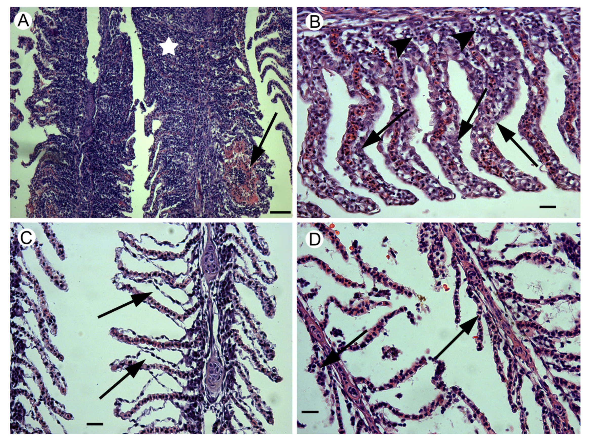
Figure 2.
Histopathological alterations observed in gills of Abramis brama during the two investigated seasons in the Tamiš River. (A) Hyperplasia of epithelium leading to fusions of lamellae (star) and the focal rupture of secondary lamellae leading to hemorrhage (arrow). (B) Epithelial hypertrophy (arrows) as well as leukocyte infiltration indicative of inflammation. (C) Epithelial lifting (arrows). (D) Rupture of the primary and secondary epithelium and necrosis (arrows). H&E. Scale bars: A = 50 µm; B, C and D = 20 µm.

Figure 3.
Semi-quantitative and statistical analyses of gill alterations in fish sampled in the Tamiš River. (A) Gill indices and reaction pattern (progressive, regressive and circulatory) indices for gills sampled in different seasons. (B) Scatterplot of canonical scores for histopathological alterations of fish captured in different seasons. Arrows indicate centroids.


Table 1.
Water quality parameters for the Jaša Tomić site in different seasons [18].
Table 1.
Water quality parameters for the Jaša Tomić site in different seasons [18].
| Parameter | Season 1 | Season 2 | WHO [21] |
|---|---|---|---|
| Water temperature (°C) | 8.2 | 14.8 | - |
| Water transparency (mm) | 150 | - | - |
| Suspended matters (mg/L) | 39 | 35 | - |
| Dissolved oxygen (mL/L) | 10.4 | 9.5 | - |
| Alkalinity (mmol/L) | 1.7 | 1.6 | - |
| pH | 7.6 | 7.5 | - |
| Electroconductivity (µS/cm) | 235 | 579 | - |
| Ammonia (mg/L) | 0.2 | 0.05 | No guidelines |
| Nitrates (mg/L) | 0.77 | 0.56 | - |
| Nitrites (mg/L) | 0.01 | 0.013 | - |
| Orthophosphates (mg/L) | 0.028 | 0.032 | - |
| Chlorides (mg/L) | 12 | 7.3 | No guidelines |
| Sulphates (mg/L) | 23 | 25 | - |
| Fe (mg/L) | 1.24 | 1.78 | 1 |
| Mn (mg/L) | 0.07 | 0.05 | 0.5 |
| Zn (mg/L) | 0.04 | 0.04 | 5 |
| Cu (mg/L) | 0.02 | 0.007 | 2 |
| Total Cr (mg/L) | 0.002 | 0.002 | 0.05 |
| Pb (mg/L) | 0.009 | <0.001 | 0.01 |
| Cd (µg/L) | 0.2 | 0.2 | 3 |
| Hg (µg/L) | 0.1 | <0.1 | 1 |
| Ni (mg/L) | 0.006 | 0.004 | 0.02 |
| As (mg/L) | 0.001 | <0.001 | 0.01 |
Table 2.
Histopathological alterations and alteration indices for gills of fish sampled in the Tamiš River in different seasons.
Table 2.
Histopathological alterations and alteration indices for gills of fish sampled in the Tamiš River in different seasons.
| Alteration (Pathological Relevance) | Season (Site) | All Effects ANOVA | ||||
|---|---|---|---|---|---|---|
| Season 1 | Season 2 | F | d.f. | p | ||
| Sečanj (N = 5) | B. Despotovac (N = 8) | Sečanj (N = 20) | ||||
| Epithelial hypertrophy (1) | 1.28 ± 0.20 a | 1.52 ± 0.20 a,b | 2.30 ± 0.20 b | 5.08 | 2 | 0.01 |
| Epithelial hyperplasia (2) | 0.48 ± 0.23 a | 3.40 ± 0.64 b | 3.01 ± 0.35 b | 6.54 | 2 | <0.01 |
| Mucous cell alterations (2) | 2.00 ± 1.56 a | 1.34 ± 0.65 a | 1.05 ± 0.33 a | 0.48 | 2 | 0.62 |
| Chloride cell alterations (2) | 0.00 ± 0.00 a | 0.05 ± 0.05 a | 0.03 ± 0.02 a | 0.37 | 2 | 0.70 |
| Epithelial lifting (1) | 0.92 ± 0.21 a | 2.02 ± 0.54 a,b | 2.65 ± 0.31 b | 3.47 | 2 | 0.04 |
| Necrosis (3) | 3.84 ± 1.05 a | 0.09 ± 0.09 b | 1.47 ± 0.61 a,b | 3.99 | 2 | 0.03 |
| Hyperemia (1) | 1.88 ± 0.38 a | 1.57 ± 0.31 a | 2.40 ± 0.31 a | 1.41 | 2 | 0.26 |
| Aneurism (1) | 0.00 ± 0.00 a | 0.17 ± 0.09 a | 0.11 ± 0.03 a | 1.51 | 2 | 0.24 |
Importance factors are indicated in parenthesis. Within rows, for a given parameter, different letters in the superscript indicate significant statistical difference (i.e., the presence of the same letter in two columns within the same row indicates no significant differences, while different letters in two columns within the same row indicate a significant difference of p < 0.05).
Table 3.
Discriminant canonical analysis (DCA) for gill histopathological alterations in fish captured in Tamiš River during different seasons.
Table 3.
Discriminant canonical analysis (DCA) for gill histopathological alterations in fish captured in Tamiš River during different seasons.
| Alterations | Canonical Function 1 | Canonical Function 2 |
|---|---|---|
| Epithelial hypertrophy | −0.086 | 0.861 |
| Epithelial hyperplasia | −0.861 | −0.427 |
| Mucous cell alterations | 0.568 | −0.154 |
| Chloride cell alterations | −0.021 | −0.184 |
| Epithelial lifting | −0.557 | 0.262 |
| Necrosis | 0.547 | 0.349 |
| Hyperemia | −0.017 | 0.255 |
| Aneurism | −0.026 | −0.023 |
| Eigenvalue | 1.407 | 0.455 |
| Cum. prop. | 0.756 | 1.000 |
| Canonical R | 0.765 | 0.559 |
| Wilks’ lambda | 0.256 | 0.687 |
| d.f. | 16 | 7 |
| p | 0.007 | 0.192 |
Publisher’s Note: MDPI stays neutral with regard to jurisdictional claims in published maps and institutional affiliations. |
© 2021 by the authors. Licensee MDPI, Basel, Switzerland. This article is an open access article distributed under the terms and conditions of the Creative Commons Attribution (CC BY) license (https://creativecommons.org/licenses/by/4.0/).
Share and Cite
MDPI and ACS Style
Marinović, Z.; Miljanović, B.; Urbányi, B.; Lujić, J. Gill Histopathology as a Biomarker for Discriminating Seasonal Variations in Water Quality. Appl. Sci. 2021, 11, 9504. https://0-doi-org.brum.beds.ac.uk/10.3390/app11209504
AMA Style
Marinović Z, Miljanović B, Urbányi B, Lujić J. Gill Histopathology as a Biomarker for Discriminating Seasonal Variations in Water Quality. Applied Sciences. 2021; 11(20):9504. https://0-doi-org.brum.beds.ac.uk/10.3390/app11209504
Chicago/Turabian StyleMarinović, Zoran, Branko Miljanović, Béla Urbányi, and Jelena Lujić. 2021. "Gill Histopathology as a Biomarker for Discriminating Seasonal Variations in Water Quality" Applied Sciences 11, no. 20: 9504. https://0-doi-org.brum.beds.ac.uk/10.3390/app11209504
Note that from the first issue of 2016, this journal uses article numbers instead of page numbers. See further details here.