Review

Molecular Basis of Irisin Regulating the Effects of Exercise on Insulin Resistance

1
College of Physical Education, Yangzhou University, Yangzhou 225009, China
2
Research Institute of Education Science, Hunan University, Changsha 410006, China
*
Author to whom correspondence should be addressed.
Submission received: 30 April 2022 / Revised: 6 June 2022 / Accepted: 6 June 2022 / Published: 8 June 2022
(This article belongs to the Special Issue New Trends in Sport and Exercise Medicine II)

Abstract

:
Insulin resistance is recognized as one major feature of metabolic syndrome, and frequently emerges as a difficult problem encountered during long-term pharmacological treatment of diabetes. Insulin resistance often causes organs or tissues, such as skeletal muscle, adipose, and liver, to become less responsive or resistant to insulin. Exercise can promote the physiological function of those organs and tissues and benefits insulin action via increasing insulin receptor sensitivity, glucose uptake, and mitochondrial function. This is done by decreasing adipose tissue deposition, inflammatory cytokines, and oxidative stress. However, understanding the mechanism that regulates the interaction between exercise and insulin function becomes a challenging task. As a novel myokine, irisin is activated by exercise, released from the muscle, and affects multi-organ functions. Recent evidence indicates that it can promote glucose uptake, improve mitochondrial function, alleviate obesity, and decrease inflammation, as a result leading to the improvement of insulin action. We here will review the current evidence concerning the signaling pathways by which irisin regulates the effect of exercise on the up-regulation of insulin action in humans and animals.

1. Introduction

Insulin resistance (IR) is recognized as a hallmark of metabolic dysfunction, and many individuals develop cell dysfunction and type 2 diabetes mellitus (T2DM) eventually [1]. T2DM has become more common in developed and developing countries over the last few decades, now affecting about 8% of the world’s population [2]. It is clear that T2DM is triggered by a combination of IR in the adipose, liver, and skeletal muscle [3], involving a variety of risk factors, including hyperlipidemia, obesity, inflammation, and physical inactivity [1].
An important goal in preventive strategies for T2DM is to reverse those adverse factors and then improve IR. Among the intervention regimens, regular exercise can increase energy consumption, muscle mass, and blood circulation between tissues, and reduce fat mass and inflammations, which ultimately lead to improvement of insulin sensitivity [4,5,6]. However, understanding the mechanism that mediates the influence of exercise on IR has been challenging.
Skeletal muscle has already been recognized as a secretory tissue for a long time [7]. Exercise stimulates the skeletal muscle to produce a large number of myokines [8], such as myostatin, fibroblast growth factor-2, L-Lactate, kynurenine, interleukin-6 (IL-6), etc.; they either exert autocrine and paracrine effects or enter the circulation and act as endocrinal regulators affecting the physiological function of organs and tissues [9,10,11,12]. As a newly identified myokine, irisin is provoked by exercise, mainly secreted from muscle, and secondarily secreted by adipose and other tissues in small amounts, to regulate energy consumption [10,13,14]. Given the close relationship between irisin and exercise and its essential role in regulating energy metabolism, irisin is expected to become a potential candidate for modulating the benefits of exercise on insulin action. This review aimed to best our understandings of the signaling pathways that regulate the interaction between exercise, irisin, and insulin action, seeking potential strategies for T2DM prevention.

2. Biology and Physiology of FNDC5/Irisin

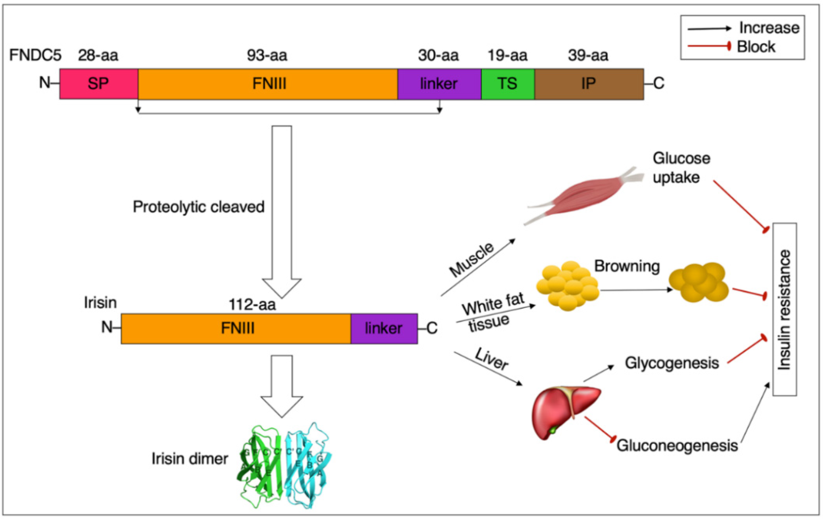
2.1. Summary Profile of FNDC5/Irisin

Two decades ago, a new gene was identified by two independent groups, which is widely distributed in the heart, skeletal muscle, brain, as well as other tissues of mice [15,16]. After a decade, the gene was named fibronectin type III domain-containing protein 5 (FNDC5). It is one of the most important target genes of peroxisome proliferator-activated receptor γ coactivator 1α (PGC1α) which is a transcriptional coactivator, frequently produced in exercised skeletal muscle [10]. Mice either overexpressing PGC-1α or subjected to endurance exercise highly upregulated FNDC5 gene expression, and conditioned medium (from FNDC5 overexpression mice) cultivated myocytes caused adipocytes to upregulate uncoupling protein 1 (UCP1), a key gene leading to brown adipose tissues (BAT). Meanwhile, a new myokine–irisin has been discovered; it contains 112 amino acids (aa) and is formed from skeletal muscle FNDC5 after being cleaved at the N-terminus [10]. The released irisin, according to this study, may be responsible for several of the positive benefits of physical activity on energy consumption via adipose tissue browning [10].

2.2. Consequence, Structure, and Detection of FNDC5/Irisin

The FNDC5 locus contains six exons and encodes a 209-aa protein, including a 28-aa signal peptide, a 93-aa fibronectin type III (FNIII) domain, a 30-aa linker, a 19-aa transmembrane region, and a 39-aa intracellular domain [17]. This transmembrane protein’s FNIII domain plus the linker has been called irisin and is thought to be cleaved (Figure 1). The molecular weight (MW) of the FNDC5 protein is predicted to be about 20,300, and without glycosylation, irisin (112 aa peptide) has a MW of 12,600.
Irisin is highly conserved, with 100 percent identity in its amino acid sequence across humans, mouse, rat, and bovines [18]. However, the start codons of humans are different from those of animals, with the human FNDC5 gene having a mutated start codon ATA rather than the standard ATG frequently seen in rodents. To test if the ATA codon has an effect on FNDC5 expression, Raschke and colleagues [18] used HEK293 cells to transfect FNDC5 with either the conventional or noncanonical codon and found that the protein level of FNDC5 in constructs with the ATA codon is only 1% that of constructs with the ATG codon. As a result, they proposed that the human FNDC5 gene would lose its ability to translate to a full-length FNDC5 protein. Under the circumstances, PCR and other more specific and sensitive procedures are more effective compared to enzyme-linked immunosorbent assays (ELISA) kits or Western blot analyses [19].
FNDC5 is proposed to be cut at the N-terminal site, and released as irisin into circulation targeting various tissues. Irisin has the typical FNIII domain structure, which is a beta-sandwich with three beta-strands on one side and four on the other [17]. Irisin can form a tight dimer through the 2 C′ strands correlating antiparallel to form a beta zipper, giving an extended eight-strand beta construct. Albrecht et al. [19] determined non-glycosylated irisin with Western blots running as a band at an MW of 13,000, approaching 12,600 for this 112-aa peptide. Though the details and the mechanism of the cleavage of FNDC5 are still unclear, there has been clear evidence to demonstrate that Irisin is found in human and mouse body fluids, and the effects of r (recombinant)-irisin treatment in cells and animal models have been extensively reported.
As an irisin receptor, αVβ5 was first reported by Kim [20], which involved the action of irisin on both osteocyte survival and sclerostin production to regulate bone remodeling. Since then, αVβ5 was detected in other tissues, such as the brain and gut [21,22]. However, as pointed out by Kim [20], αVβ5 may not be the only irisin receptor, so further studies on irisin receptors are still needed to address the concern.
There are mainly three methods, i.e., Western blot, ELISA, and qualitative and quantitative mass spectrometry (MS) used for the assessment of circulating irisin. Among those methods, MS with quantification peptides is recognized as the “holy grail” [23]. So far, the currently available reference values for circulating irisin in humans [24] and mice [20] were 3~4 ng/mL and 0.3 ng/mL, respectively.

2.3. Distribution and Production of FNDC5/Irisin

Irisin can be found in nearly all organs and tissues. In rats, immunohistochemistry has detected irisin in the kidney, brain, heart, muscle, liver, skin, retina, thyroid, and pineal gland [15,25,26,27,28,29], meanwhile its expression has already been observed in the cerebrospinal fluid, liver, pancreas, stomach, serum, saliva, and urine of humans [30,31,32,33]. Additionally, irisin is also detected in human breast and ovarian cancer cells by using flow cytometry and laser confocal microscopy [28]. Different tissues of humans show differing levels of irisin, among which the skeletal muscle has the highest expression levels of irisin, followed by other tissues including muscle, the tongue, heart, rectum, etc.; irisin expression is 100–200 times lower in adipose tissue than it is in muscle tissue [34,35,36].
Irisin secretion is influenced by a variety of stimuli, such as exercise, diet, cold, pharmacological agents, and pathological conditions [37], among which exercise is the most strong stimulating factor for the synthesis and secretion of irisin. Irisin expression was dramatically elevated in muscles of diabetic rats after a 12-week exercise regimen [38], and a prolonged exercise protocol (90-min running at an intensity of 60% VO2max) increased serum irisin concentrations of men and women by 20.4% and 24.6%, respectively [39]. Although irisin is mainly secreted by skeletal muscle, white adipose tissue (WAT) is also a major producer, accounting for about 30% of the total circulating levels of irisin [12]. Similar to skeletal muscle, the secretion of irisin in WAT significantly increases in response to exercise training [12]. It is implied that irisin is both a myokine and an adipokine.

3. Irisin and Insulin Resistance

3.1. Irisin, Mitochondrial Function, and Insulin Resistance

Mitochondrial dysfunction is a term used to describe the changes in mitochondrial content, activity, and oxidative phosphorylation that occur under a variety of physiological situations [40]. Mitochondrial dysfunction has been linked to IR in myocytes, adipocytes, and hepatocytes [41]. Irisin improves IR by increasing oxidative phosphorylation capability and mitochondrial content [41]. The phosphorylation levels of p38/mitogen-activated protein kinase (MAPK) and extracellular-signal-regulated kinase (ERK) in adipocytes increased when the mice were treated with r-irisin (0.5 µg/g/day) for 14 days, and the expression of UCP1 was also up-regulated significantly. It is suggested that irisin can boost UCP1 gene expression in white adipocytes by activating ERK and p38MAPK [9,10]. UCP1 can promote the browning of white adipocytes, and BAT has more mitochondria than WAT [42]. This enhances the biological functions of mitochondria and improves energy metabolism [43]. After the browning of white adipocytes, mice lose weight, and the glucose tolerance and IR of mice are significantly improved [44,45]. Moreover, irisin increases the expression of betatrophin which can promote pancreatic beta-cell proliferation and improve glucose tolerance and insulin action [46]. Under the stimulation of muscle contraction, the expression and activity of PGC-1α are increased, which subsequently promotes irisin secretion [47]. Irisin is recognized as a stimulator for betatrophin expression. Zhang et al. reported that irisin treatment increased betatrophin levels in obese mice [9], and betatrophin could reduce the level of serum glucose via activating the proliferation of pancreatic cells [48]. Therefore, there may exist an axis “PGC-1α/irisin/betatrophin” in the regulation of glucose homeostasis.
Aside from adipose tissue, irisin stimulated the expression of UCP3 in skeletal muscle [49]. Similar to UCP1, UCP3 enhances the thermogenesis of mitochondria and then reduces lipid accumulation, subsequently promoting insulin action [49]. Furthermore, the treatment of r-irisin (5 nM) on C2C12 myocytes markedly increased the gene expression of mitochondrial transcription factor A (TFAM) [49]. TFAM is responsible for regulating the replication and transcription of mitochondrial genes and is capable of enhancing the biological functions of mitochondria [50].
IR usually has altered hepatic energy metabolism characterized by upregulated gene expression related to gluconeogenesis, oxidative phosphorylation, and reactive oxygen species (ROS) generation [51]. Schmid et al. [52] reported that T2DM patients were featured by a 42% reduction in liver ATP synthetic flux rate, which was linked to both peripheral and liver insulin sensitivity. Irisin treatment affects the function of mitochondrial function in hepatocytes. In the liver, r-irisin treatment significantly reduced the expression of the mitochondrial fission-related proteins dynamin-related protein 1 (drp-1) and fission 1 (Fis-1) [53]. Exogenous irisin also enhanced mitochondrial density as well as promoted PGC-1α and TFAM expression in mitochondrial biogenesis [53]. Xin et al. [54] demonstrated that r-irisin administration alleviated mitochondrial dysfunction and attenuated liver damage in diabetic mice via the AMPK pathway. In C2C12 cells with IR, mitochondrial content and oxidative phosphorylation capacity were significantly increased after r-irisin (5 nM) treatment for 12 h, and the impaired glucose uptake and IR were reversed, which might be related to the regulation of p38MAPK phosphorylation [41]. Therefore, the evidence suggests that irisin improves IR via up-regulating mitochondrial content and action.

3.2. Irisin, GLUT4, and Insulin Resistance

Glucose transporter 4 (GLUT4) is a high-affinity insulin-responsive glucose transporter that is predominantly expressed in myocytes and adipocytes [55]. The failure of translocation of GLUT4 to the cell membrane in reaction to insulin is recognized as an early step in the development of IR, a pathological condition characterized by tissues showing less responsiveness or resistance to insulin [56]. In myoblast L6 cells, treatment with r-irisin increased the phosphorylation level of p38MAPK, which consequently stimulated the translocation of GLUT4 towards the membrane of myocytes to promote glucose uptake in skeletal muscle [57]. Meanwhile, glucose uptake in L6 cells was increased in the presence of r-irisin, with the maximal rise occurring at a dose of 62 ng/mL [57]. So irisin can improve glucose uptake and IR by enhancing the intracellular trafficking and action of GLUT4. Huh et al. [58] also found that exogenous irisin boosted glucose and fatty acid metabolism in human skeletal muscle cells, comparable to the insulin-induced response. In addition, r-irisin treatment up-regulated the expression of genes involved in glucose metabolism, such as GLUT4 and hexokinase 2, and the increment of gene expression coincided with enhanced phosphorylation of AMPK [58]. GLUT4 regulates insulin-induced glucose transportation within adipocytes and myocytes [55]. Following a meal, irisin promotes the translocation of GLUT4 from intracellular reserves to the cellular membranes, inhibiting gluconeogenesis in the liver and increasing glucose uptake in skeletal muscle and adipose tissue [56]. Additionally, the insulin-mediated glucose uptake of BAT after browning was 10 times that of WAT, and the expression of GLUT4 was up-regulated remarkably [59,60]. However, whether this is the same as the increased GLUT4 translocation towards the cellular membrane observed in muscle cells remains unclear [46]. Nevertheless, it is clear that irisin can increase glucose uptake by enhancing the expression of GLUT4 in skeletal muscle and adipose tissue.

3.3. Irisin, Inflammation, and Insulin Resistance

Obesity in humans and animals is featured by a substantial rise of macrophages in the liver and adipose tissue [61]. These macrophages are highly activated and as a result, gene expression associated with pro-inflammatory cytokines is up-regulated [62]. The activation of macrophages potentially affects insulin target cells including myocytes, adipocytes, and hepatocytes via a paracrine mechanism which subsequently leads to IR [63]. Irisin can improve insulin action by down-regulating inflammatory cytokines. A recent study by Zheng et al. reported that the levels of inflammatory cytokines in pancreatic islets of high-fat diet-fed (HFD) mice were significantly reduced after r-irisin (0.5 μg/g of body weight) treatment, which was related to the inhibition of the toll-like receptor 4 (TLR4)/nuclear factor kappa B (NF-KB) signaling pathway [64]. Additionally, in MIN6 cells isolated from HFD mice, irisin administration (100 ng/mL) significantly upregulated the activity of the PI3K/AKT signaling pathway, which subsequently enhanced insulin action [64]. Dong et al. [65] demonstrated that myostatin-regulated inflammation played an essential role in IR in obese mice. The anti-myostatin peptide treatment potentially improved glucose uptake and insulin tolerances via elevating fatty acid metabolism as well as energy consumption while suppressing macrophage infiltration and proinflammatory cytokine expression in obese mice myocytes and adipocytes [65]. This beneficial effect on insulin sensitivity was regulated by irisin-induced depression of inflammation, stimulation of polarization of macrophages from M1 towards M2, and up-regulation of energy consumption [65].
After treatment with r-irisin (100 nM) on mouse macrophages, the expression of tumor necrosis factor α (TNF-α) mRNA substantially decreased by 45% compared to the controls, and its effect was twice that of r-irisin at the dosage of 50 nm (22%) [66]. TNF-α can cause insulin receptor substrate (IRS) serine phosphorylation and reduce the expression of GLUT4, which subsequently inhibit insulin signaling and triggers IR [67]. The r-irisin administration also resulted in a decrease in IL-6 mRNA and protein levels [66]. IL-6 has the capacity of inhibiting the suppressor of cytokine signaling 1/3, which promotes the IRS deterioration through ubiquitination and eventually leads to IR [68,69]. Moreover, the mRNA expression of another inflammation cytokine, interleukin-1β (IL-1β), was down-regulated by the treatment of r-irisin [66]. IL-1β is known to decrease the expression of IRS through ERK1/2, which is capable of impairing the physiological function of insulin [68]. Additionally, Mazur-Bialy et al. [70] reported that the dephosphorylation of NF-KB was up-regulated in adipocytes after treatment with irisin, and the inactivation of NF-KB inhibited the further transcription of pro-inflammatory genes. Those data suggest that irisin has a strong anti-inflammatory effect which is important for restoring the action of insulin.

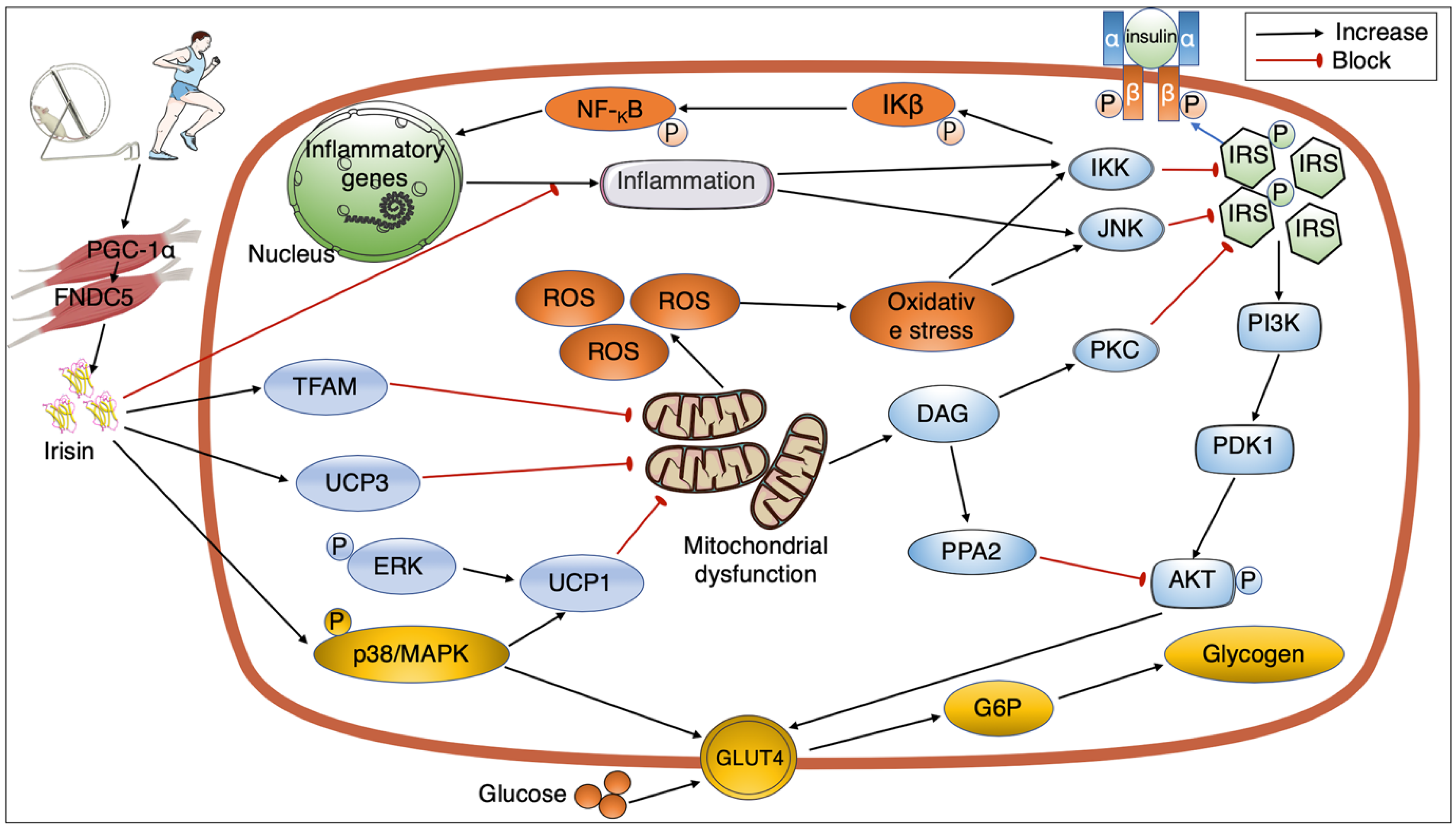
4. Signaling Pathways Mediating the Link between Exercise, Irisin, and Insulin Sensitivity

4.1. Exercise Regulating Irisin Secretion

Muscle not only acts as an effector in the motor system but also has a potential impact on the endocrine system. Exercise can promote muscles to secrete hundreds of myokines, which enter the circulatory system and activate multiple pathways to regulate energy metabolism [71]. Irisin is one of the pivotal myokines, most of which (75%) in the circulation comes from skeletal muscle, and its secretion depends on the increase in PGC-1α expression. Exercise is an important activator of PGC-1α expression upregulation, and also acts as a promoter to activate AMPK signaling pathways, which subsequently triggers the phosphorylation of PGC-1α and promotes the production of FNDC5 (Figure 2). FNDC5 was then cleaved and secreted as irisin [36,72].
Studies investigate the correlation between exercise and irisin frequently involving the changes in irisin, FNDC5, and its upstream regulating gene PGC-1α in response to exercise. The evidence from animal experiments indicated that a program of 8-week swimming training significantly increased the level of serum irisin in rats [73], and the expression of PGC-1α mRNA and FNDC5 mRNA in the muscle tissue increased in mice that received exercise intervention [10]. Pang et al. carried out 1 h treadmill exercise (at 60% VO2max) on male C57BL/6J mice, and they reported that plasma irisin quantities increased significantly during exercise and reached the peak value (20.71 ± 0.25 ng/mL) at 6 hours after the intervention. Meanwhile, PGC-1α mRNA expression levels increased significantly in muscle tissue during exercise, maintained the higher level for about 6 h, and then gradually returned to the baseline value after 12 h of exercise intervention [74]. The authors proposed that exercise promoted irisin secretion probably in a PGC-1α dependent manner [74]. In the soleus and gastrocnemius muscles, 90 min of acute swimming exercise remarkedly activated the expression of FNCD5 mRNA and protein, whereas the change in serum levels of irisin was not significant [75]. Accordingly, the swimming exercise also increased UCP1 mRNA and protein expression in subcutaneous WAT, as well as browning marker mRNA expression in visceral WAT [75].
Compared with the results from animal experiments, there is some controversy about the effects of exercise on human irisin secretion [76]. The amount of FNDC5 mRNA in 26 subjects’ skeletal muscles rose considerably after 12 weeks of continuous exercise intervention, among which the healthy control group increased by 1.4 times and the pre-diabetic group increased by 2 times, together with elevated PGC-1α mRNA expression [77]. However, no significant changes in FNDC5 mRNA levels in skeletal muscle were detected in the same subjects after 45 min of acute exercise (at 70% VO2max), even though PGC-1α mRNA levels were significantly increased by approximately 7.4-fold [77]. Additionally, in vitro experiments using electrical pulse stimulation to induce contraction of human skeletal muscle cells showed that FNDC5 mRNA levels were not changed with the upregulated PGC-1α expression [18]. These results suggest that FNDC5 may not be a direct target gene of PGC-1α. There are also different reports regarding circulating irisin levels in response to exercise intervention in humans. Nygaard et al. conducted a program of 6 months, 1 h daily exercise intervention among healthy subjects. The level of serum irisin increased by approximately 23.1% and 20.2% in the strength training and endurance exercise groups, respectively [78]. However, Hecksteden et al. demonstrated that 26 weeks of aerobic endurance or strength endurance training had no effect on circulating irisin levels in healthy subjects [79]. The divergent results might have resulted from the use of different ELISA kits that are the most common approach for measurement of serum irisin, and the ELISA kits need to be validated against tandem MS [19,23]. On the other hand, there may be differences in individuals, races, and time of collection.

4.2. GLUT4 and the Interaction between Exercise, Irisin, and Insulin Resistance

The capacity of exercise to increase glucose uptake in skeletal muscles is partly via up-regulating the expression of GLUT4 [80]. After a program of forty-five-minute treadmill exercise (running at 30 m/min with a 15% grade), GLUT4 expression on the myocyte outer membrane in rats increased by 2.5 times compared with the control [81]. The transportation of glucose to pass through the cell membrane needs a specific carrier, glucose transporters (GLUTs) [82]. Among the most significant glucose transporters proteins, GLUT4 has a strong relationship with insulin action [83]. The impaired expression or function of GLUT4 can lead to the occurrence of IR. Studies have demonstrated that the reduction of GLUT4 causes the appearance of hyperglycemia and hyperinsulinemia in mice [84,85], and mice lacking the GLUT4 gene result in IR [86].
Exercise could raise the quantity of GLUT4 in myocytes by affecting irisin production. Rats were given either high-intensity interval training or moderate-intensity continuous training for eight weeks, and the skeletal muscle expression of the FNDC5 gene both in the diabetic group and the healthy group was significantly increased [87]. Meanwhile, the levels of GLUT4 mRNA in their soleus muscle were also markedly elevated, and the moderate-intensity continuous training group showed greater effects than those of the high-intensity interval training group [87]. Moreover, several studies have shown that exercise can significantly promote the levels of PGC-1α mRNA and FNDC5 mRNA in rats [75,77], and p38 mRNA and MAPK mRNA levels in skeletal muscle are also significantly enhanced [41]. Irisin can induce the GLUT4 translocation to the myocyte membrane through phosphorylated p38/MAPK, and the uptake of glucose by skeletal muscle is subsequently increased [88]. Moreover, Song et al. reported that irisin improved the reduction of glucose consumption in insulin-resistant cardiomyocytes and activated the PI3K/AKT pathway increasing insulin action [89]. Therefore, irisin plays a crucial role in modifying the beneficial effects of exercise on IR by promoting the content and trafficking of GLUT4 within myocytes.

4.3. Mitochondrial Action and Interaction between Exercise, Irisin, and Insulin Resistance

Exercise has a positive impact on insulin action in part because it improves mitochondrial function. Exercise intervention increased mitochondrial function in BAT of rats, promoting the release of proteins that regulate mitochondrial biogenesis and fission while decreasing the level of markers that modulate mitochondrial fusion [90]. As a result, circulating insulin levels fell while insulin sensitivity rose in BAT [90]. Mitochondrial dysfunction can result in a buildup of secondary products of fat metabolism that in turn activate protein kinase C (PKC) and protein phosphatase A2 (PPA2) [91,92]. In addition, IRS tyrosine phosphorylation is impaired, and AKT is dephosphorylated, eventually weakening the physiological function of insulin [93]. Moreover, mitochondrial dysfunction is the main source of free radicals and oxidative stress, which ultimately lead to IR [94,95].
Recent evidence indicates that irisin seems to be involved in the benefits of exercise affecting the biological function of mitochondria. In a study, after four weeks of continuous training in mice (at 75% VO2max), the levels of both PGC-1α and FNDC5 increased significantly and the serum irisin expression was also higher than that of sedentary mice [96]. With the elevated expression of PGC-1α/FNDC5/irisin, mitochondrial fission and mitophagy markers increased, which help to improve mitochondria function [96]. Moreover, upregulation of irisin levels improved insulin intolerance in T2DM mice, and reduced serum insulin and glucose levels [97]. Therefore, irisin can reduce IR by enhancing the function of mitochondria. After the mice were injected with irisin (0.5 µg/g/day) for fourteen days, the phosphorylation levels of p38/MAPK and ERK in adipocytes increased, and the expression of UCP1 also elevated significantly. UCP1 can cause a browning of WAT and an increase in mitochondrial density [43]. Given that irisin is a strong stimulator for promoting mitochondrial biogenesis and insulin physiological action [53,98], it might be an essential regulator in the process of exercise improving IR via upregulating mitochondrial function. Indeed, Reisi et al. [99] reported that after eight weeks of resistance exercise, the soleus muscle expression of FNDC5 in the mice increased significantly, and UCP1 expression in the adipose tissue under the skin enhanced markedly, which subsequently increased mitochondria function and benefited insulin action.

4.4. Inflammation and Interaction between Exercise, Irisin, and Insulin Resistance

Exercise can improve IR and increase glucose uptake by reducing inflammation in fat tissue [100]. Bradley et al. [101] reported that 6-week voluntary exercise remarkably enhanced glucose tolerance and insulin sensitivity in obese mice companied by decreased levels of TNF-α, MCP-1, PAI-1 as well as I kappa kinase β (IKKβ) in adipose tissue of obese mice. Exercise-induced improvement in IR has been linked to a reduction in inflammation of adipocytes [101]. It is known that increased expression for inflammatory mediators including TNF-α, IL-6, IL-1β as well as IL-1α, and IR is often associated with elevated levels of proinflammatory cytokines [68,102]. Several studies indicated that obesity-induced high levels of inflammatory mediators were involved in the inhibition of insulin action, as well as anti-inflammation treatment effectively restored the insulin sensitivity [103,104,105].
However, it remains unclear how exercise depresses inflammation production. It is reported that, as a myokine, irisin is recognized as a strong inhibitor of inflammation cytokines [70], and its production is upregulated by exercise training [10,73]. It is proposed that exercise increases serum concentrations of irisin and it enters organs or tissues where it acts as an inhibitor of inflammatory cytokines [106]. However, how irisin regulates the production of inflammatory cytokines and leads to a consequent rise in insulin action during exercise remains to be elucidated by future studies.

5. Perspectives

Even though there has substantial progress in recent decades on disclosing the mechanisms of the effects of exercise on insulin action, the exact molecular signalings remain unclear. Recent evidence reports that the physiological and biological function of insulin is affected by a newly discovered myokine–irisin which gives rise to a novel approach for unraveling the mechanism of the correlation between exercise and insulin action. However, there are many gaps in our knowledge about the procedures, for example, the exact molecular signalings that conduct the message of irisin to stimulate irisin action during exercise have not been determined yet. The lacking steps must be addressed in the correlation between irisin and insulin-signaling network in response to exercise, and such studies will present new evidence on the exact mechanisms of cross-talk between exercise, irisin, and insulin function, perhaps providing even a more promising approach to the therapy or prevention of IR.

Author Contributions

J.L. and R.Z. designed the study. J.L., W.C., Y.P., C.Z. and B.W. performed the data collection and analysis. J.L. and B.Z. wrote the first draft of the manuscript. J.L., Y.Z. and Y.W. drew the figures. The manuscript’s intellectual content was edited by J.L. and X.L. All authors have read and agreed to the published version of the manuscript.

Funding

The Natural Science Foundation of Jiangsu Province provided funding for this research (BK20201435).

Institutional Review Board Statement

Not applicable.

Informed Consent Statement

Not applicable.

Data Availability Statement

On reasonable request, the data created and analyzed during this investigation can be obtained from the corresponding author.

Acknowledgments

We would like to acknowledge Yang for his invaluable assistance with this research.

Conflicts of Interest

The authors state that there were no commercial or financial relationships that may be considered a potential conflict of interest during the research.

References

  1. Moller, D.E.; Kaufman, K.D. Metabolic syndrome: A clinical and molecular perspective. Annu. Rev. Med. 2005, 56, 45–62. [Google Scholar] [CrossRef] [PubMed]
  2. Johnson, A.M.; Olefsky, J.M. The origins and drivers of insulin resistance. Cell 2013, 152, 673–684. [Google Scholar] [CrossRef] [PubMed] [Green Version]
  3. Stumvoll, M.; Goldstein, B.J.; van Haeften, T.W. Type 2 diabetes: Principles of pathogenesis and therapy. Lancet 2005, 365, 1333–1346. [Google Scholar] [CrossRef]
  4. Zou, Z.; Cai, W.; Cai, M.; Xiao, M.; Wang, Z. Influence of the intervention of exercise on obese type II diabetes mellitus: A meta-analysis. Prim. Care Diabetes 2016, 10, 186–201. [Google Scholar] [CrossRef]
  5. Brandt, C.; Pedersen, B.K. The role of exercise-induced myokines in muscle homeostasis and the defense against chronic diseases. J. Biomed. Biotechnol. 2010, 2010, 520258. [Google Scholar] [CrossRef]
  6. Mathur, N.; Pedersen, B.K. Exercise as a mean to control low-grade systemic inflammation. Mediat. Inflamm. 2008, 2008, 109502. [Google Scholar] [CrossRef] [Green Version]
  7. Pedersen, B.K.; Febbraio, M.A. Muscles, exercise and obesity: Skeletal muscle as a secretory organ. Nat. Rev. Endocrinol. 2012, 8, 457–465. [Google Scholar] [CrossRef]
  8. Giudice, J.; Taylor, J.M. Muscle as a paracrine and endocrine organ. Curr. Opin. Pharmacol. 2017, 34, 49–55. [Google Scholar] [CrossRef]
  9. Zhang, Y.; Li, R.; Meng, Y.; Li, S.; Donelan, W.; Zhao, Y.; Qi, L.; Zhang, M.; Wang, X.; Cui, T.; et al. Irisin stimulates browning of white adipocytes through mitogen-activated protein kinase p38 MAP kinase and ERK MAP kinase signaling. Diabetes 2014, 63, 514–525. [Google Scholar] [CrossRef] [Green Version]
  10. Bostrom, P.; Wu, J.; Jedrychowski, M.P.; Korde, A.; Ye, L.; Lo, J.C.; Rasbach, K.A.; Bostrom, E.A.; Choi, J.H.; Long, J.Z.; et al. A PGC1-α-dependent myokine that drives brown-fat-like development of white fat and thermogenesis. Nature 2012, 481, 463–468. [Google Scholar] [CrossRef]
  11. Chen, N.; Li, Q.; Liu, J.; Jia, S. Irisin, an exercise-induced myokine as a metabolic regulator: An updated narrative review. Diabetes/Metab. Res. Rev. 2016, 32, 51–59. [Google Scholar] [CrossRef] [PubMed]
  12. Roca-Rivada, A.; Castelao, C.; Senin, L.L.; Landrove, M.O.; Baltar, J.; Belen Crujeiras, A.; Seoane, L.M.; Casanueva, F.F.; Pardo, M. FNDC5/irisin is not only a myokine but also an adipokine. PLoS ONE 2013, 8, e60563. [Google Scholar] [CrossRef] [PubMed] [Green Version]
  13. Aydin, S.; Kuloglu, T.; Aydin, S. Copeptin, adropin and irisin concentrations in breast milk and plasma of healthy women and those with gestational diabetes mellitus. Peptides 2013, 47, 66–70. [Google Scholar] [CrossRef] [PubMed]
  14. Munoz, I.Y.M.; Camarillo Romero, E.D.S.; Garduno Garcia, J.J. Irisin a Novel Metabolic Biomarker: Present Knowledge and Future Directions. Int. J. Endocrinol. 2018, 2018, 7816806. [Google Scholar] [CrossRef] [Green Version]
  15. Ferrer-Martinez, A.; Ruiz-Lozano, P.; Chien, K.R. Mouse PeP: A novel peroxisomal protein linked to myoblast differentiation and development. Dev. Dyn. 2002, 224, 154–167. [Google Scholar] [CrossRef]
  16. Teufel, A.; Malik, N.; Mukhopadhyay, M.; Westphal, H. Frcp1 and Frcp2, two novel fibronectin type III repeat containing genes. Gene 2002, 297, 79–83. [Google Scholar] [CrossRef]
  17. Schumacher, M.A.; Chinnam, N.; Ohashi, T.; Shah, R.S.; Erickson, H.P. The structure of irisin reveals a novel intersubunit β-sheet fibronectin type III (FNIII) dimer: Implications for receptor activation. J. Biol. Chem. 2013, 288, 33738–33744. [Google Scholar] [CrossRef] [Green Version]
  18. Raschke, S.; Elsen, M.; Gassenhuber, H.; Sommerfeld, M.; Schwahn, U.; Brockmann, B.; Jung, R.; Wisloff, U.; Tjonna, A.E.; Raastad, T.; et al. Evidence against a beneficial effect of irisin in humans. PLoS ONE 2013, 8, e73680. [Google Scholar] [CrossRef] [Green Version]
  19. Albrecht, E.; Norheim, F.; Thiede, B.; Holen, T.; Ohashi, T.; Schering, L.; Lee, S.; Brenmoehl, J.; Thomas, S.; Drevon, C.A.; et al. Irisin-a myth rather than an exercise-inducible myokine. Sci. Rep. 2015, 5, 8889. [Google Scholar] [CrossRef] [Green Version]
  20. Kim, H.; Wrann, C.D.; Jedrychowski, M.; Vidoni, S.; Kitase, Y.; Nagano, K.; Zhou, C.; Chou, J.; Parkman, V.A.; Novick, S.J.; et al. Irisin Mediates Effects on Bone and Fat via αV Integrin Receptors. Cell 2018, 175, 1756–1768.e1717, Erratum in Cell 2019, 178, 507–508. [Google Scholar] [CrossRef] [Green Version]
  21. Bi, J.; Zhang, J.; Ren, Y.; Du, Z.; Li, T.; Wang, T.; Zhang, L.; Wang, M.; Wu, Z.; Lv, Y.; et al. Irisin reverses intestinal epithelial barrier dysfunction during intestinal injury via binding to the integrin αVβ5 receptor. J. Cell. Mol. Med. 2020, 24, 996–1009. [Google Scholar] [CrossRef] [PubMed] [Green Version]
  22. Wang, Y.; Tian, M.; Tan, J.; Pei, X.; Lu, C.; Xin, Y.; Deng, S.; Zhao, F.; Gao, Y.; Gong, Y. Irisin ameliorates neuroinflammation and neuronal apoptosis through integrin αVβ5/AMPK signaling pathway after intracerebral hemorrhage in mice. J. Neuroinflamm. 2022, 19, 82. [Google Scholar] [CrossRef] [PubMed]
  23. Polyzos, S.A.; Mathew, H.; Mantzoros, C.S. Irisin: A true, circulating hormone. Metabolism 2015, 64, 1611–1618. [Google Scholar] [CrossRef] [PubMed]
  24. Jedrychowski, M.P.; Wrann, C.D.; Paulo, J.A.; Gerber, K.K.; Szpyt, J.; Robinson, M.M.; Nair, K.S.; Gygi, S.P.; Spiegelman, B.M. Detection and Quantitation of Circulating Human Irisin by Tandem Mass Spectrometry. Cell Metab. 2015, 22, 734–740. [Google Scholar] [CrossRef] [PubMed] [Green Version]
  25. Aydin, S.; Kuloglu, T.; Ozercan, M.R.; Albayrak, S.; Aydin, S.; Bakal, U.; Yilmaz, M.; Kalayci, M.; Yardim, M.; Sarac, M.; et al. Irisin immunohistochemistry in gastrointestinal system cancers. Biotech. Histochem. 2016, 91, 242–250. [Google Scholar] [CrossRef]
  26. Dun, S.L.; Lyu, R.M.; Chen, Y.H.; Chang, J.K.; Luo, J.J.; Dun, N.J. Irisin-immunoreactivity in neural and non-neural cells of the rodent. Neuroscience 2013, 240, 155–162. [Google Scholar] [CrossRef] [Green Version]
  27. Gencer Tarakci, B.; Girgin, A.; Timurkaan, S.; Yalcin, M.H.; Gur, F.M.; Karan, M. Immunohistochemical localization of irisin in skin, eye, and thyroid and pineal glands of the crested porcupine (Hystrix cristata). Biotech. Histochem. 2016, 91, 423–427. [Google Scholar] [CrossRef]
  28. Park, M.J.; Kim, D.I.; Choi, J.H.; Heo, Y.R.; Park, S.H. New role of irisin in hepatocytes: The protective effect of hepatic steatosis in vitro. Cell. Signal. 2015, 27, 1831–1839. [Google Scholar] [CrossRef]
  29. Tastekin, E.; Palabiyik, O.; Ulucam, E.; Uzgur, S.; Karaca, A.; Vardar, S.A.; Yilmaz, A.; Aydogdu, N. The effect of high protein diet and exercise on irisin, eNOS, and iNOS expressions in kidney. Ren. Fail. 2016, 38, 1107–1114. [Google Scholar] [CrossRef] [Green Version]
  30. Aydin, S.; Kuloglu, T.; Aydin, S.; Kalayci, M.; Yilmaz, M.; Cakmak, T.; Albayrak, S.; Gungor, S.; Colakoglu, N.; Ozercan, I.H. A comprehensive immunohistochemical examination of the distribution of the fat-burning protein irisin in biological tissues. Peptides 2014, 61, 130–136. [Google Scholar] [CrossRef]
  31. Piya, M.K.; Harte, A.L.; Sivakumar, K.; Tripathi, G.; Voyias, P.D.; James, S.; Sabico, S.; Al-Daghri, N.M.; Saravanan, P.; Barber, T.M.; et al. The identification of irisin in human cerebrospinal fluid: Influence of adiposity, metabolic markers, and gestational diabetes. Am. J. Physiol. Endocrinol. Metab. 2014, 306, E512–E518. [Google Scholar] [CrossRef] [PubMed]
  32. Bakal, U.; Aydin, S.; Sarac, M.; Kuloglu, T.; Kalayci, M.; Artas, G.; Yardim, M.; Kazez, A. Serum, Saliva, and Urine Irisin with and Without Acute Appendicitis and Abdominal Pain. Biochem. Insights 2016, 9, 11–17. [Google Scholar] [CrossRef] [PubMed] [Green Version]
  33. Kuloglu, T.; Celik, O.; Aydin, S.; Hanifi Ozercan, I.; Acet, M.; Aydin, Y.; Artas, G.; Turk, A.; Yardim, M.; Ozan, G.; et al. Irisin immunostaining characteristics of breast and ovarian cancer cells. Cell. Mol. Biol. 2016, 62, 40–44. [Google Scholar] [PubMed]
  34. Huh, J.Y.; Panagiotou, G.; Mougios, V.; Brinkoetter, M.; Vamvini, M.T.; Schneider, B.E.; Mantzoros, C.S. FNDC5 and irisin in humans: I. Predictors of circulating concentrations in serum and plasma and II. mRNA expression and circulating concentrations in response to weight loss and exercise. Metabolism 2012, 61, 1725–1738. [Google Scholar] [CrossRef] [PubMed] [Green Version]
  35. Kurdiova, T.; Balaz, M.; Vician, M.; Maderova, D.; Vlcek, M.; Valkovic, L.; Srbecky, M.; Imrich, R.; Kyselovicova, O.; Belan, V.; et al. Effects of obesity, diabetes and exercise on Fndc5 gene expression and irisin release in human skeletal muscle and adipose tissue: In vivo and in vitro studies. J. Physiol. 2014, 592, 1091–1107. [Google Scholar] [CrossRef] [PubMed]
  36. Moreno-Navarrete, J.M.; Ortega, F.; Serrano, M.; Guerra, E.; Pardo, G.; Tinahones, F.; Ricart, W.; Fernandez-Real, J.M. Irisin is expressed and produced by human muscle and adipose tissue in association with obesity and insulin resistance. J. Clin. Endocrinol. Metab. 2013, 98, E769–E778. [Google Scholar] [CrossRef] [PubMed]
  37. Novelle, M.G.; Contreras, C.; Romero-Pico, A.; Lopez, M.; Dieguez, C. Irisin, two years later. Int. J. Endocrinol. 2013, 2013, 746281. [Google Scholar] [CrossRef] [Green Version]
  38. Kim, H.J.; Song, W. Resistance training increases fibroblast growth factor-21 and irisin levels in the skeletal muscle of Zucker diabetic fatty rats. J. Exerc. Nutr. Biochem. 2017, 21, 50–54. [Google Scholar] [CrossRef]
  39. Kraemer, R.R.; Shockett, P.; Webb, N.D.; Shah, U.; Castracane, V.D. A transient elevated irisin blood concentration in response to prolonged, moderate aerobic exercise in young men and women. Horm. Metab. Res. 2014, 46, 150–154. [Google Scholar] [CrossRef] [PubMed]
  40. Sivitz, W.I.; Yorek, M.A. Mitochondrial dysfunction in diabetes: From molecular mechanisms to functional significance and therapeutic opportunities. Antioxid. Redox Signal. 2010, 12, 537–577. [Google Scholar] [CrossRef] [Green Version]
  41. Ye, X.; Shen, Y.; Ni, C.; Ye, J.; Xin, Y.; Zhang, W.; Ren, Y. Irisin reverses insulin resistance in C2C12 cells via the p38-MAPK-PGC-1α pathway. Peptides 2019, 119, 170120. [Google Scholar] [CrossRef] [PubMed]
  42. Spiegelman, B.M. Banting Lecture 2012: Regulation of adipogenesis: Toward new therapeutics for metabolic disease. Diabetes 2013, 62, 1774–1782. [Google Scholar] [CrossRef] [PubMed] [Green Version]
  43. Hansen, J.B.; Kristiansen, K. Regulatory circuits controlling white versus brown adipocyte differentiation. Biochem. J. 2006, 398, 153–168. [Google Scholar] [CrossRef] [Green Version]
  44. Chen, J.Q.; Huang, Y.Y.; Gusdon, A.M.; Qu, S. Irisin: A new molecular marker and target in metabolic disorder. Lipids Health Dis. 2015, 14, 2. [Google Scholar] [CrossRef] [PubMed] [Green Version]
  45. Fisher, F.M.; Kleiner, S.; Douris, N.; Fox, E.C.; Mepani, R.J.; Verdeguer, F.; Wu, J.; Kharitonenkov, A.; Flier, J.S.; Maratos-Flier, E.; et al. FGF21 regulates PGC-1α and browning of white adipose tissues in adaptive thermogenesis. Genes Dev. 2012, 26, 271–281. [Google Scholar] [CrossRef] [PubMed] [Green Version]
  46. Perakakis, N.; Triantafyllou, G.A.; Fernandez-Real, J.M.; Huh, J.Y.; Park, K.H.; Seufert, J.; Mantzoros, C.S. Physiology and role of irisin in glucose homeostasis. Nat. Rev. Endocrinol. 2017, 13, 324–337. [Google Scholar] [CrossRef] [Green Version]
  47. Lu, J.; Xiang, G.; Liu, M.; Mei, W.; Xiang, L.; Dong, J. Irisin protects against endothelial injury and ameliorates atherosclerosis in apolipoprotein E-Null diabetic mice. Atherosclerosis 2015, 243, 438–448. [Google Scholar] [CrossRef]
  48. Tseng, Y.H.; Yeh, Y.H.; Chen, W.J.; Lin, K.H. Emerging regulation and function of betatrophin. Int. J. Mol. Sci. 2014, 15, 23640–23657. [Google Scholar] [CrossRef] [Green Version]
  49. Vaughan, R.A.; Gannon, N.P.; Barberena, M.A.; Garcia-Smith, R.; Bisoffi, M.; Mermier, C.M.; Conn, C.A.; Trujillo, K.A. Characterization of the metabolic effects of irisin on skeletal muscle in vitro. Diabetes Obes. Metab. 2014, 16, 711–718. [Google Scholar] [CrossRef]
  50. Scarpulla, R.C. Transcriptional paradigms in mammalian mitochondrial biogenesis and function. Physiol. Rev. 2008, 88, 611–638. [Google Scholar] [CrossRef] [Green Version]
  51. Misu, H.; Takamura, T.; Matsuzawa, N.; Shimizu, A.; Ota, T.; Sakurai, M.; Ando, H.; Arai, K.; Yamashita, T.; Honda, M.; et al. Genes involved in oxidative phosphorylation are coordinately upregulated with fasting hyperglycaemia in livers of patients with type 2 diabetes. Diabetologia 2007, 50, 268–277. [Google Scholar] [CrossRef] [PubMed] [Green Version]
  52. Schmid, A.I.; Szendroedi, J.; Chmelik, M.; Krssak, M.; Moser, E.; Roden, M. Liver ATP synthesis is lower and relates to insulin sensitivity in patients with type 2 diabetes. Diabetes Care 2011, 34, 448–453. [Google Scholar] [CrossRef] [PubMed] [Green Version]
  53. Bi, J.; Zhang, J.; Ren, Y.; Du, Z.; Li, Q.; Wang, Y.; Wei, S.; Yang, L.; Zhang, J.; Liu, C.; et al. Irisin alleviates liver ischemia-reperfusion injury by inhibiting excessive mitochondrial fission, promoting mitochondrial biogenesis and decreasing oxidative stress. Redox Biol. 2019, 20, 296–306. [Google Scholar] [CrossRef] [PubMed]
  54. Xin, C.; Zhang, Z.; Gao, G.; Ding, L.; Yang, C.; Wang, C.; Liu, Y.; Guo, Y.; Yang, X.; Zhang, L.; et al. Irisin Attenuates Myocardial Ischemia/Reperfusion Injury and Improves Mitochondrial Function Through AMPK Pathway in Diabetic Mice. Front. Pharmacol. 2020, 11. [Google Scholar] [CrossRef]
  55. Leto, D.; Saltiel, A.R. Regulation of glucose transport by insulin: Traffic control of GLUT4. Nat. Rev. Mol. Cell Biol. 2012, 13, 383–396. [Google Scholar] [CrossRef]
  56. Saltiel, A.R.; Kahn, C.R. Insulin signalling and the regulation of glucose and lipid metabolism. Nature 2001, 414, 799–806. [Google Scholar] [CrossRef]
  57. Lee, H.J.; Lee, J.O.; Kim, N.; Kim, J.K.; Kim, H.I.; Lee, Y.W.; Kim, S.J.; Choi, J.I.; Oh, Y.; Kim, J.H.; et al. Irisin, a Novel Myokine, Regulates Glucose Uptake in Skeletal Muscle Cells via AMPK. Mol. Endocrinol. 2015, 29, 873–881. [Google Scholar] [CrossRef] [Green Version]
  58. Huh, J.Y.; Mougios, V.; Kabasakalis, A.; Fatouros, I.; Siopi, A.; Douroudos, I.I.; Filippaios, A.; Panagiotou, G.; Park, K.H.; Mantzoros, C.S. Exercise-Induced Irisin Secretion Is Independent of Age or Fitness Level and Increased Irisin May Directly Modulate Muscle Metabolism Through AMPK Activation. J. Clin. Endocrinol. Metab. 2014, 99, E2154–E2161. [Google Scholar] [CrossRef] [Green Version]
  59. Orava, J.; Nuutila, P.; Lidell, M.E.; Oikonen, V.; Noponen, T.; Viljanen, T.; Scheinin, M.; Taittonen, M.; Niemi, T.; Enerback, S.; et al. Different metabolic responses of human brown adipose tissue to activation by cold and insulin. Cell Metab. 2011, 14, 272–279. [Google Scholar] [CrossRef] [Green Version]
  60. Huh, J.Y.; Dincer, F.; Mesfum, E.; Mantzoros, C.S. Irisin stimulates muscle growth-related genes and regulates adipocyte differentiation and metabolism in humans. Int. J. Obes. 2014, 38, 1538–1544. [Google Scholar] [CrossRef]
  61. Weisberg, S.P.; McCann, D.; Desai, M.; Rosenbaum, M.; Leibel, R.L.; Ferrante, A.W., Jr. Obesity is associated with macrophage accumulation in adipose tissue. J. Clin. Investig. 2003, 112, 1796–1808. [Google Scholar] [CrossRef] [PubMed]
  62. Nguyen, M.T.; Favelyukis, S.; Nguyen, A.K.; Reichart, D.; Scott, P.A.; Jenn, A.; Liu-Bryan, R.; Glass, C.K.; Neels, J.G.; Olefsky, J.M. A subpopulation of macrophages infiltrates hypertrophic adipose tissue and is activated by free fatty acids via Toll-like receptors 2 and 4 and JNK-dependent pathways. J. Biol. Chem. 2007, 282, 35279–35292. [Google Scholar] [CrossRef] [PubMed] [Green Version]
  63. Hotamisligil, G.S.; Shargill, N.S.; Spiegelman, B.M. Adipose expression of tumor necrosis factor-α: Direct role in obesity-linked insulin resistance. Science 1993, 259, 87–91. [Google Scholar] [CrossRef] [PubMed]
  64. Zheng, S.; Chen, N.; Kang, X.; Hu, Y.; Shi, S. Irisin alleviates FFA induced β-cell insulin resistance and inflammatory response through activating PI3K/AKT/FOXO1 signaling pathway. Endocrine 2022, 75, 740–751. [Google Scholar] [CrossRef]
  65. Dong, J.; Dong, Y.; Dong, Y.; Chen, F.; Mitch, W.E.; Zhang, L. Inhibition of myostatin in mice improves insulin sensitivity via irisin-mediated cross talk between muscle and adipose tissues. Int. J. Obes. 2015, 40, 434–442. [Google Scholar] [CrossRef] [Green Version]
  66. Mazur-Bialy, A.I.; Pochec, E.; Zarawski, M. Anti-Inflammatory Properties of Irisin, Mediator of Physical Activity, Are Connected with TLR4/MyD88 Signaling Pathway Activation. Int. J. Mol. Sci. 2017, 18, 701. [Google Scholar] [CrossRef]
  67. Peraldi, P.; Spiegelman, B. TNF-α and insulin resistance: Summary and future prospects. Mol. Cell. Biochem. 1998, 182, 169–175. [Google Scholar] [CrossRef]
  68. Tilg, H.; Moschen, A.R. Inflammatory mechanisms in the regulation of insulin resistance. Mol. Med. 2008, 14, 222–231. [Google Scholar] [CrossRef]
  69. Rui, L.; Yuan, M.; Frantz, D.; Shoelson, S.; White, M.F. SOCS-1 and SOCS-3 block insulin signaling by ubiquitin-mediated degradation of IRS1 and IRS2. J. Biol. Chem. 2002, 277, 42394–42398. [Google Scholar] [CrossRef] [Green Version]
  70. Mazur-Bialy, A.I.; Bilski, J.; Pochec, E.; Brzozowski, T. New insight into the direct anti-inflammatory activity of a myokine irisin against proinflammatory activation of adipocytes. Implication for exercise in obesity. J. Physiol. Pharmacol. 2017, 68, 243–251. [Google Scholar]
  71. Polyzos, S.A.; Kountouras, J.; Shields, K.; Mantzoros, C.S. Irisin: A renaissance in metabolism? Metabolism 2013, 62, 1037–1044. [Google Scholar] [CrossRef] [PubMed]
  72. Xu, B. BDNF (I)rising from exercise. Cell Metab. 2013, 18, 612–614. [Google Scholar] [CrossRef] [PubMed] [Green Version]
  73. Lu, Y.; Li, H.; Shen, S.W.; Shen, Z.H.; Xu, M.; Yang, C.J.; Li, F.; Feng, Y.B.; Yun, J.T.; Wang, L.; et al. Swimming exercise increases serum irisin level and reduces body fat mass in high-fat-diet fed Wistar rats. Lipids Health Dis. 2016, 15, 93. [Google Scholar] [CrossRef] [Green Version]
  74. Pang, M.; Yang, J.; Rao, J.; Wang, H.; Zhang, J.; Wang, S.; Chen, X.; Dong, X. Time-Dependent Changes in Increased Levels of Plasma Irisin and Muscle PGC-1α and FNDC5 after Exercise in Mice. Tohoku J. Exp. Med. 2018, 244, 93–103. [Google Scholar] [CrossRef] [PubMed] [Green Version]
  75. Cho, E.; Jeong, D.Y.; Kim, J.G.; Lee, S. The Acute Effects of Swimming Exercise on PGC-1α-FNDC5/Irisin-UCP1 Expression in Male C57BL/6J Mice. Metabolites 2021, 11, 111. [Google Scholar] [CrossRef]
  76. Elsen, M.; Raschke, S.; Eckel, J. Browning of white fat: Does irisin play a role in humans? J. Endocrinol. 2014, 222, R25–R38. [Google Scholar] [CrossRef] [Green Version]
  77. Norheim, F.; Langleite, T.M.; Hjorth, M.; Holen, T.; Kielland, A.; Stadheim, H.K.; Gulseth, H.L.; Birkeland, K.I.; Jensen, J.; Drevon, C.A. The effects of acute and chronic exercise on PGC-1α, irisin and browning of subcutaneous adipose tissue in humans. FEBS J. 2014, 281, 739–749. [Google Scholar] [CrossRef]
  78. Nygaard, H.; Slettalokken, G.; Vegge, G.; Hollan, I.; Whist, J.E.; Strand, T.; Ronnestad, B.R.; Ellefsen, S. Irisin in blood increases transiently after single sessions of intense endurance exercise and heavy strength training. PLoS ONE 2015, 10, e0121367. [Google Scholar] [CrossRef]
  79. Hecksteden, A.; Wegmann, M.; Steffen, A.; Kraushaar, J.; Morsch, A.; Ruppenthal, S.; Kaestner, L.; Meyer, T. Irisin and exercise training in humans-results from a randomized controlled training trial. BMC Med. 2013, 11, 235. [Google Scholar] [CrossRef] [Green Version]
  80. Hussey, S.E.; McGee, S.L.; Garnham, A.; McConell, G.K.; Hargreaves, M. Exercise increases skeletal muscle GLUT4 gene expression in patients with type 2 diabetes. Diabetes Obes. Metab. 2012, 14, 768–771. [Google Scholar] [CrossRef]
  81. Douen, A.G.; Ramlal, T.; Rastogi, S.; Bilan, P.J.; Cartee, G.D.; Vranic, M.; Holloszy, J.O.; Klip, A. Exercise induces recruitment of the “insulin-responsive glucose transporter”. Evidence for distinct intracellular insulin- and exercise-recruitable transporter pools in skeletal muscle. J. Biol. Chem. 1990, 265, 13427–13430. [Google Scholar] [CrossRef]
  82. Chen, Y.; Lippincott-Schwartz, J. Selective visualization of GLUT4 storage vesicles and associated Rab proteins using IRAP-pHluorin. Methods Mol. Biol. 2015, 1298, 173–179. [Google Scholar] [CrossRef] [PubMed]
  83. Moraes-Vieira, P.M.; Saghatelian, A.; Kahn, B.B. GLUT4 Expression in Adipocytes Regulates De Novo Lipogenesis and Levels of a Novel Class of Lipids With Antidiabetic and Anti-inflammatory Effects. Diabetes 2016, 65, 1808–1815. [Google Scholar] [CrossRef] [PubMed] [Green Version]
  84. Abel, E.D.; Peroni, O.; Kim, J.K.; Kim, Y.B.; Boss, O.; Hadro, E.; Minnemann, T.; Shulman, G.I.; Kahn, B.B. Adipose-selective targeting of the GLUT4 gene impairs insulin action in muscle and liver. Nature 2001, 409, 729–733. [Google Scholar] [CrossRef] [PubMed]
  85. Sone, H.; Suzuki, H.; Takahashi, A.; Yamada, N. Disease model: Hyperinsulinemia and insulin resistance. Part A-targeted disruption of insulin signaling or glucose transport. Trends Mol. Med. 2001, 7, 320–322. [Google Scholar] [CrossRef]
  86. Stenbit, A.E.; Tsao, T.S.; Li, J.; Burcelin, R.; Geenen, D.L.; Factor, S.M.; Houseknecht, K.; Katz, E.B.; Charron, M.J. GLUT4 heterozygous knockout mice develop muscle insulin resistance and diabetes. Nat. Med. 1997, 3, 1096–1101. [Google Scholar] [CrossRef]
  87. Rahmati-Ahmadabad, S.; Rostamkhani, F.; Meftahi, G.H.; Shirvani, H. Comparative effects of high-intensity interval training and moderate-intensity continuous training on soleus muscle fibronectin type III domain-containing protein 5, myonectin and glucose transporter type 4 gene expressions: A study on the diabetic rat model. Mol. Biol. Rep. 2021, 48, 6123–6129. [Google Scholar] [CrossRef]
  88. Xian, H.M.; Che, H.; Qin, Y.; Yang, F.; Meng, S.Y.; Li, X.G.; Bai, Y.L.; Wang, L.H. Coriolus versicolor aqueous extract ameliorates insulin resistance with PI3K/Akt and p38 MAPK signaling pathways involved in diabetic skeletal muscle. Phytother. Res. 2018, 32, 551–560. [Google Scholar] [CrossRef]
  89. Song, R.; Zhao, X.; Cao, R.; Liang, Y.; Zhang, D.Q.; Wang, R. Irisin improves insulin resistance by inhibiting autophagy through the PI3K/Akt pathway in H9c2 cells. Gene 2021, 769, 145209. [Google Scholar] [CrossRef]
  90. de las Heras, N.; Klett-Mingo, M.; Ballesteros, S.; Martín-Fernández, B.; Escribano, Ó.; Blanco-Rivero, J.; Balfagón, G.; Hribal, M.L.; Benito, M.; Lahera, V.; et al. Chronic Exercise Improves Mitochondrial Function and Insulin Sensitivity in Brown Adipose Tissue. Front. Physiol. 2018, 9, 1122. [Google Scholar] [CrossRef]
  91. Chow, L.; From, A.; Seaquist, E. Skeletal muscle insulin resistance: The interplay of local lipid excess and mitochondrial dysfunction. Metabolism 2010, 59, 70–85. [Google Scholar] [CrossRef] [PubMed] [Green Version]
  92. Dela, F.; Helge, J.W. Insulin resistance and mitochondrial function in skeletal muscle. Int. J. Biochem. Cell Biol. 2013, 45, 11–15. [Google Scholar] [CrossRef] [PubMed]
  93. Samuel, V.T.; Shulman, G.I. Mechanisms for insulin resistance: Common threads and missing links. Cell 2012, 148, 852–871. [Google Scholar] [CrossRef] [Green Version]
  94. Sebastian, D.; Hernandez-Alvarez, M.I.; Segales, J.; Sorianello, E.; Munoz, J.P.; Sala, D.; Waget, A.; Liesa, M.; Paz, J.C.; Gopalacharyulu, P.; et al. Mitofusin 2 (Mfn2) links mitochondrial and endoplasmic reticulum function with insulin signaling and is essential for normal glucose homeostasis. Proc. Natl. Acad. Sci. USA 2012, 109, 5523–5528. [Google Scholar] [CrossRef] [PubMed] [Green Version]
  95. Yaribeygi, H.; Atkin, S.L.; Sahebkar, A. A review of the molecular mechanisms of hyperglycemia-induced free radical generation leading to oxidative stress. J. Cell. Physiol. 2019, 234, 1300–1312. [Google Scholar] [CrossRef] [PubMed]
  96. He, W.; Wang, P.; Chen, Q.; Li, C. Exercise enhances mitochondrial fission and mitophagy to improve myopathy following critical limb ischemia in elderly mice via the PGC1a/FNDC5/irisin pathway. Skelet. Muscle 2020, 10, 25. [Google Scholar] [CrossRef]
  97. Wang, J.; Zhao, Y.T.; Zhang, L.; Dubielecka, P.M.; Zhuang, S.; Qin, G.; Chin, Y.E.; Zhang, S.; Zhao, T.C. Irisin Improves Myocardial Performance and Attenuates Insulin Resistance in Spontaneous Mutation (Leprdb) Mice. Front. Pharmacol. 2020, 11, 769. [Google Scholar] [CrossRef]
  98. Fan, G.-C.; Xie, C.; Zhang, Y.; Tran, T.D.N.; Wang, H.; Li, S.; George, E.V.; Zhuang, H.; Zhang, P.; Kandel, A.; et al. Irisin Controls Growth, Intracellular Ca2+ Signals, and Mitochondrial Thermogenesis in Cardiomyoblasts. PLoS ONE 2015, 10, e0136816. [Google Scholar] [CrossRef]
  99. Reisi, J.; Ghaedi, K.; Rajabi, H.; Marandi, S.M. Can Resistance Exercise Alter Irisin Levels and Expression Profiles of FNDC5 and UCP1 in Rats? Asian J. Sports Med. 2016, 7, e35205. [Google Scholar] [CrossRef] [Green Version]
  100. Ferrari, F.; Bock, P.M.; Motta, M.T.; Helal, L. Biochemical and Molecular Mechanisms of Glucose Uptake Stimulated by Physical Exercise in Insulin Resistance State: Role of Inflammation. Arq. Bras. Cardiol. 2019, 113, 1139–1148. [Google Scholar] [CrossRef]
  101. Bradley, R.L.; Jeon, J.Y.; Liu, F.-F.; Maratos-Flier, E. Voluntary exercise improves insulin sensitivity and adipose tissue inflammation in diet-induced obese mice. Am. J. Physiol. Endocrinol. Metab. 2008, 295, E586–E594. [Google Scholar] [CrossRef] [PubMed] [Green Version]
  102. Tataranni, P.A.; Ortega, E. A burning question: Does an adipokine-induced activation of the immune system mediate the effect of overnutrition on type 2 diabetes? Diabetes 2005, 54, 917–927. [Google Scholar] [CrossRef] [PubMed] [Green Version]
  103. Matsuki, T.; Horai, R.; Sudo, K.; Iwakura, Y. IL-1 plays an important role in lipid metabolism by regulating insulin levels under physiological conditions. J. Exp. Med. 2003, 198, 877–888. [Google Scholar] [CrossRef] [PubMed] [Green Version]
  104. Kopp, H.P.; Kopp, C.W.; Festa, A.; Krzyzanowska, K.; Kriwanek, S.; Minar, E.; Roka, R.; Schernthaner, G. Impact of weight loss on inflammatory proteins and their association with the insulin resistance syndrome in morbidly obese patients. Arter. Thromb. Vasc. Biol. 2003, 23, 1042–1047. [Google Scholar] [CrossRef] [Green Version]
  105. Klover, P.J.; Clementi, A.H.; Mooney, R.A. Interleukin-6 depletion selectively improves hepatic insulin action in obesity. Endocrinology 2005, 146, 3417–3427. [Google Scholar] [CrossRef] [Green Version]
  106. Zhu, W.; Sahar, N.E.; Javaid, H.M.A.; Pak, E.S.; Liang, G.; Wang, Y.; Ha, H.; Huh, J.Y. Exercise-Induced Irisin Decreases Inflammation and Improves NAFLD by Competitive Binding with MD2. Cells 2021, 10, 3306. [Google Scholar] [CrossRef]
Figure 1. Structure of irisin. The FNDC5 locus has six exons and encodes a 209-aa protein. The FNIII domain plus the linker, containing 112-aa, is thought to be split off this transmembrane protein and given the name irisin. Irisin can alleviate IR by promoting tissues or organs including skeletal muscle, adipose, or liver more sensitive or responsive to insulin, such as increasing glucose uptake, glycogenesis, and energy expenditure, and decreasing gluconeogenesis. Abbreviations: aa, amino acid.
Applsci 12 05837 g001
Figure 2. Signaling pathways for irisin regulating the benefits of physical activity upon insulin resistance. Exercise stimulates PGC1α expression in mouse and human skeletal muscle. Subsequently, the expression of FNDC5 increases, and it is then subject to cleavage, leaving the extracellular portion as irisin. Irisin is a hormone that enters the bloodstream and circulates to organs and tissues (including skeletal muscle, adipose tissue, and liver) to modulate energy metabolism and improve IR. Irisin can decrease inflammation in cells by promoting the dephosphorylation of NF-KB and down-regulating pro-inflammatory cytokines. Moreover, irisin can also enhance mitochondrial density and thermogenesis by increasing the expression of TFAM, UCP3, as well as UCP1. Additionally, irisin improves the content and trafficking of GLUT4 and subsequently increases glucose uptake via upregulating the phosphorylation level of P38/MAPK and the gene expression of GLUT4. Abbreviations: PI3K, phosphoinositide 3-kinase; PDK1, P3 dependent kinase-1; AKT, protein kinase B; G6P, glucose-6-phosphate; JNK, C-Jun N-Terminal Kinase; DAG, diacylglycerol; UCP3, uncoupling protein 3.
Applsci 12 05837 g002
Publisher’s Note: MDPI stays neutral with regard to jurisdictional claims in published maps and institutional affiliations.

