Article

Rapid-Response Magnetic Enrichment Strategy for Significantly Improving Sensitivity of Multiplex PCR Analysis of Pathogenic Listeria Species

State Key Laboratory of Food Science and Technology, Nanchang University, Nanchang 330047, China
*
Author to whom correspondence should be addressed.
Submission received: 8 June 2022 / Revised: 22 June 2022 / Accepted: 22 June 2022 / Published: 24 June 2022
(This article belongs to the Special Issue Detection and Control of Foodborne and Waterborne Pathogenic Bacteria)

Abstract

:

Featured Application

Rapid magnetic enrichment strategy can significantly improve the sensitivity of multiplex PCR analysis and shorten the pre-concentration time. The simultaneous and sensitive detection of pathogenic Listeria spp. was achieved.

Abstract

Listeria monocytogenes and Listeria ivanovii are important pathogenic Listeria spp. that cause infections in humans and animals. Establishing a rapid and sensitive method for the simultaneous screening of pathogenic Listeria spp. is of great significance for ensuring food safety. Multiplex polymerase chain reaction (mPCR) has been extensively reported to simultaneously detect several pathogens in food with high sensitivity, but a time-consuming pre-enrichment process is necessary. In this study, we report the usage of surface-modified polyethyleneimine-coated positively charged magnetic nanoparticles (PEI-MNPs) for rapid enrichment of pathogenic Listeria spp. through electrostatic interactions. The enrichment process takes only 10 min with high capture efficiency (more than 70%) at a wide pH range and ionic strength. Combined with mPCR analysis, the PEI-MNPs-mPCR strategy can simultaneously, rapidly, and sensitively detect pathogenic Listeria spp. without a time-consuming pre-concentration process. Under the optimal conditions, the detection limits of L. monocytogenes and L. ivanovii in lettuce were both as low as 101 CFU/mL, which was a hundred times lower than that without magnetic enrichment. In conclusion, the magnetic enrichment strategy based on charge interaction combined with mPCR analysis has great application potential in shortening the pre-concentration time of foodborne pathogens and improving the detection sensitivity.

Graphical Abstract

1. Introduction

Foodborne pathogens contamination is one of the main sources of food safety problems [1]. Among the genus Listeria, Listeria monocytogenes and Listeria ivanovii have been classified as important human and animal pathogens and are widely distributed in food [2]. Human infections are caused by L. monocytogenes and L. ivanovii mainly through eating contaminated food [3], such as meat products [4], vegetables [5], dairy products [6], and seafood [7], while animal infections are caused by the ingestion of organisms through contaminated vegetation. Human infection with pathogenic Listeria spp. can cause severe symptoms including sepsis, meningitis, and miscarriage [8], mainly affecting the elderly, pregnant women, newborns, and immunocompromised people [9], with a high fatality rate (about 20%) [10,11]. Some countries, such as EU member states (e.g., Germany, France, and The Netherlands), tolerate a contamination limit of 100 CFU/g of L. monocytogenes in ready-to-eat foods during their shelf life, while other countries, including the US and Italy, apply a zero-tolerance policy and require an absence of L. monocytogenes in 25 g of foodstuff [12,13]. In order to effectively reduce the harm of L. monocytogenes and L. ivanovii, it is particularly important to improve the detection sensitivity of pathogenic Listeria spp. in foodstuffs. Sensitive and rapid screening for these two pathogenic Listeria spp. to prevent outbreaks is therefore needed.
Polymerase chain reaction (PCR) and its derivatives are widely used methods for bacterial identification and detection, including real-time PCR, nested PCR, and multiplex PCR (mPCR) [14]. Among them, mPCR allows the simultaneous identification and analysis of multiple bacteria in a single reaction tube [15]. Many methods based on mPCR have been reported for simultaneous analysis and screening of pathogens. For example, Germini et al. [16] reported an mPCR assay for the concurrent screening of Escherichia coli O175:H7, Salmonella spp., and L. monocytogenes in liquid whole eggs with a detection limit of 10 cells/25 g after overnight pre-enrichment. However, although mPCR methods were capable of identifying and analyzing multiple pathogens at the same time with high sensitivity [17], the long pre-enrichment process is still necessary and is not suitable for rapid screening and diagnosis of bacterial infection. To enhance the detection sensitivity and shorten the screening time, it is particularly important to improve the sampling technology of low-level target pathogens and reduce or eliminate the interfering components in the food matrix.
The enrichment strategy based on magnetic nanoparticles (MNPs) can effectively separate the target from complex samples [18]. Among them, the immunomagnetic enrichment strategy has been generally used in pathogen separation due to its advantages of high specificity. An impedance biosensor was established by Chen et al. [19] for rapid detection of L. monocytogenes in lettuce, where the limit of detection (LOD) was 30 CFU/mL after immunomagnetic enrichment for 47 min. However, the expensive, unstable, and difficult-to-store properties of antibodies limit their application in underdeveloped regions. Therefore, a number of low-cost, stable antibody alternatives have been developed for simultaneous recognition and separation of multiple bacteria, such as antibiotics [20], lectins [21], aptamer [22], and polymers [23]. Studies have shown that the bacterial surface is rich in lipopolysaccharide, teichoic acid, and other components, which make the bacterial surface negatively charged [24]. Among many bacterial recognition reagents, polymers are a class of synthetic reagents that can be customized to contain different functional groups. Therefore, MNPs modified with positively charged polymers can bind to bacterial surfaces through electrostatic interactions. Compared with other interaction modes, charge-based interactions usually have faster response speed and broader target binding capacity.
Herein, we developed a fast-response magnetic enrichment strategy based on positively charged MNPs integrated with mPCR analysis for the sensitive detection of pathogenic Listeria spp. Amino-rich branched Polyethyleneimine (PEI) was modified on the surface of MNPs, which was adsorbed on the bacterial surface through strong electrostatic interaction [25]. Various characterizations were performed to investigate the charge characteristics and magnetic response capacity of PEI-MNPs. The factors affecting the capture of L. monocytogenes were explored. The key parameters of simultaneous capture for L. monocytogenes and L. ivanovii by PEI-MNPs were optimized. Based on the strong capture performance and fast magnetic response speed of PEI-MNPs, the proposed method has great application potential for shortening detection time and improving detection sensitivity. Given the above, the aim of this study was to develop a bacterial magnetic separation strategy based on charge interaction to improve the sensitivity of enrichment-free mPCR analysis without enrichment.

2. Materials and Methods

2.1. Materials and Reagents

Carboxylated MNPs (180 nm) were purchased from Allrun Nano Science & Technology Co., Ltd. (Shanghai, China). Brain heart infusion (BHI) broth and Luria−Bertani (LB) broth were bought from Hopebio Co., Ltd. (Qingdao, China). Ethyl(dimethylaminopropyl) carbodiimide (EDC) and N-Hydroxysulfosuccinimide sodium salt (NHSS) obtained from Aladdin Industrial Corporation (Shanghai, China) were used to activate carboxyl groups. Bovine serum albumin and agarose were bought from Solarbio Technology Co., Ltd. (Beijing, China). Polyethyleneimine (PEI, M.W 5000) was purchased from Yuanye Bio-Technology Co., Ltd. (Shanghai, China). 2 × Specific Taq Master Mix was bought from Novoprotein Technology Co., Ltd. (Shanghai, China). Distilled deionized water (ddH2O) was used for the preparation of all aqueous solutions. All DNA primers listed in Table 1 were synthesized by General Biosystems Co., Ltd. (Anhui, China).

2.2. Bacterial Strains

Bacterial strains used in this study are listed in Table 2, including 7 Listeria spp. and other common pathogenic bacteria. All bacteria were cultured in BHI broth at 37 °C overnight. Then, 100 μL of cultured solutions were spread plated on the BHI or LB agar plate after dilution with sterile PBS (10 mM, pH 7.4), and incubated at 37 °C for 18–24 h. The number of viable bacterial cells was counted by calculating colony-forming units (CFUs) on the plate.

2.3. Preparation and Characterization of PEI-MNPs

The preparation PEI-MNPs was carried out based on our previous report [29]. Briefly, 1 mL of carboxylated MNPs (10 mg/mL) were washed with sterile PBS and then re-suspended in 10 mL of sterile PBS containing 2.9 mg of EDC and 3.26 mg of NHSS for 1 h to activate the carboxyl groups. Excess activation reagents were removed by magnetic separation. Then, 7.5 mg PEI was added to the activated MNPs and coupled for 4 h. The PEI-MNPs were washed and re-suspended in 10 mL PBS after excess PEI was removed by an external magnetic field and stored at 4 °C for later use. Zeta potential of PEI-MNPs at different pH was measured by Zeta Sizer Nano ZS90 (Malvern Instruments Ltd., Malvern, UK). Lake Shore 7404 vibrating sample magnetometer (Lake Shore Cryotronics, Inc., Westerville, OH, USA) was used to measure hysteresis loops of MNPs. Scanning electronic microscopy (SEM) (JSM-6701F, JEOL Ltd., Tokyo, Japan) was used to observe the capture of Listeria by PEI-MNPs.

2.4. Procedure of Bacteria Separation

The enrichment process of pathogenic Listeria spp. is illustrated in Figure 1. 1 mL of fresh pathogenic Listeria spp. were diluted in PBS at various concentrations (101 to 106 CFU/mL), followed by the addition of 25 μg PEI-MNPs. The bacteria-PEI-MNPs complexes were formed by incubating at 37 °C for 7 min. Then the bacteria-PEI-MNPs were isolated using a magnet for 3 min and re-suspended in 1 mL of sterilized PBS. After that, the supernatant and bacteria-PEI-MNPs complexes were suitably diluted and cultured separately on BHI agar plates for bacterial enumeration. Capture efficiency (CE) was calculated based on Equation (1):
CE (%) = [Nc/ (Nc + Ns)] × 100%
Nc is the number of bacteria captured by PEI-MNPs, and Ns represents the number of unbound bacteria in the supernatant.

2.5. Effects of Bacterial Capture

Since MNP-PEI was bound to the bacterial surface mainly through electrostatic interactions, pH and ionic strength had a greater impact on the interaction. The effects of these two factors on the CE of L. monocytogenes were investigated. L. monocytogenes was added to PBS buffers at different pH (from 4 to 10) or PBS containing various concentrations of NaCl (from 100 mM to 500 mM) to a final concentration at 104 CFU/mL. Then the bacteria capture procedure was performed as described above.

2.6. DNA Extraction

The bacteria-PEI-MNPs complexes obtained after magnetic separation were used for DNA extraction by boiling method. Briefly, the bacteria-PEI-MNPs were re-suspended in 50 μL of sterile ddH2O after magnetic separation. Then, the suspension in the Eppendorf tube was put in a water bath (85 °C for 15 min) to weaken the interaction between bacteria and PEI-MNPs, and PEI-MNPs were removed by a magnet. The bacterial solution was transferred into a new tube and placed in boiling water for another 20 min, followed by 4 °C for 10 min. DNA was in the supernatant after being centrifuged for 5 min (9600× g).

2.7. mPCR Analysis

The sequences of primers used in the mPCR system and the length of mPCR amplicons were listed in Table 1. The total volume of the mPCR system was 20 μL, including 10 μL of 2 × Specific Taq Master Mix, 1.5 μL of 10 mM L. monocytogenes primer, 2 μL of 10 mM L. ivanovii primer, 0.2 μL of 10 mM internal amplification control (IAC) primer, 2 μL of target genomic DNA (including L. monocytogenes and L. ivanovii) and 1 μL of E. coli O157:H7 DNA for IAC, 3.3 μL ddH2O. The mPCR was performed with the following procedures: 95 °C for 5 min, followed by 40 cycles of 94 °C for 30 s, 59 °C for 30 s, and 72 °C for 30 s, and a final extension at 72 °C for 10 min. The mPCR product was analyzed by agarose gel electrophoresis using 1.5% agarose gel, and the images were obtained by Image Quant LAS 500 system (General Electric, Fairfield, CT, USA).

2.8. Method Performance in Artificially Contaminated Lettuce

The reliability of the PEI-MNPs magnetic enrichment strategy and mPCR analysis was evaluated in artificially contaminated lettuce samples. In a typical procedure, L. monocytogenes and L. ivanovii were added to the supernatant of the homogenized lettuce samples to a final concentration from 101 CFU/mL to 106 CFU/mL. To this was added 25 μg of PEI-MNPs into 1 mL of spiked lettuce sample before incubation for 7 min in a rotator. After separating by a magnet for 3 min, the bacteria-PEI-MNPs complexes were re-suspended into 50 μL ddH2O. Finally, DNA extraction and mPCR analysis were performed as described above.

2.9. Statistical Analysis

Each experiment was replicated three times. Data was analyzed by ANOVA followed by a t-test using SPSS v26.0 (SPSS, Inc., Chicago, IL, USA), and statistical significance was determined as p < 0.05.

3. Results and Discussion

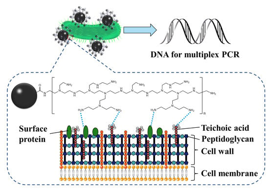
3.1. Principles of the Proposed Strategy

The schematic of the proposed fast-response magnetic enrichment strategy and mPCR analysis for detecting pathogenic Listeria spp. in lettuce was illustrated in Figure 1. Bacterial surfaces are rich in proteins and lipids, as well as complex functional groups [30]. Branched PEI can adsorb on the bacterial surface through electrostatic interactions, hydrogen bonds, and other forces [31] since PEI is rich in amino structures and has a strong positive charge [31]. Therefore, taking the advantages of the high affinity of PEI for bacteria and the rapid response ability of MNPs, PEI-modified MNPs could be used to rapidly enrich bacteria in food samples. After removing matrix interference and increasing the concentration of target bacteria through fast-response magnetic enrichment, DNA was extracted for mPCR analysis to achieve simultaneous and sensitive detection.

3.2. Characterization of PEI-MNPs

The magnetic response speed of MNP and PEI-MNPs were tested under the action of a magnet, and the results are shown in Figure 2a. PEI-MNPs achieved separation in less time (1 min), while MNPs took twice as long to achieve separation (2 min). This may be the result of partial adhesion of PEI-MNPs (in the synthesis of PEI-MNPs, one PEI molecule connected multiple MNPs). A faster magnetic response speed will facilitate the rapid separation of bacteria in samples. The Zeta potential of PEI-MNPs measured in a wide pH range (from 4 to 10) gradually decreased with the increase of pH due to the deprotonation of the surface groups [32], and the isoelectric point was around 8.6 (Figure 2b). The potential of PEI-MNPs was positive in a wide pH range (from 4 to 8), which was conducive to the electrostatic adsorption of PEI-MNPs on the negatively charged bacterial surface [33]. Moreover, MNPs and PEI-MNPs exhibited excellent superparamagnetism and fast magnetic response speed, with high magnetic saturation strengths of 30.9 emu/g and 27.8 emu/g, respectively (Figure 2c). MNPs and PEI-MNPs were observed to be uniformly spherical by SEM (Figure S1). Furthermore, PEI-MNPs with a particle size much smaller than that of pathogenic Listeria spp. were able to absorb on the surface of target bacteria in large quantities (Figure 2d), which was due to the fact the PEI-modified MNPs could interact with abundant functional groups in the surface components of pathogenic Listeria spp. The above results indicated that PEI-MNPs were able to capture Listeria rapidly.

3.3. Bacteria Captured by PEI-MNPs

In order to verify the affinity of PEI for bacteria, L. monocytogenes was selected as the model bacteria to investigate the CE by different MNPs. As shown in Figure 3a, PEI-MNPs had a higher CE for L. monocytogenes than MNPs (p < 0.01), which was due to the strong electrostatic interaction between PEI-MNPs and L. monocytogenes after MNPs were modified with PEI [34]. The number of L. monocytogenes captured by MNPs or PEI-MNPs, and the remaining bacteria in the PBS suspension after magnetic separation were cultured in BHI agar plates (Figure 3b). The distribution of single colonies indicated the excellent capture ability of MNP-PEI for L. monocytogenes, as well as the healthy viability of the bacteria.

3.4. Factors Affecting the Bacterial Capture

Since PEI-MNPs binds to bacteria mainly through electrostatic interactions, the pH and ionic strength of the sample solution have significant effects on the combination of PEI-MNPs with bacteria. The PEI-MNPs exhibited a CE higher than 70% for L. monocytogenes in the pH range of 4 to 10 (Figure 3c). The decrease in CE at pH 9 and 10 was due to the weakening of electrostatic interactions by negatively charged PEI-MNPs, where the binding of PEI-MNPs to L. monocytogenes was mainly driven by hydrogen bonding or hydrophobic interaction between functional groups [32]. In terms of ionic strength, PEI-MNPs still had a high CE (over 95%) for L. monocytogenes when the concentration of NaCl was between 100 mM and 400 mM (Figure 3d). When the concentration of NaCl reached 500 mM, the CE of L. monocytogenes by PEI-MNPs decreased due to the charge neutralization caused by excess NaCl but remained above 74% (Figure 3d). Considering the salt concentration in food samples and the dilution times during sample pretreatment, the above results indicated that the ionic strength has little influence on the separation of bacteria in food samples by PEI-MNPs. These results revealed that PEI-MNPs could capture L. monocytogenes at a wide pH range and ionic strength and achieved a relatively ideal CE (more than 70%).

3.5. Optimization of Enrichment Parameters

To achieve the optimal CE of pathogenic Listeria spp., we optimized the key parameters including dosage of PEI-MNPs, incubation time, and magnetic separation time. The results presented in Figure 4a–c, depict the CE increase with the increase in PEI-MNPs dosage, incubation time, and magnetic separation time. When the dosage of PEI-MNPs was 25 μg, then the co-incubation time of bacteria with PEI-MNPs was 7 min, and the magnetic separation time was 3 min, the CE of PEI-MNPS on pathogenic Listeria spp. mixture (104 CFU/mL of pathogenic Listeria spp.) reached a plateau (over 96%) (Figure 4a–c). Under the above optimization conditions, the CE of PEI-MNPs for different concentrations of the pathogenic Listeria spp. mixture in PBS and lettuce was evaluated (Figure 4d). The CE of PEI-MNPs for pathogenic Listeria spp. mixture from 101 CFU/mL to 104 CFU/mL was over 90%. The CE decreased significantly due to the low dosage (25 μg) of PEI-MNPs which was not enough to capture a high concentration of pathogenic Listeria spp. mixture (105 CFU/mL to 106 CFU/mL). When the dosage of PEI-MNPs increased to 100 μg, the CE of pathogenic Listeria spp. mixture in PBS and lettuce was increased to 59.5 ± 2.8% and 44.1 ± 1.9% (Figure S2), respectively. Considering the potential number of pathogenic Listeria spp. contamination in lettuce [35], PEI-MNPs have great potential for application in real food samples.

3.6. Evaluation of Method Performance in Artificially Contaminated Lettuce

The practicability of the proposed fast-response magnetic enrichment strategy integrated with mPCR analysis was assessed in spiked lettuce with pathogenic Listeria spp. at 101 CFU/mL to 106 CFU/mL. Fifteen bacterial strains were used to investigate the specificity of primers used in this study, which included 12 non-target and 3 target strains (Table 2). The results of mPCR analysis were shown in Table 2, where the selected specific primers only produced positive amplification results for target bacteria, which ensured the accuracy of mPCR analysis. Agarose gel electrophoresis was used to visualize the length of the amplicon generated by mPCR amplification. The result of conventional mPCR detection without fast-response magnetic enrichment by PEI-MNPs was shown in Figure 5a, and the limits of detection (LOD) of L. monocytogenes and L. ivanovii in artificially contaminated lettuce were both 103 CFU/mL. When mPCR was combined with the proposed fast-response magnetic enrichment strategy, the LOD of L. monocytogenes and L. ivanovii were both reduced to 101 CFU/mL (Figure 5b), which significantly improved the detection sensitivity. The enrichment strategy based on PEI-MNPs eliminated the interference of lettuce substrate, and the concentration effect increased the concentration of target bacteria. Compared with conventional mPCR analysis without enrichment, the detection sensitivity of mPCR was significantly reduced by 2 orders of magnitude after a short time of magnetic enrichment. Due to the advantages of simple operation and low cost, magnetic enrichment combined with mPCR analysis proposed in this paper has great application potential in the analysis of pathogenic Listeria spp. in food samples.

4. Conclusions

In this study, a fast-response magnetic enrichment method based on PEI-MNPs was developed to improve the sensitivity of mPCR analysis for the detection of pathogenic Listeria spp. The positively charged PEI-MNPs could separate and enrich pathogenic Listeria spp. within 10 min through electrostatic interaction with the bacteria and had a high CE at a wide pH range and ionic strength. The LOD of L. monocytogenes and L. ivanovii in lettuce were both 101 CFU/mL, which was significantly increased by 2 orders of magnitude compared with conventional mPCR. In conclusion, the proposed fast-response magnetic enrichment strategy based on PEI-MNPs combined with mPCR analysis can rapidly and sensitively detect pathogenic Listeria spp. in lettuce, which was expected to be a powerful tool for ensuring food safety. In further work, we will focus on exploring the analysis of “viable but unculturable” bacterial cells and the potential application of this method in more food samples.

Supplementary Materials

The following supporting information can be downloaded at: https://0-www-mdpi-com.brum.beds.ac.uk/article/10.3390/app12136415/s1, Figure S1: SEM images of MNP (a), PEI-MNPs (b), and pathogenic Listeria spp. (c); Figure S2: CE of pathogenic Listeria spp. at the concentrations from 105 CFU/mL to 106 CFU/mL by 100 μg PEI-MNPs in PBS and lettuce.

Author Contributions

Conceptualization, F.X. and X.B.; Methodology, F.X. and K.W.; Writing—original draft, F.X.; Writing—review & editing, H.X.; Formal analysis, X.B.; Investigation, Y.S.; Project administration, H.X.; Resources, H.X. All authors have read and agreed to the published version of the manuscript.

Funding

This research was funded by the National Key R&D Program of China (2018YFC1602500), the Research Foundation from Academic and Technical Leaders of Major Disciplines in Jiangxi Province, China (20194BCJ22004), and the Research Project of State Key Laboratory of Food Science and Technology, Nanchang University, China (SKLF-ZZB-202133).

Data Availability Statement

The datasets generated during the current study are available from the corresponding author on reasonable request.

Conflicts of Interest

The authors declare no conflict of interest.

References

  1. Huang, D.; Zhuang, Z.; Wang, Z.; Li, S.; Zhong, H.; Liu, Z.; Guo, Z.; Zhang, W. Black phosphorus-Au filter paper-based three-dimensional SERS substrate for rapid detection of foodborne bacteria. Appl. Surf. Sci. 2019, 497, 143825. [Google Scholar] [CrossRef]
  2. Rodríguez-Melcón, C.; Alonso-Calleja, C.; Capita, R. Architecture and Viability of the Biofilms Formed by Nine Listeria Strains on Various Hydrophobic and Hydrophilic Materials. Appl. Sci. 2019, 9, 5256. [Google Scholar] [CrossRef] [Green Version]
  3. Lee, B.E.; Kang, T.; Jenkins, D.; Li, Y.; Wall, M.M.; Jun, S. A single-walled carbon nanotubes-based electrochemical impedance immunosensor for on-site detection of Listeria monocytogenes. J. Food Sci. 2022, 87, 280–288. [Google Scholar] [CrossRef] [PubMed]
  4. Sun, W.; Sun, T.; Wang, X.; Liu, Q.; Dong, Q. Probabilistic model for estimating Listeria monocytogenes concentration in cooked meat products from presence/absence data. Food Res. Int. 2020, 131, 109040. [Google Scholar] [CrossRef]
  5. Kljujev, I.; Raicevic, V.; Jovicic-Petrovic, J.; Vujovic, B.; Mirkovic, M.; Rothballer, M. Listeria monocytogenes—Danger for health safety vegetable production. Microb. Pathog. 2018, 120, 23–31. [Google Scholar] [CrossRef]
  6. Olea-Rodríguez, M.d.l.Á.; Chombo-Morales, P.; Nuño, K.; Vázquez-Paulino, O.; Villagrán-de la Mora, Z.; Garay-Martínez, L.E.; Castro-Rosas, J.; Villarruel-López, A.; Torres-Vitela, M.R. Microbiological Characteristics and Behavior of Staphylococcus aureus, Salmonella spp., Listeria monocytogenes and Staphylococcal Toxin during Making and Maturing Cotija Cheese. Appl. Sci. 2021, 11, 8154. [Google Scholar] [CrossRef]
  7. Jamali, H.; Paydar, M.; Ismail, S.; Looi, C.Y.; Wong, W.F.; Radmehr, B.; Abedini, A. Prevalence, antimicrobial susceptibility and virulotyping of Listeria species and Listeria monocytogenes isolated from open-air fish markets. BMC Microbiol. 2015, 15, 144. [Google Scholar] [CrossRef] [Green Version]
  8. de Niederhäusern, S.; Bondi, M.; Camellini, S.; Sabia, C.; Messi, P.; Iseppi, R. Plant Extracts for the Control of Listeria monocytogenes in Meat Products. Appl. Sci. 2021, 11, 10820. [Google Scholar] [CrossRef]
  9. Radoshevich, L.; Cossart, P. Listeria monocytogenes: Towards a complete picture of its physiology and pathogenesis. Nat. Rev. Microbiol. 2018, 16, 32–46. [Google Scholar] [CrossRef]
  10. Swaminathan, B.; Gerner-Smidt, P. The epidemiology of human listeriosis. Microbes Infect. 2007, 9, 1236–1243. [Google Scholar] [CrossRef] [Green Version]
  11. European Food Safety Authority; European Centre for Disease Prevention and Control. The European Union one health 2020 zoonoses report. EFSA J. 2021, 19, e06971. [Google Scholar]
  12. European Commission. Commission Regulation (EC) No 2073/2005 of 15 November 2005 on microbiological criteria for foodstuffs. Off. J. Eur. Union 2005, 338, 1–26. [Google Scholar]
  13. Morganti, M.; Scaltriti, E.; Cozzolino, P.; Bolzoni, L.; Casadei, G.; Pierantoni, M.; Foni, E.; Pongolini, S. Processing-Dependent and Clonal Contamination Patterns of Listeria monocytogenes in the Cured Ham Food Chain Revealed by Genetic Analysis. Appl. Environ. Microbiol. 2016, 82, 822–831. [Google Scholar] [CrossRef] [PubMed] [Green Version]
  14. Techathuvanan, C.; D’Souza, D.H. Propidium monoazide for viable Salmonella enterica detection by PCR and LAMP assays in comparison to RNA-based RT-PCR, RT-LAMP, and culture-based assays. J. Food Sci. 2020, 85, 3509–3516. [Google Scholar] [CrossRef]
  15. Wang, H.; Zhang, C.; Xing, D. Simultaneous detection of Salmonella enterica, Escherichia coli O157:H7, and Listeria monocytogenes using oscillatory-flow multiplex PCR. Microchim. Acta 2011, 173, 503–512. [Google Scholar] [CrossRef]
  16. Germini, A.; Masola, A.; Carnevali, P.; Marchelli, R. Simultaneous detection of Escherichia coli O175:H7, Salmonella spp., and Listeria monocytogenes by multiplex PCR. Food Control 2009, 20, 733–738. [Google Scholar] [CrossRef]
  17. Xie, G.; Yu, S.; Li, W.; Mu, D.; Aguilar, Z.P.; Xu, H. Simultaneous detection of Salmonella spp., Pseudomonas aeruginosa, Bacillus cereus, and Escherichia coli O157:H7 in environmental water using PMA combined with mPCR. J. Microbiol. 2020, 58, 668–674. [Google Scholar] [CrossRef]
  18. Kotsiri, Z.; Vantarakis, A.; Rizzotto, F.; Kavanaugh, D.; Ramarao, N.; Vidic, J. Sensitive Detection of E. coli in Artificial Seawater by Aptamer-Coated Magnetic Beads and Direct PCR. Appl. Sci. 2019, 9, 5392. [Google Scholar] [CrossRef] [Green Version]
  19. Chen, Q.; Lin, J.; Gan, C.; Wang, Y.; Wang, D.; Xiong, Y.; Lai, W.; Li, Y.; Wang, M. A sensitive impedance biosensor based on immunomagnetic separation and urease catalysis for rapid detection of Listeria monocytogenes using an immobilization-free interdigitated array microelectrode. Biosens. Bioelectron. 2015, 74, 504–511. [Google Scholar] [CrossRef]
  20. Meng, X.; Yang, G.; Li, F.; Liang, T.; Lai, W.; Xu, H. Sensitive Detection of Staphylococcus aureus with Vancomycin-Conjugated Magnetic Beads as Enrichment Carriers Combined with Flow Cytometry. ACS Appl. Mater. Interfaces 2017, 9, 21464–21472. [Google Scholar] [CrossRef]
  21. Yang, G.; Huang, M.; Wang, Y.; Chen, G.; Zhao, Y.; Xu, H. Streptavidin-exposed magnetic nanoparticles for lectin magnetic separation (LMS) of Staphylococcus aureus prior to three quantification strategies. Microchim. Acta 2019, 186, 813. [Google Scholar] [CrossRef] [PubMed]
  22. Zhang, L.; Huang, R.; Liu, W.; Liu, H.; Zhou, X.; Xing, D. Rapid and visual detection of Listeria monocytogenes based on nanoparticle cluster catalyzed signal amplification. Biosens. Bioelectron. 2016, 86, 1–7. [Google Scholar] [CrossRef] [PubMed]
  23. Deng, M.; Wang, Y.; Chen, G.; Liu, J.; Wang, Z.; Xu, H. Poly-l-lysine-functionalized magnetic beads combined with polymerase chain reaction for the detection of Staphylococcus aureus and Escherichia coli O157:H7 in milk. J. Dairy Sci. 2021, 104, 12342–12352. [Google Scholar] [CrossRef]
  24. Cremin, K.; Jones, B.A.; Teahan, J.; Meloni, G.N.; Perry, D.; Zerfass, C.; Asally, M.; Soyer, O.S.; Unwin, P.R. Scanning Ion Conductance Microscopy Reveals Differences in the Ionic Environments of Gram-Positive and Negative Bacteria. Anal. Chem. 2020, 92, 16024–16032. [Google Scholar] [CrossRef] [PubMed]
  25. Xing, Q.; Zhang, M.; Xu, X.; Zhang, B.; Duan, Y.; Wei, Y.; Wang, J.; Mingyang, L. Rapid photocatalytic inactivation of E. coli by polyethyleneimine grafted O-doped g-C3N4: Synergetic effects of the boosted reactive oxygen species production and adhesion performance. Appl. Surf. Sci. 2022, 573, 151496. [Google Scholar] [CrossRef]
  26. Tao, T.; Chen, Q.; Bie, X.; Lu, F.; Lu, Z. Mining of novel species-specific primers for PCR detection of Listeria monocytogenes based on genomic approach. World J. Microbiol. Biotechnol. 2015, 31, 1955–1966. [Google Scholar] [CrossRef]
  27. Mao, Y.; Huang, X.; Xiong, S.; Xu, H.; Aguilar, Z.P.; Xiong, Y. Large-volume immunomagnetic separation combined with multiplex PCR assay for simultaneous detection of Listeria monocytogenes and Listeria ivanovii in lettuce. Food Control 2016, 59, 601–608. [Google Scholar] [CrossRef]
  28. Chiang, Y.C.; Yang, C.Y.; Li, C.; Ho, Y.C.; Lin, C.K.; Tsen, H.Y. Identification of Bacillus spp., Escherichia coli, Salmonella spp., Staphylococcus spp. and Vibrio spp. with 16S ribosomal DNA-based oligonucleotide array hybridization. Int. J. Food Microbiol. 2006, 107, 131–137. [Google Scholar] [CrossRef]
  29. Xiao, F.; Liu, J.; Feng, X.; Xie, G.; Zhang, W.; Xu, H. Rapid enrichment and detection of Staphylococcus aureus in milk using polyethyleneimine functionalized magnetic nanoparticles. Microchem. J. 2022, 178, 107388. [Google Scholar] [CrossRef]
  30. Bierne, H.; Cossart, P. Listeria monocytogenes Surface Proteins: From Genome Predictions to Function. Microbiol. Mol. Biol. Rev. 2007, 71, 377–397. [Google Scholar] [CrossRef] [Green Version]
  31. Li, Z.; Ma, J.; Ruan, J.; Zhuang, X. Using Positively Charged Magnetic Nanoparticles to Capture Bacteria at Ultralow Concentration. Nanoscale Res. Lett. 2019, 14, 195. [Google Scholar] [CrossRef] [PubMed] [Green Version]
  32. Jin, Y.; Liu, F.; Shan, C.; Tong, M.; Hou, Y. Efficient bacterial capture with amino acid modified magnetic nanoparticles. Water Res. 2014, 50, 124–134. [Google Scholar] [CrossRef] [PubMed]
  33. Hayden, S.C.; Zhao, G.; Saha, K.; Phillips, R.L.; Li, X.; Miranda, O.R.; Rotello, V.M.; El-Sayed, M.A.; Schmidt-Krey, I.; Bunz, U.H. Aggregation and interaction of cationic nanoparticles on bacterial surfaces. J. Am. Chem. Soc. 2012, 134, 6920–6923. [Google Scholar] [CrossRef] [PubMed]
  34. Thiramanas, R.; Laocharoensuk, R. Competitive binding of polyethyleneimine-coated gold nanoparticles to enzymes and bacteria: A key mechanism for low-level colorimetric detection of gram-positive and gram-negative bacteria. Microchim. Acta 2015, 183, 389–396. [Google Scholar] [CrossRef]
  35. Zhu, Q.; Gooneratne, R.; Hussain, M.A. Listeria monocytogenes in Fresh Produce: Outbreaks, Prevalence and Contamination Levels. Foods 2017, 6, 21. [Google Scholar] [CrossRef] [Green Version]
Figure 1. Schematic illustration of PEI-MNPs magnetic enrichment integrated with mPCR analysis for the simultaneous detection of pathogenic Listeria spp.
Applsci 12 06415 g001
Figure 2. Characterization of MNP and PEI-MNPs. (a) Photo of MNP and PEI-MNPs adsorption under the action of a magnet. (b) Zeta potential of PEI-MNPs at pH 4 to 10. (c) Magnetic hysteresis loops of MNP and PEI-MNPs measured at room temperature. (d) SEM image of PEI-MNPs bound to the surface of Listeria.
Applsci 12 06415 g002
Figure 3. CE of L. monocytogenes under different conditions. (a) CE of L. monocytogenes captured by MNP and PEI-MNPs, (b) Images of L. monocytogenes colonies captured by MNP and PEI-MNPs on BHI agar. (c) Effects of pH on the CE by PEI-MNPs. (d) Effects of ionic strength on the CE by PEI-MNPs. *** represents p < 0.001.
Applsci 12 06415 g003
Figure 4. Optimization of experimental parameters. (a) Dosages of PEI-MNPs. (b) Incubation time of PEI-MNPs bound to Listeria. (c) Magnetic separation time of Listeria-PEI-MNPs complexes. (d) CE of pathogenic Listeria spp. at the concentrations from 101 CFU/mL to 106 CFU/mL by PEI-MNPs in PBS and lettuce.
Applsci 12 06415 g004
Figure 5. Evaluation of the detection limit of pathogenic Listeria spp. in lettuce sample. (a) mPCR assay without PEI-MNPs magnetic enrichment. Lane M: DL1000 DNA Marker; lane C: negative control; lanes 1 to 5: 106 CFU/mL to 102 CFU/mL of Listeria. (b) PEI-MNPs magnetic enrichment combined with mPCR assay, Lane M: DL1000 DNA Marker; lane C: negative control; lanes 1 to 6: 106 CFU/mL to 101 CFU/mL of Listeria. The bands at 583 bp, 311 bp, and 475 bp represented L. monocytogenes, L. ivanovii, and IAC, respectively.
Applsci 12 06415 g005
Table 1. Primers used in the mPCR assay 1.
Target BacteriaTarget GenePrimerSequences (5′-3′)Length (bp)Reference
L. monocytogenesLMOf2365_2721lm13-FGTTCGTCGGTCCGTGGTA583[26]
lm13-RTTGGCAAGCAAGCAGTTCA
L. ivanoviiiactaiacta-FTAGAGAATGGCGAGGAGGAGTA311[27]
iacta-RTCCGGGACATTTGCACT
Bacteria DNA16s rRNA16s rRNA-FCCTACGGGAGGCAGCAGT475[28]
16s rRNA-RCGTTTACGGCGTGGACTAC
1 F = forward, R = reverse.
Table 2. Bacterial strains used in this study with results of the mPCR assay.
Bacterial StrainsSourceStrain IDResults of mPCR
lm13iacta16s rRNA
Listeria monocytogenesATCC 119115++
CMCC 254001++
Listeria ivanoviiATCC19119++
Listeria innocuaATCC11288+
Listeria seeligeriATCC35967+
Listeria grayiATCC25401+
Listeria welshimeriATCC35897+
Staphyloccocus aureusCMCC26001+
Cronobacter sakazakiiATCC29544+
Escherichia coliATCC25922+
Escherichia coli O157:H7ATCC43888+
Bacillus cereusJX-CDC 3JDZ102Y+
Salmonella EnteritidisATCC13076+
Salmonella TyphimuriumATCC13311+
Bacillus subtilisCMCC63501+
Results (+/−) represent positive and negative signals. 1 ATCC, American Type Culture Collection. 2 CMCC, China Medical Culture Collection. 3 JX-CDC, Jiang Xi Province Center for Disease Control and Prevention.
Publisher’s Note: MDPI stays neutral with regard to jurisdictional claims in published maps and institutional affiliations.

