
Disks of Oxygen Vacancies on the Surface of TiO2 Nanoparticles
1
Institute of Metal Physics UB RAS, 18 S.Kovalevskaya Street, 620108 Ekaterinburg, Russia
2
Ural Federal University, 19 Mira Street, 620002 Ekaterinburg, Russia
*
Author to whom correspondence should be addressed.
Appl. Sci. 2022, 12(23), 11963; https://0-doi-org.brum.beds.ac.uk/10.3390/app122311963
Submission received: 27 October 2022
/
Revised: 15 November 2022
/
Accepted: 18 November 2022
/
Published: 23 November 2022
(This article belongs to the Special Issue Metal-Based Composite Materials: Preparation, Structure, Properties, and Applications)
Abstract
:Oxide nanopowders are widely used in engineering, and their properties are largely controlled by the defect structure of nanoparticles. Experimental data on the spatial distribution of defects in oxide nanoparticles are unavailable in the literature, and in the work presented, to gain such information, methods of nuclear reactions and deuterium probes were employed. The object of study was oxygen-deficient defects in TiO2 nanoparticles. Nanopowders were synthesized by the sol–gel method and laser evaporation of ceramic targets. To modify the defect structure in nanoparticles, nanopowders were subjected to vacuum annealings. It was established that in TiO2 nanoparticles there form two-dimensional defects consisting of six titanium atoms that occupy the nanoparticle surface and result in a remarkable deviation of the chemical composition from the stoichiometry. The presence of such defects was observed in two cases: in TiO2 nanoparticles alloyed with cobalt, which were synthesized by the sol–gel method, and in nonalloyed TiO2 nanoparticles synthesized by laser evaporation of ceramic target. The concentration of the defects under study can be varied in wide limits via vacuum annealings of nanopowders which can provide formation on the surface of oxide nanoparticles of a solid film of titanium atoms 1–2 monolayers in thickness.
1. Introduction
The physico-chemical and functional properties of oxide nanoparticles, which are widely employed in engineering, are largely dependent on the defect structure of nanoparticles, namely, the concentration of point defects, their type, and spatial distribution. In this field, works have mainly been devoted to defects that condition oxygen deficiency in nanoparticles compared to the stoichiometry [1,2]. In addition, oxygen vacancies and clusters based on them strongly affect the electrical, magnetic, diffusion, catalytic, and other properties of oxide nanopowders [3,4,5]. For example, a concept of d0-magnetism was proposed [6], which helped to explain ferromagnetic properties observed on oxide nanopowders that did not contain any magnetic dopants by an oxygen deficiency in nanoparticles. Such results were obtained on the oxide powders of TiO2, Al2O3, ZnO, In2O3, HfO2, CuO, and others [7,8,9,10,11].
The majority of results concerning the defect structure of oxide nanoparticles are obtained for the TiO2 nanoparticles with the use of a large number of techniques [3,5,8,12,13,14,15]. They highlight a wide variety of types of point defects and the evolution of the defect structure under different treatments of nanopowders, say, oxygen annealing [3]. When investigating the defect structure, the most informative were the methods of positron annihilation [3,5,8,12,13,14,15], X-ray photoelectron spectroscopy [3,5,13], and photoluminescence [14]; as defects, there were specified oxygen and titanium vacancies [3,8], interstitial titanium atoms [12], 3D vacancy clusters [8], complexes consisting of oxygen and titanium vacancies [3], Ti3+ ions and oxygen vacancies [15], and others [12]. The diversity of defect types in the oxide nanoparticles is conditioned by a large number of atoms on the sample surface, the presence of defects - charge carriers, mixed cation valence, changing oxygen concentration upon different powder treatments, etc.
With the bulk of important results accumulated on the types of defects in the oxide nanoparticles and their impact on the magnetic properties of nanoparticles [3,16,17,18,19,20], the state-of-art in this field can hardly be considered sufficient. First, no measurements of the oxygen concentration were performed and, hence, it was not accounted for in the concentration balance. For this reason, it cannot be excluded that information on several defect types is lacking. The more probable it seems to take into account the selective sensitivity of many techniques to defects of different types. Second, there are no available experimental data on the spatial distribution of defects in nanoparticles gained by direct observations. Such experiments are necessary since the energy states of atoms near the surface and in the bulk are drastically different.
In view of the above, this work is devoted to studying the defect structure of oxide nanoparticles via measurements of the oxygen concentration and using a technique that allows the determination of the defect concentration on the surface and in the bulk of nanoparticles. The choice of TiO2 nanoparticles as the object for investigation is conditioned by a wide scope of practical applications of the dioxide titanium nanopowders [21,22,23,24,25]. The oxygen concentration was measured by the method of nuclear reactions (NRA); the error of measurements being about 0.5–1.0%, which makes it applicable only for nanopowders with rather a large oxygen deficiency compared to stoichiometry, at a level of several percent. Nanopowders of TiO2 meet this condition [26]. The defect concentration was determined by the method of deuterium probes (DP) [2]. It is based on the dependence of the deuterium solubility in the oxide particles on the concentration of defects of a specified type. The application of the DP method was reasonable since, first, it is sensitive to defects bringing about oxygen deficiency [2] and, second, owing to a drastic difference in its basic principles from those of the earlier applied techniques, it enables one to detect defects of unknown types. The NRA and DP techniques have already been used to measure the oxygen and defect concentrations in oxide nanoparticles [2,26,27,28], but in these works, the type of defects and their spatial distribution in nanoparticles were not the subject of research.
2. Experimental
2.1. Synthesis of Oxide Nanopowders
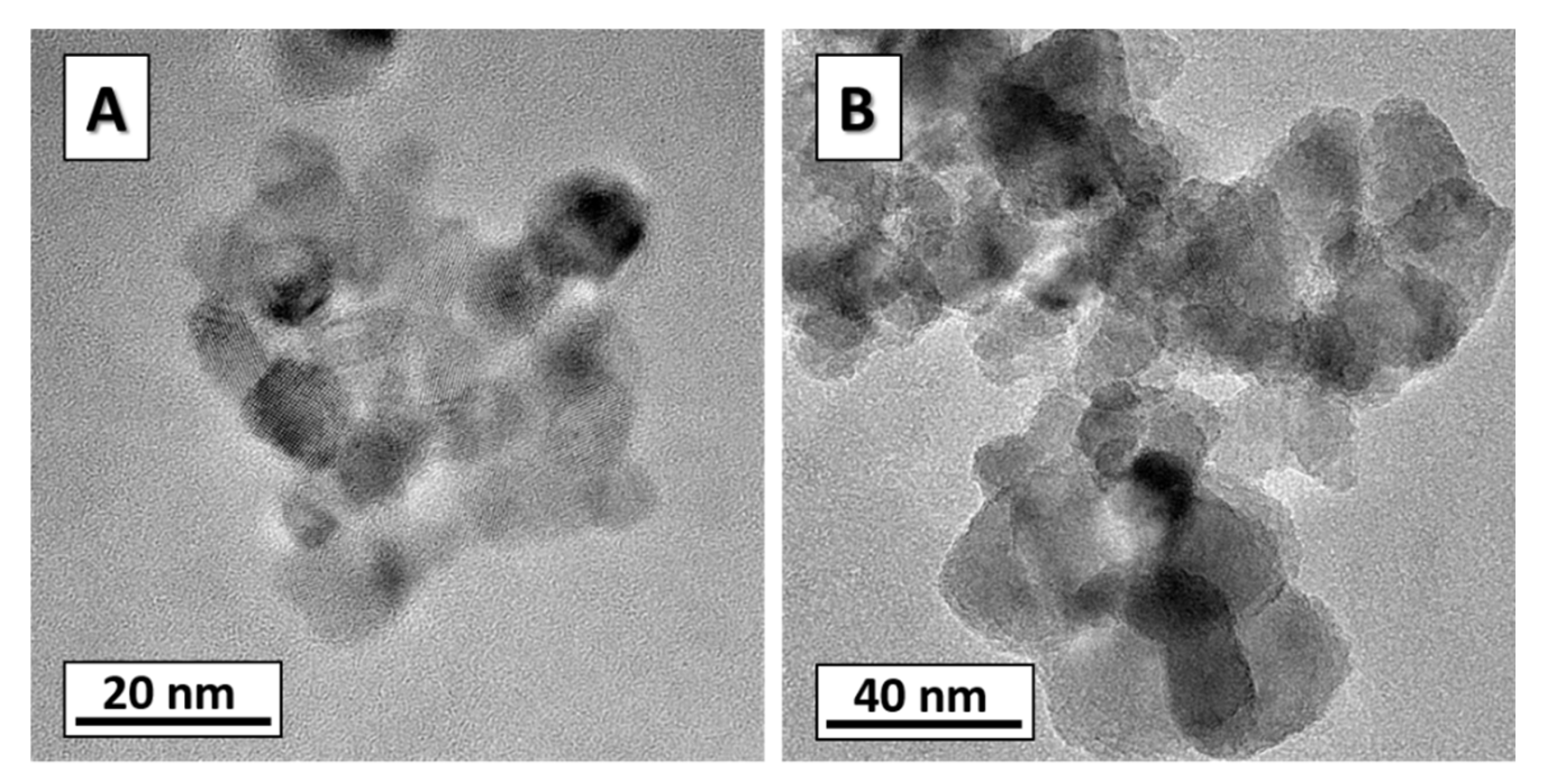
Nanopowders were produced by the technology of laser evaporation of ceramic target (LE) [26] and sol–gel method (SG) [16,29]. As an LE, pills of titanium dioxide 60 mm in diameter were produced by pressing at room temperature, with the specific surface area of micropowder being 2 m2/g. For vaporizing, a fiber-ytterbium laser was used with a wavelength of 1.07 μm and a maximal power output of 1 kW; the frequency of laser pulses was 5 kHz, duration was 50 μs. To produce nanopowders with a different specific area, the type of inert gas (argon or helium), gaseous pressure, and laser power were varied. For the synthesis of titanium oxide nanoparticles doped with cobalt ions by the SG method [29], acid hydrolysis of organometallic precursors was employed; in our case, titanium isopropyl oxide (Titanium (IV) isopropoxide) and cobalt (II) acetylacetonate as a source of metal ions. All reagents used were obtained from Sigma Aldrich. The preliminary required amounts of titanium isopropoxide (2 mL) and cobalt acetylacetonate complex were placed in a 5 mL test tube; then, 2 mL of acetone was poured into it. After vigorous shaking, a bluish-brown solution was formed. The solution was left overnight. Then, 1 mL of hydrochloric acid (0.1M) was poured into the solution. The hydrolysis reaction occurs almost instantly; however, for the process to run uniformly throughout the entire volume of the colloid, it was subjected to intensive ultrasonic action using an ultrasonic activator with a submerged titanium probe. Ultrasound exposure was 30 s. Then, the particles were separated by centrifugation (10,000 RPM) for 10 min and washed with acetone. The washing process was repeated three times. The resulting precipitate was dried at 70 °C. The precursor was further calcined in air at different temperatures (from 300 to 500 °C). Figure 1 shows micrographs of nanoparticles calcined at 400 and 500 °C, the image was obtained using transmission electron microscopy (TEM).
2.2. Methods of Nuclear Reactions and Deuterium Probes
All nanopowders predominantly contained such phases of titanium dioxide as anatase, rutile, brookite, and their mixtures. Data on the phase composition are given in more detail in Section 3.1. In the current approach, the defect structure is characterized by the oxygen concentration and defect concentration and real defects contain a certain number of elementary point defects. Hence, a real defect can comprise vacancies and interstitial atoms of oxygen and titanium. Within the approach accepted, an oxygen vacancy cannot be distinguished from an interstitial titanium atom, the same as a titanium vacancy from an interstitial oxygen atom. When interpreting the results of studying the thus prepared samples, we considered solely oxygen vacancies as the most realistic elementary point defects. In the DP technique, after annealing in gaseous deuterium, nanopowders become three-component and their chemical composition can be presented by formula , which accounts for the presence of 2 deuterium atoms per one defect [2]. Moreover, oxygen vacancies are supposed to be prone to joining in clusters, and designates the amount of vacancies in a cluster. This formula enables easily obtaining that concentrations and are connected as follows
where is the oxygen concentration in a defectless oxide; herefrom, concentrations are expressed in fractions of the number of atoms in a nanoparticle. It is seen from Expression (1) that when treating the dependences the number of vacancies in a defect N can be determined if oxygen vacancies are combined in complexes. Expression (1) is obtained for nanoparticles of arbitrary shape and does not depend on the spatial distribution of defects and mutual arrangement of vacancies in a cluster. For example, it is valid for cases when defects are distributed in a nanoparticle uniformly or only in its thin surface layer with vacancies forming in a cluster of both 2D and 3D complexes.
When using the method of LE, the average concentrations and were varied via the synthesis of nanopowders with different specific surface areas S. This approach makes it possible to produce nanopowders with different and , if the concentrations of defects in the bulk and near the surface are different. In the SG technology, nanopowders with different defect concentrations were produced using vacuum annealings. In the preliminary experiments, it was established that vacuum annealings are by no means a universal way of decreasing the oxygen concentration in the titanium dioxide nanopowders. In particular, in the case of undoped TiO2, vacuum annealings exert a poor effect on the concentrations of oxygen and defects, which did not allow the analysis of the dependence with reliable accuracy. At the same time, Co-doped nanopowders of TiO2 with widely ranging oxygen concentrations were prepared using vacuum annealings. The same result was obtained in work [5], where it was shown that alloying of TiO2 with cobalt leads to increasing vacancy concentration. In view of this, in the case of the SG method, investigation of the dependence was performed on powders alloyed with cobalt, its concentration in the cation sublattice made up of 3 at. %. Vacuum annealings were carried out after completion of the synthesis routine at temperatures varying from 200 to 500 °C and holding time, 15 to 30 min, 500 °C.
To measure the defect concentration using the DP method, first, nanopowders are annealed in gaseous deuterium, and then, the concentration of dissolved deuterium is measured using the NRA technique. The DP method is shown in work [2] to be sensitive to defects bringing about oxygen deficiency in nanoparticles and is based on the existence of unambiguous relation of the and concentrations, which is provided by the formation of nanoparticles annealed in deuterium of clusters of a strictly specified composition. In particular, in the TiO2 nanoparticles, one cluster consists of a defect and 2 deuterium atoms. The cluster composition was determined using the dependence of on the dose of deuterium irradiation. In the current work, such a procedure was applied to all powders after synthesis and vacuum annealings, and the cluster composition was the same. In general, nanoparticles can contain defects of different types simultaneously. For example, in study [2], in the TiO2 particles, along with defects detectable for the DP method, one more type of defect was observed, which consisted of Ti3+ ions and oxygen vacancies. The duration of deuterium annealing we used was, just as in [2], 1 h, with deuterium pressure being 0.6 atm. In the DP method, the determination of the defect concentration is reduced to measuring the deuterium concentration . For this reason, the metrological characteristics of the NRA technique for the reaction 2H(d,p)3H controlled the accuracy and sensitivity in the determination of the defect concentration : statistical error made up several percent of the measured value and measurement sensitivity was about 0.01 at. %.
As was noted in Section 1, no experimental data on the spatial arrangement of defects in oxide nanoparticles are available in the literature. Such a task is objectively complicated because of the small size of nanoparticles. In the current work, a particular solution was obtained. It is based on an approach that involves the characterization of the defect structure using only two defect concentrations, namely, at the surface of nanoparticle and average concentration in the nanoparticle bulk . For the powders synthesized using LE, we investigated the dependence of defect concentration on the specific surface area , which allowed us to obtain, in the model with two concentration values and , information on the spatial distribution of defects in nanoparticles. For example, the growth of with increasing means higher defect concentration near the surface then in the bulk and vice versa. If = , the defect concentration evidently is independent of . In this work, of special interest was the case when defects to which the DP method is sensitive are localized near the nanoparticle surface. In this case, the chemical composition of nanopowders can be expressed by the formula , and the following relationship works:
where is the oxide density, is the thickness of the oxygen-deficient layer whose chemical composition is denoted as TiO2−y. Expression (2) is valid for particles of arbitrary shape. Hence, with equal parameters and in the powders with different specific area the dependence will be close to linear, with the extrapolated value = 0. Since the DP method provides a high accuracy in measuring the defect concentration, in some particular cases the study of can give reliable information on the ratio of defect concentrations in the bulk and near the nanoparticle surface. Note that application of this approach is bound to solely the case when powders are synthesized by LE technology. It favors independence of the parameters , and of , which will be considered in Section 3.2.
The average concentrations of oxygen and deuterium in nanopowders were determined by the NRA technique at a 2 MB van de Graaf accelerator using reactions 16O(d,p1)17O* and 2H(d,p)3H with the deuteron energy of 900 and 650 keV, respectively. In the majority of experiments in the accelerator, the samples were at room temperature and nanoparticles of powders were pressed into an indium plate. When the concentration was measured at the temperature of 400 °C, nanoparticles were pressed into copper powder. Using Rutherford backscattering spectroscopy, it was established that indium or copper atoms were absent in the zone under analysis. The sample surface was set perpendicular to the incident beam, nuclear reaction products were registered with the use of a silicon surface barrier detector. The angle of proton registration was 160°, and the diameter of the incident beam was 1 or 2 mm. The irradiation dose was measured on the samples using a secondary monitor, with the statistical error being 0.2 to 1.0%. When mathematically processing the data, spectra from the samples under study were compared with those from reference samples having constant-in-depth isotope concentrations. For 16O, it was CuO, whereas for deuterium, ZrCr2D0.12. In more detail, the experimental setting and processing procedure are described in work [2].
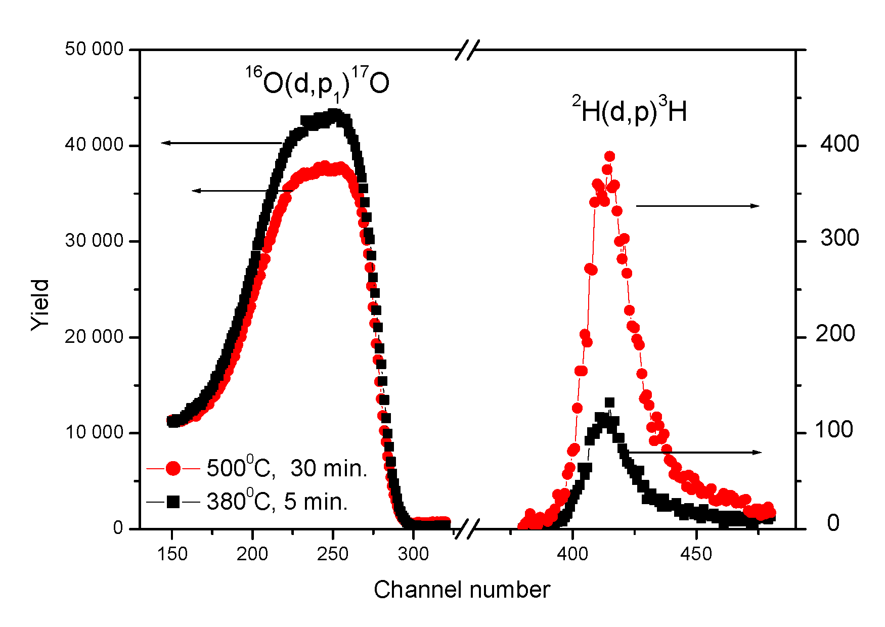
When doing this, it was necessary to provide highly accurate measurements of concentrations and , which was not trivial as the data on depended on the presence on the nanoparticle surface of water molecules. With increasing the thickness of the water layer, the oxygen concentration in factions of the number of atoms in a nanoparticle decreases since the H2O molecule contains fewer oxygen atoms than TiO2. In view of this, the powders were dried under vacuum pumping-off. To monitor the oxygen concentration , sampling measurements of the powders were performed in the accelerator chamber at 400 °C, which allows the elimination of the adsorbed water molecules. The results of the concentration and , unlike , were not changed when varying experimental conditions, temperature, pressure, and annealing time. In addition, these results did not depend on the conditions of storing the powders for a year and more, since the deuterium annealing provided the elimination of water molecules from the nanoparticle surface. Figure 2 shows the spectra of protons p for the nuclear reactions 16O(d,p1)17O* and 2H(d,p)3H, which were used to determine the oxygen and defect concentrations. The and concentrations are directly proportional to the number of registered protons, the shape of the spectra does not depend on and .
3. Results and Discussion
3.1. Number of Oxygen Vacancies in a Defect
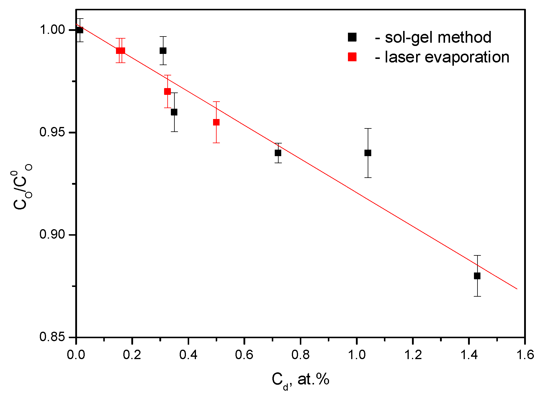
The dependence shown in Figure 3 agrees with Expression (1), which testifies to the formation in the TiO2 nanoparticles of defects consisting of oxygen vacancies. Processing by the least-square method gave the number of vacancies in a defect N = 12.5 ± 0.9.
As follows from Figure 3, the formation in the dioxide titanium nanoparticles of defects containing N = 12 oxygen vacancies is invariant with respect to experimental conditions. First, it takes place in the nanopowders synthesized by different technologies and characterized by different mechanisms of defect formation. In the technology of SG method, in the as-synthesized powders of composition close to stoichiometric, the formation of defects occurs in the course of vacuum annealing through the oxygen depletion of nanoparticles. Upon LE, oxygen-deficient defects were formed directly in the course of synthesizing. Second, the powders strongly differ in the number of defects per unit volume of nanoparticles. This follows from Figure 2 since this parameter decreases with decreasing concentration . Finally, the powders under study differed in phase composition. In the case of the SG method, it was anatase, whereas upon LE, the mixture of TiO2 phases. In particular, in the powders with S 200 m2/g, brookite with small additions of anatase and rutile predominates, whereas at S 200 m2/g, quite the opposite, brookite was not observed at all, only anatase and rutile. Since the number of vacancies in a defect N = 12 does not depend on the synthesis technology, mechanism of defect formation, phase composition, and other characteristics of nanoparticles, one can suggest that the formation of such defects, denoted as Ti6, is conditioned by thermodynamic reasons. This question needs further investigation.
3.2. Spatial Distribution of Defects Ti6 in Nanoparticles
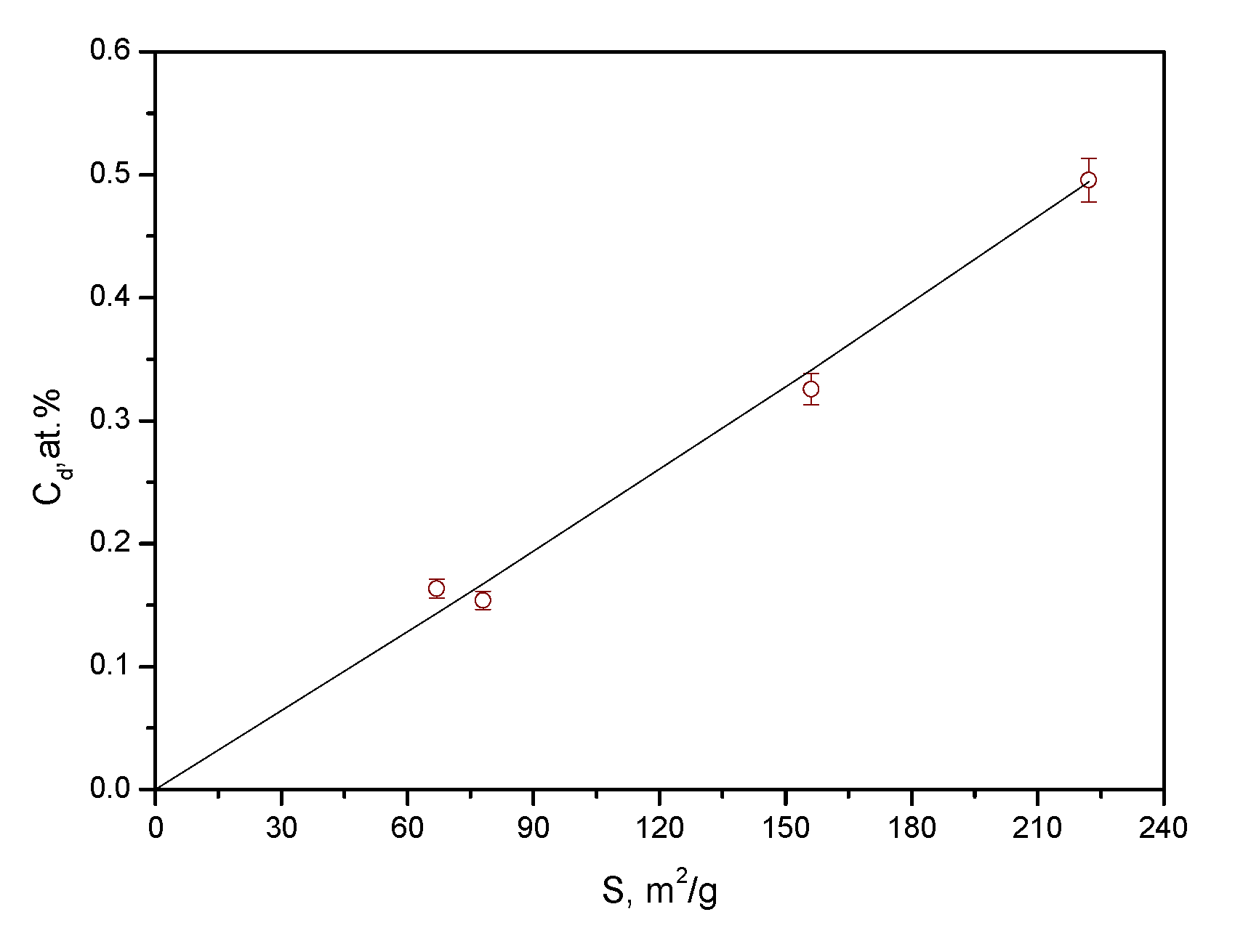
In the investigation of spatial distribution and size of defects consisting of 6 titanium atoms, different versions are considered, namely, defects located in the bulk and on the surface of nanoparticles and two-dimensional or three-dimensional ones. To obtain information on these issues, the dependence was studied for the powders synthesized by the LE. As is seen in Figure 4, the experimental dependence matches Expression (2) at constant parameters N, , and , which indicates that the defects consisting of 12 oxygen vacancies are located on the surface and absent in the bulk of TiO2 nanoparticles.
The independence of the parameter N on S was experimentally shown in Section 3.1. As for the parameters and it is conditioned by the application of technology of LE. In this case, the formation of the defect structure of the surface layer proceeds in two steps. In the first step, at high temperatures in a vacuum, there form nanoparticles of stoichiometric composition in the bulk with the surface atomic layer virtually depleted of oxygen. It was established that the thickness of the oxygen-depleted layer does not depend on S and is equal to 0.36 nm [26], which approximately matches up an oxide monolayer. In works fulfilled within the theory of density functional, the following theoretical explanation of this result was given, namely, the existence of an oxygen-free layer on the oxide surface is energetically more favorable than of stoichiometric composition TiO2 [26,30,31]. In the second step, the powder is cooled to room temperature in air and oxygen enters into the surface layer of nanoparticles. Evidently, in this case, the thickness of the oxygen-deficient layer does not depend on S. The amount of oxygen entering into nanoparticles depends on the regime of cooling the nanopowders. In this work, these regimes were the same for powders with different S.
We already mentioned above that the thickness of defects Ti6 in nanoparticles synthesized by LE technology is within the atomic scale, whereas other linear dimensions are several times as high, i.e., the defects are two-dimensional. For nanoparticles synthesized by the SG method, no data on the spatial distributions of Ti6 defects and inference on their two-dimensional or three-dimensional shape are presented in this work because of the lack of appropriate techniques. Yet, there are grounds to conclude that the Ti6 defects in nanoparticles synthesized by the SG method also are two-dimensional and localized on the nanoparticle surface. In our opinion, these defects cannot occupy internal bulk regions of nanoparticles since the crystal structure of titanium dioxide with inner defects consisting of 12 oxygen vacancies will hardly be stable. At the same time, the presence of such defects on the surface of nanoparticles does not cause destruction of the crystal lattice of the oxide, which is evidenced by the results of works [26,30,31].
In the surface layer of TiO2 particles, there are present two types of regions: (i) consisting of 6 titanium atoms and (ii) having composition close to stoichiometric TiO2. It is worth estimating fractions of these regions. If to take up a constancy of distance between the titanium atoms upon the formation of the Ti6 defect, the fraction of nanoparticle surface occupied by regions consisting of 6 titanium atoms is expressed as
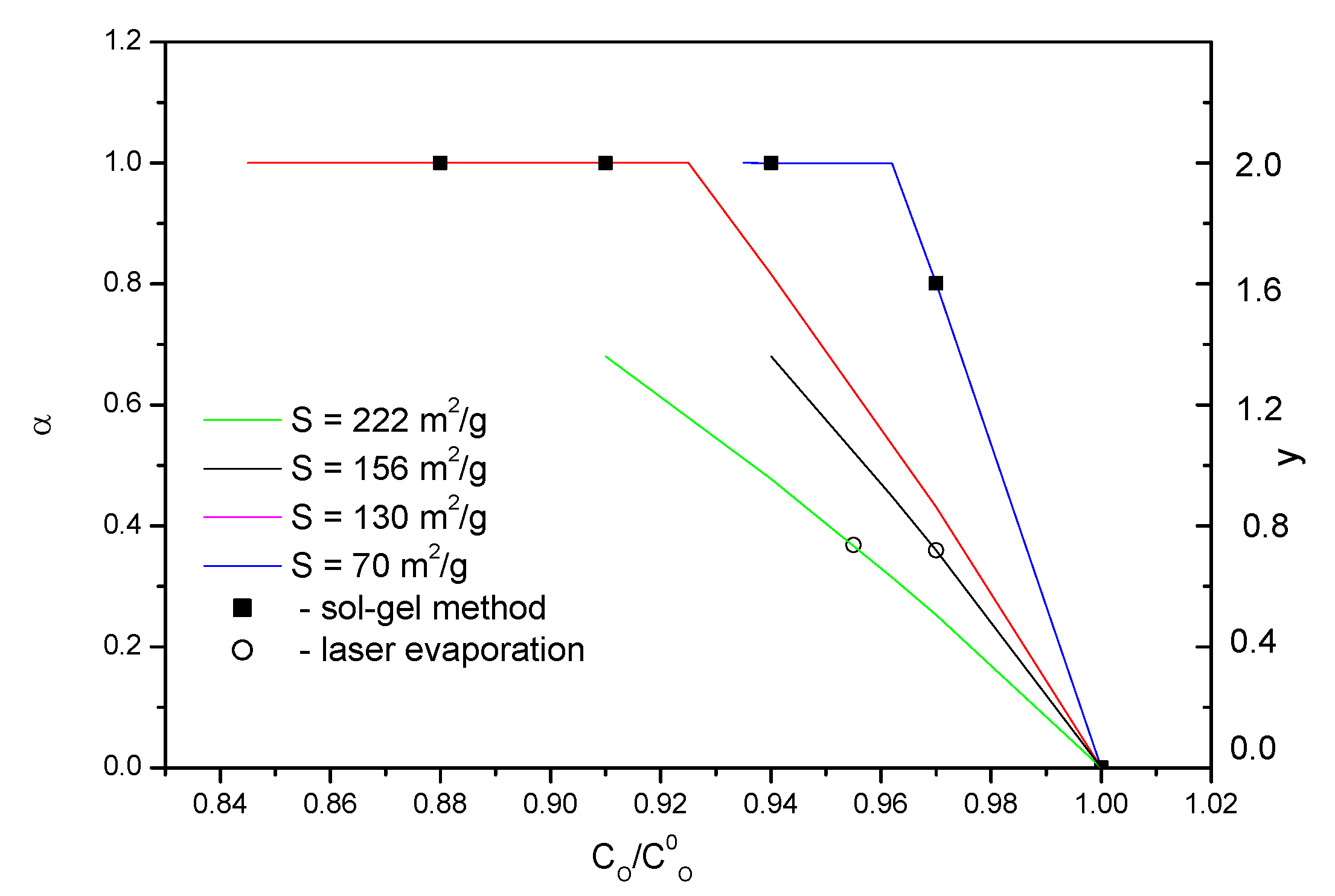
In Figure 5, the dependences calculated by Expression (3) for several values of are shown. Points in Figure 5 show sampling experimental results, data on y are also given. It is easy to show that the values of and y obey the expression y = 2. Bends on the dependences correspond to the formation on the nanoparticle surface of a solid monolayer in which oxygen atoms are absent. The horizontal portions of the dependences correspond to the formation in nanoparticles of one more monolayer deficient in oxygen. It follows from Figure 5 that the fraction that is taken by regions consisting of 6 titanium atoms can be an easily controlled parameter. In particular, this refers to nanoparticles of TiO2 produced by the SG method. In the as-synthesized state, they are virtually stoichiometric ( and by vacuum annealings and alloying with cobalt, this value can be increased to 1.
It is shown that under certain conditions, in the TiO2 nanoparticles there form two-dimensional defects consisting of 6 titanium atoms and localized on the nanoparticle surface, which results in a significant deviation of the chemical composition from the stoichiometry. Since, as noted in Section 1, in the TiO2 nanoparticles there are present defects of other types, of interest is to compare the data on the concentration of surface two-dimensional defects with those obtained in previous studies. Unfortunately, in view of the absence of such information in works on oxide nanoparticles, we have to restrict ourselves to rough estimates. In a general case, defects of almost all types cause changes in the chemical composition of nanoparticles and their presence in the samples gives rise to a deviation from the linear dependence in Figure 3. Since in the work no deviations beyond the statistical experimental errors were registered, one can conclude that the concentration of other defects was far smaller than that of defects consisting of 6 titanium atoms.
4. Conclusions
Thus, new data on the defects that condition the oxygen deficit in the oxide nanoparticles of TiO2 have been gained. The formed two-dimensional defects of atomic thickness are established to consist of six titanium atoms. They are free of oxygen atoms and located on the nanoparticle surface. The presence of such defects is detected in two cases: in the cobalt-doped nanoparticles of TiO2 synthesized by the sol–gel method and subjected to vacuum annealings and undoped nanoparticles synthesized by laser evaporation of ceramic target. The data on the defect structure were obtained by the method of deuterium probes, DP, which was applied to measure the defect concentrations, and by nuclear reactions, NRA, to determine the oxygen concentration in nanopowders. The concentration of the defects under study can be varied in wide limits; in particular, using vacuum annealings can provide gradual growth from 0 to 1.5 at. %, including the formation of a solid film of titanium atoms on the nanoparticle surface. The concentration of defects of other types in the nanoparticles under study was lower than that of two-dimensional defects located on the nanoparticle surface.
Author Contributions
Conceptualization, V.B.V.; Investigation, T.E.K. and E.V.V.; Writing—original draft, V.B.V. All authors have read and agreed to the published version of the manuscript.
Funding
The research was carried out within the state assignment of the Ministry of Science and Higher Education of the Russian Federation (theme “Function” no. AAAA-A19-119012990095-0).
Institutional Review Board Statement
Not applicable.
Informed Consent Statement
Not applicable.
Acknowledgments
Authors are very grateful to A.S. Minin, A.E. Yermakov, and M.A. Uimin for useful discussion and valuable remarks and to A.S. Minin for the synthesis of nanopowders by the sol–gel method.
Conflicts of Interest
The authors declare no conflict of interest.
References
- Diebold, U. The surface science of titanium dioxide. Surf. Sci. Rep. 2003, 48, 53–229. [Google Scholar] [CrossRef]
- Vykhodets, V.B.; Kurennykh, T.E. Characterization of the defect structure of oxide nanoparticles with the use of deuterium probes. RSC Adv. 2020, 10, 3837–3843. [Google Scholar] [CrossRef] [PubMed]
- Ghosh, S.; Nambissan, P. Evidence of oxygen and Ti vacancy induced ferromagnetism in post-annealed undoped anatase TiO2 nanocrystals: A spectroscopic analysis. J. Solid State Chem. 2019, 275, 174–180. [Google Scholar] [CrossRef]
- Mikhalev, K.N.; Germov, A.Y.; Ermakov, A.E.; Uimin, M.A.; Buzlukov, A.L.; Samatov, O.M. Crystal structure and magnetic properties of Al2O3 nanoparticles by 27Al NMR data. Phys. Solid State 2017, 59, 514–519. [Google Scholar] [CrossRef]
- Qin, X.B.; Zhang, P.; Liang, L.H.; Zhao, B.Z.; Yu, R.S.; Wang, B.Y.; Wu, W.M. Vacancy identification in Co+doped rutile TiO2crystal with positron annihilation spectroscopy. J. Physics: Conf. Ser. 2011, 262, 012051. [Google Scholar] [CrossRef]
- Bouzerar, G.; Ziman, T. Model for Vacancy-Inducedd0d0Ferromagnetism in Oxide Compounds. Phys. Rev. Lett. 2006, 96, 207602–207605. [Google Scholar] [CrossRef] [Green Version]
- Sundaresan, A.; Bhargavi, R.; Rangarajan, N.; Siddesh, U.; Rao, C.N.R. Ferromagnetism as a universal feature of nanoparticles of the otherwise nonmagnetic oxides. Phys. Rev. B 2006, 74, 161306. [Google Scholar] [CrossRef]
- Grosh, S.; Khan, G.G.; Mandal, K.; Samanta, A.; Nambissan, M.G. Evolution of Vacancy-Type Defects, Phase Transition, and Intrinsic Ferromagnetism during Annealing of Nanocrystalline TiO2 Studied by Positron Annihilation Spectroscopy. J. Phys. Chem. C 2013, 117, 8458–8467. [Google Scholar] [CrossRef]
- Sudakar, C.; Kharela, P.; Suryanarayanan, R.; Thakurc, J.S.; Naikd, V.M.; Naika, R.; Lawesa, G. Room temperature ferromagnetism in vacuum-annealed TiO2 thin films. J. Magn. Magn. Mater. 2008, 320, L31–L36. [Google Scholar] [CrossRef]
- Hong, N.H.; Sakai, J.; Poirot, N.; Brizé, V. Room-temperature ferromagnetism observed in undoped semiconducting and insulating oxide thin films. Phys. Rev. B 2006, 73, 132404. [Google Scholar] [CrossRef]
- Coey, J.M.D. High-temperature ferromagnetism in dilute magnetic oxides. J. Appl. Phys. 2005, 97, 10D313. [Google Scholar] [CrossRef]
- Murakami, H.; Onizuka, N.; Sasaki, J.; Thonghai, N. Ultra-fine particles of amorphous TiO2 studied by means of positron annihilation spectroscopy. J. Mater. Sci. 1998, 33, 5811–5814. [Google Scholar] [CrossRef]
- Gaberle, J.; Shluger, A. The role of surface reduction in the formation of Ti interstitials. RSC Adv. 2019, 9, 12182–12188. [Google Scholar] [CrossRef] [PubMed] [Green Version]
- Jiang, X.; Zhang, Y.; Jiang, J.; Rong, Y.; Wang, Y.; Wu, Y.; Pan, C. Characterization of Oxygen Vacancy Associates within Hydrogenated TiO2: A Positron Annihilation Study. J. Phys. Chem. C 2012, 116, 22619–22624. [Google Scholar] [CrossRef]
- Xu, N.N.; Li, G.P.; Pan, X.D.; Wang, Y.B.; Chen, J.S.; Bao, L.M. Oxygen vacancy-induced room-temperature ferromagnetism in D—D neutron irradiated single-crystal TiO2 (001) rutile. Chin. Phys. B 2014, 23, 106101. [Google Scholar] [CrossRef]
- Doeuff, S.; Henry, M.; Sanchez, C.; Livage, J. The gel route to Cr3+-doped TiO2, an ESR study. J. Non-Cryst. Solids 1987, 89, 84–97. [Google Scholar] [CrossRef]
- Coey, J.M.D.; Venkatesan, M.; Stamenov, P. Surface magnetism of strontium titanate. J. Phys.: Condens. Matter. 2016, 28, 485001. [Google Scholar] [CrossRef] [Green Version]
- Ermakov, A.E.; Uimin, M.A.; Korolev, A.V.; Volegov, A.S.; Byzov, I.V.; Shchegoleva, N.N.; Minin, A.S. Anomalous Magnetism of the Surface of TiO2 Nanocrystalline Oxides. Fizika Tverdogo Tela 2017, 59, 458–473. [Google Scholar] [CrossRef]
- Ahmed, S.A. Annealing effects on structure and magnetic properties of Mn-doped TiO2. J. Magn. Magn. Mater. 2016, 402, 178–183. [Google Scholar] [CrossRef]
- Sokovnin, S.Y.; Balezin, M.E. Production of nanopowders using nanosecond electron beam. Ferroelectrics 2012, 436, 108–111. [Google Scholar] [CrossRef]
- Kamat, P.V. TiO2 Nanostructures: Recent Physical Chemistry Advances. J. Phys. Chem. C 2012, 116, 11849. [Google Scholar] [CrossRef]
- Akdogan, N.; Nefedov, A.; Zabel, H.; Westerholt, K.; Becker, H.-W.; Somsen, C.; Gök, S.; Bashir, A.; Khaibullin, R.; Tagirov, L. High-temperature ferromagnetism in Co-implanted TiO2 rutile. J. Phys. D Appl. Phys. 2009, 42, 115005. [Google Scholar] [CrossRef] [Green Version]
- Chen, Z.; Zhao, Y.S.; Ma, J.; Liu, C.; Ma, Y. Detailed XPS analysis and anomalous variation of chemical state for Mn- and V-doped TiO2 coated on magnetic particles. Ceram. Int. 2017, 43, 16763–16772. [Google Scholar] [CrossRef]
- Cuaila, J.L.S.; Alayo, W.; Avellaneda, C. Ferromagnetism in spin-coated cobalt-doped TiO2 thin films and the role of crystalline phases. J. Magn. Magn. Mater. 2017, 442, 212. [Google Scholar] [CrossRef]
- Li, D.; Li, D.K.; Wu, H.Z.; Liang, F.; Xie, W.; Zou, C.W.; Chao, L.X. Defects related room temperature ferromagnetism in Cu-implanted ZnO nanorod arrays. J. Alloy. Comp. 2014, 591, 80–84. [Google Scholar] [CrossRef]
- Vykhodets, V.; Johnson, K.G.; Kurennykh, T.E.; Beketov, I.V.; Samatov, O.M.; Medvedev, A.I.; Jarvis, E.A.A. Direct Observation of Tunable Surface Structure and Reactivity in TiO2 Nanopowders. Surf. Sci. 2017, 665, 10–19. [Google Scholar] [CrossRef]
- Vykhodets, V.B.; Jarvis, E.A.A.; Kurennykh, T.E.; Beketov, I.V.; Obukhov, S.I.; Samatov, O.M.; Medvedev, A.I.; Davletshin, A.E.; Whyte, T. Inhomogeneous depletion of oxygen ions in oxide nanoparticles. Surf. Sci. 2016, 644, 141–147. [Google Scholar] [CrossRef]
- Vykhodets, V.B.; Jarvis, E.A.A.; Kurennykh, T.E.; Davletshin, A.E.; Obukhov, S.I.; Beketov, I.V.; Samatov, O.M.; Medvedev, A.I. Extreme deviations from stoichiometry in alumina nanopowders. Surf. Sci. 2014, 630, 182–186. [Google Scholar] [CrossRef]
- Uymin, M.A.; Minin, A.S.; Yermakov, A.Y.; Korolyov, A.V.; Balezin, M.Y.; Sokovnin, S.Y.; Konev, A.S.; Konev, S.F.; Molochnikov, L.S.; Gaviko, V.S.; et al. Magnetic properties and structure of TiO2 -Mn (0.73%) nanopowders: The effects of electron irradiation and vacuum annealing. Lett. Mater. 2019, 9, 91–96. [Google Scholar] [CrossRef]
- Jarvis, E.A.A.; Carter, E.A. Metallic Character of the Al2O3(0001)-(√31 × √31)R ± 9° Surface Reconstruction. J. Phys. Chem. B 2001, 105, 4045–4052. [Google Scholar] [CrossRef]
- Jarvis, E.A.A.; Carter, E.A. A nanoscale mechanism of fatigue in ionic solids. Nano Lett. 2006, 6, 505–509. [Google Scholar] [CrossRef] [PubMed]
Figure 1.
TEM micrographs of the TiO2 nanoparticles calcined at 400 °C (A) and 500 °C (B).

Figure 2.
The spectra of products of the nuclear reactions 16O(d,p1)17O* and 2H(d,p)3H for the nanopowders synthesized by sol–gel method and annealed in vacuum. The temperatures and annealing times are shown in the figure.
Figure 2.
The spectra of products of the nuclear reactions 16O(d,p1)17O* and 2H(d,p)3H for the nanopowders synthesized by sol–gel method and annealed in vacuum. The temperatures and annealing times are shown in the figure.

Figure 3.
Dependence of the oxygen concentration in TiO2 nanopowders on the defect concentration . Black points show results for powders synthesized by sol–gel method; red points, laser evaporation of ceramic target.
Figure 3.
Dependence of the oxygen concentration in TiO2 nanopowders on the defect concentration . Black points show results for powders synthesized by sol–gel method; red points, laser evaporation of ceramic target.

Figure 4.
Dependence of the defect concentration on the specific surface area of TiO2 nanoparticles. Points are experimental data for the powders synthesized by laser evaporation of ceramic targets. Line is calculated by Expression (2) at N = 12 and y = 7.56 × 10⁻⁸ g/cm2.
Figure 4.
Dependence of the defect concentration on the specific surface area of TiO2 nanoparticles. Points are experimental data for the powders synthesized by laser evaporation of ceramic targets. Line is calculated by Expression (2) at N = 12 and y = 7.56 × 10⁻⁸ g/cm2.

Figure 5.
Dependence of the parameters and y for the surface monolayer of the titanium dioxide nanoparticles on the oxygen concentration in nanopowders. Lines show the calculation results by Expression (3): green—for nanopowders with the specific area 222 m2/g; black, 156 m2/g, red, 130 m2/g, and blue, 70 m2/g. Points are sampling results for the nanoparticles under study: filled ones refer to the powders synthesized by the sol–gel method; empty, laser evaporation of ceramic target.

Publisher’s Note: MDPI stays neutral with regard to jurisdictional claims in published maps and institutional affiliations. |
© 2022 by the authors. Licensee MDPI, Basel, Switzerland. This article is an open access article distributed under the terms and conditions of the Creative Commons Attribution (CC BY) license (https://creativecommons.org/licenses/by/4.0/).
Share and Cite
MDPI and ACS Style
Vykhodets, V.B.; Kurennykh, T.E.; Vykhodets, E.V. Disks of Oxygen Vacancies on the Surface of TiO2 Nanoparticles. Appl. Sci. 2022, 12, 11963. https://0-doi-org.brum.beds.ac.uk/10.3390/app122311963
AMA Style
Vykhodets VB, Kurennykh TE, Vykhodets EV. Disks of Oxygen Vacancies on the Surface of TiO2 Nanoparticles. Applied Sciences. 2022; 12(23):11963. https://0-doi-org.brum.beds.ac.uk/10.3390/app122311963
Chicago/Turabian StyleVykhodets, Vladimir B., Tatiana E. Kurennykh, and Evgenia V. Vykhodets. 2022. "Disks of Oxygen Vacancies on the Surface of TiO2 Nanoparticles" Applied Sciences 12, no. 23: 11963. https://0-doi-org.brum.beds.ac.uk/10.3390/app122311963
Note that from the first issue of 2016, this journal uses article numbers instead of page numbers. See further details here.