Article

Members of the AP-1 Family of Transcription Factors Regulate the Expression of Gja1 in Mouse GC-1 Spermatogonial Cells

Biology Department, Université de Moncton, Moncton, NB E1A 3E9, Canada
*
Author to whom correspondence should be addressed.
Submission received: 19 December 2021 / Revised: 21 January 2022 / Accepted: 27 January 2022 / Published: 28 January 2022

Abstract

:
Gap junctions, mainly formed by Gja1 (Connexin43), play an essential role in the regulation of proliferation and differentiation of spermatogonia in the testis. Regulation of the abundance of Gja1 in spermatogonia involves various processes, including gene transcription, mRNA maturation, protein synthesis, post-translational modifications, plasma membrane integration and protein degradation. However, gene expression of Gja1 is abnormally decreased in most testicular germ cell tumors. Hence, a better understanding of the mechanisms of transcriptional regulation of Gja1 in spermatogonia is essential to understand how the loss of its expression occurs during the development of testicular cancer. As in other cell types, activator protein-1 (AP-1) transcription factors may be involved in such regulatory process. Thus, AP-1 members were overexpressed in GC-1 cells to assess their impact on Gja1 expression. We showed that Jun and Fosl2 cooperate to activate the Gja1 promoter in GC-1 cells. Furthermore, the recruitment of Jun to the proximal region (−153 to +46 bp) of the Gja1 promoter has been confirmed via chromatin immunoprecipitation. Protein kinase A and calcium-calmodulin protein kinase I also contribute to the activation of Gja1 expression by improving the cooperation between AP-1 factors. Therefore, the reduction in Gja1 expression in testicular germ cell tumors may involve a loss of cooperation between AP-1 factors.

1. Introduction

In the seminiferous tubules of the testis, spermatogonia differentiate into spermatocytes and then spermatids during spermatogenesis, leading to the formation of spermatozoa. Spermatogonia are intimately associated with the supporting Sertoli cells. The communication between these cells involves the formation of gap junctions, of which Gja1 (Connexin43) is the main unit. Each cell participating in the formation of a gap junction harbors a connexon, made of six connexin protein units, on its plasma membrane. The gap junction is made by the interaction between two connexons from adjacent cells and allows the exchange of different second messengers and small molecules (<1 kDa), including the cyclic nucleotides cyclic adenosine monophosphate (cAMP) and cyclic guanosine monophosphate (cGMP), inositol trisphosphate, glutathione, and calcium ions.
The expression of Gja1 in spermatogonia has been confirmed in several studies [1,2]. Absence of Gja1 in mice results in an important depletion of primordial germ cells in fetal testes [3]. Sertoli cells’ specific inactivation of Gja1 results in an abnormal formation of the blood-testis barrier, infertility and/or Sertoli cells only syndrome [4,5,6,7]. However, mice with germ cells with specific disruption of Gja1 expression are fertile and do not have apparent changes in the assembly of the blood-testis barrier [8], most likely due to the compensatory expression of other connexins in germ cells.
Regulation of the abundance of Gja1 in spermatogonia involves various processes, including gene transcription, mRNA maturation, protein synthesis, post-translational modifications, connexon assembly, cytoskeletal anchoring, plasma membrane integration and protein degradation [9]. Gene expression of Gja1 is abnormally decreased in most testicular germ cell tumors (TGCT) [10]. In fact, various types of human testicular tumors exhibit reduced or even absent Gja1 expression in Sertoli cells and germ/tumor cells [11,12]. Hence, a better understanding of the mechanisms of transcriptional regulation of Gja1 in spermatogonia will provide a better understanding of how this connexin is deregulated during the development of testicular cancer. Previously, we have shown that members of the activator protein-1 (AP-1) family of transcription factors, Fos and Jun, cooperate to activate the Gja1 promoter in TM3 Leydig and TM4 Sertoli cells [13]. Moreover, Jun also cooperates with Sox8/Sox9 to increase Gja1 expression in Sertoli cell lines [14]. Different signaling pathways and kinases, including the cAMP/protein kinase A (PKA) and Ca2+/calmodulin-dependent protein kinases (CaMK), can modulate the activity of AP-1 members [15].
Here, we propose that the Ca2+/calmodulin-dependent signaling and/or cAMP/PKA pathways may be involved in the regulation of Gja1 in GC-1 spermatogonial cells. Such regulation may involve the activation of AP-1 members, followed by their recruitment to the proximal region of the Gja1 promoter.

2. Materials and Methods

2.1. Chemicals

Forskolin (FSK) was purchased from Sigma-Aldrich Canada (Oakville, ON, Canada).

2.2. Plasmids

Luciferase reporter containing the mouse Gja1 promoter construct of −1697 to +72 bp was described previously [13]. The Jun, Junb, Fosl1 and Fosl2 expression vectors [16] were obtained from Dr Dany Chalbos (Institut National de la Santé et de la Recherche Médicale, Endocrinologie Moléculaire et Cellulaire des Cancers, Montpellier, France). Expression vectors encoding constitutively active forms of CaMKI and CaMKIV [17] were obtained from Dr. Thomas Soderling (Oregon Health Sciences University, Portland, OR, USA). The mouse Sox4 expression vector was purchased from OriGene (Cat #: MR207005, OriGene Technologies, Inc., Rockville, MD, USA). Expression vectors encoding the catalytic α-subunit of PKA [18] and the mouse Sp1 were obtained from Dr Robert Viger (CHUQ Research Centre, Laval University, QC, Canada).

2.3. Cell Culture and Transfections

Mouse GC-1 spermatogonia cells (ATCC® CRL-2053) [19] were grown in Dulbecco modified Eagle medium (DMEM) supplemented with 10% fetal bovine serum (Wisent Inc., St-Bruno, QC, Canada). Cells were cultured at 37 °C and 5% CO2. GC-1 cells were transfected using polyethylenimine (Sigma-Aldrich, Oakville, ON, Canada) at a ratio of 3:1 with DNA [20]. Briefly, 0.75 μg of DNA (1:2 ratio of reporter:effector plasmids) was mixed with 50 μL of serum-free DMEM and 2.25 μL of polyethylenimine 1 μg/μL, followed by a brief vortex, incubation for 15 min and distribution of 14 μL per well of a 96-well plate containing GC-1 cells at 70% confluence. Cells were lysed 48 h following transfection and luciferase activities were measured using a Varioskan luminometer (Thermo Scientific, Waltham, MA, USA).

2.4. RNA-Seq Analyses

The publicly available dataset (PRJNA491649) [21] was analyzed for the quantification of genes encoding gap junctions, CaMKs, AP-1, Sox and Sp1 members from GC-1 cells. Single-end raw reads in FASTQ format were trimmed using Trimmomatic (version 0.36.5) [22] with a sliding window across 4 bases. Trimmed reads were aligned to mouse reference genome (mm10) using RNA-STAR (version 2.6.0b-1) [23]. Gene counts were obtained using StringTie (version 1.3.4d) [24], followed by counts normalization using the DESeq2 tool (version 2.11.40.2) [25].
For gene expression comparisons between mouse spermatogonial stem cells, spermatogonia and spermatocytes, the publicly available dataset (PRJNA523810) [26] was analyzed. Cells can be distinguished based on the expression of specific markers such as Pou5f1/Oct4 for spermatogonial stem cells, Kit and Sohlh1 for spermatogonia and Sycp3 for spermatocytes. Paired-end raw reads in FASTQ format were trimmed using Trimmomatic (version 0.36.5) [22] with sliding window across 4 bases. Trimmed reads were aligned to reference mouse genome (mm10) using HISAT2 (version 2.1.0) [27]. Gene counts were obtained using featureCounts (version 1.6.2) [28], followed by differential gene expression and normalized counts analyses using the DESeq2 tool (version 2.11.40.2) [25]. All transcriptomic analyses were completed using GenAP’s Galaxy platform (Compute Canada).

2.5. Chromatin Immunoprecipitation Assays

Chromatin immunoprecipitation (ChIP) assays were performed using GC-1 cells and the SimpleChIP® Plus Sonication Chromatin IP Kit (Cell Signaling Technology, Danvers, MA, USA) according to the manufacturer’s instructions with minor modifications. Forty-eight hours after plating cells in two 75 cm2 cell culture flasks, proteins were cross-linked to DNA by adding 1% formaldehyde directly to the media and incubating for 30 min at 37 °C, followed by quenching with 0.4 M glycine for 5 min. Cells were washed twice with ice-cold PBS, pelleted, followed by nuclei preparation and chromatin fragmentation. Sonication was performed on ice using a Q125 Sonicator (Qsonica, Newtown, CT, USA) with 1 s ON/1 s OFF pulses at 45% amplitude for 4 min of sonication time to obtain DNA fragments between 200 and 700 bp in size. Protein-DNA complexes were incubated using 1 µL of normal rabbit antibody (Cat.: 2729, Cell Signaling), 10 µL of Jun rabbit monoclonal antibody (Cat.: 9165, Cell Signaling Technology, Danvers, MA, USA) or 3 µL of Jund rabbit polyclonal antibody (Cat.: GTX79259, GeneTex, Irvine, CA, USA) overnight at 4 °C with rotation, followed by immunoprecipitation using ChIP-Grade Protein G Magnetic Beads. Beads were washed using low salt and high salt buffers. Cross-links were reversed by addition of 200 mM of NaCl and proteins digested using 50 µg/mL of proteinase K and incubation at 65 °C for 2 h. DNA fragments were purified using spin columns. Quantitative PCRs were performed using previously described primers targeting the proximal region of the mouse Gja1 promoter [13]. Amplification of a region of the second intron of Rpl30 (Cat.: 7015, Cell Signaling Technology, Danvers, MA, USA) served as a negative control for Jun and Jund DNA recruitments. Quantitative PCRs were performed using the PerfeCTa® SYBR® Green FastMix® Reaction Mix (Quanta Biosciences) on a CFX Connect™ Real-Time PCR Detection System (Bio-Rad, Hercules, CA, USA). ChIP results were confirmed via three separate experiments.

2.6. Western Blot Assays

Following treatments of GC-1 cells with forskolin (FSK) 10 µM for 8 h, total protein extractions were obtained using the radioimmunoprecipitation assay (RIPA) lysis buffer. Protein concentrations were determined using the Bradford method [29]. Thirty µg of total protein extracts were separated using sodium dodecyl sulfate (SDS)–polyacrylamide gel electrophoresis and transferred onto a polyvinylidene fluoride (PVDF) membrane. Membranes were blocked with 5% non-fat dry milk in Tris-buffered saline with Tween 20 (TBST) buffer (TBS buffer containing 0.05% Tween 20) for 1 h at room temperature, followed by an incubation with a specific primary antibody overnight at 4 °C. Target proteins were detected using a rabbit polyclonal anti-Gja1 (1:1000, Cat.: 3512; Cell Signaling Technology, Danvers, MA, USA), a rabbit monoclonal anti-Jun (1:1000, Cat.: 9165, Cell Signaling Technology, Danvers, MA, USA), a rabbit monoclonal anti-Junb (1:1000, Cat.: 3753, Cell Signaling Technology, Danvers, MA, USA), a mouse monoclonal anti-Jund (1:500, Cat.: MAB5526-SP, R&D Systems Inc., Minneapolis, MN, USA) or a rabbit monoclonal anti-Fosl2 (1:1000, Cat.: 19967, Cell Signaling Technology, Danvers, MA, USA). The membranes were washed three times in TBST buffer for 5 min, followed by incubation with horseradish peroxidase-conjugated secondary antibody (1:5000, New England BioLabs, Whitby, ON, Canada) for 1 h. Tubulin-b (TUBB) was detected for total protein extracts control purposes using a mouse monoclonal anti-Tubb (1:100,000, Cat.: 66240-1-IG, ProteinTech Group, Inc., Rosemont, IL, USA). Final revelation was performed using the Luminata Forte chemiluminescence detection system (Millipore, Billerica, MA, USA). Images were taken using the ChemiDoc MP imaging system (Bio-Rad, Hercules, CA, USA).

2.7. Statistical Analyses

To identify significant differences between groups, statistical analyses were done on fold activation results using a one-way or two-way analysis of variance followed by the appropriate multiple comparison test as indicated in the figure legends. Single comparisons between two experimental groups were done using Student’s t-test. For all statistical analyses, p < 0.05 was considered significant. All statistical analyses were done using the GraphPad Prism 9.2.0 software package (GraphPad Software Inc., La Jolla, CA, USA).

3. Results

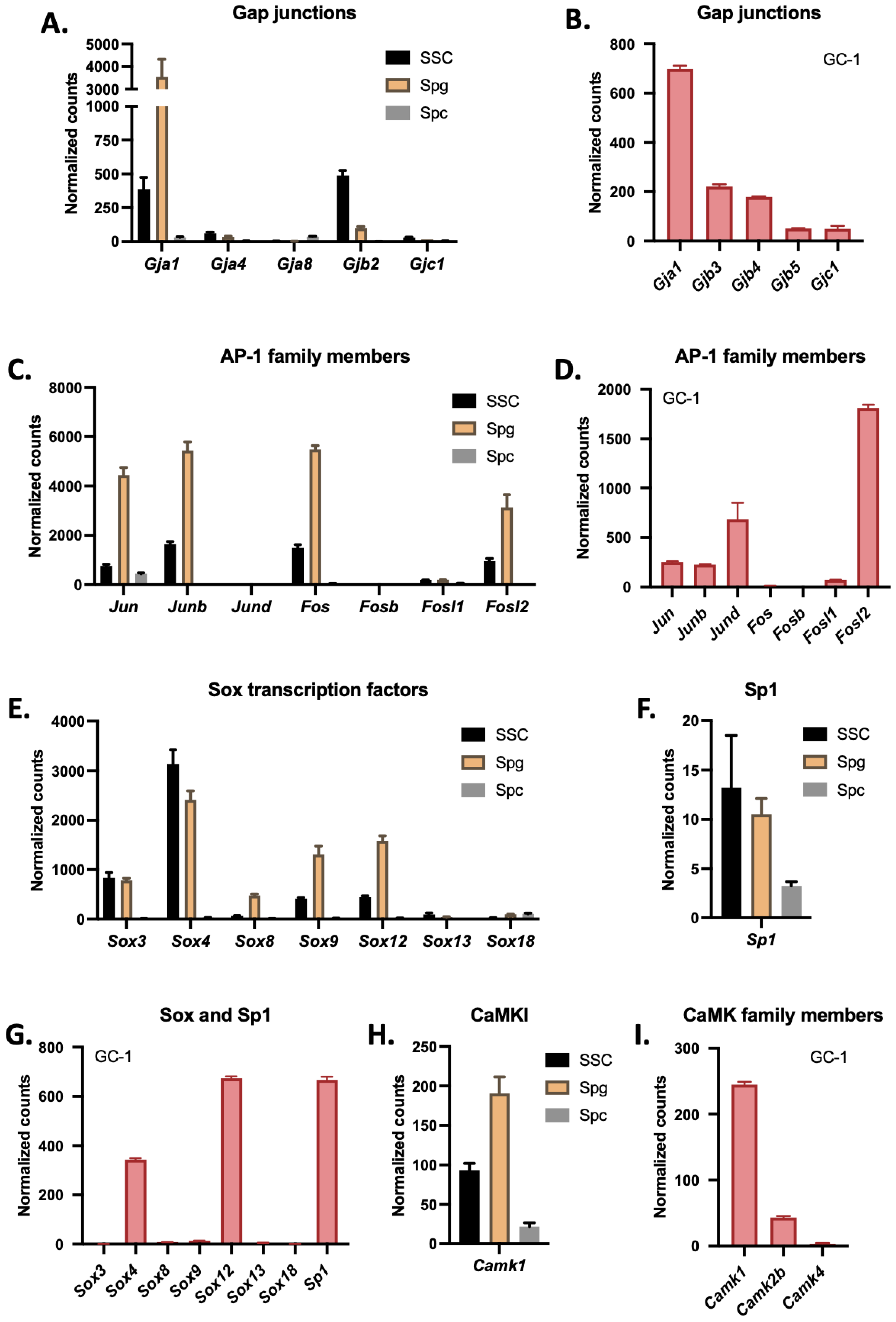
3.1. Gja1 Is the Most Highly Expressed Connexin in Spermatogonia and GC-1 Cells

Based on previously published RNA-Seq data (SRA: PRJNA523810 and PRJNA491649) [21,26], expression levels for different connexins, AP-1 family members and CaMKs are presented in spermatogonial stem cells, spermatogonia, spermatocytes and GC-1 cells (Figure 1). Interestingly, Gja1 is the most highly expressed connexin in spermatogonia (Figure 1A) and GC-1 cells (Figure 1B). Among transcription factors potentially involved in the regulation of Gja1 expression, AP-1 family members Jun, Junb, Fos and Fosl2 are highly expressed in spermatogonia (Figure 1C), whereas Jun, Junb, Jund and Fosl2 are predominantly expressed in GC-1 cells (Figure 1D). For Sox family members, Sox3, Sox4, Sox8, Sox9 and Sox12 are highly expressed in spermatogonia (Figure 1E), but only Sox4 and Sox12 are expressed in GC-1 cells (Figure 1G). Although involved in the regulation of Gja1 in other cell types, Sp1 is weakly expressed in spermatogonia (Figure 1F), whereas it is highly abundant in GC-1 cells (Figure 1G). Being a potential regulator of Gja1 expression, CaMKI, encoding the calcium/calmodulin-dependent protein kinase I, is highly expressed in spermatogonia (Figure 1H) and GC-1 cells (Figure 1I).

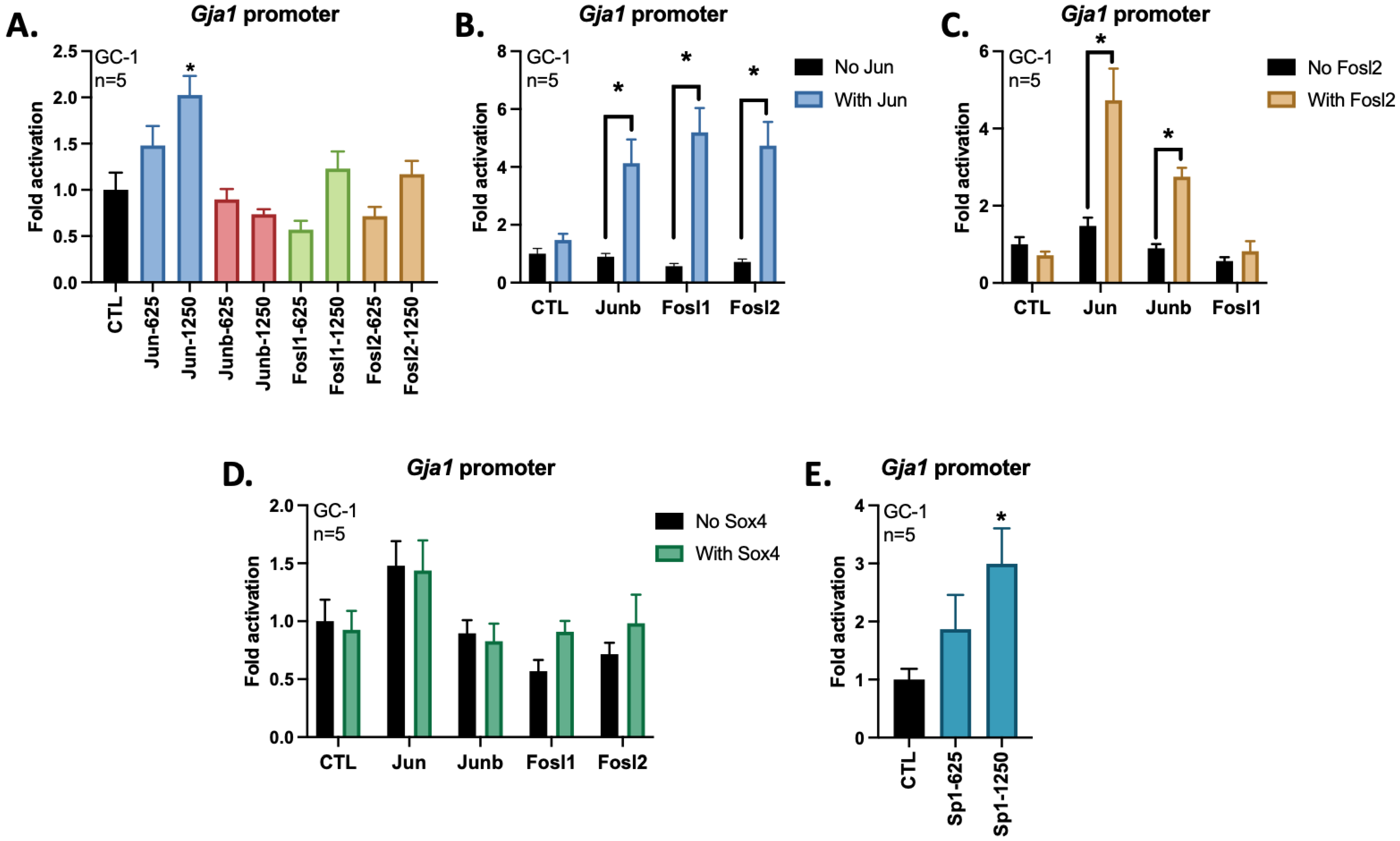
3.2. AP-1 Members Jun and Fosl2 Cooperate to Activate the Gja1 Promoter in GC-1 Cells

To determine if members of the AP-1 family of transcription factors are involved in the regulation of Gja1 promoter activity, we performed co-transfections with increasing concentrations of Jun, Junb, Fosl1 or Fosl2 expression vectors along with the mouse −1697 to +72 bp Gja1 promoter/Firefly luciferase reporter construct (Figure 2A). Interestingly, only Jun at an expression plasmid concentration of 1250 ng/mL was able to activate the Gja1 promoter. However, when combined with other AP-1 members, such as Junb, Fosl1 or Fosl2, such Jun-dependent activation of the Gja1 promoter was increased to 4–5 folds (Figure 2B). To better define the implication of Fosl2 in the regulation of Gja1 promoter activity and heterodimer formation, expression plasmids for AP-1 members, such as Jun, Junb and Fosl1, were combined with Fosl2. Interestingly, it is the overexpressions of Jun and Fosl2 that resulted in greater activation of the Gja1 promoter in GC-1 cells (Figure 2C). Since the gene encoding Sox4 is highly expressed in spermatogonia and GC-1 cells, we investigated if this transcription factor could also cooperate with AP-1 members to activate the Gja1 promoter as previously reported in Sertoli cells [14]. However, overexpression of Sox4 has no effect on Gja1 promoter activation in GC-1 cells (Figure 2D). As previously reported in myometrial cells [30,31], Sp1 can activate the Gja1 promoter in GC-1 cells (Figure 2E).

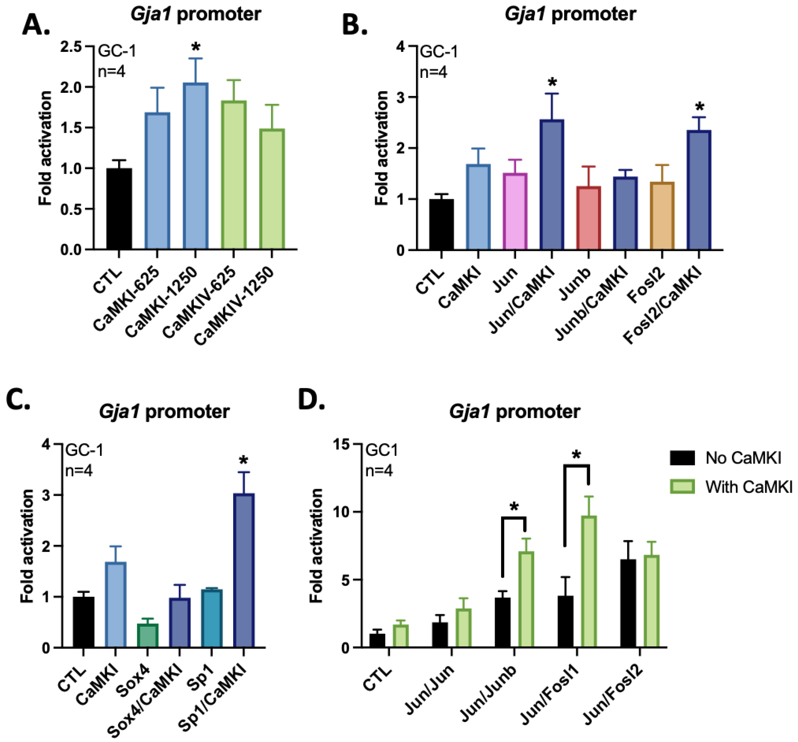
3.3. CaMKI Activates the Gja1 Promoter in GC-1 Cells

Since CaMKI is highly expressed in spermatogonia and GC-1 cells, we evaluated if a constitutively active form of this kinase could contribute to AP-1-dependent activation of Gja1 expression. Indeed, CaMKI overexpressed via transfection activates the Gja1 promoter (Figure 3A). Furthermore, CaMKI cooperates with Jun and Fosl2, but not with Junb, to activate the Gja1 promoter in GC-1 cells (Figure 3B). CaMKI was also able to cooperate with Sp1, but not with Sox4, to increase Gja1 promoter activity (Figure 3C). Based on previously reported cooperations between AP-1 members to activate the Gja1 promoter (Figure 2), we also investigated if overexpression of constitutively active CaMKI could further increase such activation. Interestingly, the cooperations between Jun and Junb or Fosl1 to activate the Gja1 promoter were further increased by CaMKI (Figure 3D).

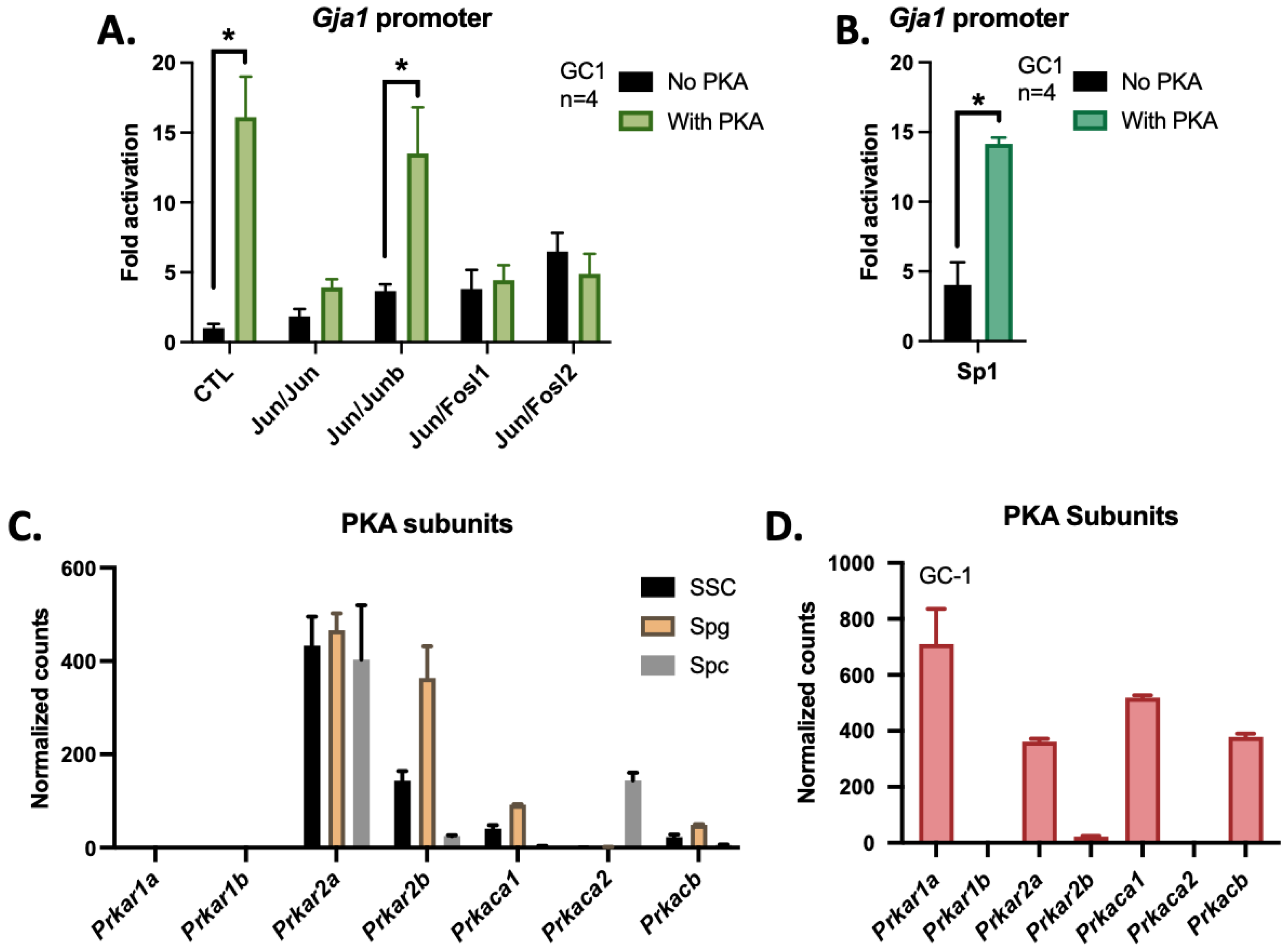
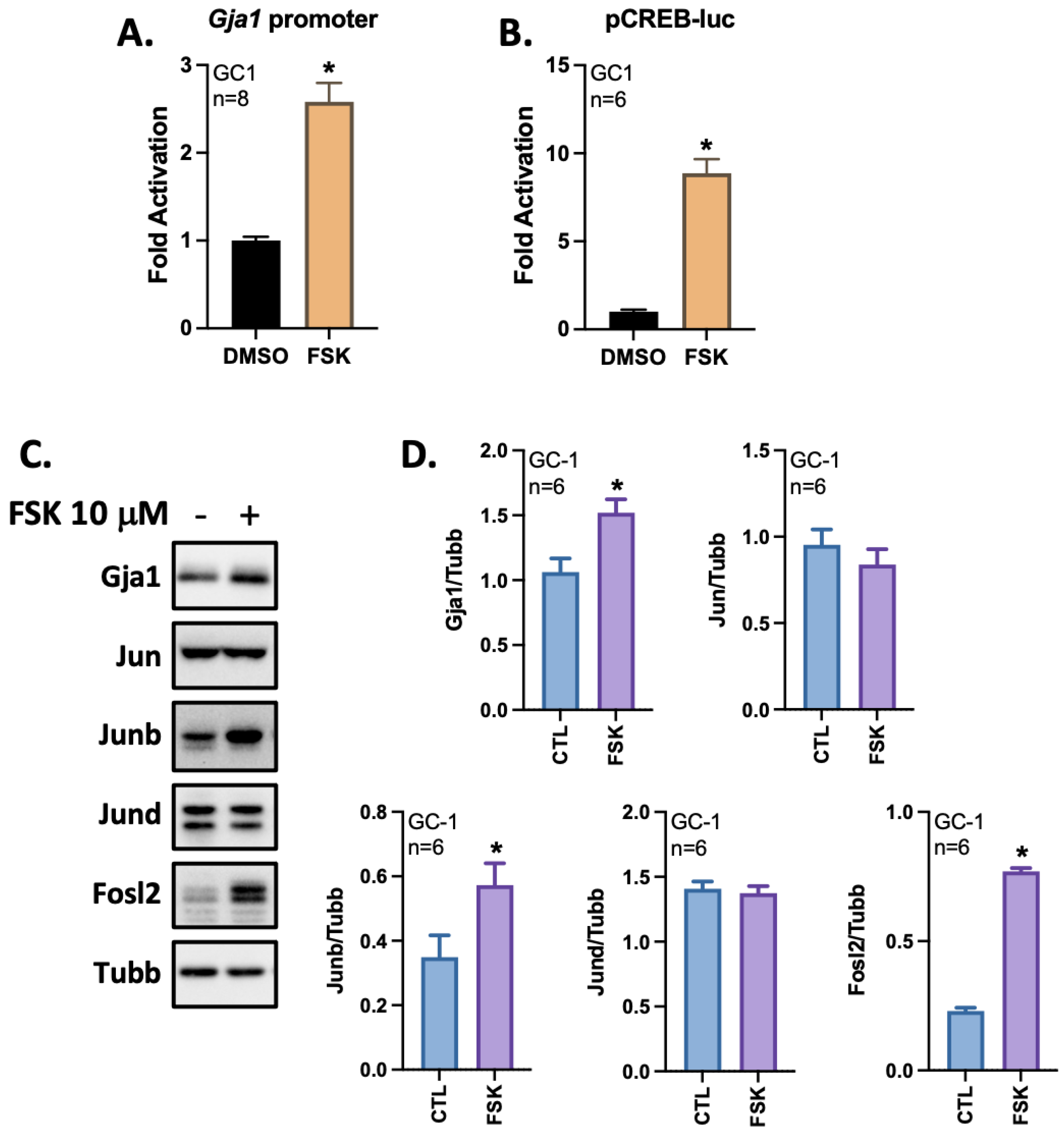
3.4. The cAMP/PKA Pathway Activates the Gja1 Promoter in GC-1 Cells

Since Jun can be activated by PKA [32], we investigated if overexpression of constitutively active PKA enzyme could influence Gja1 promoter activity. Interestingly, PKA overexpression alone upregulated the activity of the Gja1 promoter by 15.5 folds (Figure 4A). In addition, the activation of the Gja1 promoter by PKA was maintained by the cooperation between Jun and Junb in GC-1 cells (Figure 4A). In addition, PKA overexpression increased Sp1-dependent activation of the Gja1 promoter (Figure 4B). According to previously published RNA-Seq data (SRA: PRJNA523810 and PRJNA491649) [21,26], PKA subunits Prkar2a and Prkaca were highly expressed in spermatogonia (Figure 4C) and GC-1 cells (Figure 4D). Hence, these subunits may participate in the assembly of PKA in spermatogonial cells. Interestingly, the testis-specific Prkaca variant (Prkaca2) is highly expressed in spermatocytes as reported previously [33].
Since the transcriptional activities of AP-1 members can be modulated by activation of the cAMP/PKA pathway, we evaluated the ability of forskolin, an activator of adenylate cyclase, to regulate Gja1 expression in GC-1 cells. Interestingly, treatments with forskolin 10 µM for 8 h resulted in the activation of the Gja1 promoter in GC-1 cells (Figure 5A). Use of reporter plasmid pCREB-luc harboring CREB regulatory elements upstream of the Firefly luciferase served as a positive control for activation of the cAMP/PKA pathway by forskolin, leading to phosphorylation of Creb1 in GC-1 cells (Figure 5B). To better assess the effects of forskolin on the regulation of Gja1, GC-1 cells were treated, followed by total protein extractions and quantification of protein levels using Western blots. Consistent with the increase in Gja1 promoter activity in response to forskolin, Gja1 protein levels are also increased under these same treatment conditions (Figure 5C,D). In addition, the protein levels of Junb and Fosl2 are increased in response to forskolin (Figure 5C,D), suggesting that the increase of these AP-1 members by forskolin contributes, in part, to the activation of Gja1 expression in GC-1 cells.

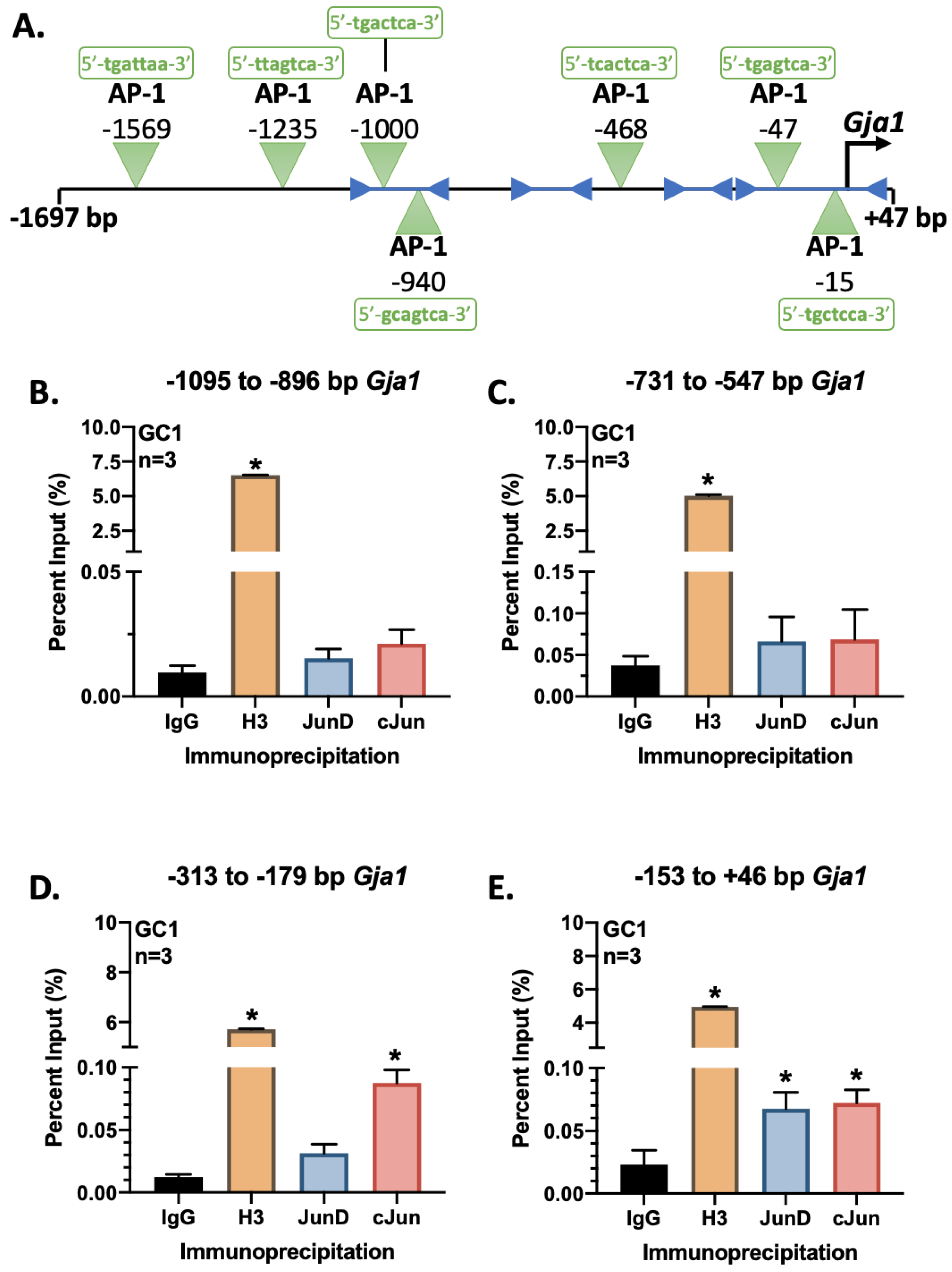
3.5. Jun Is Recruited to the Proximal Region of the Gja1 Promoter

Different potential AP-1 DNA regulatory elements along the Gja1 promoter have been reported previously (Figure 6A) [13]. To better define which of these elements are involved in Gja1 promoter activation by AP-1 members in GC-1 cells, chromatin immunoprecipitation analyses were performed using antibodies against Jund and Jun followed by qPCR quantification. Interestingly, Jun protein-DNA interactions are enriched for the −313 to −179 bp and −153 to +46 bp regions of the Gja1 promoter (Figure 6D,E). Although Jund is being recruited to the −153 to +46 bp region of the Gja1 promoter, such interaction may be attributed to the high levels of expression of Jund in GC-1 cells and may not be relevant as this AP-1 member is not expressed in normal spermatogonia cells.

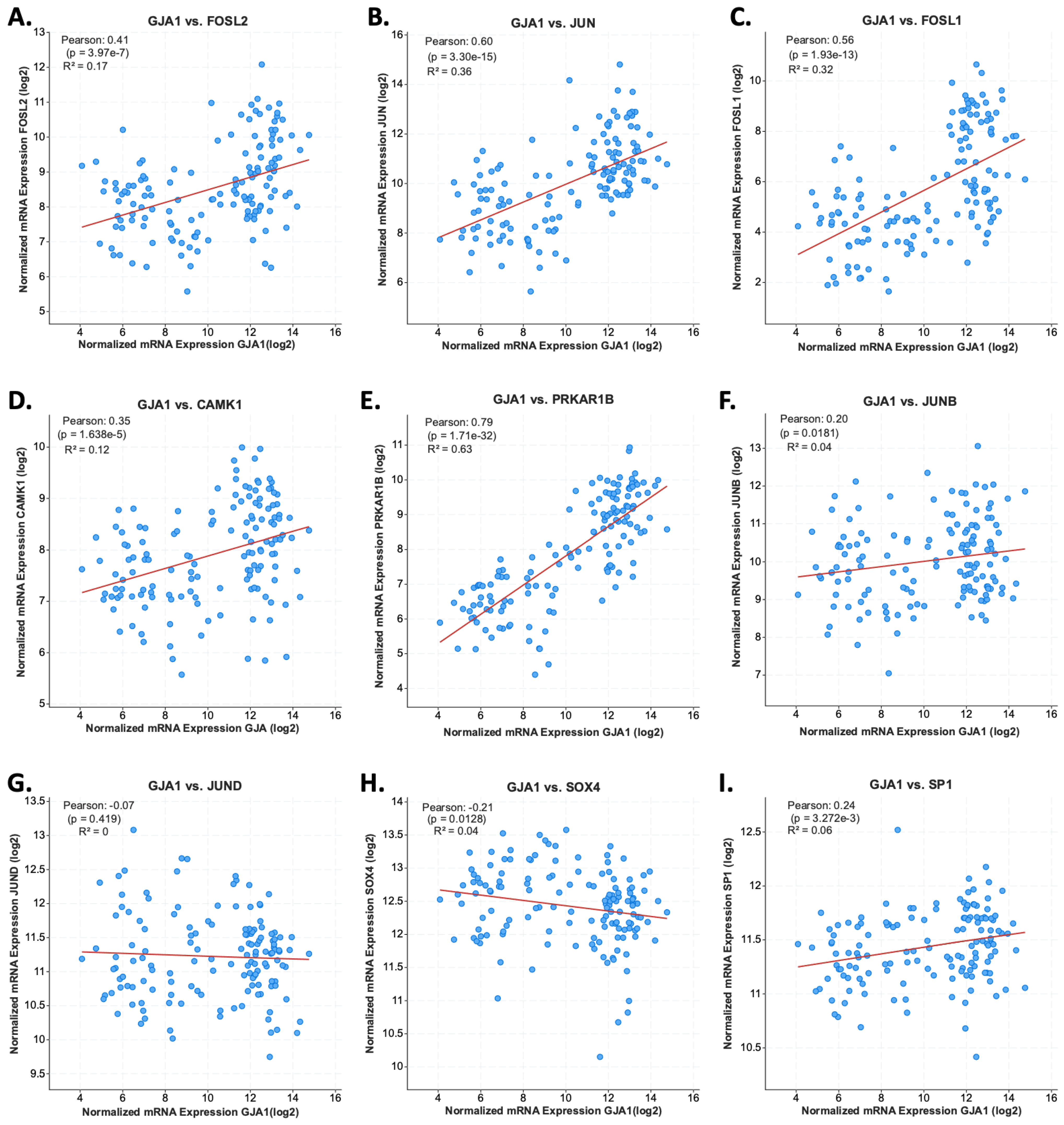
3.6. JUN and FOSL2 Are Correlated with GJA1 Expression in Testicular Germ Cells Tumors

To investigate whether FOSL2 and JUN participate in the regulation of GJA1 in TGCT, we investigated for correlations between gene expressions by searching the TCGA—PanCancer Atlas database (https://portal.gdc.cancer.gov/, accessed on 19 December 2021) through the cBioPortal platform [34,35]. Interestingly, FOSL2 and JUN expressions are correlated with that of GJA1 in the transcriptomes of TGCT (Figure 7A,B). Moreover, the expression of FOSL1, also capable of cooperating with Jun, correlates with that of GJA1 (Figure 7C). Furthermore, the expression of CAMKI, being able to enhance Jun/Fosl1-dependent activation of Gja1 promoter, correlates with that of GJA1 (Figure 7D). Interestingly, the expression of the gene encoding the subunit PRKAR1B highly correlates with GJA1 expression in TGCT (Figure 7E). However, the expressions of AP-1 members JUNB (Figure 7F) and JUND (Figure 7G), as well as that of SOX4 (Figure 7H) and SP1 (Figure 7I), do not correlate with GJA1 expression.

4. Discussion

According to RNA-Seq data, AP-1 members Jun, Junb, Jund and Fosl2 are the most expressed in GC-1 cells, whereas Jun, Junb, Fos and Fosl2 are highly expressed in spermatogonial cells. Hence, the GC-1 cell line is an excellent model to evaluate the abilities of AP-1 members Jun, Junb and Fosl2, commonly abundant in spermatogonia and GC-1 cells, to regulate Gja1 expression. For Jun and Junb, their expression levels are low in intact testes and much higher in isolated cells [36], suggesting that disruption of cell-to-cell contact increases their abundance in isolated spermatogonia. Here, we show that Jun and Junb cooperate with Fosl2 to activate the Gja1 promoter in GC-1 cells. While AP-1 family members are important regulators of Gja1 expression in different cell types [13,37], the combinations of these transcription factors vary in a cell-dependent manner. Indeed, we show that Jun and Fosl2 are important regulators of Gja1 in GC-1 spermatogonial cells, while Jun and Fos are more involved in the regulation of Gja1 in somatic cells of the testis [13]. Precisely, we and others have demonstrated that Gja1, being highly expressed in Leydig and Sertoli cells, is regulated by Jun and Fos [13].
According to our ChIP results, the proximal region of the Gja1 promoter is required for AP-1-dependent activation in spermatogonia. Although Jund is not expressed in germ cells, we showed that it is being recruited to the proximal region of the Gja1 promoter by ChIP assay on GC-1 cells. Interestingly, Jund knockout mice have an impaired spermatogenesis linked to an important reduction in Gja1 expression in seminiferous tubules [2]. Hence, Jund-dependent regulation of Gja1 may be relevant for other cell populations having high levels of this transcription factor such as Sertoli and Leydig cells (our unpublished data). In our ChIP assays, the recruitments of Junb and Fosl2 to the proximal region of the Gja1 promoter could not be observed, as opposed to Jun. This may be attributed to an inability of immunoprecipitations of Junb or Fosl2 by their respective antibodies due to the inaccessibility of the epitopes. Indeed, Junb and/or Fosl2 may be hidden in a major complex involving AP-1 members, cofactors and other transcription factors such as Sp1. It is noteworthy that both 32- and 35-kDa isoforms of Fosl2 are equivalently expressed in GC-1 cells. The 32 kDa isoform has a substitution of amino acids 1–33 of the N-terminus with MSFSLF amino acids. The impact of this substitution on the transcriptional regulatory function of Fosl2 remains to be elucidated.
Although Sp1 is highly expressed in GC-1 cells, this transcription factor is rather weakly expressed in germ cells. As previously reported in human myometrial cells [30], the Sp1 factor can regulate Gja1 promoter activity in GC-1 cells. Putative Sp1 regulatory elements have been characterized between −77 to −69 and −59 to −48 bp of the human GJA1 promoter [37]. Thus, this transcription factor may be an important regulator of GJA1 expression in certain types of TGCT. However, the expression of SP1 is not correlated with GJA1 in TGCT according to TCGA PanCancer Atlas data [38].
Sox4 and Sox12 are highly expressed in spermatogonia and GC-1 cells. Unlike what has been reported in Sertoli cells [14], Sox4 does not cooperate with AP-1 members to activate the Gja1 promoter in GC-1 cells. In addition, SOX4 and SOX12 are not correlated with GJA1 in TGCT according to TCGA PanCancer Atlas data [38]. However, regulation of GJA1 expression in TGCT may involve other members of the SOX family. Indeed, SOX8 and SOX9, known to regulate Gja1 expression in Sertoli cell lines [14], show correlated expressions with that of GJA1 in TGCT (p < 1.04 × 10⁻⁶).
CaMKI is a serine/threonine kinase highly expressed in spermatogonia and GC-1 cells. In addition, CaMKI can activate Gja1 expression. However, the cooperation between Jun and Fosl2 to regulate Gja1 promoter activity seems to be independent of CaMKI. Indeed, it is rather the cooperation between Jun and Junb or Fosl1 to activate the Gja1 promoter that is enhanced by CaMKI. Interestingly, Jun has a potential CaMKI phosphorylation site at S295 in mouse and S292 in human, whereas Fosl2 contains seven potential CaMKI phosphorylation sites in mouse and in human (S16, S17, S19, T149, S308, S309, S310) as reported using GPSv5.0 [39]. Thus, the activity of these AP-1 family members may be regulated by CaMKI and contribute to activate Gja1 expression in other cell types. Importantly, the increase in Gja1 promoter activation by Jun and Fosl1 in the presence of constitutively activated CaMKI is not relevant for spermatogonia because Fosl1 is not expressed in these cells. However, four potential CaMKI phosphorylation sites (S254, S255, S256, S257) can be identified in mouse Fosl1 using GPSv5.0 [39], suggesting that the regulation of Fosl1 activity by CaMKI, leading to regulation of Gja1 expression, may be important in other cell types. In addition, the expression of FOSL1 is correlated with that of GJA1 in TGCT. CaMKI phosphorylates a variety of substrates, including CREB [40]. Hence, regulation of Gja1 expression by CaMKI in GC-1 cells, and possibly TGCT, may involve phosphorylation of CREB1 and its recruitment to a potential CRE regulatory element within the proximal region of the Gja1 promoter. Indeed, CREB1 and its co-activator CBP are being recruited to the Gja1 promoter in rat ovarian granulosa cells [41]. In addition, CREB1 can also regulate Gja1 promoter activity indirectly by activating the expressions of Jun [42] and possibly Fosl2. However, the confirmation of such a regulatory mechanism of Gja1 expression by CaMKI phosphorylation of CREB1 will require further investigation. CaMKI can also participate in the activation of extracellular signal-regulated kinase (ERK) [43]. Interestingly, activated ERK1/2 can phosphorylate Jun and Fosl2, influencing their transcriptional activation capabilities [44,45,46,47].
As suggested by forskolin treatments, activations of adenylate cyclase and of the cAMP/PKA pathway leads to increased Gja1 promoter activity, resulting in increased protein levels. Although PKA contributes to increased activation of the Gja1 promoter through cooperation between Jun and Junb, such activation does not exceed that by PKA alone. However, forskolin-dependent increase in Gja1 protein levels may be attributed to increased Fosl2 protein levels and activation of other signaling pathways dependent on membrane calcium influx [48,49]. Although treatment of GC-1 cells with forskolin leads only to increases in Junb and Fosl2 protein levels, the activity of other AP-1 members could be increased via phosphorylation. Moreover, the cAMP/PKA signaling pathway could contribute to the activation and recruitment of a transcription factor complex involved in the activation of the Gja1 promoter as suggested by an increase in Gja1 expression in response to forskolin. Additionally, the Ras signaling pathway, having AP-1 members among its downstream targets, has been reported to be involved in the activation of the human GJA1 promoter in NIH3T3 cells [50] and could be relevant in the regulation of this connexin in TGCT.
Using mouse transgenic models, Gja1 has been reported to play an essential role during spermatogenesis, especially for the stage of primary spermatocytes differentiation [51]. Germ cell-specific Gja1 knockout mice have been developed to assess the importance of this connexin in spermatogenesis [8]. Surprisingly, inactivation of Gja1 in germ cells has no effect on spermatogenesis and the animals are fertile, suggesting that the critical function of Gja1 is probably related to the differentiation of Sertoli cells and interactions between these cells, rather than interactions with developing germ cells. However, the reduced expression of Gja1 in germ cells has been associated with increased risks of testicular cancer development. Indeed, this tumor suppressor gene is downregulated during development of testicular seminoma [12]. Importantly, the lack of infertility phenotype in germ cell-specific Gja1 knockout mice suggests compensation by the expression of other connexins, such as Gjb2 (Cx25) or Gjc1 (Cx45) [8]. Indeed, Gjc1 is expressed in peritubular, Sertoli and germ cells [52] and its protein levels are increased in testes of germ cell-specific Gja1 knockout mice [8]. Additionally, the transfer of molecules through gap junctions is selective and unidirectional from Sertoli cells to spermatogonia [53,54,55]. Since spermatogenesis is compromised in Sertoli cell-specific Gja1 knockout mice, whereas it is preserved in germ cell-specific Gja1 knockout mice [4,5,8,56], it is most likely that the connexons of spermatogonia are heterotypic in nature [56,57].
TGCT are the most common cancer in young men. Indeed, more than 90% of all testicular cancers are germ cell tumors. This type of cancer develops from transformed gonocytes or undifferentiated spermatogonia. Seminomas generally have a good prognosis, while non-seminomatous tumors are much more likely to present with metastases and mixed germ cell tumors composition. Interestingly, GJA1 has been proposed as a potential diagnostic and prognostic marker for TGCT [58]. Although GJA1 is expressed in seminoma, its protein localization is aberrantly detected in the Golgi apparatus and has a molecular weight of 70 kDa [10], suggesting that its protein maturation is being compromised. Overall, connexins have been considered tumor-suppressor genes, and pharmacological up-regulation of their expression has therapeutic implication in cancer treatments [59]. In this study, we show that Jun and Fosl2 cooperate to activate Gja1 expression in GC-1 spermatogonial cells. In addition, the expression of these AP-1 members is correlated to that of GJA1 in TGCT RNA-Seq data. Importantly, most of these samples have low levels of expression of GJA1. Hence, this gene could be a potential biological marker linked to the severity of TGCT development. However, the causal relationship between the loss of AP-1 members’ cooperation and the reduced expression of GJA1 in TGCT cannot be made solely based on the correlations. Such regulatory mechanism of GJA1 expression in TGCT will require further investigation.

5. Conclusions

Overall, the AP-1 members Jun and Fosl2 cooperate to activate Gja1 expression in GC-1 cells. In addition, the CaMKI and cAMP/PKA pathways upregulate the Gja1 promoter activity. Furthermore, the expressions of JUN, FOSL1, FOSL2 and CAMKI are correlated with that of GJA1 in TGCT. Therefore, the reduction in GJA1 expression in TGCT may involve loss of cooperation between AP-1 members.

Author Contributions

Conceptualization, L.J.M.; methodology, M.N. and L.J.M.; validation, M.N., H.T.N. and L.J.M.; formal analysis, L.J.M.; investigation, M.N., A.B., H.T.N., M.D. and L.J.M.; writing—original draft preparation, L.J.M.; writing—review and editing, M.N., H.T.N., M.D., A.B. and L.J.M.; visualization, L.J.M.; supervision, L.J.M.; project administration, L.J.M.; funding acquisition, L.J.M. All authors have read and agreed to the published version of the manuscript.

Funding

This research was funded by the Natural Sciences and Engineering Research Council (NSERC) of Canada, grant number 386557.

Institutional Review Board Statement

Not applicable.

Informed Consent Statement

Not applicable.

Data Availability Statement

The RNA-Seq data presented in this study are available publicly at: https://0-www-ncbi-nlm-nih-gov.brum.beds.ac.uk/bioproject/PRJNA491649 accessed on 19 December 2021, https://0-www-ncbi-nlm-nih-gov.brum.beds.ac.uk/bioproject/PRJNA523810 accessed on 19 December 2021, and https://portal.gdc.cancer.gov accessed on 19 December 2021.

Acknowledgments

We would like to thank Dany Chalbos (Institut National de la Santé et de la Recherche Médicale, Endocrinologie Moléculaire et Cellulaire des Cancers, Montpellier, France), Thomas Soderling (Oregon Health Sciences University, Portland, OR, USA) and Robert Viger (CHUQ Research Centre, Laval University, QC, Canada) for providing expression plasmids used in this study.

Conflicts of Interest

The authors declare no conflict of interest. The funders had no role in the design of the study; in the collection, analyses, or interpretation of data; in the writing of the manuscript, or in the decision to publish the results.

References

  1. Risley, M.S.; Tan, I.P.; Roy, C.; Sáez, J.C. Cell-, Age- and Stage-Dependent Distribution of Connexin43 Gap Junctions in Testes. J. Cell. Sci. 1992, 103, 81–96. [Google Scholar] [CrossRef] [PubMed]
  2. Batias, C.; Defamie, N.; Lablack, A.; Thepot, D.; Fenichel, P.; Segretain, D.; Pointis, G. Modified Expression of Testicular Gap-Junction Connexin 43 during Normal Spermatogenic Cycle and in Altered Spermatogenesis. Cell Tissue Res. 1999, 298, 113–121. [Google Scholar] [CrossRef] [PubMed]
  3. Juneja, S.C.; Barr, K.J.; Enders, G.C.; Kidder, G.M. Defects in the Germ Line and Gonads of Mice Lacking Connexin43. Biol. Reprod. 1999, 60, 1263–1270. [Google Scholar] [CrossRef] [PubMed]
  4. Brehm, R.; Zeiler, M.; Rüttinger, C.; Herde, K.; Kibschull, M.; Winterhager, E.; Willecke, K.; Guillou, F.; Lécureuil, C.; Steger, K.; et al. A Sertoli Cell-Specific Knockout of Connexin43 Prevents Initiation of Spermatogenesis. Am. J. Pathol. 2007, 171, 19–31. [Google Scholar] [CrossRef] [PubMed] [Green Version]
  5. Sridharan, S.; Simon, L.; Meling, D.D.; Cyr, D.G.; Gutstein, D.E.; Fishman, G.I.; Guillou, F.; Cooke, P.S. Proliferation of Adult Sertoli Cells Following Conditional Knockout of the Gap Junctional Protein GJA1 (Connexin 43) in Mice. Biol. Reprod. 2007, 76, 804–812. [Google Scholar] [CrossRef] [Green Version]
  6. Carette, D.; Weider, K.; Gilleron, J.; Giese, S.; Dompierre, J.; Bergmann, M.; Brehm, R.; Denizot, J.-P.; Segretain, D.; Pointis, G. Major Involvement of Connexin 43 in Seminiferous Epithelial Junction Dynamics and Male Fertility. Dev. Biol. 2010, 346, 54–67. [Google Scholar] [CrossRef]
  7. Noelke, J.; Wistuba, J.; Damm, O.S.; Fietz, D.; Gerber, J.; Gaehle, M.; Brehm, R. A Sertoli Cell-Specific Connexin43 Knockout Leads to Altered Interstitial Connexin Expression and Increased Leydig Cell Numbers. Cell Tissue Res. 2015, 361, 633–644. [Google Scholar] [CrossRef]
  8. Günther, S.; Fietz, D.; Weider, K.; Bergmann, M.; Brehm, R. Effects of a Murine Germ Cell-Specific Knockout of Connexin 43 on Connexin Expression in Testis and Fertility. Transgenic Res. 2013, 22, 631–641. [Google Scholar] [CrossRef]
  9. Saez, J.C.; Berthoud, V.M.; Branes, M.C.; Martinez, A.D.; Beyer, E.C. Plasma Membrane Channels Formed by Connexins: Their Regulation and Functions. Physiol. Rev. 2003, 83, 1359–1400. [Google Scholar] [CrossRef] [Green Version]
  10. Roger, C.; Mograbi, B.; Chevallier, D.; Michiels, J.F.; Tanaka, H.; Segretain, D.; Pointis, G.; Fenichel, P. Disrupted Traffic of Connexin 43 in Human Testicular Seminoma Cells: Overexpression of Cx43 Induces Membrane Location and Cell Proliferation Decrease. J. Pathol. 2004, 202, 241–246. [Google Scholar] [CrossRef]
  11. Defamie, N.; Berthaut, I.; Mograbi, B.; Chevallier, D.; Dadoune, J.-P.; Fénichel, P.; Segretain, D.; Pointis, G. Impaired Gap Junction Connexin43 in Sertoli Cells of Patients with Secretory Azoospermia: A Marker of Undifferentiated Sertoli Cells. Lab. Investig. 2003, 83, 449–456. [Google Scholar] [CrossRef] [PubMed] [Green Version]
  12. Brehm, R.; Rüttinger, C.; Fischer, P.; Gashaw, I.; Winterhager, E.; Kliesch, S.; Bohle, R.M.; Steger, K.; Bergmann, M. Transition from Preinvasive Carcinoma in Situ to Seminoma Is Accompanied by a Reduction of Connexin 43 Expression in Sertoli Cells and Germ Cells. Neoplasia 2006, 8, 499–509. [Google Scholar] [CrossRef] [PubMed] [Green Version]
  13. Ghouili, F.; Martin, L.J. Cooperative Regulation of Gja1 Expression by Members of the AP-1 Family CJun and CFos in TM3 Leydig and TM4 Sertoli Cells. Gene 2017, 635, 24–32. [Google Scholar] [CrossRef] [PubMed]
  14. Ghouili, F.; Roumaud, P.; Martin, L.J. Gja1 Expression Is Regulated by Cooperation between SOX8/SOX9 and CJUN Transcription Factors in TM4 and 15P-1 Sertoli Cell Lines. Mol. Reprod. Dev. 2018, 85, 875–886. [Google Scholar] [CrossRef]
  15. Tratner, I.; Ofir, R.; Verma, I.M. Alteration of a Cyclic AMP-Dependent Protein Kinase Phosphorylation Site in the c-Fos Protein Augments Its Transforming Potential. Mol. Cell. Biol. 1992, 12, 998–1006. [Google Scholar] [CrossRef] [Green Version]
  16. Teyssier, C.; Belguise, K.; Galtier, F.; Chalbos, D. Characterization of the Physical Interaction between Estrogen Receptor Alpha and JUN Proteins. J. Biol. Chem. 2001, 276, 36361–36369. [Google Scholar] [CrossRef] [Green Version]
  17. Wayman, G.A.; Kaech, S.; Grant, W.F.; Davare, M.; Impey, S.; Tokumitsu, H.; Nozaki, N.; Banker, G.; Soderling, T.R. Regulation of Axonal Extension and Growth Cone Motility by Calmodulin-Dependent Protein Kinase I. J. Neurosci. 2004, 24, 3786–3794. [Google Scholar] [CrossRef]
  18. Tremblay, J.J.; Hamel, F.; Viger, R.S. Protein Kinase A-Dependent Cooperation between GATA and CCAAT/Enhancer-Binding Protein Transcription Factors Regulates Steroidogenic Acute Regulatory Protein Promoter Activity. Endocrinology 2002, 143, 3935–3945. [Google Scholar] [CrossRef] [Green Version]
  19. Hofmann, M.C.; Narisawa, S.; Hess, R.A.; Millán, J.L. Immortalization of Germ Cells and Somatic Testicular Cells Using the SV40 Large T Antigen. Exp. Cell Res. 1992, 201, 417–435. [Google Scholar] [CrossRef]
  20. Boussif, O.; Lezoualc’h, F.; Zanta, M.A.; Mergny, M.D.; Scherman, D.; Demeneix, B.; Behr, J.P. A Versatile Vector for Gene and Oligonucleotide Transfer into Cells in Culture and in Vivo: Polyethylenimine. Proc. Natl. Acad. Sci. USA 1995, 92, 7297–7301. [Google Scholar] [CrossRef] [Green Version]
  21. Zagore, L.L.; Sweet, T.J.; Hannigan, M.M.; Weyn-Vanhentenryck, S.M.; Jobava, R.; Hatzoglou, M.; Zhang, C.; Licatalosi, D.D. DAZL Regulates Germ Cell Survival through a Network of PolyA-Proximal MRNA Interactions. Cell Rep. 2018, 25, 1225–1240.e6. [Google Scholar] [CrossRef] [PubMed] [Green Version]
  22. Bolger, A.M.; Lohse, M.; Usadel, B. Trimmomatic: A Flexible Trimmer for Illumina Sequence Data. Bioinformatics 2014, 30, 2114–2120. [Google Scholar] [CrossRef] [PubMed] [Green Version]
  23. Dobin, A.; Davis, C.A.; Schlesinger, F.; Drenkow, J.; Zaleski, C.; Jha, S.; Batut, P.; Chaisson, M.; Gingeras, T.R. STAR: Ultrafast Universal RNA-Seq Aligner. Bioinformatics 2013, 29, 15–21. [Google Scholar] [CrossRef] [PubMed]
  24. Pertea, M.; Pertea, G.M.; Antonescu, C.M.; Chang, T.-C.; Mendell, J.T.; Salzberg, S.L. StringTie Enables Improved Reconstruction of a Transcriptome from RNA-Seq Reads. Nat. Biotechnol. 2015, 33, 290–295. [Google Scholar] [CrossRef] [PubMed] [Green Version]
  25. Love, M.I.; Huber, W.; Anders, S. Moderated Estimation of Fold Change and Dispersion for RNA-Seq Data with DESeq2. Genome Biol. 2014, 15, 550. [Google Scholar] [CrossRef] [PubMed] [Green Version]
  26. Chen, J.; Gao, C.; Lin, X.; Ning, Y.; He, W.; Zheng, C.; Zhang, D.; Yan, L.; Jiang, B.; Zhao, Y.; et al. The MicroRNA MiR-202 Prevents Precocious Spermatogonial Differentiation and Meiotic Initiation during Mouse Spermatogenesis. Development 2021, 148, dev199799. [Google Scholar] [CrossRef]
  27. Kim, D.; Langmead, B.; Salzberg, S.L. HISAT: A Fast Spliced Aligner with Low Memory Requirements. Nat. Methods 2015, 12, 357–360. [Google Scholar] [CrossRef] [Green Version]
  28. Liao, Y.; Smyth, G.K.; Shi, W. FeatureCounts: An Efficient General Purpose Program for Assigning Sequence Reads to Genomic Features. Bioinformatics 2014, 30, 923–930. [Google Scholar] [CrossRef] [Green Version]
  29. Bradford, M.M. A Rapid and Sensitive Method for the Quantitation of Microgram Quantities of Protein Utilizing the Principle of Protein-Dye Binding. Anal. Biochem. 1976, 72, 248–254. [Google Scholar] [CrossRef]
  30. Echetebu, C.O.; Ali, M.; Izban, M.G.; MacKay, L.; Garfield, R.E. Localization of Regulatory Protein Binding Sites in the Proximal Region of Human Myometrial Connexin 43 Gene. Mol. Hum. Reprod. 1999, 5, 757–766. [Google Scholar] [CrossRef] [Green Version]
  31. Geimonen, E.; Boylston, E.; Royek, A.; Andersen, J. Elevated Connexin-43 Expression in Term Human Myometrium Correlates with Elevated c-Jun Expression and Is Independent of Myometrial Estrogen Receptors. J. Clin. Endocrinol. Metab. 1998, 83, 1177–1185. [Google Scholar] [CrossRef] [PubMed] [Green Version]
  32. de Groot, R.P.; Sassone-Corsi, P. Activation of Jun/AP-1 by Protein Kinase A. Oncogene 1992, 7, 2281–2286. [Google Scholar] [PubMed]
  33. Agustin, J.T.; Wilkerson, C.G.; Witman, G.B. The Unique Catalytic Subunit of Sperm CAMP-Dependent Protein Kinase Is the Product of an Alternative Calpha MRNA Expressed Specifically in Spermatogenic Cells. Mol. Biol. Cell 2000, 11, 3031–3044. [Google Scholar] [CrossRef] [PubMed] [Green Version]
  34. Cerami, E.; Gao, J.; Dogrusoz, U.; Gross, B.E.; Sumer, S.O.; Aksoy, B.A.; Jacobsen, A.; Byrne, C.J.; Heuer, M.L.; Larsson, E.; et al. The CBio Cancer Genomics Portal: An Open Platform for Exploring Multidimensional Cancer Genomics Data. Cancer Discov. 2012, 2, 401–404. [Google Scholar] [CrossRef] [Green Version]
  35. Gao, J.; Aksoy, B.A.; Dogrusoz, U.; Dresdner, G.; Gross, B.; Sumer, S.O.; Sun, Y.; Jacobsen, A.; Sinha, R.; Larsson, E.; et al. Integrative Analysis of Complex Cancer Genomics and Clinical Profiles Using the CBioPortal. Sci. Signal. 2013, 6, pl1. [Google Scholar] [CrossRef] [Green Version]
  36. Alcivar, A.A.; Hake, L.E.; Hardy, M.P.; Hecht, N.B. Increased Levels of JunB and C-Jun MRNAs in Male Germ Cells Following Testicular Cell Dissociation. Maximal Stimulation in Prepuberal Animals. J. Biol. Chem. 1990, 265, 20160–20165. [Google Scholar] [CrossRef]
  37. Geimonen, E.; Jiang, W.; Ali, M.; Fishman, G.I.; Garfield, R.E.; Andersen, J. Activation of Protein Kinase C in Human Uterine Smooth Muscle Induces Connexin-43 Gene Transcription through an AP-1 Site in the Promoter Sequence. J. Biol. Chem. 1996, 271, 23667–23674. [Google Scholar] [CrossRef] [Green Version]
  38. Hoadley, K.A.; Yau, C.; Hinoue, T.; Wolf, D.M.; Lazar, A.J.; Drill, E.; Shen, R.; Taylor, A.M.; Cherniack, A.D.; Thorsson, V.; et al. Cell-of-Origin Patterns Dominate the Molecular Classification of 10,000 Tumors from 33 Types of Cancer. Cell 2018, 173, 291–304.e6. [Google Scholar] [CrossRef] [Green Version]
  39. Wang, C.; Xu, H.; Lin, S.; Deng, W.; Zhou, J.; Zhang, Y.; Shi, Y.; Peng, D.; Xue, Y. GPS 5.0: An Update on the Prediction of Kinase-Specific Phosphorylation Sites in Proteins. Genom. Proteom. Bioinform. 2020, 18, 72–80. [Google Scholar] [CrossRef]
  40. Sheng, M.; Thompson, M.A.; Greenberg, M.E. CREB: A Ca2+-Regulated Transcription Factor Phosphorylated by Calmodulin-Dependent Kinases. Science 1991, 252, 1427–1430. [Google Scholar] [CrossRef]
  41. Fang, W.-L.; Lai, S.-Y.; Lai, W.-A.; Lee, M.-T.; Liao, C.-F.; Ke, F.-C.; Hwang, J.-J. CRTC2 and Nedd4 Ligase Involvement in FSH and TGFβ1 Upregulation of Connexin43 Gap Junction. J. Mol. Endocrinol. 2015, 55, 263–275. [Google Scholar] [CrossRef] [PubMed] [Green Version]
  42. Lamph, W.W.; Dwarki, V.J.; Ofir, R.; Montminy, M.; Verma, I.M. Negative and Positive Regulation by Transcription Factor CAMP Response Element-Binding Protein Is Modulated by Phosphorylation. Proc. Natl. Acad. Sci. USA 1990, 87, 4320–4324. [Google Scholar] [CrossRef] [PubMed] [Green Version]
  43. Schmitt, J.M.; Wayman, G.A.; Nozaki, N.; Soderling, T.R. Calcium Activation of ERK Mediated by Calmodulin Kinase I. J. Biol. Chem. 2004, 279, 24064–24072. [Google Scholar] [CrossRef] [Green Version]
  44. Monje, P.; Hernández-Losa, J.; Lyons, R.J.; Castellone, M.D.; Gutkind, J.S. Regulation of the Transcriptional Activity of C-Fos by ERK. A Novel Role for the Prolyl Isomerase PIN1. J. Biol. Chem. 2005, 280, 35081–35084. [Google Scholar] [CrossRef] [PubMed] [Green Version]
  45. Sánchez, I.; Hughes, R.T.; Mayer, B.J.; Yee, K.; Woodgett, J.R.; Avruch, J.; Kyriakis, J.M.; Zon, L.I. Role of SAPK/ERK Kinase-1 in the Stress-Activated Pathway Regulating Transcription Factor c-Jun. Nature 1994, 372, 794–798. [Google Scholar] [CrossRef]
  46. Chalmers, C.J.; Gilley, R.; March, H.N.; Balmanno, K.; Cook, S.J. The Duration of ERK1/2 Activity Determines the Activation of c-Fos and Fra-1 and the Composition and Quantitative Transcriptional Output of AP-1. Cell Signal. 2007, 19, 695–704. [Google Scholar] [CrossRef] [PubMed]
  47. Karin, M. The Regulation of AP-1 Activity by Mitogen-Activated Protein Kinases. J. Biol. Chem. 1995, 270, 16483–16486. [Google Scholar] [CrossRef] [Green Version]
  48. Yanagibashi, K.; Papadopoulos, V.; Masaki, E.; Iwaki, T.; Kawamura, M.; Hall, P.F. Forskolin Activates Voltage-Dependent Ca2+ Channels in Bovine but Not in Rat Fasciculata Cells. Endocrinology 1989, 124, 2383–2391. [Google Scholar] [CrossRef]
  49. Morita, K.; Dohi, T.; Kitayama, S.; Koyama, Y.; Tsujimoto, A. Stimulation-Evoked Ca2+ Fluxes in Cultured Bovine Adrenal Chromaffin Cells Are Enhanced by Forskolin. J. Neurochem. 1987, 48, 248–252. [Google Scholar] [CrossRef]
  50. Carystinos, G.D.; Kandouz, M.; Alaoui-Jamali, M.A.; Batist, G. Unexpected Induction of the Human Connexin 43 Promoter by the Ras Signaling Pathway Is Mediated by a Novel Putative Promoter Sequence. Mol. Pharmacol. 2003, 63, 821–831. [Google Scholar] [CrossRef] [Green Version]
  51. Winterhager, E.; Pielensticker, N.; Freyer, J.; Ghanem, A.; Schrickel, J.W.; Kim, J.-S.; Behr, R.; Grümmer, R.; Maass, K.; Urschel, S.; et al. Replacement of Connexin43 by Connexin26 in Transgenic Mice Leads to Dysfunctional Reproductive Organs and Slowed Ventricular Conduction in the Heart. BMC Dev. Biol. 2007, 7, 26. [Google Scholar] [CrossRef] [PubMed] [Green Version]
  52. Risley, M.S. Connexin Gene Expression in Seminiferous Tubules of the Sprague-Dawley Rat. Biol. Reprod. 2000, 62, 748–754. [Google Scholar] [CrossRef] [PubMed] [Green Version]
  53. Goldberg, G.S.; Valiunas, V.; Brink, P.R. Selective Permeability of Gap Junction Channels. Biochim. Biophys. Acta 2004, 1662, 96–101. [Google Scholar] [CrossRef] [Green Version]
  54. Decrouy, X.; Gasc, J.-M.; Pointis, G.; Segretain, D. Functional Characterization of Cx43 Based Gap Junctions during Spermatogenesis. J. Cell. Physiol. 2004, 200, 146–154. [Google Scholar] [CrossRef] [PubMed]
  55. Risley, M.S.; Tan, I.P.; Farrell, J. Gap Junctions with Varied Permeability Properties Establish Cell-Type Specific Communication Pathways in the Rat Seminiferous Epithelium. Biol. Reprod. 2002, 67, 945–952. [Google Scholar] [CrossRef] [Green Version]
  56. Rode, K.; Langeheine, M.; Seeger, B.; Brehm, R. Connexin43 in Germ Cells Seems to Be Dispensable for Murine Spermatogenesis. Int. J. Mol. Sci. 2021, 22, 7924. [Google Scholar] [CrossRef]
  57. Kidder, G.M.; Cyr, D.G. Roles of Connexins in Testis Development and Spermatogenesis. Semin. Cell Dev. Biol. 2016, 50, 22–30. [Google Scholar] [CrossRef]
  58. Chevallier, D.; Carette, D.; Segretain, D.; Gilleron, J.; Pointis, G. Connexin 43 a Check-Point Component of Cell Proliferation Implicated in a Wide Range of Human Testis Diseases. Cell. Mol. Life Sci. 2013, 70, 1207–1220. [Google Scholar] [CrossRef]
  59. Yamasaki, H.; Omori, Y.; Krutovskikh, V.; Zhu, W.; Mironov, N.; Yamakage, K.; Mesnil, M. Connexins in Tumour Suppression and Cancer Therapy. Novartis Found. Symp. 1999, 219, 241–254; discussion 254–260. [Google Scholar] [CrossRef]
Figure 1. Expression profiles of genes encoding members of the AP-1 family and gap junctions in mouse germ cells and GC-1 spermatogonia cells. Data was obtained from publicly available RNA-Seq data sets (PRJNA491649 and PRJNA523810) and analyzed for the determination of normalized counts. The expression profiles for genes encoding gap junctions (A,B), AP-1 family members (C,D), Sox members (E,G), Sp1 (F,G) and CaMKs family members (H,I) are presented. The numbers of biological replicates are three for SSC, two for Spg, three for Spc and six for GC-1 cells. Abbreviations: SSC, spermatogonial stem cells; Spg, spermatogonia; Spc, spermatocytes.
Applsci 12 01408 g001
Figure 2. Regulation of the activity of Gja1 promoter by AP-1 family members and Sp1 in GC-1 cells. GC-1 cells were transfected with the full-length (−1697 to +72 bp) Gja1 promoter/luciferase reporter plasmid in combination with different expression plasmids. (A) Increasing concentrations (625 and 1250 ng/mL) of Jun, Junb, Fosl1 and Fosl2 expression plasmids were evaluated for their effects on Gja1 promoter activity. (B) Jun cooperates with other AP-1 family members to activate the Gja1 promoter/reporter construct. (C) Fosl2 cooperates with Jun or Junb to activate the Gja1 promoter/reporter construct. (D) The transcription factor Sox4 has no effect on the activity of Gja1 promoter. (E) High levels of Sp1 expression plasmid (1250 ng/mL) activate the Gja1 promoter/reporter construct. Results are presented as means relative to the control (±SEM). The number of experiments, each performed in triplicate, is indicated (n). Statistics were performed using a two-way ANOVA followed by a Holm-Sidak’s multiple comparison test. Statistically significant differences are indicated by an asterisk (* p < 0.05).
Applsci 12 01408 g002
Figure 3. CaMK-dependent activation of the Gja1 promoter in mouse GC-1 cells. GC-1 cells were co-transfected with an empty expression vector (CTL) or expression vectors for constitutively active forms of CaMKI and CaMKIV along with the mouse Gja1 promoter/Firefly luciferase reporter plasmid. (A) Increasing concentrations of CaMKI or IV (625 or 1250 ng/mL) are co-transfected with the mouse Gja1 promoter/Firefly luciferase reporter plasmid. (B) Expression plasmids encoding Jun, Junb or Fosl2 were co-transfected with CaMKI to evaluate their potentials to activate the Gja1 promoter/Firefly luciferase reporter plasmid. (C) Expression plasmids encoding Sp1 or Sox4 were co-transfected with CaMKI to evaluate their potentials to activate the Gja1 promoter/Firefly luciferase reporter plasmid. In (D), GC-1 cells were co-transfected with an empty expression vector (CTL) or expression vectors for Jun and other AP-1 members in the absence or presence of an expression vector encoding a constitutively active form of CaMKI along with the mouse Gja1 promoter/Firefly luciferase reporter plasmid. The data is presented as fold activations compared to control (CTL) (±SEM). The number of experiments, each performed in triplicate, is indicated (n). Statistics were performed using a two-way ANOVA followed by a Holm-Sidak’s multiple comparison test. Statistically significant differences from control (CTL) are indicated by an asterisk (* p < 0.05).
Applsci 12 01408 g003
Figure 4. PKA promotes the activation the Gja1 promoter by AP-1 members in mouse GC-1 cells. In (A), GC-1 cells were co-transfected with an empty expression vector (CTL) or expression vectors for Jun and other AP-1 members in the absence or presence of an expression vector encoding the constitutively active form of PKA (catalytic α-subunit) along with the mouse Gja1 promoter/Firefly luciferase reporter plasmid. In (B), GC-1 cells were co-transfected with an empty expression vector (CTL) or expression vectors for Sp1 in the absence or presence of an expression vector encoding constitutively active form of PKA along with the mouse Gja1 promoter/Firefly luciferase reporter plasmid. Expression profiles of genes encoding PKA subunits in mouse germ cells (C) and GC-1 spermatogonia cells (D) were obtained from publicly available RNA-Seq data sets (PRJNA491649 and PRJNA523810). Prkaca1 and Prkaca2 are variant isoforms of the Prkaca transcript. The data is presented as fold activations compared to CTL (±SEM). The number of experiments, each performed in triplicate, is indicated (n). Statistics were performed using a two-way ANOVA followed by a Holm-Sidak’s multiple comparison test. Statistically significant differences are indicated by an asterisk (* p < 0.05). Abbreviations: SSC, spermatogonial stem cells; Spg, spermatogonia; Spc, spermatocytes.
Applsci 12 01408 g004
Figure 5. Influence of forskolin (FSK) on Gja1 promoter activity and proteins’ levels in GC-1 cells. GC-1 cells were transfected with the mouse Gja1 promoter/Firefly luciferase (A) or the pCREB-luc (B) reporter plasmid, followed by treatments with 10 µM FSK for 8 h. In (C), GC-1 cells were stimulated with 10 μM FSK for 8 h, followed by total protein extractions for detections of Gja1, Jun, Junb, Jund and Fosl2 using Western blots. The protein Tubulin-β (Tubb) was used as a loading control for total protein extracts. Associated normalized densitometries are presented in (D) and are compiled from four biological replicates. The number of experiments is indicated (n). Statistically significant differences from control (CTL) are indicated by an asterisk (* p < 0.05).
Applsci 12 01408 g005
Figure 6. Recruitment of AP-1 members to the proximal region of the mouse Gja1 promoter. In (A), the potential AP-1 DNA regulatory elements and amplified regions between blue arrows are identified on the Gja1 promoter. (BE) Chromatin immunoprecipitation (ChIP) assays for Jun and Jund recruitments on the Gja1 promoter were performed with GC-1 cells. Indicated Gja1 promoter regions were amplified via qPCR following ChIP assays. Recruitment using a normal rabbit IgG was used as a negative control. Histone H3 was used as a positive control for protein-DNA complexes. Results are presented as percentages of input (± SEM). The number of biological replicates is indicated (n). Statistical analyses were performed using an ANOVA, followed by a Holm-Sidak’s multiple comparisons test. Statistically significant differences from control (IgG) are indicated by an asterisk (* p < 0.05).
Applsci 12 01408 g006
Figure 7. Correlations between the expressions of GJA1, AP-1 members (AC,F,G), CAMKI (D), PRKAR1B (E), SOX4 (H) and SP1 (I) in TGCT. Normalized mRNA expression data was obtained from the TCGA—PanCancer Atlas database (https://portal.gdc.cancer.gov/, accessed on 19 December 2021) and analyzed for correlations between gene expressions using the cBioPortal platform [34,35].
Applsci 12 01408 g007
Publisher’s Note: MDPI stays neutral with regard to jurisdictional claims in published maps and institutional affiliations.

