Article

Magnetic Field Action on Limnospira indica PCC8005 Cultures: Enhancement of Biomass Yield and Protein Content

by
Kricelle Mosquera Deamici
1,
Michele Greque de Morais
2,
Lucielen Oliveira dos Santos
3,
Fabrice Gros
1,
Jorge Alberto Vieira Costa
2 and
Céline Laroche
1,*
1
Clermont Auvergne INP, CNRS, Institut Pascal, Université Clermont Auvergne, F-63000 Clermont-Ferrand, France
2
Laboratory of Biochemical Engineering, College of Chemistry and Food Engineering, Federal University of Rio Grande, Rio Grande 96203-900, RS, Brazil
3
Laboratory of Biotechnology, College of Chemistry and Food Engineering, Federal University of Rio Grande, Rio Grande 96203-900, RS, Brazil
*
Author to whom correspondence should be addressed.
Submission received: 3 January 2022 / Revised: 27 January 2022 / Accepted: 28 January 2022 / Published: 31 January 2022

Abstract

:
The effects of a magnetic field (MF) on the cyanobacteria Limnospira indica PCC 8005 growth rate and biomass composition were investigated. A device to apply the MF during the cultivation was built and the cyanobacteria were exposed to a steady 11 mT transverse MF. The growth increased with MF application, and when it was applied for 1 h per day, 123% more biomass was produced than in the control group. The protein content in the biomass cultured under this condition increased, achieving 60.4 w/w, while the Chl-a increased by 326%. The MF application for 1 h per day was found to be more efficient than when applied continuously for 24 h per day, in addition to being more economical and sustainable. This study showed an inexpensive and non-toxic way to enhance biomass concentration, leading to amounts more than 100% higher than those obtained in the control group. Furthermore, the high protein content in the biomass gave us several possibilities to increase the nutritional value of food.

1. Introduction

One of the most important points in microalgae culture is the reduction in the cost of biomass production. Despite the numerous properties of microalgae as feedstock for biofuel production and the huge application areas, the costs greatly influence the scale-up and biomass production. Since getting the biomass is the starting point to achieve the main properties of the rich nutritional value of microalgae, a production strategy that results in an increased growth rate is required [1,2]. On the other hand, producing biomass with high productivity will consequently allow for obtaining extractable compounds with high added value.
New technologies may be tested to increase protein concentration and biomass yield in microalgae cultivations, among them, the use of magnetic fields (MFs) [3]. Although this area of study is promising and innovative, it is not well explored. The MF and electromagnetic fields (EMF) in microalgae cultures are being studied both for the biomass production process itself for the improvement of growth kinetics, but also for the downstream processes, such as harvesting and extraction methods [2,4,5]. These effects may be observed in different classes of microorganisms, as in microalgae cultures. Among them, cyanobacteria are a large and widespread group of photo-autotrophic microorganisms that combine the ability to perform oxygenic photosynthesis with typical prokaryotic features [6]. Arthrospira platensis, traditionally known as spirulina, is a blue-green cyanobacterium [7] and is probably the most cultivated photosynthetic microorganism worldwide. In a recent publication [8], phylogenetic analyses based on the 16S rRNA gene has allowed for establishing a new classification of most of the commercially and bank-deposited strains, including the creation of a new genus under the name Limnospira. The strain studied in the lab for many years under the name Arthrospira platensis [9,10,11], from Institut Pasteur Collection (France), has now been reclassified as Limnospira indica PCC8005. These cyanobacteria have a high content of biologically active compounds, with many applications in human and animal nutrition; cosmetics; and the production of high-value molecules for pharmaceuticals with, for example, antioxidants, anti-inflammatory, antiviral, antiparasitic or anticancer effects [12,13,14]. Moreover, it can also be considered as a suitable feedstock for bioethanol production due to its high glycogen content [15]. Microalga proteins stand out favorably with conventional sources regarding quality and quantity, demonstrating that microalgae biomass is a promising source of dietary protein [16].
Numerous studies are being conducted to maximize the worldwide Arthrospira biomass production, mainly through focusing on abiotic factors, such as light intensity, temperature, medium composition, pH and salinity, in different cultivation systems [17,18,19,20].
The growing interest in the effects of magnetic or electromagnetic fields in biological systems, along with more efficient technologies, are driving the research and application of electrotechnologies [4,21]. In this way, effective techniques with electromagnetic biostimulation [2,22] and static or modulated MF were studied by different authors [4,5,23,24]. The presence of this kind of technology may result in different outcomes and practical applications depending on its characteristics, e.g., configuration, electric current intensity, intensity and treatment duration. Furthermore, for each biological system that is in contact with the magnetic treatment, it will have a different effect [4].
In this sense, the magnetic treatment may be considered a new approach that aims to improve the parameters evaluated in microalgae cultures. However, there are some gaps in these studies because it is difficult to compare previous investigations from one study to another since the heterogeneity of microalgae strains is huge. To have a database about this relevant topic, it becomes important to evaluate different conditions and strains. For this purpose, in this study, a magnetic field generator was designed and applied for the treatment of microalgae in different conditions. The Limnospira indica PCC 8005 growth and biomass yield were monitored after exposition to a moderate magnetic field (MF) and the effects of this treatment were studied.

2. Materials and Methods

2.1. Strain and Culture Conditions

Limnospira indica PCC 8005 from the Culture Collection of Institute Pasteur (France) was studied. The strain was cultivated in modified Zarrouk medium [25] with the following composition (g L−1): 16.8 NaHCO3, 2.5 NaNO3, 0.5 K2HPO4, 1.0 K2SO4, 1.0 NaCl, 0.04 CaCl2, 0.08 EDTA, 0.08 MgSO4·7H2O, 0.01 FeSO4·7H2O and 1.0 mL of trace elements stock solutions (g L−1): 0.23 MnCl2·4H2O, 0.03 CuSO4·H2O and 0.11 ZnSO4·7H2O.
Cultures were conducted in batch mode for 20 days in 600 mL rectangular photobioreactors (PBR) equipped with a temperature probe to control an external water bath (RE415, Lauda) and circulation in a thermal jacket, a condenser to limit water losses, an injection site for sterile CO2/air mix (1% CO2) and a sampling system. Photobioreactors were placed on a magnetic stirrer and homogeneity of the cultures was achieved with magnetic stirring bars. Light was provided by 3 halogen lamps (Sylvania professional 25, BAB 38, 12 V, 20 W), with the light irradiance controlled by a power supply. A calibration was performed via measurements of irradiance as a function of the power supply and distance between the lamps and photobioreactors using an LI-COR quantum sensor (LI-190 SA), and for this study, the irradiance applied was set to 120 μmol photons m−2 s−1 with continuous illumination.
For all conditions (control and MF application), four replicates were tested. Pictures and a scheme of experimental devices are presented in Figure 1.

2.2. Magnetic Field (MF) Application

An MF can be generated by the circulation of an electric current through a conductor. Hence, coils using enameled copper wire (0.8 mm diameter) were specifically designed and created to generate a homogenous transverse MF in the region of interest (ROI), i.e., in the volume of the PBR. To ensure this homogeneity, two coils (with 400 turns) were used on each side of the flasks, as shown in Figure 1A. A schematic diagram is provided in Figure 2. Each coil was connected to a power source that could provide an adjustable electrical current (in this study, a 3 A current was used). The MF intensity was mapped using an FH 51 gaussmeter (Magnet-Physik) equipped with an HS-TB51 sensor in the ROI. This mapping was performed by measuring the MF intensity at 11 positions along the x-axis (width), 5 positions along the y-axis (thickness) and 5 positions along the z-axis (height) (Figure 3).
Microalgae cultures were exposed to the MF effects for 24 and 1 h per day and they were compared to the control culture (Cc), where no MF was applied and the same conditions of temperature, illumination, aeration and nutrients were used. These application times were chosen in order to evaluate the L. indica behavior under the same conditions as in previous studies on other strains [5,23,26,27] in order to obtain an effective comparison of the results.

2.3. Monitoring of Microalgal Growth

The microalga growth was monitored daily via the optical density at 750 nm in a UV-vis spectrophotometer (V730, Jasco) and the biomass concentration (g L−1) was compared to an OD/dry weight correlation obtained for this cyanobacterium. The pH was also monitored daily using a pHmeter (SevenEasy S20, Mettler Toledo).
Biomass concentration (X, g L−1), biomass productivity (P, g L−1 d−1), specific growth rate (µmax, d−1) and doubling time (Dt, d) were calculated from these data. Biomass productivity was obtained following the equation P = (Xt − X0)/(t − t0), where X is the biomass concentration at time t (d) and time t0 (day 0). The average productivity (Pav) was the mean value obtained during all the linear phases of growth. The µmax corresponds to the exponential regression in the logarithmic phase of cell multiplication by lnX (g L−1) versus t (d). The Dt was determined using ln(2)/µmax.

2.4. Nitrate Consumption

Nitrogen in the form of NaNO3 was quantified in the culture medium every 72 h, according to the methodology of [28] modified according to [29]. In 400 µL of culture medium, 3.6 mL of 5% perchloric acid was added. Samples at room temperature were measured spectrophotometrically at 210 and 275 nm in relation to a standard curve of NaNO3. The removal rate was calculated by defining the initial content of nitrate as 100% and finding each day’s content relative to the initial content.

2.5. Characterization of the L. indica Biomass

The biomass obtained after 20 days of culture was centrifuged at 10,000× g for 20 min (Multifuge 1S, Thermo Scientific, Courtaboeuf, France) to remove the culture medium, washed with distilled water to remove salts, centrifugated again and lyophilized (Heto Powerdry LL1500, Thermo Scientific, Courtaboeuf, France). The characterization was performed on 5 mg of lyophilized biomass resuspended in 10 mL of distilled water and sonicated with an ultrasonic probe (UP100H, Hielscher, Teltow, Germany) for 10 min with a cycle of 59 s (on/off), amplitude 25%. This procedure was used to release the intracellular material into the liquid medium. Protein content was determined by the colorimetric method described by Lowry et al. [30], with a standard curve of bovine serum albumin. Carbohydrate content was determined via the method described by Dubois et al. [31] using a glucose standard curve. The extraction of chlorophyll was performed at the end of the cultivation using methanol 99.8% (v/v) according to the method proposed by Litchtenthaler [32]. The chlorophyll-a content was obtained according to Equation (1):
Chl a (μg mL−1) = 16.72A665.2 − 9.16A652.4

2.6. Statistical Analysis

Results were evaluated using Tukey’s test at a probability level of p ≤ 0.05. The error bars on the figures correspond to standard deviations. Results of the growth parameters and biomass composition were compared using the efficiency of MF equation: ɳ = (CMF − CC)/CC · 100, where ɳ corresponds to the relative difference between the responses evaluated with MF (CMF) and responses obtained by the control cultures (CC).

3. Results

3.1. Mapping of Magnetic Field

The raw values of this mapping are available in Supplementary Table S1 and the results at 7 cm height are shown in Figure 3. Analyses of this mapping showed that by using a 3 A current, an 11 ± 2 mT field in the ROI was obtained and it was shown that the MF was transverse to the flask, with the collinear component being negligible.

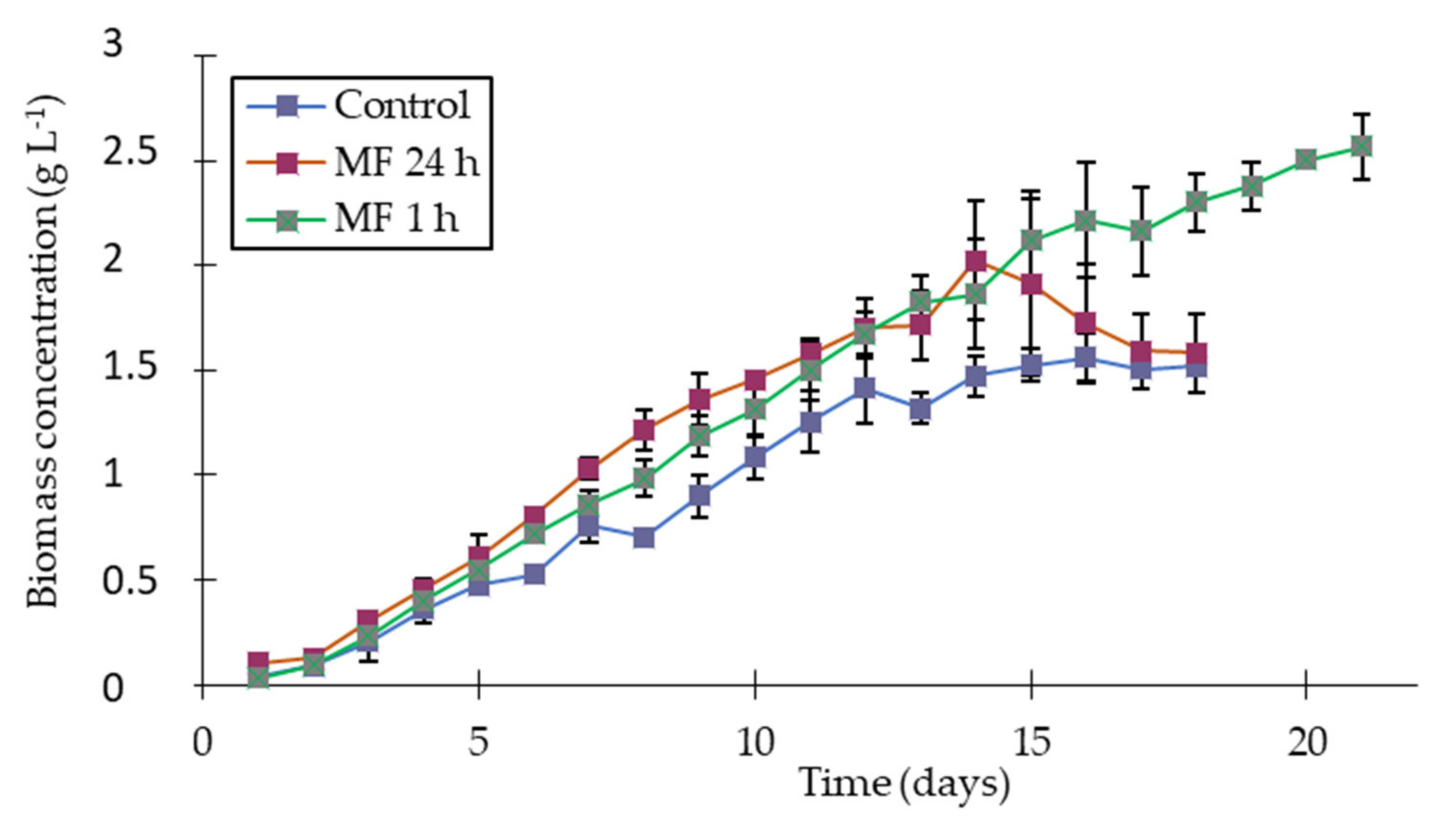
3.2. L. indica Growth and pH Evolution under Magnetic Field

The growth of L. indica PCC 8005 is shown in Figure 4, where the curves represent the daily biomass concentration. All assays started with 0.1 g L−1 and the growth showed a small lag phase until the second day. After this, the growth phase was observed and it continued until the 16th day, when the stationary phase began for all conditions. Despite the same behavior shown in the growth phases starting in the same period, under the MF treatment, there was a significant increase (p ≤ 0.05) in the growth rate from the sixth day and it remained until the end with the MF treatment for 1 h per day and until the 15th day with the MF treatment for 24 h per day. According to other studies that applied an MF in Spirulina assays [4,26,27], the action during a short period (1 h per day) was enough and it worked better if the main objective was biomass production.
The Xmax obtained on the 13th day with an MF applied for 1 h per day (2.12 ± 0.16 g L−1) was 39.5% higher than in the control assay (1.52 ± 0.01 g L−1), while with an MF for 24 h per day (2.02 ± 0.28 g L−1), the increase was 32.9%. As the growth was observed until the end of the cultivation for the assay with an MF applied for 1 h per day, this biomass concentration even reached 2.57 g L−1. Veiga et al. [4] also obtained an enhancement in the biomass concentration when the cyanobacteria Spirulina was cultured under 60 mT, achieving 37% more biomass than the control group. An MF of 11 mT was demonstrated to be more efficient than high intensities (60 mT, as studied by [26]) since the highest biomass concentration of L. indica was 2.57 g L−1 under 11 mT. In the study of Chu et al. [33], the high intensities tested (30 and 40 mT) did not stimulate the growth of Nannochloropsis oculata, while the lowest intensity, 20 mT, was enough to increase the growth rate. As demonstrated in some papers, there is no linearity regarding the intensity used and the effect obtained, which led us to conclude that these modifications depend on the strain, MF intensity, frequency and functional time. According to [34,35], there is potential damage in the algal cell at high MF intensities applied for a long period due to the increase in the free radicals. Li et al. [36] stated that the increase in microalgae growth rates may be associated with the higher nutrient consumption since the permeability of the membrane can increase under some MF treatments with an electromagnetic field. Wang et al. [37] reported that high MF intensities can change the enzyme conformation, which impacts the intracellular biochemical reactions. By measuring the O2 production rate, Hirano et al. [38] showed that an MF can have a positive effect by enhancing the photosynthesis rate and carbon fixation, with an optimal field intensity of around 100 gauss (equivalent to 10 mT). Furthermore, it is difficult to make an effective comparison since, in those studies, different MF application modes were tested, such as magnets, which do not present a homogeneous field, as in the present study. At this step, it is thus difficult to provide a hypothesis for the mechanism by which MF could influence biomass growth rates and concentrations as it may imply several targets. Further research remains necessary to go further into the elucidation of this mechanism. Although the assay with MF applied for 24 h per day produced less biomass than the one for 1 h per day, the mean biomass productivity obtained for both conditions were similar (0.14 g L−1 day−1) but significantly greater (p ≤ 0.05) than that obtained in the control group (0.11 g L−1 day−1). The same conclusions can be made for the growth rates, which were found to be about 52% and 56% greater for cultures conducted under 1 h and 24 h per day of MF treatment, respectively, as well as a reduction in the doubling time by about 35% compared with the control.
The pH was monitored daily and increased during all days of cultivation. The control culture varied from an initial pH of 9.72 up to 10.68 for the control assay, 9.74 up to 11.2 for the 24 h per day MF application and 9.73 up to 11.14 for 1 h per day MF application. During the cyanobacteria culture, the pH gradually increased due to the mechanism of inorganic carbon assimilation. At the initial pH, the inorganic carbon is mainly in the HCO3- form. These bicarbonate ions can enter the cells via an active transporter before being internally converted as CO2 by carbonic anhydrase. Hydroxyl ions are thus released, leading to an increase in pH in the medium [39]. At a high pH, the carbon species equilibrium is modified, with an increase in CO32− the concentration and a decrease in the HCO3- concentration (pKa = 10.3). In our experiments, the pH increased above this value, thus probably leading to a limitation in the carbon availability for all conditions. This may explain the entry into the stationary phase of the control assay, as already observed by [40] for Spirulina cultures with a stop in the growth observed in the pH range of 10.5–10.8. However, while using the MF, the final pH was found to be greater, suggesting a lower carbon limitation or a better uptake efficiency of the cells. Several studies suggested that an MF could play a role in the physicochemical properties of aqueous electrolytic solutions [41,42]. In particular, [43] proposed that MF has an impact on CO2 solubility in seawater, with it being increased for greater MF intensities. Those experiments were conducted with different kinds of magnetic fields and intensities, making them difficult to compare, and this study is controversial [44]. Even if it remains to be verified, the application of MF in our experiments may thus have increased this CO2 solubility, allowing for delaying the carbon limitation and supporting growth for an extended period. Nevertheless, the hypothesis of a better carbon uptake efficiency should also be considered. According to [45], an MF can affect the bioelectrocatalytic transformations of several enzyme sets in algal cells through enhancing electron transfer. These intracellular biochemical reactions could have a further impact on the microalgae cultures, modifying the CO2 biofixation rate [23] and O2 production rate [38,45], as well as increasing the surface area of chloroplasts and thylakoids [34].

3.3. Nitrogen Consumption and Removal Rate

Nitrogen in the nitrate form is a source for microalgal growth, with it being the second most important inorganic nutrient for cyanobacteria according to [46]. When the nitrate is absorbed by cyanobacterial cells, it is reduced to nitrite by nitrate reductase and then to ammonium by nitrite reductase [47]. In this study, the nitrogen consumption by L. indica was evaluated during the cultivation and the results are shown in Figure 5.
The nitrogen removal rates were calculated to be 51.2 mg L−1 day−1 and 53.4 mg L−1 day−1 for the cultures under MF for 1 and 24 h per day, respectively, which were significantly different (p ≤ 0.05) from the control (Table 1). This finding appears in accordance with the results of biomass productivities, which were observed to be greater for cultures in an MF than for the control. Moreover, [36] stated that the enhancement in growth rates may be associated with a high uptake of nutrients due to the permeability of the membrane in Spirulina platensis exposed to electromagnetic fields. According to [48], the physiological and biochemical constitution of microorganisms are usually susceptible to the action of electromagnetic forces and, in the present study, we demonstrated that Limnospira indica’s growth rate could be altered by an MF application. Likewise, all metabolic reactions are based on the difference in electrical charges and system ions. In microalgae, the movement of electrons and ions may cause changes in biomolecule concentrations, and it can modify free radical activities, cell growth and enzymatic activity [4]. Different ways to apply magnetics forces in the microalgae culture may cause different effects; furthermore, each strain behaves in a different way in response to an MF, which is why it is essential to study distinct conditions to obtain comparable studies.

3.4. Biomass Composition

Regarding the biomass composition, the proteins, carbohydrates and chlorophyll-a content were evaluated (Table 1). Spirulina is considered an ideal food supplement since it has a good nutritional profile, making it an alternative source of protein for human and animal consumption [49] since its biomass has a high protein content (around 60–70%), in addition to vitamins and pigments. This high protein content gives several possibilities to increase the nutritional value of food. Some studies demonstrated the protein power of Spirulina in food supplements for both sports performance [50] and for the elderly to consume to provide health benefits [51,52,53]. Thus, it appeared important to evaluate the protein content of the biomass produced under MF. The protein content increased by 12% (67.08 w w−1; p ≤ 0.05) under MF action applied for 1 h per day in relation to the control culture (60.4 w w−1). However, the protein content decreased significantly when the MF was applied for 24 h per day (53.6% w w−1). Veiga et al. [3] also obtained high protein content in the Spirulina biomass cultured with MF (30 or 60 mT), remaining in the range from 56 to 73% (w w−1), but with no significant differences between conditions, stating the possibility that the MF acts on protein metabolism. On the other hand, [23] evaluated the phycocyanin content from the biomass of Arthrospira platensis SAG 21.99 cultured under MF action and the authors concluded that in assays with MF action of 24 h per day, its content decreased (p ≤ 0.05), compared with the control and using an MF for 1 h per day. However, [38] noticed a differential effect of an MF on Spirulina platensis phycocyanin content, depending on the intensity applied, with a strong increase for 100 gauss, which is approximately the intensity of the MF applied in our study. Although in the present study, the phycocyanin content was not evaluated, this pigment presents protein characteristics and could be linked to the protein increase/decrease through the MF action.
Table 1 also shows the concentration of Chl-a in the L. indica biomass. The chlorophyll content usually varies in response to physical factors, such as temperature, mixing and light intensity, as well as chemical factors, i.e., nutrient availability [54]. In this study, the Chl-a content increased significantly (p ≤ 0.05) under conditions with MF application. The MF enhanced the content for the assays by about 162% and 135% with MF action for 1 and for 24 h per day, respectively. This finding was in accordance with previous studies [5,23,26,27,38]. As Chl-a is the major component of cyanobacteria photosynthetic systems with functions to conduct the photosynthesis [55], the hypothesis of a better photosynthesis rate explaining the increased growth rate observed in our experiments could thus be formulated.
On the other side, the carbohydrate content of L. indica was not modified by the MF action, and the amount observed was statistically similar (p ≤ 0.05) to the control group (14.1%, w w−1). In the study of [26], the carbohydrate content in the Spirulina sp. biomass was significantly increased for 30 and 60 mT static magnetic fields (SMFs) applied for 24 h per day. The same observation was made for Chlorella fusca cultivated under a 60 mT SMF [23]. Further experiments focused on carbohydrate repartition (quantification of glycogen and exopolysaccharide amounts for Spirulina biomass and starch for Chlorella fusca) suggested that an SMF could enhance the intracellular polysaccharide accumulation [56]. However, for lower SMF intensity (5 mT), the carbohydrate content of Spirulina biomass was found unchanged [26] and similar to those obtained in the present study (~14% w w−1), suggesting that carbohydrate accumulation was enhanced only for high MF intensities.

4. Conclusions

The effects of an MF on the growth and biomass composition of L. indica PCC 8005 were investigated. The application of 11 mT increased the final biomass concentration and the growth rate, especially when applied for 1 h per day. Moreover, protein content was significantly improved due to the action of MF applied for 1 h per day (67.08 w w−1), and a strong increase in chlorophyll content was also noticed. The MF application strategy thus appeared as a low-cost approach for an increase in biomass production with improved nutritional value.

Supplementary Materials

The following supporting information can be downloaded at: https://0-www-mdpi-com.brum.beds.ac.uk/article/10.3390/app12031533/s1, Table S1: Mapping of the magnetic field intensities.

Author Contributions

Conceptualization, F.G., J.A.V.C. and C.L.; methodology, F.G., J.A.V.C. and C.L.; validation, K.M.D., F.G. and C.L.; investigation, K.M.D.; writing—original draft preparation, K.M.D.; writing—review and editing, K.M.D., M.G.d.M., L.O.d.S., J.A.V.C., F.G. and C.L.; supervision, F.G., J.A.V.C. and C.L.; funding acquisition, J.A.V.C. and C.L. All authors have read and agreed to the published version of the manuscript.

Funding

This research was funded by Program CAPES-COFECUB (Sv 945/19); the National Council for Scientific and Technological Development (CNPq), and the Ministry of Science, Technology, Innovations and Communication (MCTIC). This study was financed in part by the Coordenação de Aperfeiçoamento de Pessoal de Nível Superior-Brasil (CAPES)-Finance Code 001.

Conflicts of Interest

The authors declare no conflict of interest.

References

  1. Geada, P.; Rodrigues, R.; Loureiro, L.; Pereira, R.; Fernandes, B.; Teixeira, J.A.; Vasconcelos, V.; Vicente, A.A. Electrotechnologies applied to microalgal biotechnology—Applications, techniques and future trends. Renew. Sustain. Energy Rev. 2018, 94, 656–668. [Google Scholar] [CrossRef] [Green Version]
  2. Haberkorn, I.; Buchmann, L.; Hiestand, M.; Mathys, A. Continuous nanosecond pulsed electric field treatments foster the upstream performance of Chlorella vulgaris-based biorefinery concepts. Bioresour. Technol. 2019, 293, 122029. [Google Scholar] [CrossRef] [PubMed]
  3. Veiga, M.C.; Fontoura, M.M.; Oliveira, M.G.; Costa, J.A.V.; Santos, L.O. Magnetic fields: Biomass potential of Spirulina sp. for food supplement. Bioprocess Biosys. Eng. 2020, 43, 1231–1240. [Google Scholar] [CrossRef] [PubMed]
  4. Santos, L.O.; Deamici, K.M.; Menestrino, B.C.; Garda-Buffon, J.; Costa, J.A.V. Magnetic treatment of microalgae for enhanced product formation. World J. Microbiol. Biotechnol. 2017, 33, 169–172. [Google Scholar] [CrossRef]
  5. Deamici, K.M.; Santos, L.O.; Costa, J.A.V. Magnetic field as promoter of growth in outdoor and indoor assays of Chlorella fusca. Bioprocess Biosyst. Eng. 2021, 44, 1453–1460. [Google Scholar] [CrossRef]
  6. Whitton, B.A.; Potts, M. The Ecology of Cyanobacteria: Their Diversity in Time and Space; Kluwer Academic Publishers: Dordrecht, The Netherlands; Springer: Dordrecht, The Netherlands, 2002; 669p. [Google Scholar]
  7. Ali, S.K.; Saleh, A.M. Spirulina: An overview. Int. J. Pharm. Pharm. Sci. 2012, 4, 9–15. [Google Scholar]
  8. Nowicka-Krawczyk, P.; Mühlsteinová, R.; Hauer, T. Detailed characterization of the Arthrospira type species separating commercially grown taxa into the new genus Limnospira (Cyanobacteria). Sci. Rep. 2019, 9, 1–11. [Google Scholar] [CrossRef]
  9. Farges, B.; Laroche, C.; Cornet, J.-F.; Dussap, C.-G. Spectral kinetic modelling and long-term behaviour assessment of Arthrospira platensis growth in photobioreactor under red (620 nm) light illumination. Biotechnol. Prog. 2009, 25, 151–162. [Google Scholar] [CrossRef]
  10. Phélippe, M.; Gonçalves, O.; Thouand, G.; Cogne, G.; Laroche, C. Characterization of the polysaccharides chemical diversity of the cyanobacteria Arthrospira platensis. Algal Res. 2019, 38, 101426. [Google Scholar] [CrossRef]
  11. Poughon, L.; Laroche, C.; Creuly, C.; Dussap, C.-G.; Paille, C.; Lasseur, C.; Monsieurs, P.; Heylen, W.; Coninx, I.; Mastroleo, F.; et al. Limnospira indica PCC8005 growth in photobioreactor: Model and simulation of the ISS and ground experiments. Life Sci. Space Res. 2020, 25, 53–65. [Google Scholar] [CrossRef]
  12. Priyadarshani, I.; Rath, B. Commercial and industrial applications of microalgae—A review. J. Algal Biomass. Utln. 2012, 3, 89–100. [Google Scholar]
  13. Makhlouf, R.; Makhlouf, I. Evaluation of the effect of Spirulina against Gamma irradiation induced oxidative stress and tissue injury in rats. Int. J. Appl. Sci. Eng. Res. 2012, 1, 152–164. [Google Scholar]
  14. Wu, L.; Ho, J.A. Antioxidative and hepatoprotective effects of Spirulina. In Spirulina in Human Nutrition and Health; Gershwin, M.E., Belay, A., Eds.; CRC Press, Taylor and Francis Group: Boca Raton, FL, USA, 2008; pp. 119–151. [Google Scholar]
  15. Gonzalez Bautista, E.; Laroche, C. Arthrospira platensis as a feasible feedstock for bioethanol production. Appl. Sci. 2021, 11, 6756. [Google Scholar] [CrossRef]
  16. Pereira, A.M.; Lisboa, C.R.; Costa, J.A.V. High protein ingredients of microalgal origin: Obtainment and functional properties. Innov. Food Sci. Emerg. Technol. 2018, 47, 187–194. [Google Scholar] [CrossRef]
  17. Vonshak, A. Spirulina platensis (Arthrospira) Physiology, Cell-Biology and Biotechnology; Taylor & Francis: London, UK, 1997; 233p. [Google Scholar]
  18. Sukumaran, P.; Bin Omar, H.; Nulit, R.B.; Halimoon, N.B.; Simoh, S.B.; Ismail, A.B. The prospects of the cultivation of Arthrospira platensis under outdoor conditions in Malaysia. Jordan J. Biol. Sci. 2018, 11, 419–426. [Google Scholar]
  19. Ljubic, A.; Safafar, H.; Holdt, S.; Jacobsen, C. Biomass composition of Arthrospira platensis during cultivation on industrial process water and harvesting. J. Appl. Phycol. 2018, 30, 943–954. [Google Scholar] [CrossRef] [Green Version]
  20. Karemore, A.; Yuan, Y.; Porubsky, W.; Chance, R. Biomass and pigment production for Arthrospira platensis via semi-continuous cultivation in photobioreactors: Temperature effects. Biotechnol. Bioeng. 2020, 117, 3081–3093. [Google Scholar] [CrossRef]
  21. Sarraf, M.; Kataria, S.; Taimourya, H.; Santos, L.O.; Menegatti, R.D.; Jain, M.; Ihtisham, M.; Liu, S. Magnetic Field (MF) Applications in Plants: An Overview. Plants 2020, 9, 1139. [Google Scholar] [CrossRef]
  22. Hunt, R.W.; Zavalin, A.; Bhatnagar, A.; Chinnasamy, S.; Das, K.C. Electromagnetic biostimulation of living cultures for biotechnology, biofuel and bioenergy applications. Int. J. Mol. Sci. 2009, 10, 4719–4722. [Google Scholar] [CrossRef] [Green Version]
  23. Deamici, K.M.; Santos, L.O.; Costa, J.A.V. Use of static magnetic fields to increase CO2 biofixation by the microalga Chlorella Fusca. Bioresour. Technol. 2019, 276, 103–109. [Google Scholar] [CrossRef] [PubMed]
  24. Nezammahalleh, H.; Ghanati, F.; Adams, T.A., II; Nosrati, M.; Shojaosadati, S.A. Effect of moderate static electric field on the growth and metabolism of Chlorella vulgaris. Bioresour. Technol. 2016, 218, 700–711. [Google Scholar] [CrossRef] [PubMed] [Green Version]
  25. Cogne, G.; Lehmann, B.; Dussap, C.-G.; Gros, J.-B. Uptake of macrominerals and trace elements by the cyanobacterium Spirulina platensis (Arthrospira platensis PCC 8005) under photoautotrophic conditions: Culture medium optimization. Biotechnol. Bioeng. 2003, 81, 588–593. [Google Scholar] [CrossRef]
  26. Deamici, K.M.; Costa, J.A.V.; Santos, L.O. Magnetic fields as triggers of microalga growth: Evaluation of its effect on Spirulina sp. Bioresour. Technol. 2016, 220, 62–67. [Google Scholar] [CrossRef] [PubMed]
  27. Deamici, K.M.; Santos, L.O.; Costa, J.A.V. Magnetic field action on outdoor and indoor cultures of Spirulina: Evaluation of growth, medium consumption and protein profile. Bioresour. Technol. 2018, 249, 168–174. [Google Scholar] [CrossRef] [PubMed]
  28. Cawse, P.A. The determination of nitrate in soil solutions by ultraviolet spectrophotometry. Analyst 1967, 92, 311–315. [Google Scholar] [CrossRef]
  29. American Public Health Association. Standard Methods for the Examination of Water and Wastewater; American Public Health Association: Washington, DC, USA, 1971; pp. 237–239. [Google Scholar]
  30. Lowry, O.H.; Rosebrough, N.J.; Farr, A.L.; Randall, R.J. Protein measurement with the Folin phenol reagent. J. Biol. Chem. 1951, 193, 265–275. [Google Scholar] [CrossRef]
  31. Dubois, M.; Gilles, K.A.; Hamilton, J.K.; Rebers, P.A.; Smith, F. Colorimetric method for determination of sugars and related substances. Anal. Chem. 1956, 28, 350–356. [Google Scholar] [CrossRef]
  32. Lichtenthaler, H.K. Chlorophylls and carotenoids: Pigments of photosynthetic biomembranes. Meth. Enzymol. 1987, 148, 350–382. [Google Scholar]
  33. Chu, F.-J.; Wan, T.-J.; Pai, T.-Y.; Lin, H.-W.; Liu, S.-H.; Huang, C.-F. Use of magnetic fields and nitrate concentration to optimize the growth and lipid yield of Nannochloropsis oculate. J. Environ. Manage. 2020, 253, 109680. [Google Scholar] [CrossRef]
  34. Small, D.P.; Huüner, N.P.; Wan, W. Effect of static magnetic fields on the growth, photosynthesis and ultrastructure of Chlorella kessleri microalgae. Bioelectromagnetics 2012, 33, 298–308. [Google Scholar] [CrossRef]
  35. Wang, H.-Y.; Zeng, X.-B.; Guo, S.-Y.; Li, Z.-T. Effects of magnetic field on the antioxidant defense system of recirculation-cultured Chlorella vulgaris. Bioelectromagnetics 2008, 29, 39–46. [Google Scholar] [CrossRef] [PubMed]
  36. Li, Z.-Y.; Guo, S.-Y.; Li, L.; Cai, M.-Y. Effects of electro- magnetic field on the batch cultivation and nutritional composition of Spirulina platensis in an air-lift photobioreactor. Bioresour. Technol. 2007, 98, 700–705. [Google Scholar] [CrossRef] [PubMed]
  37. Wang, H.Y.; Zeng, X.B.; Guo, S.Y. Growth of Chlorella vulgaris under different magnetic treatments. Prog. Mod. Biomed. 2006, 6, 106–108. [Google Scholar]
  38. Hirano, M.; Ohta, A.; Abe, K. Magnetic field effects on photosynthesis and growth of the cyanobacterium Spirulina platensis. J. Ferm. Bioeng. 1998, 86, 313–316. [Google Scholar] [CrossRef]
  39. Shiraiwa, Y.; Goyal, A.; Tolbert, N.E. Alkalization of the medium by unicellular green algae during uptake of dissolved inorganic carbon. Plant Cell Physiol. 1993, 34, 649–657. [Google Scholar] [CrossRef]
  40. Binaghi, L.; Borghi, A.D.; Lodi, A.; Converti, A.; Borghi, M.D. Batch and fed-batch uptake of carbon dioxide by Spirulina platensis. Process. Biochem. 2003, 38, 1341–1346. [Google Scholar] [CrossRef]
  41. Beruto, D.T.; Botter, R.; Perfumo, F.; Scaglione, S. Interfacial effect of extremely low frequency electromagnetic fields (EM-ELF) on the vaporization step of carbon dioxide from aqueous solutions of body simulated fluid (SBF). Bioelectromagnetics 2003, 24, 251–261. [Google Scholar] [CrossRef]
  42. Vallée, P.; Lafait, J.; Legrand, L.; Mentre, P.; Monod, M.-O.; Thomas, Y. Effects of pulsed low frequency electromagnetic fields on water characterized by light scattering techniques: Role of bubbles. Langmuir 2005, 21, 2293–2299. [Google Scholar] [CrossRef] [Green Version]
  43. Pazur, A.; Winklhofer, M. Magnetic effect on CO2 solubility in seawater: A possible link between geomagnetic field variations and climate. Geophys. Res. Lett. 2008, 35, L16710. [Google Scholar] [CrossRef] [Green Version]
  44. Köhler, P.; Muscheler, R.; Richter, K.-U.; Snowball, I.; Wolf-Gladrow, D.A. Comment on ‘‘Magnetic effect on CO2 solubility in seawater: A possible link between geomagnetic field variations and climate’’ by Alexander Pazur and Michael Winklhofer. Geophys. Res. Lett. 2009, 36, L03705. [Google Scholar] [CrossRef] [Green Version]
  45. Tu, R.; Jin, W.; Xi, T.; Yang, Q.; Han, S.F.; Abomohra, A.-F. Effect of static magnetic field on the oxygen production of Scenedesmus obliquus cultivated in municipal wastewater. Water Res. 2015, 86, 132–138. [Google Scholar] [CrossRef] [PubMed]
  46. Mann, N.H. Protein phosphorylation in cyanobacteria. Microbiology 1994, 40, 3207–3215. [Google Scholar] [CrossRef] [Green Version]
  47. Liotenberg, S.; Campbell, D.; Rippka, R.; Houmard, J.; Marsac, N.T. Effect of the nitrogen source on phycobiliprotein synthesis and cell reserves in a chromatically adapting filamentous cyanobacterium. Microbiology 1996, 142, 611–622. [Google Scholar] [CrossRef] [PubMed] [Green Version]
  48. Teng, H.C. A Puzzle of the effect of magnetic field on biological cells. Life Sci. J. 2005, 2, 16–21. [Google Scholar]
  49. Spolaore, P.; Joannis-Cassan, C.; Duran, E.; Isamber, A. Commercial applications of microalgae. J. Biosci. Bioeng. 2006, 101, 87–96. [Google Scholar] [CrossRef] [Green Version]
  50. Milasius, K.; Malickaite, R.; Dadeliene, R. Effect of Spirulina food supplement on blood morphological parameters, biochemical composition and on the immune function of sportsmen. Biol. Sport 2009, 26, 157–172. [Google Scholar] [CrossRef] [Green Version]
  51. Bauer, J.; Biolo, G.; Cederholm, T.; Cesari, M.; Cruz-Jentoft, A.J.; Morley, J.E.; Phillips, S.; Sieber, C.; Stehle, P.; Teta, D.; et al. Evidence-based recommendations for optimal dietary protein intake in older people: A position paper from the protage study group. J. Am. Med. Dir. Assoc. 2013, 14, 542–559. [Google Scholar] [CrossRef]
  52. Deutz, N.E.P.; Bauer, J.M.; Barazzoni, R.; Biolo, G.; Boirie, Y.; Bosy-Westphal, A.; Cederholm, T.; Cruz-Je, A.; Krznariç, Z.; Nair, K.S.; et al. Protein intake and exercise for optimal muscle function with aging: Recommendations from the ESPEN expert group. Clin. Nutr. 2014, 33, 929–936. [Google Scholar] [CrossRef] [Green Version]
  53. Santos, T.D.; Freitas, B.C.B.; Moreira, J.B.; Zanfonato, K.; Costa, J.A.V. Development of powdered food with the addition of Spirulina for food supplementation of the elderly population. Innov. Food Sci. Emerg. Technol. 2016, 37, 216–220. [Google Scholar] [CrossRef]
  54. Ferreira, V.S.; Pinto, R.F.; Sant’Anna, C. Low light intensity and nitrogen starvation modulate the chlorophyll content of Scenedesmus dimorphus. J. Appl. Microbiol. 2015, 120, 661–670. [Google Scholar] [CrossRef] [Green Version]
  55. Jeon, H.J.; Choi, Y.-K.; Eom, H.S.; Kang, G.-H.; Kim, K.J.; Yang, Y.-H.; Lee, A.H.; Kim, K.H.; Kim, S.J.; Kim, H.J. Comparison among dry cell weight, chlorophyll a concentration, and amperometric signal during a batch cultivation of Spirulina maxima. Sens. Actuators B Chem. 2014, 205, 9–11. [Google Scholar] [CrossRef]
  56. Deamici, K.M.; de Morais, M.G.; Santos, L.O.; Koenraad, M.; Gardarin, C.; Costa, J.A.V.; Laroche, C. Static Magnetic Fields effects on polysaccharides production by different microalgae strains. Appl. Sci. 2021, 11, 5299. [Google Scholar] [CrossRef]
Figure 1. Experimental device used for the L. indica PCC8005 cultivation. (A) Cultures with magnetic field application through coils and (B) cultures control without MF application but in the same temperature, luminosity and aeration conditions. (1) Coils with 400 turns used to generate the MF, (2) illumination provided by 3 halogen lamps and (3) 600 mL photobioreactors. (C) Scheme of the experimental devices (control).
Applsci 12 01533 g001
Figure 2. Schematic diagram of the experimental device used for the L. indica PCC8005 cultivation with magnetic field application through coils. Red arrows indicate the transverse magnetic field.
Applsci 12 01533 g002
Figure 3. MF intensity mapping at a 7 cm height (middle of the culture vessel). Raw values are available in the Supplementary Materials.
Applsci 12 01533 g003
Figure 4. Growth curves of L. indica PCC8005 cultivated under an MF with different application times and the control culture.
Applsci 12 01533 g004
Figure 5. Consumption of nitrate during the L. indica PCC8005 cultivation under MF action.
Applsci 12 01533 g005
Table 1. Growth parameters, protein, carbohydrates and Chl-a contents in L. indica PCC8005 biomass cultivated under control conditions and the MF applied for 24 and 1 h per day. ɳ values represent the increase (+) or decrease (−) according to the MF effect as compared with the control.
ParametersUnitControlMF 1 h ɳ (%)MF 24 h ɳ (%)
Xmax *g L−11.52 ± 0.01 a2.12 ± 0.16 c+123.52.02 ± 0.28 b *+32.9
avPg L−1 day−10.11 ± 0.01 a0.14 ± 0.01 b +27.30.14 ± 0.03 b +27.3
µmaxday−10.550 ± 0.01 a0.638 ± 0.01 ab+15.90.859 ± 0.03 b+56.0
Dtday1.26 ± 0.07 a0.81 ± 0.04 a−34.10.83 ± 0.05 b−35.7
N removal ratemg L−1 day−141.8 ± 0.3 a 51.2 ± 0.2 b+22.553.4 ± 0.4 b+27.7
Proteins%, w w−160.41 ± 3.41 b67.08 ± 1.72 c+12.6953.6 ± 2.71 a −11.3
Carbohydrates%, w w−114.01 ± 1.84 a,b11.42 ± 0.55 a−18.4814.99 ± 2.09 b+6.9
Chlorophyll-aµg mg−1 DW1.11 ± 0.15 a2.91 ± 0.31 b+161.12.63 ± 0.31 b+135.8
* Xmax obtained in the 13th day. Means ± standard deviations. Different letters in the same column correspond to significant differences (p ≤ 0.05).
Publisher’s Note: MDPI stays neutral with regard to jurisdictional claims in published maps and institutional affiliations.

