Article

Heat Transfer Analysis and Effects of (Silver and Gold) Nanoparticles on Blood Flow Inside Arterial Stenosis

1
Department of Mathematics, University of Gujrat, Gujrat 50700, Pakistan
2
Department of Mechanical Engineering, College of Engineering, King Khalid University, P.O. Box 960, Abha 61421, Saudi Arabia
3
Department of Electrical Engineering, Ardahan University, Ardahan 75002, Turkey
4
Department of Civil Engineering, College of Engineering, Taif University, P.O. Box 11099, Taif 21944, Saudi Arabia
5
Department of Mathematics and Statistics, College of Science, Taif University, P.O. Box 11099, Taif 21944, Saudi Arabia
*
Author to whom correspondence should be addressed.
Submission received: 14 December 2021 / Revised: 19 January 2022 / Accepted: 28 January 2022 / Published: 2 February 2022
(This article belongs to the Special Issue New Trends of Silver Nanoparticles in Biomedicine)

Abstract

:
The current investigation was based on a simulation employing CFD in COMSOL Multiphysics. The base fluid that was used in this simulation was blood. The flow was considered as a laminar, unsteady and incompressible Newtonian fluid, and the Newtonian nature of blood is acceptable at high shear rate. The behavior of blood flow was analyzed with the objective of obtaining pressure, temperature and velocity effects through an arterial stenosis. Two types of nanoparticles were used in this work: silver (Ag) and gold (Au). The equations of mass, momentum and energy were solved by utilizing the CFD technique. A fine element size mesh was generated through COMSOL. The results of this analysis show that velocity changes through confined parts of the artery, the velocity in a diseased region is higher and the velocity decreases before and after the stenotic region. In the heat transfer feature, the upper and lower boundary temperature was set to 24.85 °C and 27.35 °C, respectively. The nanoparticles affected the physical properties of blood, such as thermal conductivity, density, dynamic viscosity and specific heat. The addition of gold and silver nanoparticles prevented overheating because both nanoparticles have a high thermal conductivity, which has a principal role in dissipating temperature quickly. Nusselt number variations were also calculated and the results show that the curve decreases inside the stenosis. It could be concluded that the streamlines show abnormal behavior and recirculation occurs just after the stenosed area at t = 0.7 s and 1 s. These results will help greatly in the treatment of stenosed arteries.

1. Introduction

Nanoparticles help in many diagnostic applications and in the effective treatment of cardiovascular diseases. The physical properties of nanoparticles, such as shape, surface and size modifications, provide better treatment strategies and the delivery of nanoparticles into the artery is mostly dependent on these physical properties. T. Hayat and S. Nadeem [1] studied the enhancement of heat transfer by adding silver and copper oxide nanoparticles into the base fluid water. S. Nadeem and S. Ijaz [2] studied single wall carbon nanotubes in the flow of blood through multiple stenoses by considering variable viscosity. Three-dimensional flow models under different effects and with the addition of nanoparticles have been studied by [3,4]. S. Nadeem et al. [5] analyzed Prandtl magnetohydrodynamic nanofluid flow through arterial stenoses. An important and different utilization of nanoparticles can be seen in the modern field of medicine. Gold nanoparticles are the most impactful material in nanomedicine and nanoscience. They are being used as photovoltaic agents, radio infections, drug carriers and contrast agents. In addition, there are many properties of gold and silver nanoparticles that are attractive for use in biomedical diagnosis and treatment. The treatment of arterial diseases using nanoparticles is still under development. T. Elnaqeeb et al. [6] investigated blood flow through stenosed vessels and presented results for different flow parameters. They concluded that by adding gold nanoparticles, the velocity of blood increases and they also compared their results to Cu and TiO2 models. A number of researchers [7,8,9,10] have paid attention to the study of arterial diseases by applying different methods.
Blood vessels are important components in the circulatory system. The primary objective of the blood circulatory system is to transport oxygen and important nutrients to active body tissues and remove waste products. The dynamics of blood flow play an important role in the expansion and formation of cardiovascular diseases. Cardiovascular diseases are responsible for the majority of deaths in underdeveloped countries. One of the most serious and common arterial diseases is arterial stenosis. An arterial stenosis blocks the flow of blood and also affects blood pressure. It is a progressive disease that affects up to 2.5 million people over the age of 75. The main causes, symptoms and complications of stenosis are:
CausesSymptomsComplications
Birth defects, build-up of calcium, radiation therapy and rheumatic fever.Chest pain, heart palpitations, swollen ankles or feet and difficulty in walking short distances.Stroke, heart failure, blood clots, arrhythmias and endocarditis.
For problems related to physiological flows, the computational fluid dynamics (CFD) tool has become useful over recent years. The main purpose of CFD is to numerically simulate the motion of fluid and heat transfer by using computational prowess. J. Liu et al. [11] examined the solutions to problems governing equations numerically by using COMSOL. A. Hussain et al. [12] studied the heat transfer consequences for laminar and unsteady fluid flow through distinct elliptic cylinders and found the maximum and minimum values for temperature, pressure and velocity.
In this article, the behavior of blood flow through a stenosed region is studied. We assumed blood to be a Newtonian fluid because blood shows Newtonian nature in large cavities, veins and arteries. The law of conservation of mass, momentum and energy were solved using COMSOL Multiphysics software and the results were calculated for the velocity, temperature and pressure of the blood flow inside the affected region by utilizing CFD. This article introduces the mathematical study of the addition of gold and silver nanoparticles into the blood flowing through a stenosed region in order to investigate that how hybrid NPs can help in improving blood flow.

2. Materials and Methods

In this research, blood was assumed to be an incompressible and unsteady Newtonian fluid flowing through an arterial stenosis. A sketch of the blood flow problem in arterial stenosis is shown in Figure 1, and the coordinate system was selected in such a way that the blood flow was along the z-axis and r was taken as perpendicular to the flow.
The width and height of the two-dimensional artery were 1.1 m and 0.8 m, respectively, with coordinate positions of r = 0.1 and z = 0. The physics of laminar blood flow and heat transfer was added through the “add physics” feature in COMSOL 5.4. At the boundary wall, the velocity was zero and the entrance and exit positions were selected. In the laminar flow and heat transfer, the reference temperature was taken as 19.85 °C. In the heat transfer, the initial value of temperature was considered as −73.15 °C and at the upper boundary, temperature 1 was taken as 24.85 °C and at the lower boundary, temperature 2 was 27.35 °C. The material properties, i.e., thermophysical properties, of blood were added. In the next step, the time-dependent model governing equations, i.e., mass, momentum [13] and heat transfer equations, [14] were added.

2.1. Continuity Equation

1 r r r u + z w = 0 ,
In COMSOL, the equation of continuity that described the blood mass transport was expressed as:
    . u = 0 ,
where u and w are the velocities of the nanofluid denoted in r and z directions, respectively.

2.2. Equation of Motion

ρ h n f u t + u u r + w u z = p r + μ h n f z w r u z ,
ρ h n f w t + u w r + w w z = p z μ h n f r + 1 r w r u z .  
In COMSOL, the model governing the momentum equation was shown as:
ρ u t + ρ u . u = . P I + K + F ,
K = μ u + u T ,
where ρ h n f ,   μ h n f and P are the density, viscosity and pressure of the nanofluid, respectively.

2.3. Energy Equation

ρ C p h n f t + u r + w z T = k h n f 2 r 2 + r r + 2 z 2 T ,
In COMSOL, the governing heat transfer equation was
d z ρ C p T t + d z ρ C p u . T + . q = d z Q + q o + d z Q p + d z Q v d ,
where q = d z k T , q o = q A s Δ T ,   Q v d = τ . u ,   Q p = α P T P t + u . P , A s = 0.8 m2, α P = 1 ρ P t , τ = P I + K ,   d z is the thickness of the fluid, which is equal to 1 m, Q is the heat source, Q v d is the viscous dissipation heat source, T shows the temperature gradient, C p , n f shows the heat capacitance of the fluid and k n f is specific heat. The considered initial values are:
u = w = 0 ,   T = T o   and   P = 0 .
The thermophysical properties of the hybrid nanofluids were defined as follows [15]:
ρ h n f = 1 ϕ 2 1 ϕ 1 ρ f + ϕ 1 ρ s 1 + ϕ 2 ρ s 2 , μ h n f = μ f 1 ϕ 1 2.5 1 ϕ 2 2.5 , ρ C p h n f = 1 ϕ 2 1 ϕ 1 ρ C p f + ϕ 1 ρ C p s 1 + ϕ 2 ρ C p s 2 , k h n f k f = k s 1 + 2 k f 2 ϕ 1 k f k s 1 k s 1 + 2 k f + ϕ 1 k f k s 1 × k s 2 + 2 k n f 2 ϕ 2 k n f k s 2 k s 2 + 2 k n f + ϕ 2 k n f k s 2 . }

2.4. Boundary Conditions

The boundary conditions contained inlet, outlet, wall and thermal insulation.

2.4.1. The Inlet

The blood flow rate, or velocity, was simulated at the inlet of the artery. The blood volume could be controlled by the velocity at the inlet and the area of inflow cross-section. Figure 2 shows how the inlet in the model was defined.
The inlet boundary condition was:
u r , z , t = U o n ,
where U o is the normal inflow velocity, which is equal to 0.5.

2.4.2. The Outlet

We presented the pressure of the blood flow model at the outlet to add a more realistic approach to the simulation. Figure 3 represents the outlet that was on the opposite side from the inlet and was where the blood flowed out.
In COMSOL, the equation at the outlet was given as:
τ   n = P   n , where   P = P o ,
where n = n r , n z is the pointing normal outflow velocity and the absolute pressure is P o = 0   P a .

2.4.3. At the Wall

Blood cannot flow through the wall because it sticks to the wall due to its viscosity. Thus, at the wall, the boundary conditions were:
u = 0 ,   w = 0 .
and no slip condition was considered. Figure 4 represents the wall of arterial stenosis.

2.4.4. Thermal Insulation

The insulated boundaries in the COMSOL heat transfer feature are shown in Figure 5.
The thermal insulation equation used in COMSOL was:
n . q = 0

2.5. Computational Mesh

Mesh is one of the important factors in computational fluid dynamics. The rate of convergence and the accuracy of the solution can be determined by the quality of the mesh. The “physics controlled mesh” function in COMSOL automatically generates mesh. Fine element size mesh produces more accurate results and provides better performance than the normal size. Figure 6 and Figure 7 show “normal” and “fine” element size meshes, and it was observed that the mesh was more refined in the stenosed region, as can be seen by the stenosed area, and was less refined when far away from the stenosis.

3. Validation

For the accuracy of this problem, the obtained results with the addition of hybrid nanoparticles were compared to those of [15]. They selected the initial velocities of 0.1 and 0.05. In our simulation, we considered an initial velocity of 0.5. The other conditions were same, but in our simulation addition of nanoparticles improved the results for velocity, which can be seen from the obtained results. The current results have a good agreement with [15].

4. Results

The mathematical study of blood flow in the arteries is an essential and challenging problem. In the arterial system, the most common disease is the contraction and hardening of the walls of blood vessels, which is called arterial stenosis. A mathematical model to find the velocity, temperature and pressure of the blood through arteries in the presence of stenosis when adding hybrid nanoparticles was studied. The addition of hybrid nanoparticles affected the physical properties of blood, such as dynamic viscosity, specific heat, density and thermal conductivity, resulting in a change in the simulation results. The heat transfer phenomenon was observed by investigating Nusselt number variation.
Figure 1 shows the two-dimensional sketch of the problem. Figure 2 represents the inlet point of the blood flow and the velocity of the fluid was also defined by this boundary condition. Figure 3 shows the outlet point of the blood flow and was where the fluid flowed out. Figure 4 and Figure 5 show the wall and thermal insulation boundaries of the problem respectively. The thermal insulation boundary conditions implied that the conditions were not affected by the heat source in the simulation.
In Figure 6 and Figure 7, normal and fine element size meshes are shown, and we studied their effects on the results. The mesh vertices of the normal element size were 948, the total triangles were 1315, the quad entities were 214, the maximum element size was 0.0737, the minimum element size was 3.3 × 10⁻⁴ and the total number of elements was 1529. The mesh vertices of the fine element size were 1195, the total triangles were 1747, the quad entities were 234, the vertex elements were 8, the total number of elements was 1981, the minimum element size was 8 × 10⁻⁴, the maximum element size was 0.028 and the resolution of the narrow region was 1. The fine element size had a better performance and provided more accurate results.
Figure 8, Figure 9, Figure 10, Figure 11, Figure 12, Figure 13, Figure 14, Figure 15, Figure 16, Figure 17, Figure 18, Figure 19, Figure 20 and Figure 21 display the variations of time at t = 0.1 s, 0.7 s and 1 s for velocity, pressure, temperature, isothermal contours and streamlines. Figure 8, Figure 9 and Figure 10 describe the consequences of the velocity profile. In Figure 8, the velocity is 1.59 m/s at t = 0.1 s. In Figure 9, the maximum velocity is 1.57 m/s at 0.7 s. In Figure 10, the maximum velocity is 1.58 m/s at 1 s. The results show that the maximum velocity was found at t = 0.1 s, i.e., 1.59 m/s. It can be seen that the flow pattern at the inlet was smooth. It is also noted that the diseased region had a higher velocity compared to before and after the stenosis. This was anticipated because Bernoulli’s equation results prove that the velocity of the narrow region should be higher. Figure 11, Figure 12 and Figure 13 show the profiles of temperature for the heat transfer. The temperature varied at different times but the maximum and minimum temperature limit remained the same with respect to time at 26.85 °C and −73.15 °C, respectively. The results show that the addition of nanoparticles effectively enhanced the heat transfer ratio.
Figure 14, Figure 15 and Figure 16 show the pressure profiles, and the maximum and minimum values of the pressure were observed at t = 1 s. The maximum pressure was obtained just before the stenosed region and the minimum pressure was at the end of the stenosis. Figure 17, Figure 18 and Figure 19 show the isothermal contours for the temperature profiles. The maximum range of the isothermal contours remained the same and the minimum contours were found at t = 0.1 s. The contour pattern scattered and moving away from the narrow region. Figure 20, Figure 21 and Figure 22 display the streamlines that recirculated after the stenosis. The overall heat transfer was examined with the help of the Nusselt number (Nu). In the present problem, Figure 23 depicts the varying Nuav number with respect to time. It can be clearly observed that the Nusselt number dropped when the fluid flow reached the stenosis boundaries and also that the average Nusselt number increased before and after that point, with varying times. Figure 24 is for the validation of the current model.
Table 1 represents the computed mesh element size and area for the normal and fine options in m2. We used the fine element size mesh for more accurate results and better performance. Table 2 shows the description of the mesh statistics for the mesh elements, quality of elements, mesh area, total number of entities of triangles and quadrilaterals and total number of elements. Table 3 shows the mesh size of the stenosed artery, i.e., the maximum and minimum mesh element size, resolution of narrow region, maximum element growth rate, geometric entity level and curvature factor. The quality and resolution of the mesh elements are important aspects of the simulation. In Table 4, the properties of the blood base fluid and gold and silver nanoparticles are utilized to find the solution, and these properties affected the dynamic viscosity, thermal conductivity, heat capacity and density of the blood.

5. Conclusions

We investigated the consequences of using hybrid nanoparticles in the blood flow through a diseased artery. The main objective of this study was to describe the CFD results for the velocity, temperature and pressure through the narrow area of the artery. From the results, it can be observed that the addition of gold and silver nanoparticles prevented overheating and disfavored the maximum velocity. Our simulations could be used to obtain a finer image of hemodynamics. Some of the main results were:
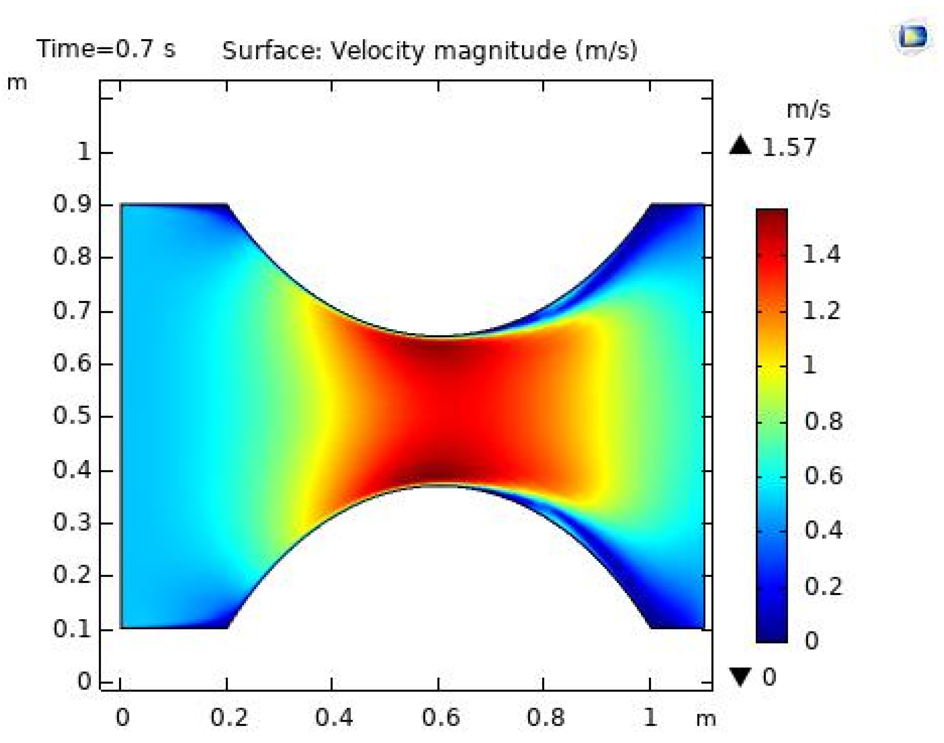
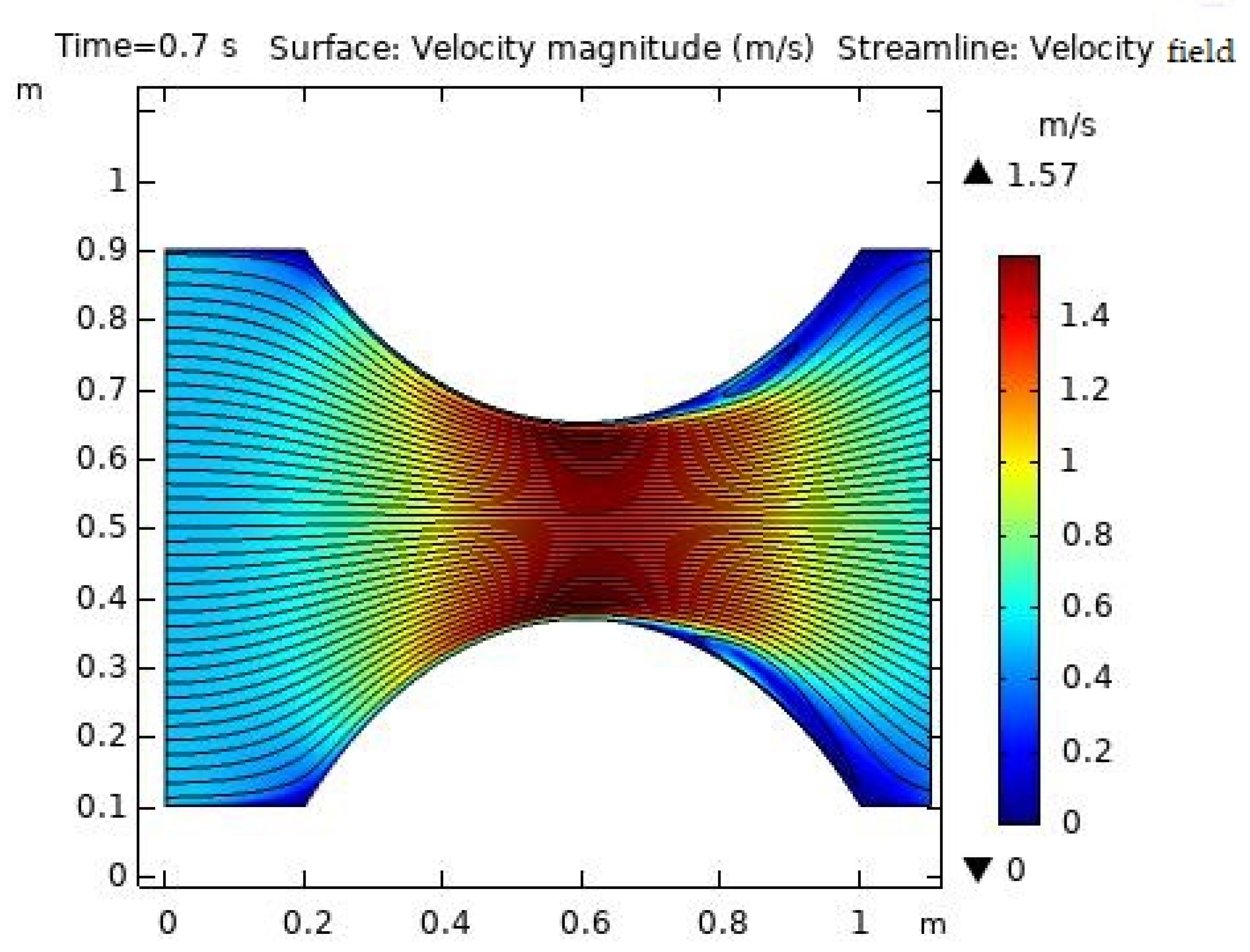
  • The laminar flow study showed that the velocity of the blood flow varies throughout the model due to arterial plaque. The maximum velocity of the blood flow was 1.59 m/s at t = 0.1 s;
  • The streamlines showed abnormal behavior near the stenosis area and that recirculation occurred as the intensity of the stenosis increased; however, the flow was normal when nanoparticles were added;
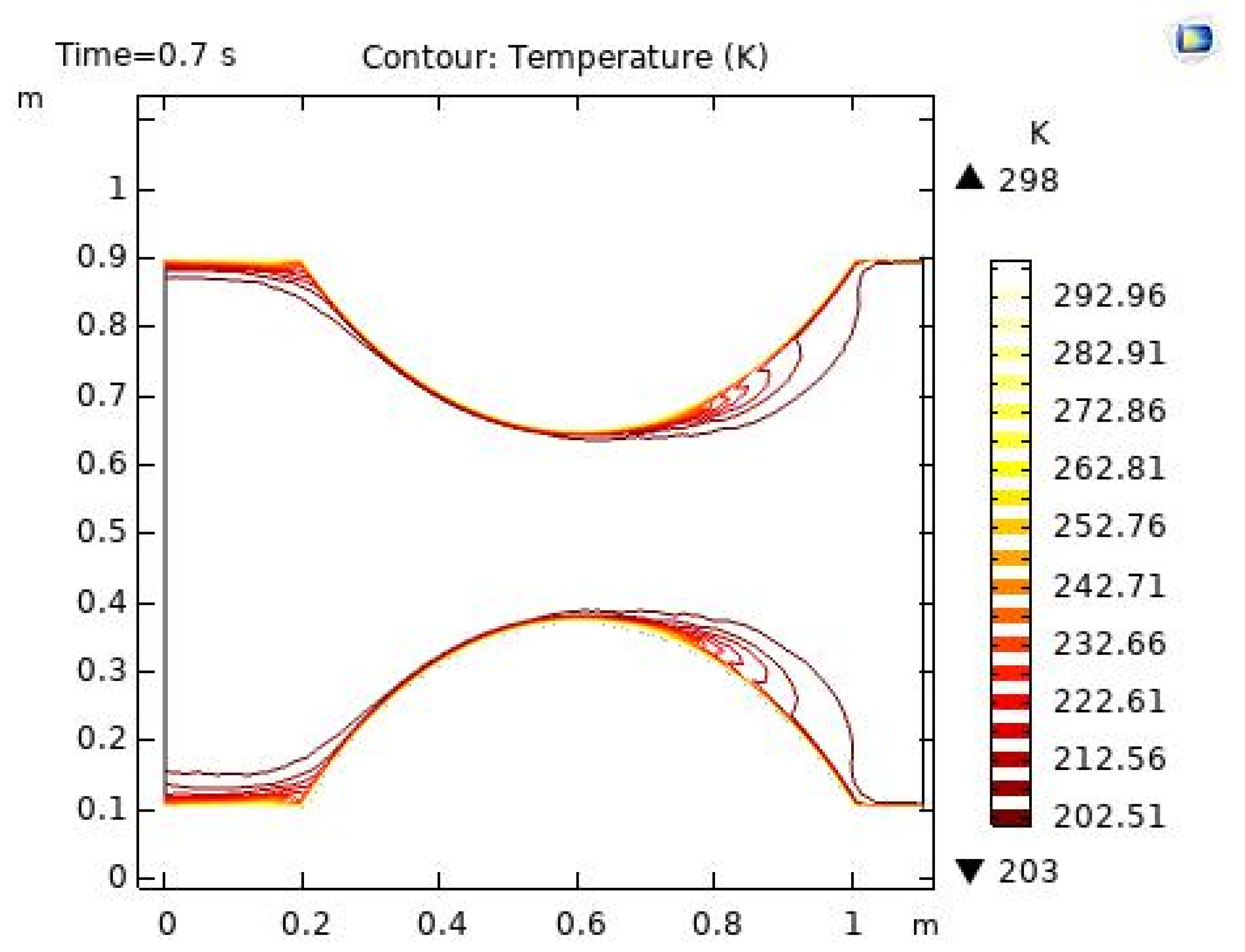
  • The isothermal contours displayed the results clearly on colored surfaces at t = 0.1, 0.7 and 1 s;
  • The temperature and Nusselt number curves varied for slight variations of time;
  • The pressure profiles also manifested clear patterns near the stenosed region;
  • We can further study the physical qualities, such as skin friction coefficient, and also analyze the problem using radiation and magnetohydrodynamic effects to understand the reasons for stenosis, which may help in the treatment of arterial stenosis.

Author Contributions

A.H.: conceptualization, supervision, L.S.: data curation, writing—original draft; A.R.: formal analysis, investigation, writing—original draft; S.A.: methodology, software; F.G.: visualization, validation, funding acquisition, writing—review & editing; H.H.C.: validation, resources, writing—review & editing; A.H.A.: investigation, formal analysis, funding acquisition, writing—review & editing; M.S.A.: visualization, validation, writing—review & editing. All authors have read and agreed to the published version of the manuscript.

Funding

This project was funded by the Taif University Researchers Supporting Project (TURSP-2020/252), Taif University, Taif, Saudi Arabia.

Institutional Review Board Statement

Not applicable.

Informed Consent Statement

Not applicable.

Data Availability Statement

Data available from corresponding author.

Acknowledgments

The authors would like to extend their appreciation to the Deanship of Scientific Research at King Khalid University, Saudi Arabia, for funding this work through the Research Group Program under grant no. RGP.1/11/43. Additionally, the authors would like to acknowledge financial support from the Taif University Researchers Supporting Project (TURSP-2020/252), Taif University, Taif, Saudi Arabia.

Conflicts of Interest

The authors declare no conflict of interest.

References

  1. Hayat, T.; Nadeem, S. Heat transfer enhancement with Ag-CuO/water hybrid nanofluid. Results Phys. 2017, 7, 2317–2324. [Google Scholar] [CrossRef]
  2. Nadeem, S.; Ijaz, S. Single wall carbon nanotube (SWCNT) examination on blood flow through a multiple stenosed artery with variable nanofluid viscosity. AIP Adv. 2015, 5, 107217. [Google Scholar] [CrossRef] [Green Version]
  3. Ghadikolaei, S.; Gholinia, M. Terrific effect of H2 on 3D free convection MHD flow of C2H6O2H2O hybrid base fluid to dissolve Cu nanoparticles in a porous space considering the thermal radiation and nanoparticle shapes effects. Int. J. Hydrog. Energy 2019, 44, 17072–17083. [Google Scholar] [CrossRef]
  4. Ghadikolaei, S.; Gholinia, M.; Hoseini, M.; Ganji, D. Natural convection MHD flow due to MoS2-Ag nanoparticles suspended in C2H6O2H2O hybrid base fluid with thermal radiation. J. Taiwan Inst. Chem. Eng. 2019, 97, 12–23. [Google Scholar] [CrossRef]
  5. Ijaz, S.; Sadiq, M.A. Inspiration of Induced Magnetic Field on a Blood Flow of Prandtl Nanofluid Model with Stenosis. Curr. Nanosci. 2014, 10, 753–765. [Google Scholar] [CrossRef]
  6. Elnaqeeb, T.; Shah, N.A.; Mekheimer, K. Hemodynamic Characteristics of Gold Nanoparticle Blood Flow Through a Tapered Stenosed Vessel with Variable Nanofluid Viscosity. BioNanoScience 2019, 9, 245–255. [Google Scholar] [CrossRef]
  7. Nadeem, S.; Ijaz, S.; Akbar, N.S. Nanoparticle analysis for blood flow of Prandtl fluid model with stenosis. Int. Nano Lett. 2013, 3, 35. [Google Scholar] [CrossRef] [Green Version]
  8. Ahmed, A.; Nadeem, S. Biomathematical study of time-dependent flow of a Carreau nanofluid through inclined catheterized arteries with overlapping stenosis. J. Cent. South Univ. 2017, 24, 2725–2744. [Google Scholar] [CrossRef]
  9. Ijaz, S.; Nadeem, S. Examination of nanoparticles as a drug carrier on blood flow through catheterized composite stenosed artery with permeable walls. Comput. Methods Programs Biomed. 2016, 1339, 83–94. [Google Scholar] [CrossRef] [PubMed]
  10. Akhtar, S.; McCash, L.B.; Nadeem, S.; Saleem, S.; Issakhov, A. Mechanics of non-Newtonian blood flow in an artery having multiple stenosis and electroosmotic effects. Sci. Prog. 2021, 104, 1–15. [Google Scholar] [CrossRef] [PubMed]
  11. Liu, J.; Wang, G.; Zhang, L.; Shi, Y.; Zhang, H.; Yao, S.-C. Numerical simulation of single bubble boiling behavior. Propuls. Power Res. 2017, 6, 117–125. [Google Scholar] [CrossRef]
  12. Hussain, A.; Hassan, A.; Al Mdallal, Q.; Ahmad, H.; Rehman, A.; Altanji, M.; Arshad, M. Heat transportation enrichment and elliptic cylindrical solution of time-dependent flow. Case Stud. Therm. Eng. 2021, 27, 101248. [Google Scholar] [CrossRef]
  13. Choudhari, P.; Panse, M. Finite Element Modeling and Simulation of Arteries in the Human Arm to Study the Aortic Pulse Wave Propagation. Procedia Comput. Sci. 2016, 93, 721–727. [Google Scholar] [CrossRef] [Green Version]
  14. Durantes, R.; Moon, J.; Pacheco, R.; Pacheco-Vega, A. Numerical Modeling of Single-Phase Fluid-Flow in Wavy Micro-Channels. In White Papers and Application Notes; California State University-Los Angeles: Los Angeles, CA, USA, 2020; Available online: https://www.comsol.com/paper/numerical-modeling-of-single-phase-fluid-flow-in-wavy-micro-channels-9371 (accessed on 28 November 2021).
  15. Ghadikolaei, S.; Gholinia, M. 3D mixed convection MHD flow of GO-MoS2 hybrid nanoparticles in H2O-(CH2OH)2 hybrid base fluid under the effect of H2 bond. Int. Commun. Heat Mass Transf. 2020, 110, 104371. [Google Scholar] [CrossRef]
  16. Tripathi, J.; Vasu, B.; Bég, O.A.; Gorla, R.S.R. Unsteady hybrid nanoparticle-mediated magneto-hemodynamics and heat transfer through an overlapped stenotic artery: Biomedical drug delivery simulation. Proc. Inst. Mech. Eng. Part H J. Eng. Med. 2021, 235, 1175–1196. [Google Scholar] [CrossRef] [PubMed]
  17. Nasrin, R.; Hossain, A.; Zahan, I. Blood flow analysis inside a stenotic artery using Power-Law fluid model. RDMS 2020, 13, 1–10. [Google Scholar] [CrossRef]
Figure 1. The geometry of arterial stenosis.
Applsci 12 01601 g001
Figure 2. The geometry of the inlet.
Applsci 12 01601 g002
Figure 3. The geometry of the outlet.
Applsci 12 01601 g003
Figure 4. The wall of arterial stenosis.
Applsci 12 01601 g004
Figure 5. The thermal insulated boundaries of the problem.
Applsci 12 01601 g005
Figure 6. Normal element size mesh.
Applsci 12 01601 g006
Figure 7. Fine element size mesh.
Applsci 12 01601 g007
Figure 8. The velocity profile at t = 0.1 s.
Applsci 12 01601 g008
Figure 9. The velocity profile at t = 0.7 s.
Applsci 12 01601 g009
Figure 10. The velocity profile at t = 1 s.
Applsci 12 01601 g010
Figure 11. The temperature profile at t = 0.1 s.
Applsci 12 01601 g011
Figure 12. The temperature profile at t = 0.7 s.
Applsci 12 01601 g012
Figure 13. The temperature profile at t = 1 s.
Applsci 12 01601 g013
Figure 14. The pressure profile at t = 0.1 s.
Applsci 12 01601 g014
Figure 15. TThe pressure profile at t = 0.7 s.
Applsci 12 01601 g015
Figure 16. The pressure profile at t = 1 s.
Applsci 12 01601 g016
Figure 17. The isothermal contour profile at t = 0.1 s.
Applsci 12 01601 g017
Figure 18. The isothermal contour profile at t = 0.7 s.
Applsci 12 01601 g018
Figure 19. The isothermal contour profile at t = 1 s.
Applsci 12 01601 g019
Figure 20. The streamlines at t = 0.1 s.
Applsci 12 01601 g020
Figure 21. The streamlines at t = 0.7 s.
Applsci 12 01601 g021
Figure 22. The streamlines at t = 1 s.
Applsci 12 01601 g022
Figure 23. The Nusselt number variation with time.
Applsci 12 01601 g023
Figure 24. The validation figures with [17] and our obtained results.
Applsci 12 01601 g024
Table 1. The mesh elements computed using COMSOL.
ElementsElement SizeMesh Area
Mesh 1Normal0.5949 m2
Present studyMesh 2Fine0.5948 m2
Table 2. A description of the mesh statistics.
PropertyValue
Mesh vertices1195
Edge elements173
Quadrilateral entities234
Triangles1747
Vertex elements8
Average element quality0.8291
Minimum element quality0.3539
Mesh area0.5948 m2
Total no. of elements1981
Ratio area of element0.06767
Table 3. A description of the mesh sizes.
PropertyValue
Maximum element growth rate1.13
Maximum element size0.028
Minimum element size8 × 10−4
Curvature factor0.3
Resolution of narrow region1
Geometric entity levelEntire geometry
Table 4. The thermophysical properties of the blood base fluid and the silver and gold nanoparticles [16].
PropertyVariableBloodGoldSilverUnit
Dynamic viscosity μ 0.0030.004640.005Pa. s
Heat capacity C p 3746129235J/(kg. K)
Thermal conductivityK0.52310429W/(m. K)
Density ρ 106319,30010,500Kg/m2
Publisher’s Note: MDPI stays neutral with regard to jurisdictional claims in published maps and institutional affiliations.

