
Effect of Gamma Irradiation on the Optical Properties of the Conjugated Copolymer B-co-MP


1
Department of Physics, College of Science, Imam Mohammad Ibn Saud Islamic University (IMSIU), Riyadh 13318, Saudi Arabia
2
Department of Physics, College of Women for Arts, Science and Education, Ain-Shams University, Cairo 11757, Egypt
*
Author to whom correspondence should be addressed.
Appl. Sci. 2022, 12(3), 1606; https://0-doi-org.brum.beds.ac.uk/10.3390/app12031606
Submission received: 16 January 2022
/
Revised: 29 January 2022
/
Accepted: 31 January 2022
/
Published: 3 February 2022
(This article belongs to the Special Issue Spectroscopic Characterization of Polymers and Composite Materials)
Abstract
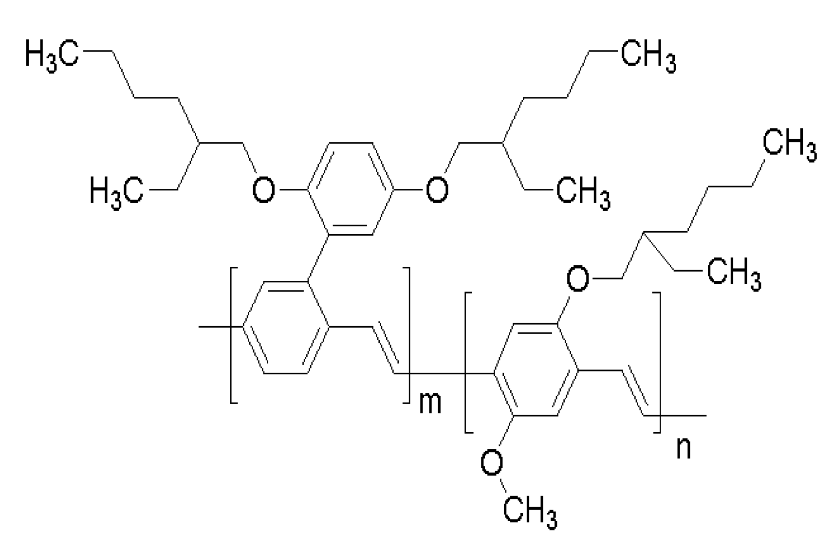
:The conjugated polymer poly {[2-[2′,5′-bis(2″-ethylhexyloxy)phenyl]-1,4-phenylenevinylene]-co-[2-methoxy-5-(2′-ethylhexyloxy)-1,4-phenylenevinylene]} (B-co-MP) has been proven to be an excellent laser medium with a high photochemical stability. Moreover, the impact of γ-irradiation on its optical and chemical properties has been investigated. Herein, the spectral and amplified spontaneous emission (ASE) characteristics of B-co-MP at various concentrations under γ-irradiation doses are studied. Various concentrations of B-co-MP in tetrahydrofuran (THF) were prepared. The samples were irradiated with various γ-doses from 5 to 20 kGy using a Co-60 source at room temperature. The absorption, fluorescence, and ASE spectra were dramatically blue-shifted after the γ-irradiation. This indicates that the increment in the γ-irradiation dose led to a widening in the energy gap and reduction in the number of carbon atoms (N). The change in the spectral profiles could be attributed to chain conformational alterations and/or chain scission induced by the γ-irradiation. We anticipate this study to boost our understanding of optical and structural profiles of B-co-MP under various conditions, including γ-irradiation and the potential utility of this copolymer in a variety of applications.
1. Introduction
Conjugated polymers have drawn considerable attention due to their great number of applications in different disciplines such as light-emitting diodes (LEDs) and luminescent solar cells, as well as their low cost and facile synthesis [1,2,3,4,5]. Among conjugated polymers, the poly(phenylene vinylene) (PPV) group has fascinated researchers due to its novel optical and electrical features [6,7,8,9,10,11]. Conjugated polymers have been foreseen as potential radiation-sensing materials [12,13,14]. Conjugated polymers can be stimulated by injecting charges or utilizing photo-excitation. Optically active organic dyes, such as laser dyes, have spectral features similar to these macromolecules. However, many fluorescent organic compounds, including laser dyes, have a low fluorescence quantum yield under high concentrations due to reabsorption processes, and cannot emit laser light [15]. At variance, the PPV conjugated polymers have a strong absorption and emission, a significant Stokes shift, a high luminescence, and a fluorescence quantum yield, and emit laser light even at higher concentrations. In 1992, researchers discovered that a liquid conjugated polymer (MEH-PPV) produced yellow/red wavelengths that generated laser action.
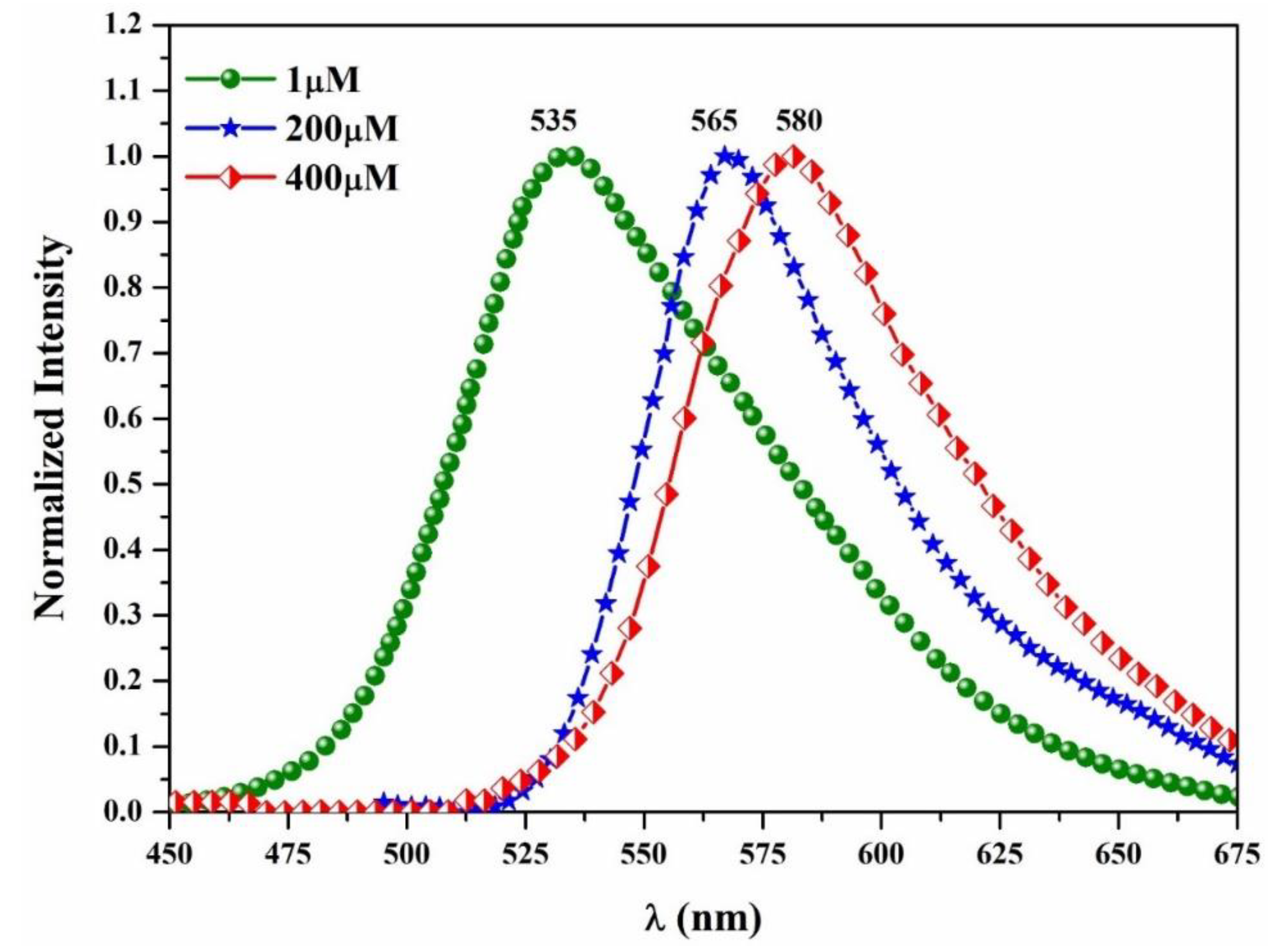
Furthermore, the efficiency of MEH-PPV was comparable to that of the conventional laser dye rhodamine 6G [16]. The spectral properties of B-co-MP were studied in a few organic solvents. The fluorescence spectrum of B-co-MP for low concentrations showed one peak at 535 nm. The intensity decreased by increasing the concentration and the spectrum’s peak position red-shifted towards and became fixed at 580 nm [17,18]. Under laser pump excitation (Nd: YAG (355 nm)), B-co-MP exhibits an amplified spontaneous emission (ASE) spectrum at 560 nm. This peak may be related to the monomeric state of MEH- PPV as a segment [19].
On the other hand, the ionizing radiation technique is widely used to induce changes in structural and optical properties of polymers subjected to irradiation [20]. According to the experimental findings, ionizing radiation may induce a cross-link or break in polymers’ molecular bonds based on the polymer structure, type of ionizing radiation, and absorbed dose [21,22]. Therefore, investigating the impact of radiation on these polymers is highly needed, which can be potentially exploited in diverse fields. For instance, A.M. Abdul-Kader et al. reported that the optical and electrical properties of poly allyl diglycol carbonate (CR-39) were enhanced upon gamma irradiation in the dose range of up to nearly 1 MGy [23]. In a previous study, gamma irradiation influenced both films and solutions in the chloroform of poly[2-methoxy-5-(2′-hexyloxy)-p-phenylenevinylene] (MEH-PPV). MEH-PPV in solution was sensitive to low doses of ionizing radiation. Recently, polypyrrole and polyvinylpyrrolidone hydrogels with varying concentrations and content were irradiated with gamma rays at 25 kGy to facilitate polymerization and cross-linking, resulting in PPy/PVP hydrogels with optimal mechanical properties [24]. This work attempts to provide a deeper insight into the optical properties of the conjugated copolymer of B-co-MP in tetrahydrofuran (THF) as a function of concentrations and irradiation doses. We highlight that the spectral profiles of B-co-MP and ASE wavelengths change remarkably upon varying the γ-irradiation dose. The low-cost, easy preparation, and the proper dosimetric response of B-co-MP render it an effective tool for biomedical applications.
2. Experimental
2.1. Materials
The copolymer poly B-co-MP was purchased from Sigma-Aldrich, which was used as received. The molecular structure of this compound is seen in Figure 1. The copolymer’s two components, BEHP-PPV and MEH-PPV, had a 60:40 fractional ratio. By dissolving B-co-MP in tetrahydrofuran (THF, with a purity of 99.8%), solutions with variable concentrations were prepared.
2.2. γ-Irradiation and Characterization of Prepared Samples
Gamma cells (type GC-220 Excel (manufactured by MDS Nordion, Ottawa, ON, Canada) at the King Abdul Aziz City for Science and Technology (KACST), Atomic Energy Institute, were used for γ-irradiation using a Co-60 source. The irradiation procedure was accomplished in air at room temperature. B-co-MP solutions were exposed to various doses (5, 10, 15, 20 kGy) at a 12.67 kGy/h dose rate. The JASCO V-770 spectrophotometer was used to obtain the UV–Vis absorption spectra; scans were performed at 37 nm/min with a bandwidth of 1 nm throughout the range from 100 to 1100 nm. The fluorescence spectra were recorded using a PerkinElmer LS45 spectrofluorometer. All of these measurements were obtained at room temperature.
3. Results and Discussion
3.1. Absorption Spectra of B-co-MP
Figure 2 reveals the UV–vis absorption spectra of B-co-MP in the THF solution at a concentration of 1 µM, pre-and post-different doses of γ-irradiation. The non-irradiated sample showed double peaks, one around 305 nm and the other around 450 nm. These absorption peaks were attributed to π–π* (HOMO–LUMO) transitions [4]. The same sample was then irradiated with doses ranging from 5 to 20 kGy at a constant temperature (25 °C). The results showed that the central peak at 450 nm was blue-shifted (shorter wavelength), with a significant decrease in the absorbance after irradiation, as demonstrated in Figure 2. As shown in the inset of Figure 2, the peak position shift was linearly proportional to the gamma radiation dose.
Following the same procedure, five different solutions of various concentrations (1, 10, 100, 200, and 400 µM) were prepared and irradiated with four different doses (5, 10, 15, and 20 kGy) for each solution. Figure 3 displays the shift in the central maximum wavelength peak (450 nm) measured as a function of the solution concentration. The shift in the peak was linearly dependent on the gamma dose for each concentration studied. Furthermore, it is noteworthy that the shift amount also relied linearly on the concentration of the prepared solutions; for example, at a fixed γ-irradiation dose (10 kGy), the concentrations of 1, 10, 100, 200, and 400 µM yielded blue shifts corresponding to 7, 11, 14, 22, and 23 nm, respectively (see inset Figure 3). The plausible explanation for the blue shift observed in the primary absorption peak is the change in the polymer structure caused by a reduction in the copolymer’s conjugation length due to a free radical interaction with the polymer backbone caused by solvent radiolysis [25,26].
3.2. Energy Band-Gap of B-co-MP
The energy band-gap (), the number of carbon atoms per conjugation length (N), and the refractive index () all had an impact on the material’s optical properties. The energy band-gap of B-co-MP in the THF was calculated from the absorption spectra before and after γ-irradiation, using the Tauc equation [27].
where α is the absorption coefficient, is the incident photons energy, B is the constant, and is an empirical power representing the electronic transition type. The value of corresponds to 1/2 or 3/2 for the direct allowed or direct forbidden transitions, respectively. However, it took values of two and three for the indirect allowed and indirect forbidden transitions, respectively [23]. The studied samples showed a direct allowed transition by plotting (αhν)2 versus hv as demonstrated in Figure 4. The straight line’s extrapolation obtained the values to the hν-axis, and its values are mentioned in Table 1. It could be seen that the energy band-gap of the copolymer increased from 2.28 eV for the non-irradiated to 2.53 eV for the highest dose γ-irradiated samples. The expansion of the energy band-gap can be ascribed to bond breaking in the polymer caused by the interaction of γ-irradiation with a polymer, decreasing the effective conjugation length.
3.3. Carbon Atoms Number of B-co-MP
The carbonaceous cluster is one of the essential factors that influence the optical properties of polymer materials. For example, it is known that the following relation (Equation (2)) is a correlation between the carbon atoms number N per conjugation length and the band-gap, which can be calculated [28].
where β denotes the band structure energy of a neighboring π sites pair, and β is correlated with π–π* optical transitions in a –C = C– structure and equal to 2.9 eV. The reduction in the conjugation length caused a blue shift in the prominent absorption spectra of the irradiated samples [21]. The carbon atoms number per conjugation length decreased slightly from eight for the non-irradiated to seven for the irradiated sample of the dose of 20 kGy, as shown in Table 1. The reduction in the unsaturated conjugation length in polymer samples resulted in coloration, where samples became red to blue/green with the increasing in the γ-irradiation dose (data not shown). The values of can be used to compute the values of the refractive index, , for the studied B-co-MP in THF via the following equation [29,30]:
The calculated refractive index is listed in Table 1. Results showed that the index of the refractive value decreased as the γ-irradiation increased, and its value for the studied samples was noticeably high. Thus, these studied samples with a high refractive index could be employed in manufacturing optoelectronic devices and anti-reflective coatings [31].
3.4. Fluorescence and ASE Spectra
On the other hand, B-co-MP’s fluorescence spectra in THF at various concentrations (1, 200, and 400 µM) pre-irradiation were recorded. The result shows that there was only one fluorescence peak around 535 nm. The spectrum’s wavelength was red-shifted and fixed at 580 nm when the concentration increased, as shown in Figure 5.
After that, a solution of 1 µM was irradiated with four different doses under a constant temperature of 25 °C. The results showed that the spectrum exhibited a blue shift as the γ-irradiation dose increased. For example, at the 5 kGy dose, the profile of the fluorescence spectrum blue-shifted 9 nm. Once the exposure dose increased to 10 kGy, the fluorescence spectrum shifted about 16 nm. Eventually, at a dose of 15 and 20 kGy, the fluorescence spectrum moved 26 and 35 nm, respectively, as shown in Figure 6. These findings were consistent with the absorption profiles as described earlier.
B-co-MP’s fluorescence peak in THF for the five concentrations (1, 10, 100, 200, and 400 µM) with four different doses (5, 10, 15, and 20 kGy) are presented in Figure 7. The study revealed that the solutions with high concentrations had more substantial blue shifts than diluted ones under the same operational conditions (see the slope of the equations displayed in Figure 7). This result was in line with our hypothesis that the B-co-MP is an excellent material for gamma detectors that can be used for dosimetric purposes.
3.5. ASE Spectra
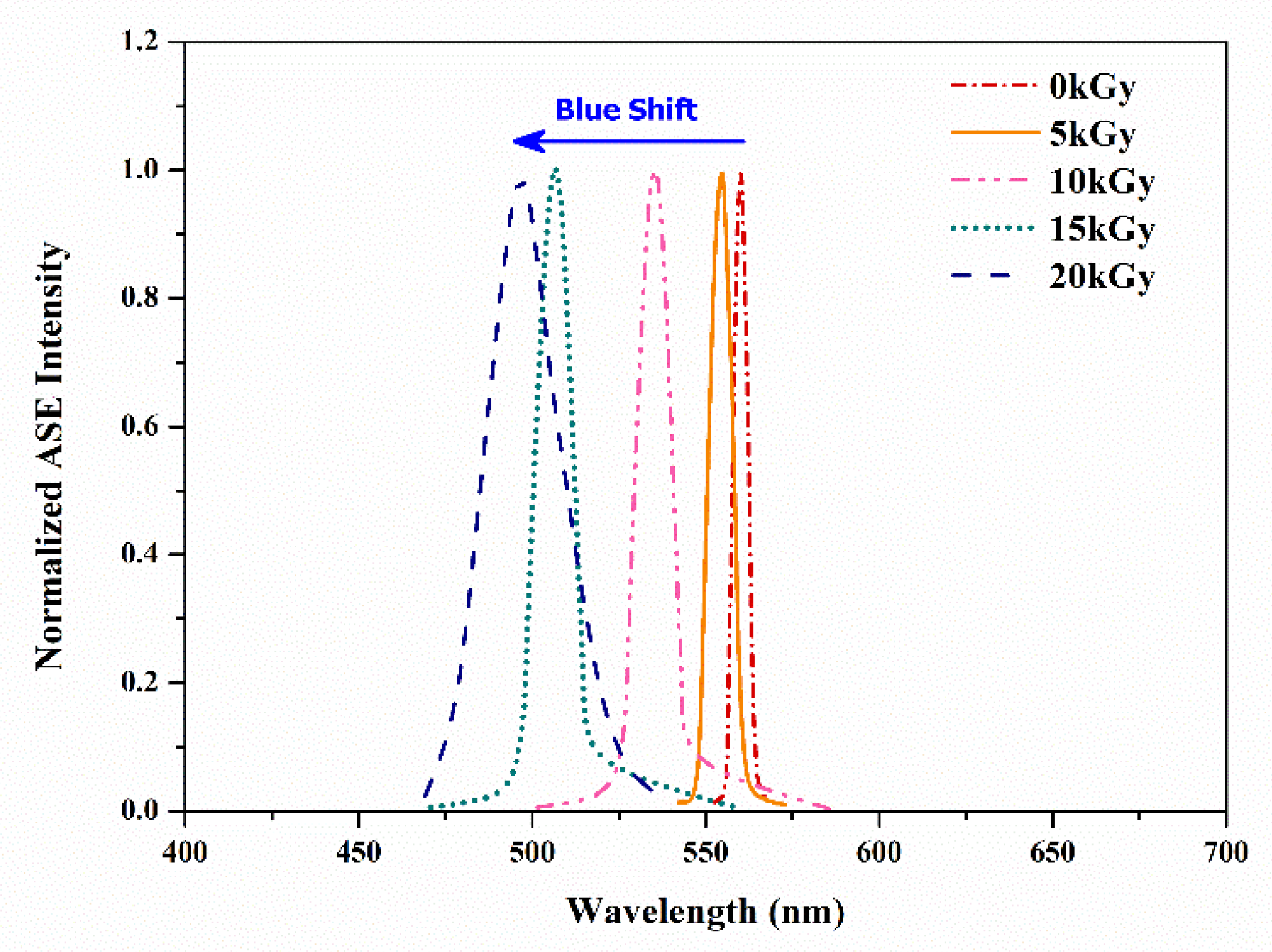
The ASE spectrum of B-co-MP in THF at a 400 µM concentration pre-γ-irradiation is shown in Figure 8. An Nd:YAG laser (λ = 355 nm) was used to excite the solution. The ASE peak was centered at 560 nm with a spectral bandwidth of 5 nm at the full-width half maximum. At this concentration, the ASE at 560 nm did not coincide with the fluorescence peak (580 nm). Previously, we demonstrated that B-co-MP formed ASE only as a monomer (560 nm) due to the MEH-PPV segment [17]. We exposed the same solution to different gamma doses (5 to 20 kGy), followed by laser excitation at an energy of 12 mJ. In Figure 8, the ASE peak was observed at 554 nm with an FWHM of 8 nm at 5 kGy. Doubling the gamma dose to 10 kGy yielded a blue shift to 535 nm with an FWHM of 11 nm. At 15 kGy, the ASE peak shifted to 506 nm with an FWHM of 13 nm. Interestingly, for the 20 kGy, we observed laser-induced fluorescence with an FWHM of 25 nm and no ASE detected.
4. Conclusions
To sum up, B-co-MP’s optical characteristics in THF at different concentrations were obtained pre-and post-γ-irradiation with varying doses. The results showed that a dose greater than 5 kGy directly affected the peak positions of the absorption and fluorescence spectra. Under high γ-irradiation doses, the absorption, fluorescence, and ASE spectra shifted to the blue region. These effects extended to the energy band-gap value and the number of carbon atoms per cluster in the polymeric samples. In addition to its low cost and ease of preparation, the polymer B-co-MP is an excellent candidate for medical, dosimetric, and optoelectronic devices.
Author Contributions
Conceptualization, B.A.E.-B. and K.H.I.; methodology, O.A.A., B.A.E.-B., M.K.M.A. and K.H.I.; validation, K.H.I.; formal analysis, O.A.A., B.A.E.-B., M.K.M.A. and K.H.I.; investigation, O.A.A., B.A.E.-B., M.K.M.A. and K.H.I.; resources, O.A.A., B.A.E.-B., M.K.M.A. and K.H.I.; data curation, O.A.A., B.A.E.-B., M.K.M.A. and K.H.I.; writing—original draft preparation, O.A.A., B.A.E.-B., M.K.M.A. and K.H.I.; writing—review and editing, O.A.A., B.A.E.-B., M.K.M.A. and K.H.I.; visualization, O.A.A.; supervision, K.H.I.; project administration, O.A.A. and K.H.I.; funding acquisition, K.H.I. All authors have read and agreed to the published version of the manuscript.
Funding
This research was funded by the Deanship of Scientific Research at the Imam Mohammad Ibn Saud Islamic University for funding this work through Research Group no. RG-21-09-46.
Institutional Review Board Statement
Not applicable.
Informed Consent Statement
Not applicable.
Acknowledgments
The authors extend their appreciation to the Scientific Research at the Imam Mohammad Ibn Saud Islamic University for funding this work through Research Group no. RG-21-09-46. The authors gratefully acknowledge Ahmed Alsadig (University of Trieste) for his kind assistance throughout this work.
Conflicts of Interest
The authors declare no conflict of interest.
References
- Tessler, N. Lasers based on semiconducting organic materials. Adv. Mater. 1999, 11, 363–370. [Google Scholar] [CrossRef]
- Neureiter, H.; Gebauer, W.; Väterlein, C.; Sokolowski, M.; Bäuerle, P.; Umbach, E. Study of charge carrier injection and luminescence processes in oligothiophene-based light-emitting diodes. Synth. Met. 1994, 67, 173–176. [Google Scholar] [CrossRef]
- Graham, S.C.; Friend, R.H.; Fung, S.; Moratti, S.C. The effect of X-ray irradiation on poly (p-phenylene vinylene) and derivatives. Synth. Met. 1997, 84, 903–904. [Google Scholar] [CrossRef]
- Silva, E.A.B.; Borin, J.F.; Nicolucci, P.; Graeff, C.F.O.; Netto, T.G.; Bianchi, R.F. Low dose ionizing radiation detection using conjugated polymers. Appl. Phys. Lett. 2005, 86, 131902. [Google Scholar] [CrossRef]
- Lobez, J.M.; Swager, T.M. Radiation detection: Resistivity responses in functional poly (olefin sulfone)/carbon nanotube composites. Angew. Chem. Int. Ed. 2010, 49, 95–98. [Google Scholar] [CrossRef] [Green Version]
- Friend, R.H.; Gymer, R.W.; Holmes, A.B.; Burroughes, J.H.; Marks, R.N.; Taliani, C.; Bradley, D.D.C.; Dos Santos, D.A.; Brédas, J.L.; Lögdlund, M.; et al. Electroluminescence in conjugated polymers. Nature 1999, 397, 121–128. [Google Scholar] [CrossRef]
- Ho, P.K.; Kim, J.S.; Burroughes, J.H.; Becker, H.; Li, S.F.; Brown, T.M.; Cacialli, F.; Friend, R.H. Molecular-scale interface engineering for polymer light-emitting diodes. Nature 2000, 404, 481–484. [Google Scholar] [CrossRef]
- Martens, H.; Huiberts, J.; Blom, P. Simultaneous measurement of electron and hole mobilities in polymer light-emitting diodes. Appl. Phys. Lett. 2000, 77, 1852–1854. [Google Scholar] [CrossRef] [Green Version]
- Crone, B.K.; Campbell, I.H.; Davids, P.S.; Smith, D.L. Charge injection and transport in single-layer organic light-emitting diodes. Appl. Phys. Lett. 1998, 73, 3162–3164. [Google Scholar] [CrossRef]
- Jang, J.W.; Lee, C.E.; Lee, D.W.; Jin, J.I. Transient electroluminescence study of mobility balancing in organic light-emitting diodes based on poly (p-phenylenevinylene) derivatives. Solid State Commun. 2004, 130, 265–268. [Google Scholar] [CrossRef]
- Lee, H.M.; Oh, D.K.; Lee, C.H.; Lee, C.E.; Lee, D.W.; Jin, J.I. Time-of-flight measurements of charge-carrier mobilities in a poly (p-phenylenevinylene) derivative carrying an electron-transporting moiety. Synth. Met. 2001, 119, 473–474. [Google Scholar] [CrossRef]
- Isa, N.M.; Baharin, R.; Majid, R.A.; Rahman, W.A.W. Optical properties of conjugated polymer: Review of its change mechanism for ionizing radiation sensor. Polym. Adv. Technol. 2017, 28, 1559–1571. [Google Scholar] [CrossRef]
- Desiraju, G.R.; Hulliger, J. Current Opinion in Solid State & Materials Science-Molecular Crystals and Materials. Curr. Opin. Solid State Mater. Sci. 2001, 2, 105–106. [Google Scholar]
- Zhong, H.; Zhao, Y.; Li, Y.; Pei, Q. Photoluminescence quenching of conjugated polymer nanocomposites for gamma ray detection. Nanotechnology 2008, 19, 505503. [Google Scholar] [CrossRef]
- Mylnikov, V.S. Photoconducting polymers. In Photoconducting Polymers/Metal-Containing Polymers; Springer: Berlin/Heidelberg, Germany, 1994; pp. 1–88. [Google Scholar]
- Moses, D. High quantum efficiency luminescence from a conducting polymer in solution: A novel polymer laser dye. Appl. Phys. Lett. 1992, 60, 3215–3216. [Google Scholar] [CrossRef]
- AlSalhi, M.S.; Ibnaouf, K.H.; Masilamani, V.; Yassin, O.A. Amplified spontaneous emission from internal energy transfer process in the copolymer BEHP-co-MEH-PPV. J. Lumin. 2012, 132, 484–490. [Google Scholar] [CrossRef]
- Ibnaouf, K.H.; Prasad, S.; Masilamani, V.; AlSalhi, M.S.; Mustapha, N.; Alyamani, A. Triple amplified spontaneous emissions from a conjugated copolymer BEHP-co-MEH-PPV in solution. Phys. E Low-Dimens. Syst. Nanostruct. 2013, 53, 66–71. [Google Scholar] [CrossRef]
- Ibnaouf, K.H.; Prasad, S.; Al Salhi, M.S.; Hamdan, A.; Zaman, M.B.; El Mir, L. Influence of the solvent environments on the spectral features of CdSe quantum dots with and without ZnS shell. J. Lumin. 2014, 149, 369–373. [Google Scholar] [CrossRef]
- Mariani, M.; Consolati, G.; Quasso, F.; Lotti, N.; Munari, A.; Galletta, M.; Macerata, E. Effects of gamma irradiation on poly (ethylene isophthalate). J. Radioanal. Nucl. Chem. 2010, 286, 625–629. [Google Scholar] [CrossRef]
- Batagin-Neto, A.; Bronze-Uhle, E.S.; Fernandes, D.M.; Fratoddi, I.; Venditti, I.; Decker, F.; Bodo, E.; Russo, M.V.; Graeff, C.F.O. Optical behavior of conjugated Pt-containing polymetallaynes exposed to gamma-ray radiation doses. J. Phys. Chem. B 2011, 115, 8047–8053. [Google Scholar] [CrossRef]
- Slimani, K.; Moine, L.; Aymes-Chodur, C.; Laurent, A.; Labarre, D.; Yagoubi, N. Determination of scission, crosslinking and branching parameters of electron beam irradiated methacrylate–acrylamide copolymer. Polym. Degrad. Stab. 2009, 94, 584–590. [Google Scholar] [CrossRef]
- Abdul-Kader, A.; Zaki, M.; El-Badry, B.A. Modified the optical and electrical properties of CR-39 by gamma ray irradiation. J. Radiat. Res. Appl. Sci. 2014, 7, 286–291. [Google Scholar] [CrossRef] [Green Version]
- El-Badry, B.A.; Zaki, M.F.; Abdul-Kader, A.M.; Hegazy, T.M.; Morsy, A.A. Ion bombardment of poly-allyl-diglycol-carbonate (CR-39). Vacuum 2009, 83, 1138–1142. [Google Scholar] [CrossRef]
- Jeong, J.-O.; Park, J.S.; Kim, Y.A.; Yang, S.J.; Jeong, S.I.; Lee, J.Y.; Lim, Y.M. Gamma ray-induced polymerization and cross-linking for optimization of PPy/PVP hydrogel as biomaterial. Polymers 2020, 12, 111. [Google Scholar] [CrossRef] [Green Version]
- Bronze-Uhle, E.S.; Batagin-Neto, A.; Lavarda, F.C.; Graeff, C.F.O. Ionizing radiation induced degradation of poly (2-methoxy-5-(2’-ethyl- hexyloxy) -1,4-phenylene vinylene) in solution. J. Appl. Phys. 2011, 110, 073510. [Google Scholar] [CrossRef] [Green Version]
- Alwan, T.J. Effects of gamma irradiation on the physical properties of PAni.MWCNT/PMMA films. J. Phys. Stud. 2019, 23, 6–11. [Google Scholar] [CrossRef]
- Romanova, E.; Melnikov, A.; Kuzutkina, Y.; Shiryaev, V.; Guizard, S.; Mouskeftaras, A. Nonlinear optical properties of amorphous semiconductors. In Proceedings of the 2012 International Conference on Mathematical Methods in Electromagnetic Theory, MMET, Kharkiv, UKraine, 28–30 August 2012; pp. 521–526. [Google Scholar] [CrossRef]
- Al-Naggar, T.I.; El-Badry, B.A.; All, N.F.A. Study the modifications induced by alphaparticles in cellulose nitrate NTD. Vacuum 2018, 160, 31–36. [Google Scholar] [CrossRef]
- Rammah, Y.S.; Ibrahim, S.E.; Awad, E.M. Electrical and optical properties of Makrofol DE 1-1 polymeric films induced by gamma irradiation. Bull. Natl. Res. Cent. 2019, 43, 1–10. [Google Scholar] [CrossRef]
- Kurt, A. Influence of Alcl3 on the optical properties of new synthesized 3-Armed poly(methyl methacrylate) films. Turk. J. Chem. 2010, 34, 67–79. [Google Scholar] [CrossRef]
Figure 1.
Molecular structures of the copolymer B-co-MP.

Figure 2.
UV–vis absorption spectra of B-co-MP in THF at a concentration of 1 µM at different gamma doses.
Figure 2.
UV–vis absorption spectra of B-co-MP in THF at a concentration of 1 µM at different gamma doses.

Figure 3.
The main absorption peak wavelength (λmax) of the B-co-MP in THF versus gamma doses at different concentrations.
Figure 3.
The main absorption peak wavelength (λmax) of the B-co-MP in THF versus gamma doses at different concentrations.

Figure 4.
Photon energy (hν) and (αhν)2 of B-co-MP in THF at a concentration of 1 µM at different gamma doses.
Figure 4.
Photon energy (hν) and (αhν)2 of B-co-MP in THF at a concentration of 1 µM at different gamma doses.

Figure 5.
The fluorescence spectra of B-co-MP in THF measured for different concentrations pre-irradiation.
Figure 5.
The fluorescence spectra of B-co-MP in THF measured for different concentrations pre-irradiation.

Figure 6.
Fluorescence spectra of B-co-MP in THF at a concentration of 1 µM at different γ-irradiation doses.
Figure 6.
Fluorescence spectra of B-co-MP in THF at a concentration of 1 µM at different γ-irradiation doses.

Figure 7.
The main fluorescence peak wavelength (λmax) of the B-co-MP in THF versus gamma doses for different concentrations.
Figure 7.
The main fluorescence peak wavelength (λmax) of the B-co-MP in THF versus gamma doses for different concentrations.

Figure 8.
ASE spectra of B-co-MP in THF at concentration of 400 µM for different doses.


Table 1.
The variation of optical energy band-gap (), the number of carbon atoms per conjugated length (N), and refractive index () for B-co-MP in THF at a concentration of 1 µM at different gamma doses.
Table 1.
The variation of optical energy band-gap (), the number of carbon atoms per conjugated length (N), and refractive index () for B-co-MP in THF at a concentration of 1 µM at different gamma doses.
| Gamma Dose (KGy) | Optical Band-Gap Energy (eV) | Number of Carbon Atoms per Conjugated Length (N) | Refractive Index (n) |
|---|---|---|---|
| 0 | 2.28 | ~ 8.0 | 2.62 |
| 5 | 2.34 | ~7.8 | 2.60 |
| 10 | 2.4 | ~7.6 | 2.58 |
| 15 | 2.45 | ~7.4 | 2.56 |
| 20 | 2.53 | ~7.0 | 2.54 |
Publisher’s Note: MDPI stays neutral with regard to jurisdictional claims in published maps and institutional affiliations. |
© 2022 by the authors. Licensee MDPI, Basel, Switzerland. This article is an open access article distributed under the terms and conditions of the Creative Commons Attribution (CC BY) license (https://creativecommons.org/licenses/by/4.0/).
Share and Cite
MDPI and ACS Style
Aldaghri, O.A.; El-Badry, B.A.; Ali, M.K.M.; Ibnaouf, K.H. Effect of Gamma Irradiation on the Optical Properties of the Conjugated Copolymer B-co-MP. Appl. Sci. 2022, 12, 1606. https://0-doi-org.brum.beds.ac.uk/10.3390/app12031606
AMA Style
Aldaghri OA, El-Badry BA, Ali MKM, Ibnaouf KH. Effect of Gamma Irradiation on the Optical Properties of the Conjugated Copolymer B-co-MP. Applied Sciences. 2022; 12(3):1606. https://0-doi-org.brum.beds.ac.uk/10.3390/app12031606
Chicago/Turabian StyleAldaghri, Osamah A., Basma A. El-Badry, Mohammed Khalil M. Ali, and Khalid H. Ibnaouf. 2022. "Effect of Gamma Irradiation on the Optical Properties of the Conjugated Copolymer B-co-MP" Applied Sciences 12, no. 3: 1606. https://0-doi-org.brum.beds.ac.uk/10.3390/app12031606
Note that from the first issue of 2016, this journal uses article numbers instead of page numbers. See further details here.