Article

Enhancement of the Neuroprotective Effect of Fermented Spirulina maxima Associated with Antioxidant Activities by Ultrasonic Extraction

1
Department of Medical Biomaterials Engineering, Kangwon National University, Chuncheon 24341, Korea
2
The Jeju International Marine Science Center for Research & Education, Korea Institute of Ocean Science and Technology (KIOST), Jeju 49111, Korea
3
Department of Food Science and Engineering, Seowon University, Cheongju 28674, Korea
*
Author to whom correspondence should be addressed.
Submission received: 9 November 2018 / Revised: 28 November 2018 / Accepted: 29 November 2018 / Published: 3 December 2018
(This article belongs to the Collection Ultrasound in Extraction Processing)

Abstract

:
This study is the first demonstration of the neuroprotective activity of the marine alga, Spirulina maxima, fermented by Lactobacillus plantarum HY-08, which was freshly isolated from fermented vegetables. The combined process of the fermentation with ultrasonic extraction at 40 kHz for 4 hr greatly increased the amounts of β-carotene in the extract up to 1.62 mg/g, along with a high extraction yield of 18.26%, compared to 1.03 mg/g and 12.8% from ultrasonic extraction itself, and even much higher than 0.81 mg/g and 10.6% from conventional water extraction. It was also proved that the neuroprotective activities of the extract were enhanced possibly by higher contents of β-carotene in the extract, compared to those from water extraction or ultrasonic extraction itself. This extract was found to have strong antioxidant properties, with 49.01% 1,1-diphenyl-2-picrylhydrazyl (DPPH) radical scavenging activity, which resulted in 90.30% neuroprotective activity. Specifically, the fermented extract markedly enhanced the brain-derived neurotrophic factor (BDNF)/p-CREB signaling pathways, which can effectively prevent memory impairment in mice caused by oxidative stress. β-Carotene also increased the expression of p-CREB and BDNF, and a mechanism of their upregulation by β-carotene is first proposed in this study. It can be concluded that the strong neuroprotective effect of the extract was markedly influenced by the antioxidant activities, due to the synergistic effects of high amounts of β-carotene and other biologically active substances in the extract that can be obtained by lactic acid fermentation, combined with ultrasonic processing.

Graphical Abstract

1. Introduction

Dementia is a degenerative brain disease characterized by a decline in learning, and there is a considerable increase of social interest in its prevention and treatment [1]. In general, the mechanisms underlying the phenomena of the decline in memory and cognitive functions have been understood to be an excessive accumulation of amyloid β protein in the brain [2]. In additions to this, neurological degeneration resulting from nerve cell damage occurring during the decomposition of acetylcholine (ACh) by acetylcholinesterase (AChE) has also been considered to be a major mechanism that is associated with dementia [2,3]. Prolyl endopeptidase (PEP) is a protease that is known to be highly involved in the generation of amyloid β protein, and it digests proline, and it is one of key neuropeptides in the cerebral cortex of the brain [4,5]; therefore, PEP inhibitors have been developed as therapeutic agents to overcome declines in memory and cognitive function. In addition, several chemicals that act as acetylcholinesterase inhibitors, such as donepezil, rivastigmine, and galantamine, have also been developed to improve memory in dementia patients. However, these chemicals have demonstrated side effects such as hepatotoxicity, vomiting, diarrhea, and gastrointestinal disorders. Therefore, research has been actively conducted to develop therapeutic agents from natural resources to replace these medicines [6,7,8]. For this purpose, multiple studies have been conducted to develop health functional foods that help to improve cognitive function or memory impairment, such as Korean angelica roots, ginkgo leaves, and Codonopsis lanceolata [9,10,11]. The primary mechanism of these plants in enhancing cognitive function has been found to be their strong antioxidant activities, effectively inhibiting both oxidative stress and free radical generation [9,10,11]. However, no effective functional food that helps to improve cognitive function or memory impairment has been developed from marine resources.
The marine microalga Spirulina maxima, with a high antioxidant activity, has been shown to have various biological effects, including anticancer, anti-inflammatory, detoxification, and anti-cholesterol activities [12,13]. These activities of S. maxima stem from biologically active substances such as C-phycocyanin (C-PC), chlorophyll, carotenoid, novel sulfated polysaccharides, and vitamin B12 [14,15,16]. However, C-PC and chlorophyll, which are major bioactive components in Spirulina, have the disadvantage of being easily broken down by light and high temperatures during conventional processing [17,18]. Most conventional methods of processing S. maxima would inevitably be affected by physical factors such as exposure to sunlight, oxygen, and high temperatures [19]. In addition to these difficulties, the utilization of the fermentation process has not been employed to process S. maxima, and except for kelp, fermentation has seldom been used for marine microalgae, even though lactic acid fermentation has been widely employed to develop other functional food materials [20]. Among bacteria, the lactic acid bacterium has been the most commonly used for the fermentation of functional foods. In general, the fermentation process has been proven to increase the biological activity of this bacteria, including its anticancer, immunological enhancement, and antibacterial activities. Recent studies have also reported that lactic acid bacteria have antioxidant and whitening effects [21]. S. maxima has been fermented by lactic acid bacteria isolated from fermented vegetables (i.e., kimchi) (MG547899, National Center for Biotechnology Information, GenBank, USA) in an effort to derive greater memory improvement effects [21]. To ensure the enhancement of its biological activities, in the current study, the fermentation of S. maxima used a nonthermal process and ultrasonic extraction in combination with lactic acid fermentation, which enabled the effective elution of heat-sensitive bioactive substances such as β-carotene from the S. maxima. An ultrasonic process has been shown to be effective in extracting heat-labile components from many natural resources at relatively low process temperatures [19]. Therefore, utilizing the advantages of such a combined process, the neuroprotective activities associated with the antioxidant activities of S. maxima extracts were evaluated to explore new areas for the application of the marine microalga, S. maxima.

2. Materials and Methods

2.1. Preparation of Fermented S. maxima Fermentation

The marine alga Spirulina maxima used in this study was supplied by the Korea Institute of Ocean Science & Technology (KIOST, Jeju, Korea). To prepare the S. maxima fermentation extract, 100 g of S. maxima powder was mixed with 1 L of distilled water containing 0.5% (w/v) yeast extract, 1% peptone, 2% glucose, 0.01% magnesium sulfate, 0.005% manganese sulfate, 0.2% potassium phosphate and 0.1% (v/v) polysorbate 80 (All chemicals were purchased from Sigma, St. Louis, MO, USA). The culture medium was inoculated with 1 × 10⁵ CFU of Lactobacillus plantarum HY-08 (Accession number: MG547899) as an initial seed volume, which was incubated at 37 °C for four days under anaerobic conditions. The whole culture broth was then treated by an ultrasonic extractor (AUG-R3-900, ASIA ULTRASONIC, Gyeonggi-do, Korea). Based on the previous research, we set the ultrasound conditions as follows: 40 kHz for 6 hr at room temperature [22]. To prepare two different control samples, 100 g of S. maxima powder was first added to 1 L of distilled water and extracted at 100 °C for 12 h, and the ultrasonic extract was prepared only by ultrasonic extractor treatment (AUG-R3-900, ASIA ULTRASONIC, Gyeonggi-do, Korea) at 40 kHz for 12 hr at room temperature. Both of the obtained extracts were filtered using a vacuum filtration pump (KNF Laboport Pressure Pump, Cole-Parmer, Vernon Hills, IL, USA) and 20–25 μm filter paper from Whatman Co. (Piscataway, NJ, USA). The filtered extracts were concentrated using a rotary vacuum evaporator (Rotary Vacuum Evaporator N-N series, EYELA, Rikakikai Co., Tokyo, Japan), and powdered using a freeze dryer (PVTFA 10AT, ILSIN, Suwon, Korea).

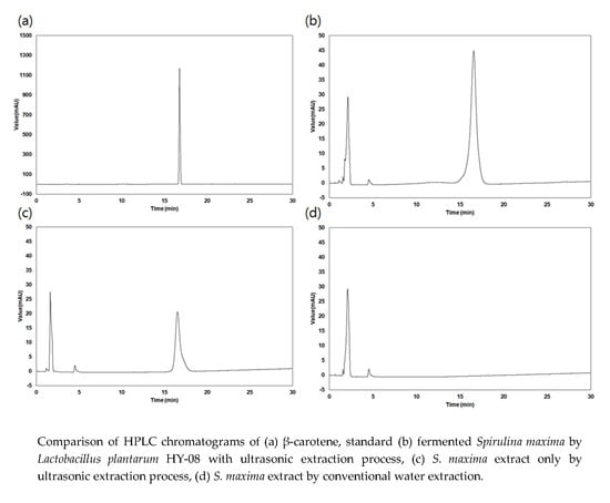
2.2. Determination of β-Carotene Content in the Extracts

The amount of β-carotene in the S. maxima extracts from the three different processes was analyzed using high-performance liquid chromatography (HPLC, Dionex 3000, Dionex Co., Sunnyvale, CA, USA) with a C18 column (Jupiter C18 column, 4.6 × 250 mm, 5 µm, 300 A, 00G-4053-E0, Phenomenex, CA, USA). In the analysis, the mobile phases of solvent A (60% methanol, 10% butanol, 30% water) and solvent B (89.5% methanol, 10% butanol, 0.5% water) were flowed under gradient conditions of 0–8 min (75:25), 8–50 min (90:10), 50–55 min (90:10), 55–57 min (75:25), and 57–65 min (75:25) at a flow velocity of 1.0 mL/min, along with a standard β-carotene (C4582, Sigma-Aldrich, St. Louis, MO, USA). The analysis was conducted by simultaneously evaluating the wavelengths 452 nm and 325 nm [17].

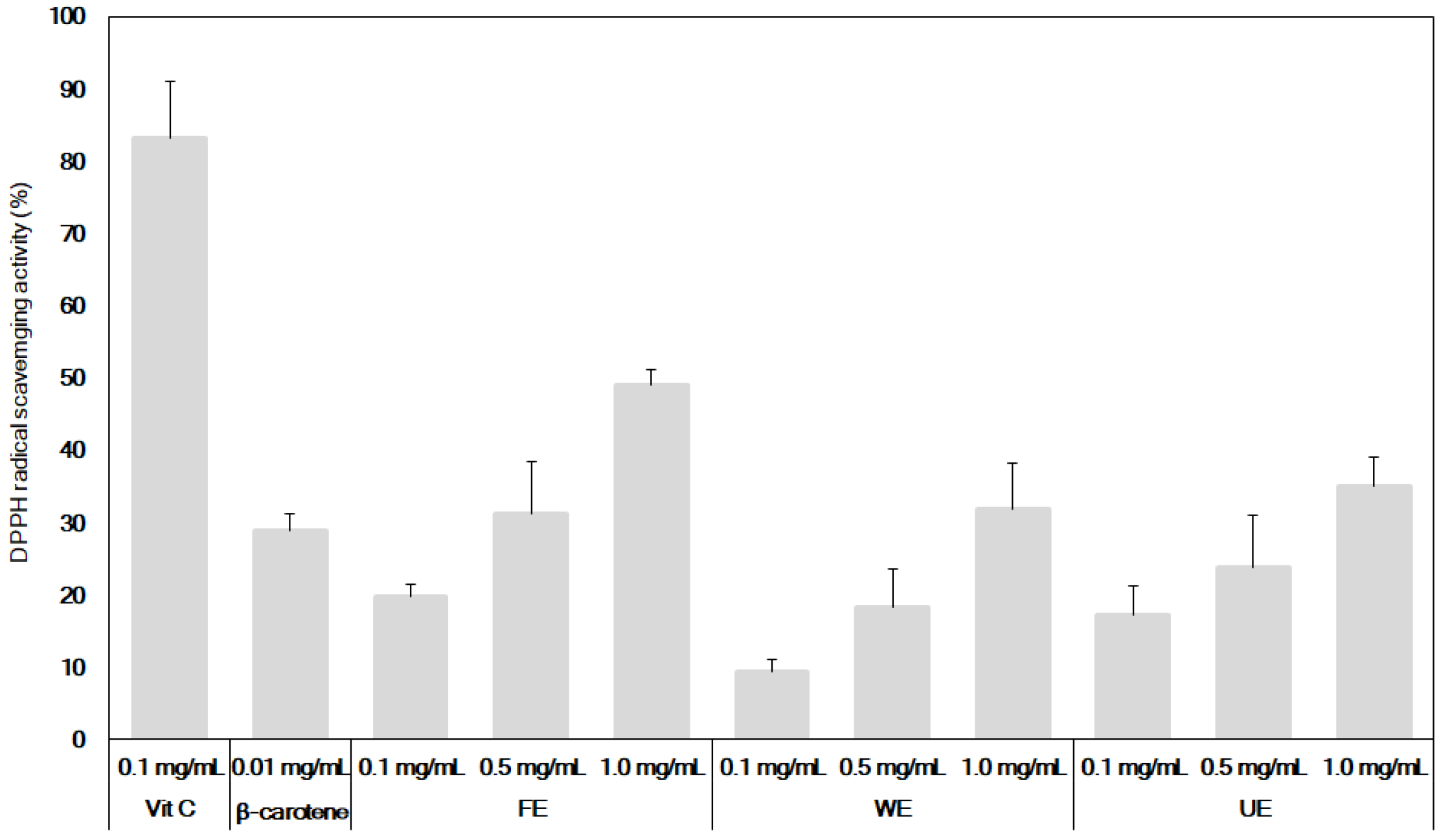
2.3. Measurement of the DPPH Free Radical-Scavenging Activity of the Extracts

The antioxidant activity of the extracts was evaluated by measuring α,α-diphenyl-β-picrylhydrazyl (DPPH) free radical-scavenging activity, which is a representative method for measuring the antioxidant activity of the extracts. In a 96-well plate, 200 μL of a 0.1 mM DPPH solution was prepared in ethanol as a solvent, and 80 μL of the sample at various concentrations (0.156, 0.313, 0.625, and 1.250 mg/mL) was added. The mixtures were left unattended in a dark room at 25 °C for 20 min, and the absorbance was then measured at 525 nm. The optical density was measured at 517 nm using a microplate reader (Thermo Fisher Scientific, Waltham, MA, USA). The relevant values were estimated as DPPH radical scavenging activity (%) [23].
DPPH radical scavenging activity (%) = (Control O.D. − Sample O.D.)/(Control O.D.) × 100

2.4. Measurement of the Neuroprotective Activity of the Extracts

A 3-(4,5-dimethylthiazo-2-yl)-2,5-diphenyltetrazolium bromide (MTT) assay was performed to measure the neuroprotective activity of the extracts from the various extraction processes. First, a 96-well plate was inoculated with mouse hippocampus-derived cells (HT22, donated by Prof. Choong Je Ma, Department of Medical Biomaterials Engineering, College of Biomedical Science, Kangwon National University) at a concentration of 1.5 × 10⁴ cells/well, and the cells were divided into an experimental group that was treated with glutamate at approximately 80% cell confluency, to induce apoptosis and a second group that was not treated with glutamate. The samples were treated with different concentrations (200 µL per concentration), and the cells were cultivated for 24 hr in a CO2 incubator. Thereafter, a MTT solution at a concentration of 5 μg/mL was added to each well. The supernatant was removed 4 hr later, 10 µL of acid-isopropanol (0.04 N HCl in isopropanol) was added to each well, and the absorbance was measured at a wavelength of 565 nm using a microplate reader (Tecan, Mannedorf, switzerland). The degree of neuroprotective effect of the extracts was expressed as the relative protection by estimating the ratio of the absorbance of cell growth treated with glutamate alone (control) to the absorbance of cell growth treated with glutamate and the extract [11].

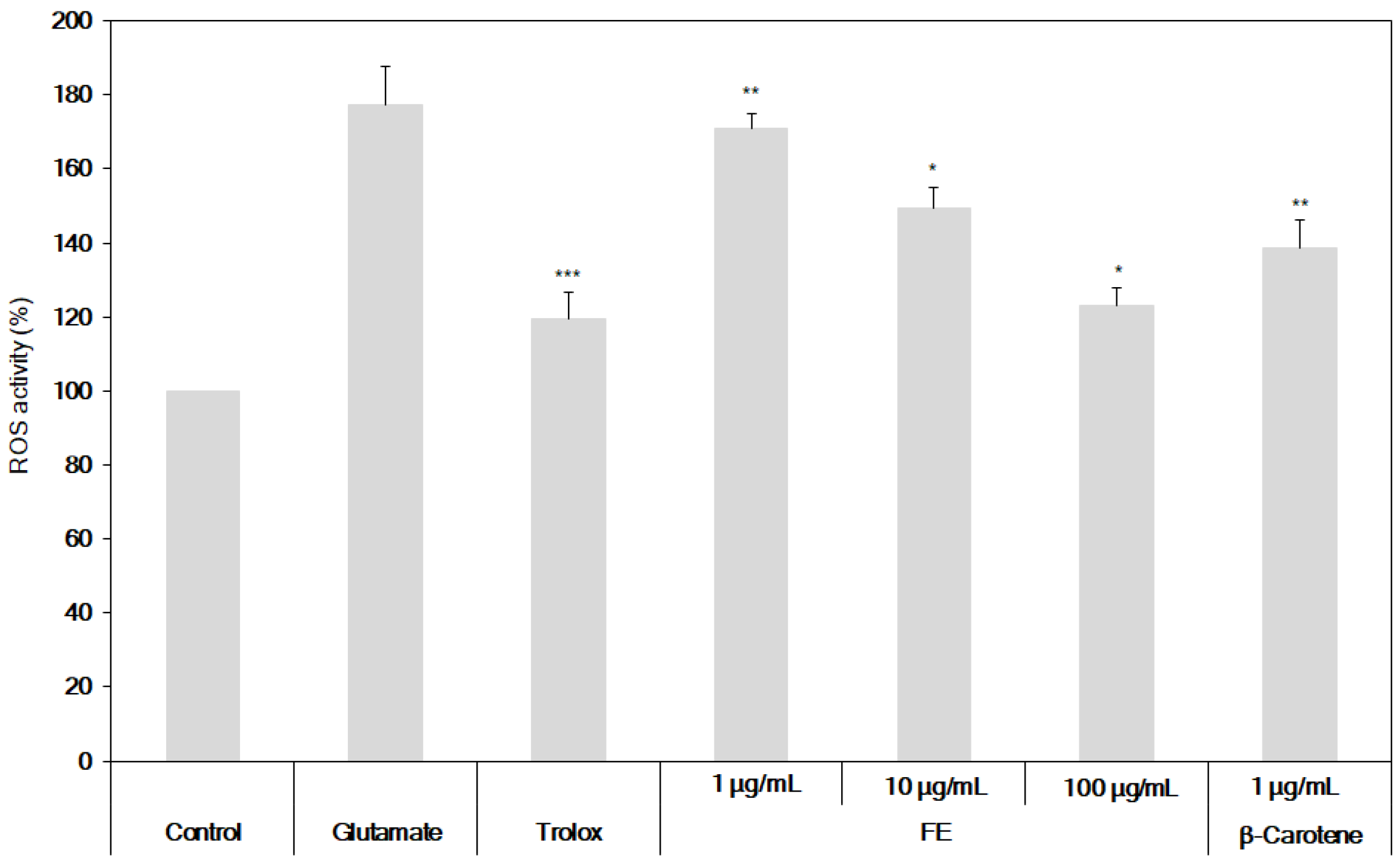
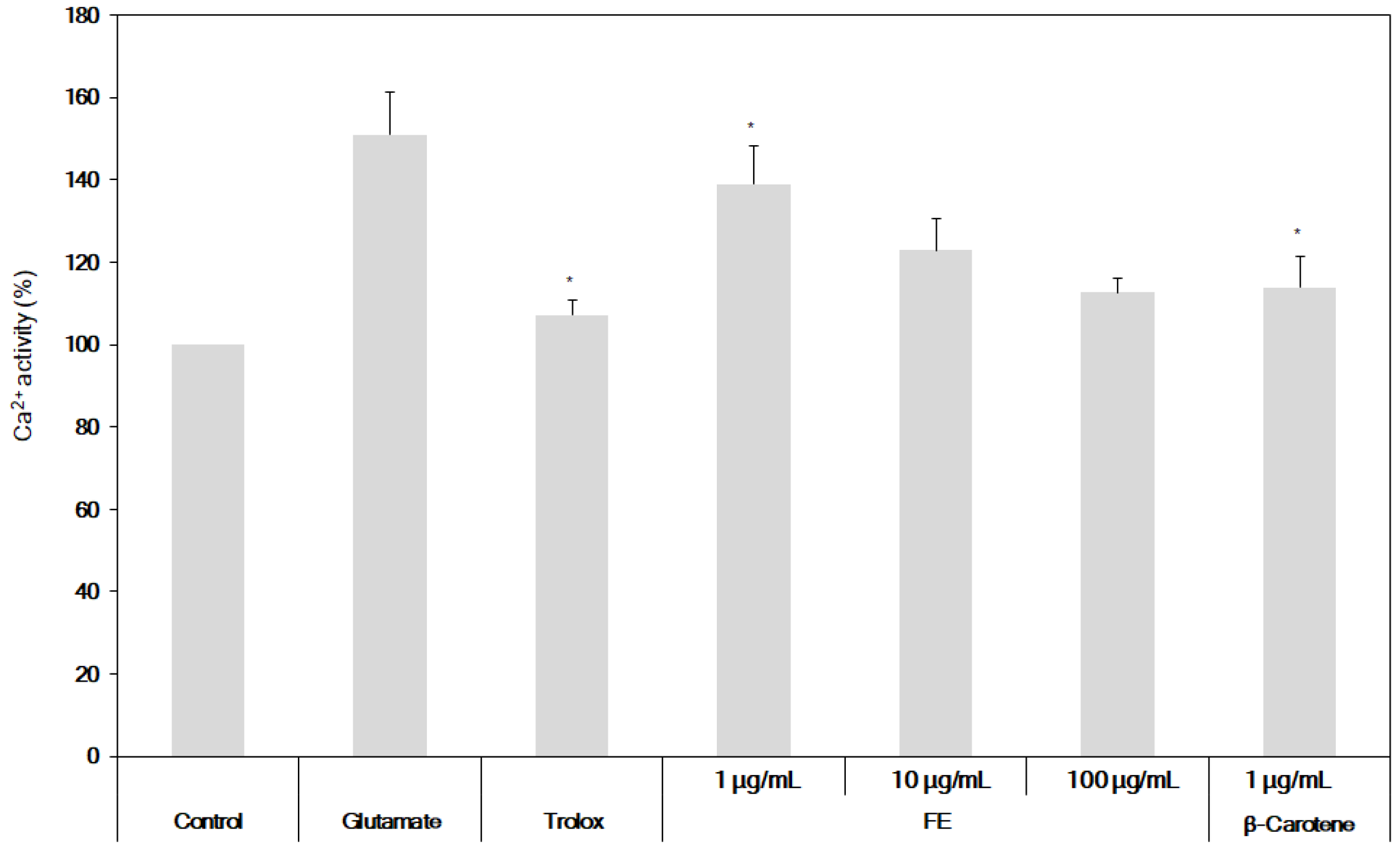
2.5. Determination of the Inhibition of ROS and Ca2+ Production from HT22 Cells

To measure reactive oxygen species (ROS) activity, the glutamate-induced ROS in HT22 cells was measured using 2′,7′-dichlorofluorescein diacetate (DCF-DA). The HT22 cells were seeded into a 48-well plate at a concentration of 4.02 × 10³ cells/well, cultured at 37 °C for 24 hr, treated with Trolox (positive control) and glutamate, and cultured for an additional 8 hr. Thereafter, the cells were treated with 10 μM DCF-DA, cultured for 1 hr, and treated with 1% Triton X-100. The ROS activity was measured at an excitation wavelength of 488 nm and an emission wavelength of 530 nm using a fluorescence analyzer [24]. Fura-2AM was used to measure calcium ions (Ca2+) in the HT22 cells. After treatment with either the sample, Trolox (positive control), or glutamate, the HT22 cells were treated with 2 μM Fura-2AM before harvesting. Two hours later, the cells were washed with PBS and treated with 1% Triton X-100 before extraction. The calcium ions were measured at an excitation wavelength of 400 nm and an emission wavelength of 535 nm using a calcium ion fluorescence analyzer [24].

2.6. Measurement of Glutathione Peroxidase and Glutathione Reductase Activities via Treatment of the Extracts

First, a 96-well plate was inoculated with HT22 cells at a concentration of 1.5 × 10⁴ cells/well. After incubation, 1, 10, and 100 μg/mL of sample and Trolox (50 μM) were added to the cells and incubated for 1 hr, followed by treatment with 2 mM glutamate. To measure the glutathione peroxidase activity, 100 µL/mL of the reaction mixture was well mixed with 500 µL of 0.1 M potassium phosphate buffer (pH 7.0) and 100 µL of 0.24 U glutathione, allowed to incubate for 10 min at 37 °C, and treated with 100 µL of 1.5 mM Nicotinamide Adenine Dinucleotide Phosphate Hydrogen (NADPH). The peroxide-independent consumption of NADPH was then measured at 340 nm for 1 min using a spectrophotometer. Next, 1 mM potassium cyanide and 100 µL of 1.5 mM hydrogen peroxide were added to inhibit the catalase activity. The peroxide-independent NADPH concentration was determined by measuring the decrement of peroxide for 3 min to assess the enzyme activity. The enzyme activity of glutathione was measured at 412 nm after adding 1 mM NADPH to 1.0 mL of a solution containing 100 µg of the sample, 0.1 M sodium phosphate buffer (pH 7.6), 1 mM EDTA, 1 mM GSSG and 0.6 mM 5,5′-dithiobis-2-nitrobenzoic acid (DTNB). The activity of glutathione reductase was also measured by calculating the ratio of the production of 2-nitro-3-thiobenzoic acid from DTNB during the reduction of glutathione [25,26].

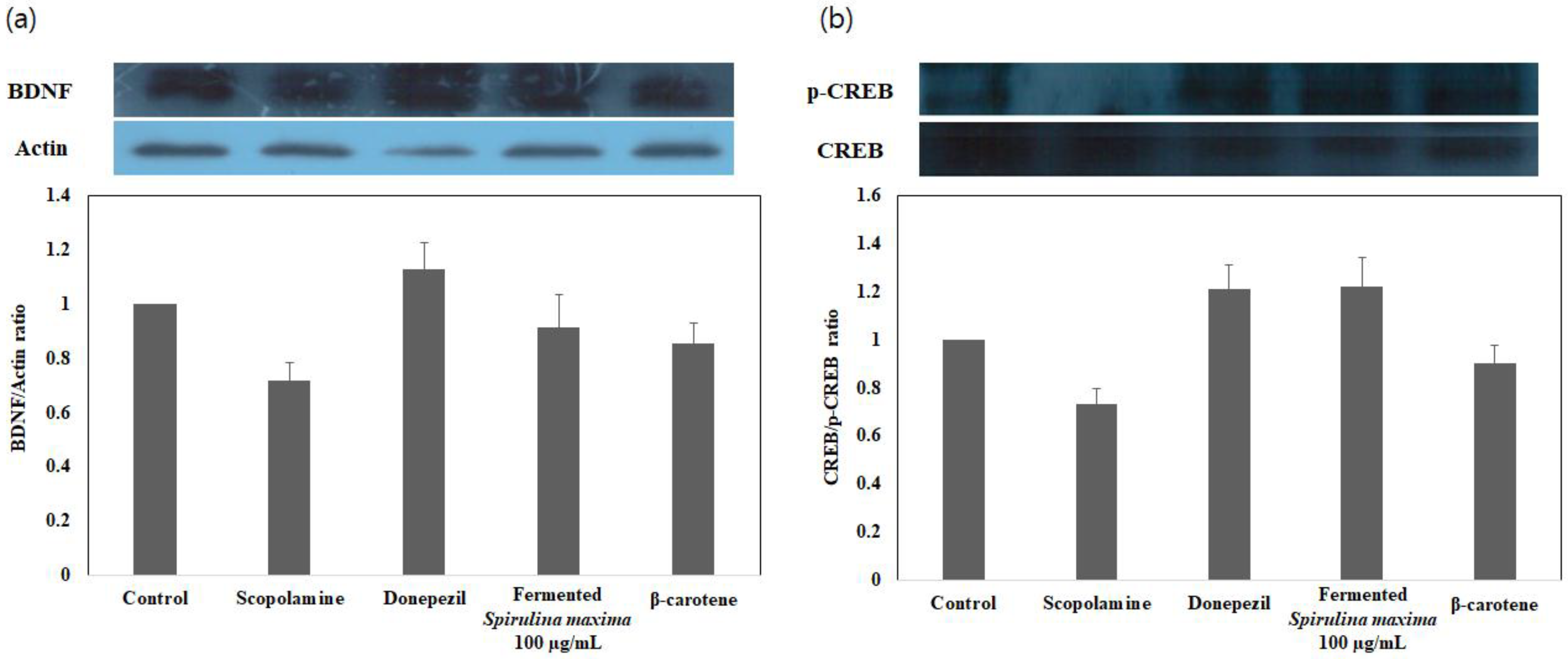
2.7. Western Blot Analysis of BDNF (Brain-Derived Neurotrophic Factor) and p-CREB (cAMP-Responsive Element-Binding Protein) Gene Expression Induced by the Extract

Proteins were quantitated from HT22 cells that had been treated with the extracts, and the proteins were separated by electrophoresis on a 12% polyacrylamide gel, and transferred to a polyvinylidene fluoride (PVDF) membrane. The membrane was blocked with 5% skim milk solution for 1 h, and incubated with primary antibodies BDNF (SC-546, Santa Cruz Biotechnology, Dallas, TX, USA), p-CREB (SC-81486, Santa Cruz Biotechnology, Dallas, TX, USA) at 4 °C for 24 hr. The membrane was then washed three times using TBST (Tris-buffered saline with 0.1% Tween-20), and incubated with the secondary antibody at room temperature for 1 hr. The separated proteins were observed using an LAS 4000 mini system (GE Healthcare, Richmond, VA, USA) [27,28].

2.8. Statistical Analysis

The data are expressed as the mean ± SD (standard deviation), and the mean is the average of five test results per experiment. The data were analyzed using Student’s t-test (SAS 9.1, SAS, Cary, NC, USA). The data were also analyzed by the one-way analysis of variance (ANOVA) test, and the mean values were considered to be significantly different at p < 0.01, p < 0.05 and p < 0.1.

3. Results

3.1. Extraction Yield and β-Carotene Content of the Extracts

HPLC analysis exhibited the highest β-carotene content in the S. maxima fermentation products. The concentrations of β-carotene in individual processes were analyzed, and the results are presented in Table 1 and Figure 1.
To identify the enhancement of the β-carotene content in the S. maxima extracts, three different extraction processes (ultrasonic, conventional hot water, and fermentation process with ultrasonic extraction processes) were compared and analyzed. The β-carotene content of the S. maxima fermentation products was estimated as 1.62 mg/g, and the lowest β-carotene content at 0.81 mg/g was observed in the hot water extract. A β-carotene content of 1.03 mg/g was obtained when only the ultrasonic process was applied, as well as 0.06 mg/g of negligible amount of β-carotene in the culture broth of only the fermentation as another control (data not shown). This result clearly indicate that a larger quantity of β-carotene was eluted through the fermentation with ultrasonic processing, possibly by first softening the cell walls of Spirulina, and bioconversion during the fermentation and then accelerating the elution of β-carotene in applying ultrasonic treatment. The highest extraction yield, at approximately 18.26%, was obtained from the S. maxima fermentation products, and the lowest yield (10.66%) was obtained from hot water extraction, showing similar trends in the amount of β-carotene in the extracts. Interestingly, the HPLC chromatogram of the extract from both fermentation and ultrasonic extraction contained more small peaks, which could represent the existence of other components in the extracts that are associated with the highest extraction yield. Therefore, it was shown that the fermentation extraction process was effective in processing the marine alga, S. maxima.

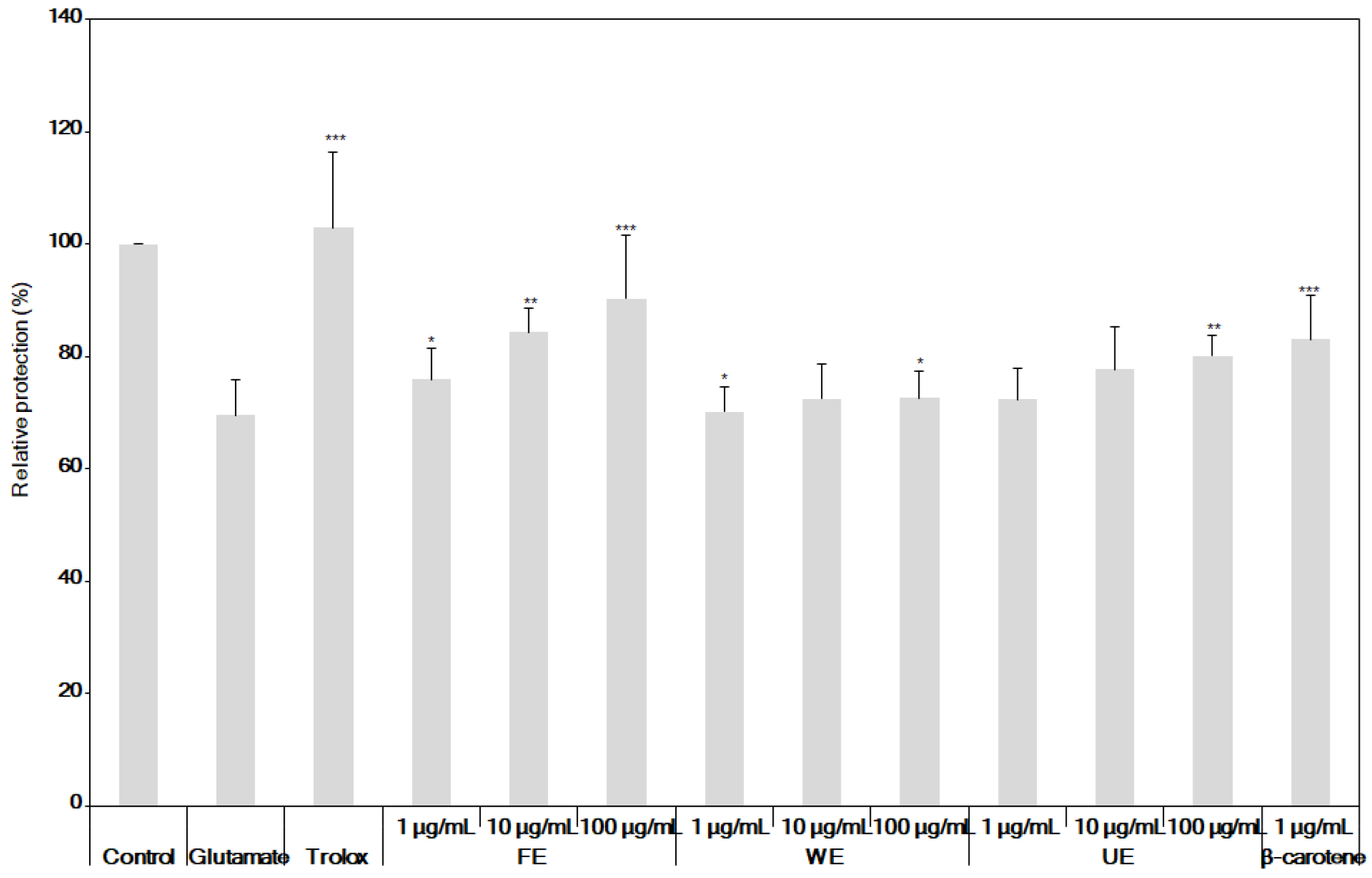
3.2. Comparison of Antioxidant and Neuroprotective Activities of β-Carotene and the Fermented S. maxima Extract

To identify the antioxidant effects of the S. maxima fermentation products based on the above results, the DPPH activities were measured, and the results are presented in Figure 2. In the case of the hot water extract and ultrasonicated extracts, the highest DPPH free radical scavenging activity (approximately 31.98% and 35.12%, respectively) were observed at the highest concentration of 1.0 mg/mL. On the other hand, the S. maxima fermentation products showed a DPPH free radical scavenging activity of approximately 49.01% at the same dosage, indicating that the S. maxima fermentation products exhibited the highest antioxidant activity. Therefore, the neuroprotective activities were measured based on the excellent antioxidant effect of the S. maxima fermentation products analyzed, as described above.
The neuroprotective activities were compared and analyzed in the HT22 cells. The results are presented in Figure 3.
The neuroprotective activities were measured in HT22 cells treated with glutamate under the extraction conditions of 12 hr at 100 °C, which is a hot water extraction process, and these results indicated neuroprotective activities of 70.10%, 72.39%, and 72.60% in treating 1 μg/mL, 10 μg/mL, and 100 μg/mL of the WE, respectively. In the case of the S. maxima fermentation products, the neuroprotective activities in the glutamate-treated HT22 cells were shown to be 75.84%, 84.33%, and 90.30%, depending on the treatment concentrations, and in the case of the ultrasonicated extracts, the neuroprotective activities were shown to be 72.30%, 77.60%, and 80.12%, depending on the treatment concentrations. The fermented extracts exhibited markedly increased neuroprotective activities compared to those of the S. maxima extracts obtained through conventional extraction methods. In particular, the result of treatment of the glutamate-treated HT22 cells with β-carotene from S. maxima at a concentration of 1 µg/mL, showed a high neuroprotective activity of approximately 82.96%, indicating that the β-carotene contained in the S. maxima directly affected the neuroprotective activity.

3.3. Neuroprotective Effects of the Fermented S. maxima Extract against Brain Oxidative Stress

The results of the ROS formation inhibitory activity of the S. maxima fermentation products are presented in Figure 4.
In the case of the S. maxima fermentation products, as the concentration increased, the ROS activity was significantly inhibited from 170.96%, to 149.27% and 123.26%. In the case of β-carotene, the ROS activity was significantly inhibited to 138.59% when 1 μg/mL β-carotene was administered. The levels of glutathione, glutathione reductase, and glutathione peroxidase, which inhibit ROS formation, are presented in Table 2.
According to the results in Table 2, when treating the S. maxima fermentation extracts along with 2 mM of glutamate, the production of glutathione was significantly increased from 51.02% to 63.76% and 80.38% as the treatment dosages were increased, and even similarly, to 91.88% from treatment with a strong antioxidant, Trolox, as a positive control. However, in the case of treating 1 μg/mL of β-carotene, its activity was observed to be approximately 47.73%, which was lower than that of treating 1 μg/mL of the fermented extract. In addition to the production of glutathione, the enzyme activities of both glutathione reductase and glutathione peroxidase were also significantly increased as the concentrations of the S. maxima fermentation products increased. These results strongly support our data that the fermented extracts have good antioxidant activities, which can work to improve the enzyme activities that are associated with the glutathione production pathway. Therefore, this sequential process represents antioxidant recovery reaction in memory-impaired brain systems. The measurements of the intracellular Ca2+ concentration, which induce ROS activity within the cells, are shown in Figure 5.
Upon treating the S. maxima fermentation products, the calcium concentration significantly decreased, from 138.98% to 122.85% and 112.63%, as the concentration of the S. maxima fermentation products increased. The effect of β-carotene in reducing the calcium concentration was approximately 113.93%, which was close to the effect of treating 100 μg/mL of the fermented extract.

3.4. Enhancement of BDNF/p-CREB Signaling Pathways in Glutamate-Induced, Oxidatively Stressed HT22 Cells with the Fermented S. maxima Extract

The results of the measurement of the BDNF/p-CREB signaling pathway of BDNF and the parent transcription factor p-CREB from the S. maxima fermentation are presented in Figure 6.
According to the results, the BDNF expression levels in the experimental group treated with the S. maxima fermentation products increased to 0.91. In addition, the BDNF expression levels in the experimental group treated with β-carotene increased to 0.85. The p-CREB expression levels in the experimental group treated with the S. maxima fermentation products increased to 1.22. Therefore, to accurately measure the induction of the signaling pathways by the S. maxima fermentation products, according to the gene expression, the BDNF/p-CREB expression ratio was measured. The results are presented in Table 3. The p-CREB/BDNF expression ratio was determined to be approximately 1.338, indicating that the p-CREB expression was higher.

4. Discussion

Among the many biologically active substances in S. maxima, β-carotene was selected and analyzed for content determination in the extracts of S. maxima from various extraction processes because it is known to be effective for improving cognitive ability and memory [29]. Although a fat-soluble β-carotene is normally extracted using an organic solvent such as methanol, ethanol, or acetone, water should be used in this case, as organic solvents are not suitable for use in fermentation [30]. Therefore, the concentrations of β-carotene in the extracts processed with only hot water and from ultrasonic extraction with water solvent should also be measured as controls, to compare with those from a combined process of fermentation and ultrasonic extraction [17]. The result of this work proved this combined process is effective in eluting β-carotene with water solvent. These are considered to be the first convincing results where an increase of β-carotene content from S. maxima was observed by using a combination of lactic acid fermentation and ultrasonic extraction with only water solvents, which is critically important in industrial applications, although previous reports have indicated that the β-carotene content and the content of the indicator components in natural products could be increased through fermentation [31]. It was also clearly shown that the highest antioxidant activity exhibited in the fermentation extracts can be attributed to the highest β-carotene content in the extracts from a combined process compared to the other extracts, because β-carotene has mainly worked on strong antioxidant effects in the extracts β-carotene from various processes [29,32,33]. Also, from previous reports of the function of β-carotene, such as a high neuroprotective activity [29,34], the above results strongly indicate that the extract with high amounts of β-carotene could be expected to improve cognitive function and memory. More interestingly, the neuroprotective activities of the extracts that contained lower amounts of β-carotene performed better than when treating with pure β-carotene, possibly due to the synergistic effects between the β-carotene and other biologically active substances, along with the high extraction yield. Similar results were also shown in other works [35]. However, more detailed experiments investigating these effects are required.
β-carotene is known to be a potent antioxidant, and its strong antioxidant activity is known to be effective for ischemia treatment and neuroprotective activity [27,29,30], which can result in neuroprotective effects of S. maxima. These features are considered to be the reasons for the high neuroprotective activity observed above [29,34]. Since the fermentation extracts and β-carotene showed high neuroprotective activity in the above results, it is possible to expect the correlation between the activities against oxidative stress and the neuroprotective activity, which can derive a neuroprotective mechanism that is associated with antioxidant activities, whose mechanism was also proposed in other works [25,35,36]. To confirm this hypothesis, the reduction of ROS production was also observed in this work, because this treatment induces oxidative damage, Ca2+ ions induce ROS activity, and Glutathione (GSH) inhibits ROS formation [36]. Therefore, this clearly proved that that such high ROS activity and Ca2+ concentration still led to a reduced oxidative stress in the fermentation extract is considered to be attributable to the high β-carotene content. Based on such a strong antioxidant effect, S. maxima fermentation products are considered to be effective for diverse chronic degenerative brain diseases such as Alzheimer’s disease, Parkinson’s disease, Huntington’s disease, and stroke [29,30,36]. With regard to the BDNF/p-CREB signaling pathway for neuroprotective and memory enhancement activities, BDNF is known to produce new neurites to reinforce connections among nerve cells, and increase the number of connections to improve abilities. cAMP-responsive element-binding protein (p-CREB) is a gene required in the process of learning and memorizing, and it is known to act as a parent transcription factor for BDNF expression [37]. On the other hand, β-carotene has been reported to amplify the expression of genes such as BDNF to improve depression [27]. In particular, the increase in the expression of BDNF was proven for the first time with memory improvement using β-carotene. The p-CREB/BDNF expression ratio was 1.055, indicating that the expression of BDNF was more strongly upregulated than p-CREB, indicating that the extract has memory-improving effects, mainly by boosting up the production of BDNF, compared to the phosphorylation of CREB, and similar results were also reported elsewhere [37]. Therefore, by combining fermentation and an ultrasonic extraction process, the S. maxima extracts were proven to positively affect the memory-improving activity because they increased the expression of the parent transcription factor (p-CREB) and exhibited antioxidant effects, thereby increasing the expression of brain-derived neurotrophic factor (BDNF).

5. Conclusions

This study clearly showed, for the first time, that the extraction yield of β-carotene in the extracts of S. maxima was significantly increased by combining lactic acid fermentation with a nonthermal ultrasonic extraction process, using water as a solvent. This study was also the first to prove that the increase of β-carotene content in the extracts greatly enhances its neuroprotective effects by efficiently suppressing the production of ROS and intracellular calcium ions in glutamate-induced HT22 cells, in addition to enhancing glutathione synthesis enzyme activity. The overall neuroprotective effects of the extract from a combined process were considered to be strongly correlated with its strong antioxidant activities. The effects of this extract were more pronounced than those of intact Spirulina sp. or of extracts from other conventional extraction processes, such as water or ethanol extraction, possibly due to the lower yields of β-carotene in these other processes. This study also reported in detail that neuroprotective mechanisms of the extract effectively suppressed oxidative stress by the cascading effects of an initial increase in the expression of the parent transcription factor p-CREB, along with antioxidant effects, and then an induction of the expression of BDNF. A similar mechanism of β-carotene was determined to explain its neuroprotective effects, even though in general, its activities were lower than those of the extracts, due to the synergistic effects of the extract from the combined process, rather than the effect of β-carotene as a single component. Conclusively, the S. maxima extract from both fermentation and ultrasonic extraction processes exhibited a strong neuroprotective effect mainly caused by suppressing oxidative stress via markedly inducing the brain antioxidative pathway of BDNF/p-CREB expression in glutamate-induced HT22 cells. It was additionally revealed that the effects of the extract were closely correlated with the synergistic effects from β-carotene and other biologically active components in the extract. Therefore, the results of this work could provide useful information for upgrading the biological activities of natural resources by employing nonthermal processes of fermentation and ultrasonic extraction.

Author Contributions

W.Y.C., S.-J.H., and D.H.K. carried out all of the experiments, and H.Y.L. designed the experiments and drafted the manuscript. All authors read and approved the final manuscript.

Funding

This research was funded by [Ministry of Oceans and Fisheries, Korea] grant number [20170326].

Acknowledgments

This research was a part of the project titled ‘Development of integrated technologies for developing biomaterials using by magma seawater’, funded by the Ministry of Oceans and Fisheries, Korea (20170326).

Conflicts of Interest

The authors declare no conflict of interest.

References

  1. Citron, M. Alzheimer’s disease: Treatments in discovery and development. Nat. Neurosci. 2002, 5, 1055–1057. [Google Scholar] [CrossRef] [PubMed]
  2. Dawson, G.R.; Iversen, S.D. The effects of novel cholinesterase inhibitors and selective muscarinic receptor agonists in tests of reference and working memory. Behav. Brain Res. 1993, 57, 143–153. [Google Scholar] [CrossRef]
  3. Murphy, M.P.; LeVine, H. Alzheimer’s Disease and the β-amyloid peptide. J. Alzheimers Dis. 2010, 19, 311–328. [Google Scholar] [CrossRef] [PubMed]
  4. Hannula, M.J.; Myöhänen, T.T.; Tenorio-Laranga, J.; Männistö, P.T.; Garcia-Horsman, J.A. Prolyl oligopeptidase colocalizes with α-synuclein, β-amyloid, tau protein and astroglia in the post-mortem brain samples with Parkinson’s and Alzheimer’s diseases. Neuroscience 2013, 242, 140–150. [Google Scholar] [CrossRef] [PubMed]
  5. Toide, K.; Shinoda, M.; Fujiwara, T.; Iwamoto, Y. Effect of a novel prolyl endopeptidase inhibitor, JTP-4819, on spatial memory and central cholinergic neurons in aged rats. Pharmacol. Biochem. Behav. 1997, 56, 427–434. [Google Scholar] [CrossRef]
  6. Fukunari, A.; Kato, A.; Sakai, Y.; Yoshimoto, T.; Ishiura, S.; Suzuki, K.; Nakajima, T. Colocalization of prolyl endopeptidase and amyloid beta-peptide in brains of senescence-accelerated mouse. Neurosci. Lett. 1994, 176, 201–204. [Google Scholar] [CrossRef]
  7. Takeda, A.; Loveman, E.; Clegg, A.; Kirby, J.; Picot, J.; Payne, E.; Green, C. A systematic review of the clinical effectiveness of donepezil, rivastigmine and galantamine on cognition, quality of life and adverse events in Alzheimer’s disease. Int. J. Geriatr. Psychiatry 2006, 21, 17–28. [Google Scholar] [CrossRef] [PubMed]
  8. Rodda, J.; Morgan, S.; Walker, Z. Are cholinesterase inhibitors effective in the management of the behavioral and psychological symptoms of dementia in Alzheimer’s disease? A systematic review of randomized, placebo-controlled trials of donepezil, rivastigmine and galantamine. Int. Psychogeriatr. 2009, 21, 813–824. [Google Scholar] [CrossRef] [PubMed]
  9. Yan, J.J.; Kim, D.H.; Moon, Y.S.; Jung, J.S.; Ahn, E.M.; Baek, N.I.; Song, D.K. Protection against β-amyloid peptide-induced memory impairment with long-term administration of extract of Angelica gigas or decursinol in mice. Prog. Neuropsychopharmacol. Biol. Psychiatry 2004, 28, 25–30. [Google Scholar] [CrossRef]
  10. Oken, B.S.; Storzbach, D.M.; Kaye, J.A. The Efficacy of Ginkgo biloba on cognitive function in Alzheimer Disease. Arch. Neurol. 1998, 55, 1409–1415. [Google Scholar] [CrossRef] [PubMed]
  11. Weon, J.B.; Lee, B.; Yun, B.R.; Lee, J.; Lee, H.Y.; Park, D.S.; Chung, H.C.; Chung, J.Y.; Ma, C.J. Memory enhancing effect of Codonopsis lanceolata by high hydrostatic pressure process and fermentation. Korean J. Pharmacogn. 2013, 44, 41–46. [Google Scholar]
  12. Hosseini, S.M.; Shahbazizadeh, S.; Khosravi-Darani, K.; Mozafari, M.R. Spirulina paltensis: Food and Function. Curr. Nutr. Food Sci. 2013, 9, 189–193. [Google Scholar] [CrossRef]
  13. Lee, J.C.; Hou, M.F.; Huang, H.W.; Chang, F.R.; Yeh, C.C.; Tang, J.Y.; Chang, H.W. Marine algal natural products with anti-oxidative, anti-inflammatory, and anti-cancer properties. Cancer Cell. Int. 2013, 13, 55–61. [Google Scholar] [CrossRef] [PubMed]
  14. Kay, R.A. Microalgae as food and supplement. Crit. Rev. Food Sci. Nutr. 1991, 30, 555–783. [Google Scholar] [CrossRef] [PubMed]
  15. Son, M.H.; Park, K.H.; Choi, A.R.; Yoo, G.; In, M.J.; Kim, D.H.; Chae, H.J. Investigation of biological activities of enzymatic hydrolysate of Spirulina. J. Korean Soc. Food Sci. Nutr. 2009, 38, 136–141. [Google Scholar] [CrossRef]
  16. Yang, H.N.; Lee, E.H.; Kim, H.M. Spirulina platensis inhibits anaphylactic reaction. Life Sci. 1997, 61, 1237–1244. [Google Scholar] [CrossRef]
  17. Lee, Y.J.; Kim, S.H.; Kim, J.S.; Han, J.A.; Seo, H.J.; Lim, H.J.; Choi, S.Y. Studies on simultaneous determination of chlorophyll a and b, pheophorbide a, and β-carotene in Chlorella and Spirulina products. J. Food Hyg. Saf. 2005, 20, 141–146. [Google Scholar]
  18. Yamada, T.; Sakaguchi, K. Comparative studies on Chlorella cell walls: Induction of protoplast formation. Arch. Microbiol. 1982, 132, 10–13. [Google Scholar] [CrossRef]
  19. Oh, S.H.; Ahn, J.H.; Kang, D.H.; Lee, H.Y. The effect of ultrasonificated extracts of Spirulina maxima on the anticancer activity. Mar. Biotechnol. 2011, 13, 205–214. [Google Scholar] [CrossRef]
  20. Eom, S.H.; Kang, Y.M.; Park, J.H.; Yu, D.U.; Jeong, E.T.; Lee, M.S.; Kim, Y.M. Enhancement of polyphenol content and antioxidant activity of brown alga Eisenia bicyclis extract by microbial fermentation. Fish. Aquat. Sci. 2011, 14, 192–197. [Google Scholar] [CrossRef]
  21. Tsai, C.C.; Chan, C.F.; Huang, W.Y.; Lin, J.S.; Chan, P.; Liu, H.Y.; Lin, Y.S. Applications of Lactobacillus rhamnosus spent culture supernatant in cosmetic antioxidation, whitening and moisture retention applications. Molecules 2013, 18, 14161–14171. [Google Scholar] [CrossRef] [PubMed]
  22. Choi, W.Y.; Lee, H.Y. Enhancement of Chlorophyll a Production from Marine Spirulina maxima by an Optimized Ultrasonic Extraction Process. Appl. Sci. 2018, 8, 26. [Google Scholar] [CrossRef]
  23. Dietz, B.M.; Kang, Y.H.; Liu, G.; Eggler, A.L.; Yao, P.; Chadwick, L.R.; Pauli, G.F.; Farnsworth, N.R.; Mesecar, A.D.; Breemen, R.B.; et al. Xanthohumol isolated from humulus lupulus inhibits menadione-induced DNA damage through induction of quinone reductase. Chem. Res. Toxicol. 2005, 18, 1296–1305. [Google Scholar] [CrossRef]
  24. Lee, J.; Weon, J.B.; Ma, C.J. Neuroprotective activity of phytosterols isolated from Artemisia apiacea. Korean J. Pharmacogn. 2014, 45, 214–219. [Google Scholar]
  25. Dar, M.A.; Khan, A.M.; Raina, R.; Beigh, S.A.; Sultana, M. Effect of repeated oral administration of bifenthrin antioxidant status and acetylcholinesterase activity in brain of rats. Toxicol. Environ. Chem. 2015, 97, 961–967. [Google Scholar] [CrossRef]
  26. Lee, M.R.; Moon, S.H.; Choi, A.R.; Lee, S.C.; Ahn, K.H.; Park, H.R. Neuroprotective effects of extracts from Diospyros kaki L. Peel. Korean J. Food Cookery Sci. 2011, 27, 67–73. [Google Scholar] [CrossRef]
  27. Kim, N.R.; Kim, H.Y.; Kim, M.H.; Kim, H.M.; Jeong, H.J. Improvement of depressive behavior by Sweetme Sweet Pumpkin™ and its active compound, β-carotene. Life Sci. 2016, 147, 39–45. [Google Scholar] [CrossRef]
  28. Ying, S.W.; Futter, M.; Rosenblum, K.; Webber, M.J.; Hunt, S.P.; Bliss, T.V.; Bramham, C.R. Brain-derived neurotrophic factor induces long-term potentiation in intact adult hippocampus: Requirement for ERK activation coupled to CREB and upregulation of Arc synthesis. J. Neurosci. 2002, 22, 1532–1540. [Google Scholar] [CrossRef]
  29. Li, F.J.; Shen, L.; Ji, H.F. Dietary intakes of vitamin E, vitamin C, and β-carotene and risk of Alzheimer’s Disease: A Meta-Analysis. J. Alzheimers Dis. 2012, 31, 253–258. [Google Scholar] [CrossRef]
  30. Craft, N.E.; Soares, J.H. Relative solubility, stability, and absorptivity of lutein and beta-carotene in organic solvents. J. Agric. Food Chem. 1992, 40, 431–434. [Google Scholar] [CrossRef]
  31. Juan, M.Y.; Chou, C.C. Enhancement of antioxidant activity, total phenolic and flavonoid content of black soybeans by solid state fermentation with Bacillus subtilis BCRC 14715. Food Microbiol. 2010, 27, 586–591. [Google Scholar] [CrossRef]
  32. Sergio, A.R.P.; Robert, M.R. β-Carotene and Other Carotenoids as Antioxidants. J. Am. Coll. Nutr. 1999, 18, 426–433. [Google Scholar] [CrossRef]
  33. Tsuchihashi, H.; Kigoshi, M.; Iwatsuki, M.; Niki, E. Action of β-Carotene as an Antioxidant against Lipid Peroxidation. Arch. Biochem. Biophys. 1995, 323, 137–147. [Google Scholar] [CrossRef]
  34. Mitchell, J.J.; Paiva, M.; Heaton, M.B. Vitamin E and β-carotene protect against ethanol combined with ischemia in an embryonic rat hippocampal culture model of fetal alcohol syndrome. Neurosci. Lett. 1999, 263, 189–192. [Google Scholar] [CrossRef]
  35. Mitchell, J.J.; Paiva, M.; Heaton, M.B. The Antioxidants Vitamin E and β-Carotene Protect Against Ethanol-Induced Neurotoxicity in Embryonic Rat Hippocampal Cultures. Alcohol 1999, 17, 163–168. [Google Scholar] [CrossRef]
  36. Coyle, J.T.; Puttfarcken, P. Oxidative stress, glutamate, and neurodegenerative disorders. Science 1993, 262, 689–695. [Google Scholar] [CrossRef]
  37. Chen, D.Y.; Bambah-Mukku, D.; Pollonini, G.; Alberini, C.M. Glucocorticoid receptors recruit the CaMKIIα-BDNF-CREB pathways to mediate memory consolidation. Nat. Neurosci. 2012, 15, 1707–1714. [Google Scholar] [CrossRef] [Green Version]
Figure 1. High-performance liquid chromatography (HPLC) chromatograms of (a) β-carotene standard (b) fermented Spirulina maxima by Lactobacillus plantarum HY-08 with ultrasonic extraction process, (c) S. maxima extracted by only ultrasonic extraction process, (d) S. maxima extracted by conventional water extraction.
Applsci 08 02469 g001
Figure 2. Comparison of the DPPH radical scavenging activity of several extraction processes. FE: fermented Spirulina maxima by Lactobacillus plantarum HY-08, WE: S. maxima extract by conventional extraction conditions. UE: Ultrasonication process.
Applsci 08 02469 g002
Figure 3. The neuroprotective activities of fermented Spirulina maxima on glutamate-induced oxidative stress in HT22 cells. The values shown are the mean. * p < 0.05, ** p < 0.01 and *** p < 0.001 show significant differences in comparison to the glutamate group (n = 6).
Applsci 08 02469 g003
Figure 4. The effect of fermented Spirulina maxima on glutamate-induced reactive oxygen species (ROS) production in HT22 cells. The values shown are the mean. * p < 0.05, ** p < 0.01 and *** p < 0.001 show significant differences in comparison to the glutamate group (n = 6).
Applsci 08 02469 g004
Figure 5. The effect of fermented Spirulina maxima on the level of Ca2+ in HT22 cells. The values shown are the mean. * p < 0.05 show significant differences in comparison to the glutamate group (n = 6).
Applsci 08 02469 g005
Figure 6. (a) Brain-derived neurotrophic factor (BDNF) and (b) p-CREB expression levels from fermented Spirulina maxima after glutamate-induced ROS production in HT22 cells.
Applsci 08 02469 g006
Table 1. Estimation of β-carotene content from S. maxima in the extracts from various extraction conditions.
Extraction Processβ-Carotene Content (mg/g)Extraction Yield (%)
FE 11.62 ± 0.2218.26 ± 1.93
WE 20.81 ± 0.1310.60 ± 1.44
UE 31.03 ± 0.3412.77 ± 1.68
1 FE: Fermented Spirulina maxima with ultrasonic extraction process at room temperature for 6 hr. 2 WE: Conventional water extraction at 100 °C for 12 hr. 3 UE: Ultrasonic extraction process at room temperature for 6 hr.
Table 2. The effect of fermented Spirulina maxima on the levels of glutathione, glutathione reductase, and glutathione peroxidase in HT22 cells treated by 2 mM glutamate.
SampleTreated Concentration (µg/mL)Glutathione (% of Control)Glutathione Reductase (% of Control)Glutathione Peroxidase (% of Control)
Control-100.00100.00100.00
Trolox50 μM91.88 ± 8.9093.32 ± 7.2191.26 ± 1.67
Fermented Spirulina maxima extracts151.02 ± 3.3161.42 ± 8.2367.94 ± 5.50
1063.76 ± 5.1870.23 ± 4.4975.07 ± 7.36
10080.38 ± 9.3081.27 ± 5.1180.33 ± 7.18
β-carotene147.73 ± 2.8548.08 ± 1.0743.75 ± 6.35
Table 3. Effect of neuroprotective activities of fermented Spirulina maxima on the BDNF/p-CREB expression ratio in HT22 cells.
Extraction Processp-CREB/BDNF Expression Ratio
FE 11.338
β-carotene1.055
1 FE: Fermented Spirulina maxima with ultrasonication process at 25 °C for 6 hr

