Review

Rehabilitative Ultrasound Imaging Evaluation in Physiotherapy: Piloting a Systematic Review

by
Samuel Fernández Carnero
1,2,3,*,
José Luis Arias Buria
3,4,
Juan Nicolás Cuenca Zaldivar
1,2,
Alejandro Leal Quiñones
1,2,
Cesar Calvo-Lobo
5 and
Carlos Martin Saborido
6
1
Department of Physical Therapy, Universidad Francisco de Vitoria, Pozuelo de Alarcon, 28223 Madrid, Spain
2
Grupo de Investigación en Fisioterapia e Imagen Intervencionista (GIFIMI), Universidad Francisco de Vitoria, Pozuelo de Alarcón, 28223 Madrid, Spain
3
Spanish Society for Ultrasound in Physiotherapy (SEEFi), 28006 Madrid, Spain
4
Professor of Physiotherapy, Occupational Therapy, Physical Medicine and Rehabilitation Department, Universidad Rey Juan Carlos, 28922 Alcorcon, Spain
5
Nursing and Physical Therapy Department, Faculty of Health Sciences, Universidad de León, 24401 Ponferrada, Spain
6
Fundación San Juan de Dios, Centro de Ciencias de la Salud San Rafael, Universidad Antonio de Nebrija, 28036 Madrid, Spain
*
Author to whom correspondence should be addressed.
Submission received: 25 November 2018 / Revised: 28 December 2018 / Accepted: 29 December 2018 / Published: 6 January 2019
(This article belongs to the Special Issue Ultrasound Technology for Clinical Diagnosis and Decisions Making)

Abstract

:

Featured Application

This pilot study/systematic review provides an opportunity to improve the methodology of the research group, identify gaps or pitfalls to avoid, and develop a posterior and complete systematic review with no limitations that is the best application in order to begin using Cochrane’s guidelines.

Abstract

Background: Research of ultrasound use in physiotherapy and daily practice has led to its use as an everyday tool. Methods: The aims were: (1) Checking the proposed systematic review protocol methodology; (2) evaluating the evidence from the last five years; and (3) coordinating the work of the team of reviewers in performing a complete systematic review. Thus, this is a pilot study prior to a full systematic review. The findings in databases related to health sciences with the meta-search engine Discovery EBSCO, Covidence, and Revman were used. Inclusion and exclusion criteria were described for eligibility. Results: Search provided 1029 references regarding the lumbar region on ultrasound scans. Of these, 33 were duplicates. After Covidence, 996 studies were left for screening. A full-text reading brought one randomized clinical trial (RCT). Conclusions: Validity and reliability references were found. The most suitable points were novice versus expert, and ultrasound versus electromyography (EMG) with just one RCT cohort, and observational and case reports. The lines of investigation increasingly endorsed the validity of using ultrasound in physiotherapy. Post-acquisition image analysis could also be a future line of research.

1. Introduction

There is a lot of evidence on the use of ultrasound from an aspect that greatly diverges from that used by doctors, and for which the objectives are also different. The beginnings of the ultrasound technique, known as rehabilitative ultrasound imaging (RUSI), arose when ultrasound was used to assess tissue morphology rather than the pathological cross-sections [1]. The technique evolved during the 1980s at the hands of certain researchers [2], and underwent further development in subsequent years up to the first edition of the International Symposium of RUSI (Rehabilitative Ultrasound Imaging) [3] held in 2006 in San Antonio, Texas. During this time, the technique has evolved for its use in exploring the musculoskeletal tissues from a morphological and functional point of view in an attempt to explain activity disorders in some cases, and morphology with regard to the feelings of pain or disability in others.
The second edition of this symposium was held in Madrid in 2016, and its conclusions [4] showed a greater scope of ultrasound implementation in physiotherapy.
Musculoskeletal pain is the second highest cause of disability worldwide [5]. This fact has been supported since its first publication, and many factors influence its high incidence and prevalence of back pain, the anatomical region which is most affected. These factors include the increase in degenerative disorders such as osteoarthritis, inability to exercise, and increase in population age [6].
The technical advancements in physiotherapy are essential for reflecting the work on a clinical level, and to be able to perform research in physiotherapy. Ultrasound is particularly relevant due to the high number of publications which validate it [7,8,9,10,11,12,13,14,15,16,17,18,19,20,21,22,23,24,25,26,27,28,29,30,31,32,33,34,35,36]. Currently, it is a tool recognized by the World Confederation of Physical Therapy (WCPT) at its congress in Las Vegas, Nevada (2009), and recognized on its website through an International Scientific Society known as the International Society in Electrophysical Agents in Physical Therapy (ISEAPT) that supports and endorses its use [37].
During this entire period, up until now, only two systematic reviews of the RUSI technique on the lumbar pelvic area have been performed [38,39], in which we can find different results, given that the first recognizes it as a valid technique for measuring the changes in the musculature in maximal and submaximal contractions, while the other speculates that it has to improve despite having good inter-tester validity studies.
What is certain is that these systematic reviews were performed in 2009, and since then, the technique has not been reviewed again with almost a decade passing in-between them. The present study is confined to the lumbar region, although the complete review evaluates the lumbar pelvic region (lumbar, abdomen, and pelvic floor).
The aims of this pilot study were two-fold: (1) To evaluate the scientific evidence on the RUSI technique in the lumbar region from 2012 to 2017 by performing a systematic review and meta-analysis when possible and (2) To coordinate the group of reviewers for the following phase in which a full systematic review of the lumbar pelvic area was conducted.

2. Methods

2.1. Protocol and Registration

The protocol was registered in the International Prospective Register of Systematic Reviews (PROSPERO) in the Center for Reviews and Dissemination from the University of York with the number CRD42017078326 and accessible in CRD York PROSPERO website.
As it is a pilot systematic review and an attempt to evaluate the work of a team of reviewers, the time frame used was five years (2012–2017), and the search strategy included only the lumbar region without language limitations.

2.2. Eligibility Criteria

Several inclusion criteria were considered: (1) Adults >18 with and without lumbopelvic pain; (2) randomized clinical trials (RCT) that contain the population detailed in point 1 and use of sonography as a diagnostic tool in morphology and muscle view in the treatment (biofeedback tool) of the lumbopelvic region; (3) RCTs that contain the population detailed in point 1 or controlled prospective designs; (4) studies that compare magnetic resonance imaging (MRI); (5) electromyography (EMG) versus ultrasound (US); and (5) validity and reliability and quantitative and/or reliability of lumbopelvic region.
Several exclusion criteria were considered: (1) Non-randomized studies; (2) ultrasound for medical purposes (tissue injuries), e.g., tumors, tears, inflammatory disease; (3) letters, editorials, comments, case-studies; and (4) symposium, congresses, and abstracts reports.

2.3. Information Sources

The search was implemented using Discovery EBSCO with the search strategy described, and the health sciences databases, which included related studies: (1) ScienceDirect; (2) Medline; (3) SportDiscus; (4) CINHAL; (5) Cochrane Database of Systematic Review; and (6) SciELO.

2.4. Search

The following word combinations were used for searching the required information: (1) “rehabilitative ultrasound imaging” or “real time ultrasound imaging” or “sonography” or “echography” or “ultrasound” AND (2) “physiotherapy” or “physical therapy” AND (3) “lumbar spine” or “lumbar region” or “lumbar multifidus” or “lowback”.

2.5. Study Selection

Once the file (.ris) was extracted, it was exported to the specific tool, Covidence systematic review software (Covidence™), Veritas Health Innovation, Melbourne, Australia program, in order to coordinate the team of reviewers, so that one of the authors reviewed all of the articles, others worked as peer reviewers, and the last author resolved potential conflicts. The disagreements were solved by a third author.

2.6. Data Collection Process

Data were extracted by one reviewer and checked by others using customized forms.

2.7. Risk of Bias in Individual Studies

For the analysis of risk of bias and data analysis, Revman [40] was used. In the title and abstract screening, the reviewers just chose YES, NO, or MAYBE, but in the full-text screening they chose reason for exclusion.

2.8. Synthesis of Results

In situations in which we considered studies to be sufficiently homogenous in terms of participants, interventions, and outcomes, we planned to synthesize results in a meta-analysis using the random-effect model. We forecasted that we would perform statistical analysis using the Cochrane Collaboration’s statistical software, Review Manager.

2.9. Additional Analyses

In addition, the studies were organized by years, study types, and country precedence, and thus have more perspective about the direction of this technique.

2.10. Ethical Considerations
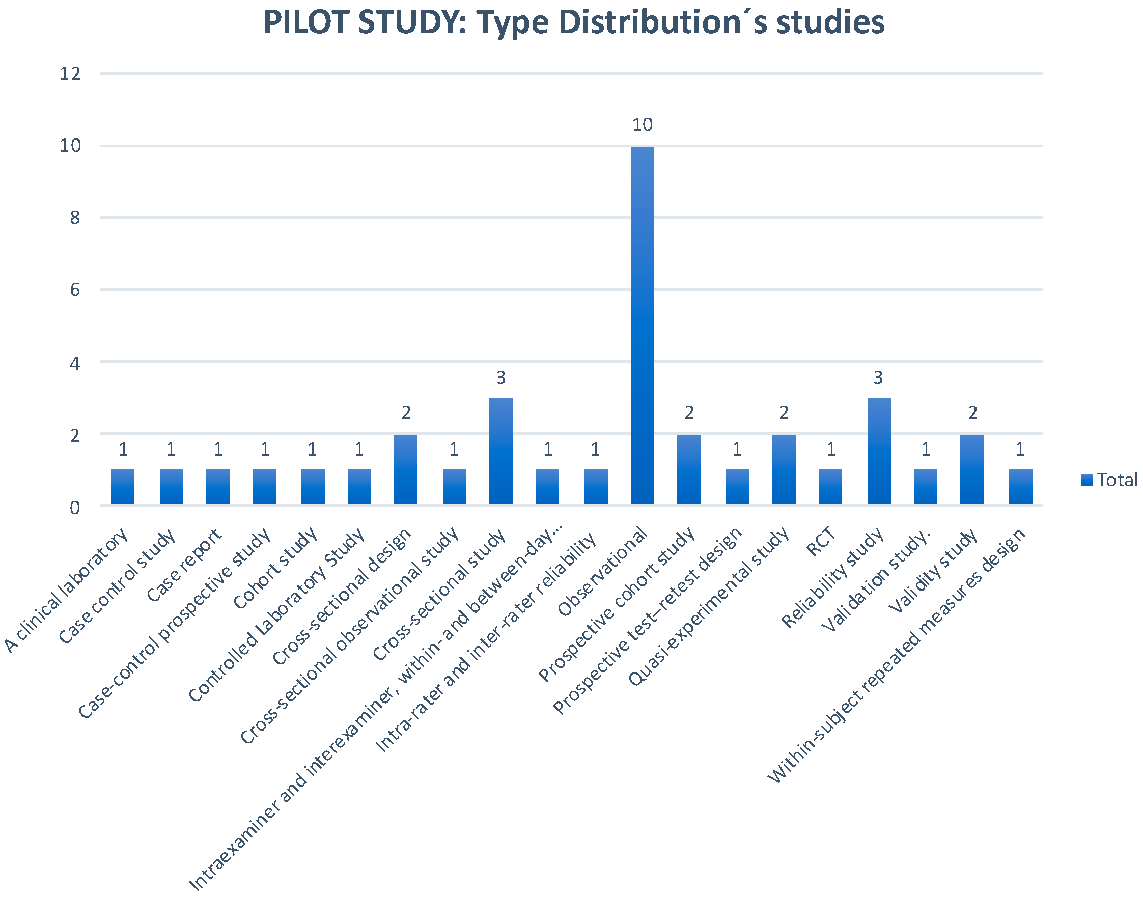
There were no ethical considerations for this project.

3. Results

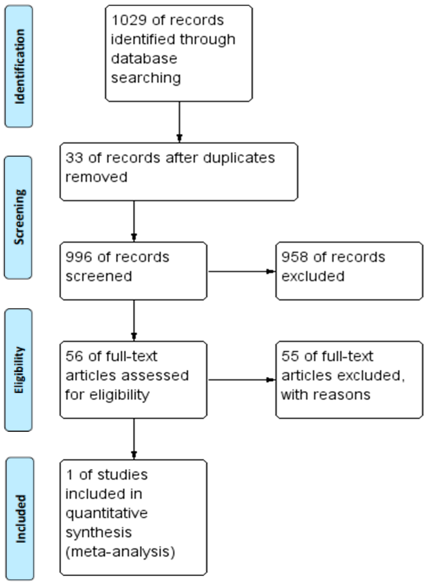
3.1. Study Selection

In order to carry out the systematic review, a search strategy was established with terms obtained from the PubMed library of control terms, Medical Subjects Headings (MeSH). The Preferred Reporting Items for Systematic Reviews and Meta-Analyses (PRISMA) method was followed [41] for generating the flow diagram (Figure 1).
The database search provided a total of 1029 references Of these, 33 were duplicates. CovidenceTM filtered them automatically, leaving 996 studies to assess. A full-text reading gave an end-result of one randomized clinical trial that passed into the extraction phase (Figure 1), and that was the reason metanalysis could not be done.
Just one study was included with the inclusion and exclusion criteria, and the characteristics of the included study are shown in (Table 1). The other studies were organized according the type of study and are detailed in additional analysis section.

3.2. Study Characteristics

The risk of bias analysis conducted with the REVMANTM tool of the included RCTs gave a global result of low risk of bias (Table 2). Finally, the bias analysis chart was prepared as follows (Figure 2).

3.3. Risk of Bias within Studies

Although we only found one RCT, it turned out to be of great interest to observe the published studies on ultrasound in the lumbar region in physiotherapy.

3.4. Additional Analysis

The number of studies is increasing every year (Figure 3), with an upward trend and a peak in publications in 2014 and 2015. Activity continued in the subsequent years, and picked up significantly in 2017. Evidently, it is not just a passing trend [42] even today. Therefore, it is a line of research on a global level, which is of interest, and in the light of the evidence analyzed, can be potentially very useful for the evaluation and evidence of the techniques used in physiotherapy.
The global distribution of the scientific production according to the articles reviewed over the last five years as full-texts shows a predominance in the United States and Australia (30% and 17%, respectively) followed by China and Japan (7%), and finally the rest of the countries in a bracket ≤4.9%, which includes Spain (Figure 4).
The graph gives an idea of the main research drivers and the countries involved in this lumbar region technique at present.
The analysis of the articles according to the type of study provided us with evidence that can be split into two groups:
1. Randomized Clinical Trial. Hebert 2015.
In this paper, a parallel RCT comparing two post-operative rehabilitation protocols following lumbar discectomy was developed. The methodology concerning design, recruiting processes, randomization, allocation, intervention, and measurements were done according with the objective and primary purposes. The results were expressed as a percentage change in thickness, but it would be wise if the measurements were included. There was an excellent study on follow-ups after samples along six months. This research demonstrates that exercise is an excellent approach for low back pain treatment, and ultrasound is a perfect tool with validity to obtain measurements related to the patient’s improvement. Interesting results described no differences between specific and general exercise. The cross-sectional areas and muscle morphology were evaluated by sonography, and the two groups improved in the measurements.
An important limitation was recognized by the author in that there was no control group.
2. Non-Randomized Trials.
This section ranges from case reports to quasi-experimental studies. Most of them are prospective observational studies, cross-sectional, and valid by intra and inter-tester, which support or confirm working methods (Figure 5).
The intraclass correlation results were compiled in a comprehensive table for direct observation and better evaluation was performed (Table 3).

4. Discussion

4.1. Summary of Evidence

To the author’s knowledge, this is the first systematic review that addressed the knowledge about ultrasound in physiotherapy beginning with a pilot study in order to avoid gaps of pitfalls in the final systematic review. The evidence was organized depending on the type of study, and this analysis has been described in this discussion.

4.2. Case Control Studies

The studies of cases and controls provide two reference points. The first compares patients with lumbar pain against asymptomatic cases in which the thickness of the lumbar muscles is measured in different positions [43]. On the one hand, it rules out certain concepts that could be labeled as wrong in that, in the prone position, there are no differences between control groups and actual cases. Although it is true that there are significant differences between the groups when they are standing up, it is only at the L5/S1 level. It would be interesting for future research that measurements could also be obtained at this level given the popularity of taking measurements at L4/L5. On the other hand, we found publications in which three instructions are assessed when asking the patient to contract the lumbar muscles after comparing healthy patients with symptomatic ones [44]. The aim of these studies was to determine if the contraction was better depending on the point of exploration and given instructions. In this case, the best result arose from the instructions to do a pelvic tilt in comparison to the others, and the best point at which greater contraction is observed was at L4/L5.

4.3. Case Report

It is well-known that inactivity leads to atrophy in the lumbar muscles in addition to infiltration of adipose tissue. In this case report, the muscles of subjects who underwent microgravity were assessed [45], and it was found that the muscle size remained constant at the level L2/L4, but once again was reduced in size at the level of L5. As the aim of this case, changes in individuals submitted to microgravity and exercises both before and after flights were reported, and different behavior of the abdominal muscles was found with the reduction in internal and transverse oblique muscles. Once again, the greater changes were found at the L5 level.

4.4. Cohort Studies

The correlation of variables among the clinical history, physical examination, changes in the lumbar multifidus, and spinal manipulation was evaluated in a cohort [46] performing a prospective study during a follow-up week. The manual therapy was combined with lumbar stabilization exercises. Changes were found in the Oswestry Disability Index and in the thickness of the multifidus muscle, although this last parameter yielded somewhat contradictory results. The linear regression analysis helped us to conclude that the combination of exercises with manipulation would be the most convenient way to achieve the best results on pain, disability, and/or functionality.
In line with the previous study, in another cohort of patients with lumbar pain, stabilizing exercise programs was performed over six weeks [47], showing significant changes in muscle contractions and improvement in functionality and pain. However, these factors were not associated based on statistical analysis.

4.5. Observational Studies

The observational studies have allowed us to test the real effects of certain physiotherapeutic interventions and compare them in some cases with other unknown variables, such as the activation of the muscles by using ultrasound. Therefore, the effects of exercises on unstable surfaces, lifting weights, or walking were evaluated in this section.
The unstable base of support (BOS) exercises, combined with weight-bearing [48], gave better contraction of the lumbar muscles when the exercises were performed in combination than alone on an unstable base; these results were contrary to what was thought at the time. The lumbar stabilization exercises are another frequent approach used in physiotherapy, given that they have shown an improvement in patients with pain assessed by ultrasound [49] in the thickening and activation in relation to the improvement in pain and functionality.
On some occasions, evaluating the local muscles compared to the regional ones could be of great interest with regard to the common synergies, both in the asymptomatic and symptomatic states. For this evaluation, in a study of patients with sacroiliac joint dysfunction [50], the changes were evaluated, both in the multifidus muscles as well as the abdominal ones. It would have been logical to think there would be a change, but it was demonstrated that it was associated with the sacroiliac joint dysfunction side. Along this line, however, correlations were made between the pain through the visual analogue scale (VAS) and ultrasound, in which the healthy side was compared with the affected side [2]. The linear regression of this comparison showed that the greater the ratio between the healthy and pathological sides, the harsher the symptoms.
In view of this discovery, we could be led to believe that the review of all the evidence (complete systematic review) could give more consistent results concerning correlations between ultrasound and other variables.
The lines of research which are beginning to develop are of great interest, in which new variables such as echo intensity and the variety of greys evaluated in the image post-acquisition phase through software are correlated [51]. These findings could then be correlated with variables such as muscle thickness or pain.
The comparisons between electromyography (EMG) and ultrasound have also been performed and have led to the ultrasound being considered a useful tool for evaluating functionality. Although in some studies, it has been performed on a healthy population [52], a correlation has been found in ultrasound output and EMG results between the increase in external oblique muscle activation and thickening abdominal transverse muscle thickening, and changes in the multifidus muscle that represents progress in monitoring muscle activity.
The comparison between ultrasound and EMG in muscle activation is worth highlighting [53]. In this study, muscles at rest and at maximum voluntary contraction were assessed in 30 healthy volunteers, yielding a correlation between ultrasound measurements and EMG monitoring of r = 0.51–0.61, which leads to the ultrasound increasingly being considered as a tool for measuring the activity.

4.6. Validity and Reliability Studies

We appreciate that the validity studies are of great importance and usefulness, as in some cases they have been able to offer security and confidence for clinical applications in addition to assessing the security in the monitoring processes or for the use in future research projects.
The intraclass correlation coefficient (ICC) is one of the principles in many areas, and the lumbar region has had several situations with different strategies such as the use of proprioceptive neuromuscular facilitation in order to assess muscular contractions at rest and contractions in patients with lumbar pain [54], finding an ICC > 0.93–0.99. These types of correlations in all studies reach 0.90, and even exceed it in some cases. The comparisons in ultrasound measurements between healthy and pathological subjects and comparisons on the same day and between days during contractions and at rest [55] give an ICC intra-rater (0.78–0.95 and 0.74–0.94) for older people and an ICC inter-rater (0.87–0.97 and 0.80–0.95) for younger people.
Comparisons were also carried out to the point of assessing the confidence of experts against novices. This study suggests that it is one of the only ones which compares two measurements in two assessors over several days, reaching a conclusion that an inter-rater ICC of 0.99–1.00 in healthy subjects. At the same time, they achieved an intra-rater ICC of 0.99–1.00 in healthy subjects. The sampling method and size of the sample may possibly have yielded some of the highest data.
Along this line, in another study, the healthy population data was compared to the pathological population with respect to ICC within the same day and between days [24]. However, on this occasion, it involved comparing ultrasound measurements of a static image with measurements from a video clip. In the first place, no significant differences were found in this regard, and in the second place, the ICCs on the same day were 0.99 and between days were 0.93–0.98. This work could provide great validity for measurements of post-acquisition muscle activations as measurements based on ultrasound in certain patients’ delayed sampling.
The ICC calculation was also useful for specific tasks [56] such as a maximum isometric contraction in healthy patients compared with those with lumbar pain. However, it is true that the population was very small. The ICC range was between 0.84 and 1.00.
Authors should discuss the results and the way in which they can be interpreted with respect to the perspective of previous studies and of the working hypotheses. The findings and their implications should be discussed in the broadest context possible. Future research directions may also be highlighted.

4.7. Future Studies

Future study lines must be a full review with no year’s limitations, languages, body region, or other factors. This could be affordable with the present research group and the experience gained from this study. Along this line, we could probably explain the relationship between the ultrasound measurements and other variables such as pain or disability during some interventions such as exercise.
Post-acquisition image analysis could also be a future line of research.
It is worthwhile designing more RCTs in order to obtain better results analysis.

5. Conclusions

This piloting exercise seems to show that rehabilitative ultrasound imaging (RUSI) technique in the lumbar region from 2012 to 2017 years by performing a systematic review demonstrated improvement of the muscles in people with low back pain who developed two exercises programs.
Despite the lack of a control group, a limitation of the study, RUSI may be considered as a potential technique for evaluating exercise programs in the physical therapy field regarding patients who suffer low back pain.

Limitations

Considering this was a pilot study, we could not obtain enough samples for an extractive phase and reach metanalysis development.

Author Contributions

Conceptualization, S.F.C. and C.M.S.; methodology, S.F.C. and C.M.S.; software, S.F.C.; formal analysis, S.F.C.; investigation, S.F.C., J.L.A.B., J.N.C.Z., A.L.Q., C.C.L., C.M.S.; resources, S.F.C.; writing—original draft preparation, S.F.C.; writing—review and editing, S.F.C., J.L.A.B., J.N.C.Z., A.L.Q., C.C.L., C.M.S.; visualization, S.F.C.; supervision, C.M.S.; project administration, S.F.C.

Funding

This research received no external funding.

Acknowledgments

The Medical Center “Clínica Centro Sur” in Ciempozuelos (Madrid, SPAIN) for the support given to get the resources used in this research.

Conflicts of Interest

The authors declare no conflict of interest.

Abbreviations

BOSBase of support
EMGElectromyography
ICCIntraclass Correlation Coefficient
ISEAPTInternational Society for Electrophysical Agents in Physical Therapy
MRIMagnetic Resonance Imaging
MESHMedical Subjects Headings
PROSPEROPreferred Reporting Items for Systematic Reviews and Meta-Analyses
RCTRandomized Clinical Trial
RUSIRehabilitative Ultrasound Imagine
USUltrasound
VASVisual Analogue Scale
WCPTWorld Confederation for Physical Therapy

References

  1. Ikai, M.; Fukunaga, T. Calculation of muscle strength per unit cross-sectional area of human muscle by means of ultrasonic measurement. Eur. J. Appl. Physiol. 1968, 26, 26–32. [Google Scholar] [CrossRef]
  2. Huang, Q.; Zhang, Y.; Li, D.; Yang, D.; Huo, M.; Maruyama, H. The Evaluation of Chronic Low Back Pain by Determining the Ratio of the Lumbar Multifidus Muscle Cross-sectional Areas of the Unaffected and Affected Sides. J. Phys. Ther. Sci. 2014, 26, 1613–1614. [Google Scholar] [CrossRef] [PubMed] [Green Version]
  3. Teyhen, D. Rehabilitative Ultrasound Imaging Symposium San Antonio, TX, 8–10 May 2006. J. Orthop. Sports Phys. Ther. 2006, 36, A1–A3. [Google Scholar] [CrossRef] [PubMed]
  4. Fernández-Carnero, S.; Calvo-Lobo, C.; Garrido-Marin, A.; Arias-Buría, J.L. 2nd Rehabilitative Ultrasound Imaging Symposium in Physiotherapy—Madrid, Spain, 3–5 June 2016. Br. J. Sports Med. 2018, 52, A1. [Google Scholar]
  5. Woolf, A.D. Bone and Joint Decade report: Moving together beyond the decade. Preface. Best Pract. Res. Clin. Rheumatol. 2012, 26, 167–168. [Google Scholar] [CrossRef]
  6. Briggs, A.M.; Cross, M.J.; Hoy, D.G.; Sànchez-Riera, L.; Blyth, F.M.; Woolf, A.D.; March, L. Musculoskeletal Health Conditions Represent a Global Threat to Healthy Aging: A Report for the 2015 World Health Organization World Report on Ageing and Health. Gerontologist 2016, 56 (Suppl. 2), S243–S255. [Google Scholar] [CrossRef]
  7. Djordjevic, O.; Djordjevic, A.; Konstantinovic, L. Interrater and intrarater reliability of transverse abdominal and lumbar multifidus muscle thickness in subjects with and without low back pain. J. Orthop. Sports Phys. Ther. 2014, 44, 979–988. [Google Scholar] [CrossRef]
  8. Hides, J.A.; Richardson, C.A.; Jull, G.A. Magnetic resonance imaging and ultrasonography of the lumbar multifidus muscle: Comparison of two different modalities. Spine 1995, 20, 54–58. [Google Scholar] [CrossRef]
  9. Storheim, K.; Bø, K.; Pederstad, O.; Jahnsen, R. Intra-tester reproducibility of pressure biofeedback in measurement of transversus abdominis function. Physiother. Res. Int. 2002, 7, 239–249. [Google Scholar] [CrossRef]
  10. Kidd, A.W.; Magee, S.; Richardson, C.A. Reliability of real-time ultrasound for the assessment of transversus abdominis function. J. Gravit. Physiol. 2002, 9, P131–P132. [Google Scholar]
  11. Dankaerts, W.; O’Sullivan, P.B.; Burnett, A.F.; Straker, L.M.; Danneels, L.A. Reliability of EMG measurements for trunk muscles during maximal and sub-maximal voluntary isometric contractions in healthy controls and CLBP patients. J. Electromyogr. Kinesiol. 2004, 14, 333–342. [Google Scholar] [CrossRef] [PubMed]
  12. Teyhen, D.S. Reliability of ultrasound imaging to measure muscle thickness of the lateral abdominal muscles. J. Orthop. Sports Phys. Ther. 2006, 36, A-8–A-9. [Google Scholar]
  13. O’Sullivan, C.; Bentman, S.; Bennett, K.; Stokes, M. Rehabilitative ultrasound imaging of the lower trapezius muscle: Technical description and reliability. J. Orthop. Sports Phys. Ther. 2007, 37, 620–626. [Google Scholar] [CrossRef] [PubMed]
  14. Hides, J.A.; Miokovic, T.; Belavy, D.L.; Stanton, W.R.; Richardson, C.A. Ultrasound imaging assessment of abdominal muscle function during drawing-in of the abdominal wall: An intrarater reliability study. J. Orthop. Sports Phys. Ther. 2007, 37, 480–486. [Google Scholar] [CrossRef]
  15. Wallwork, T.L.; Hides, J.A.; Stanton, W.R. Intrarater and interrater reliability of assessment of lumbar multifidus muscle thickness using rehabilitative ultrasound imaging. J. Orthop. Sports Phys. Ther. 2007, 37, 608–612. [Google Scholar] [CrossRef]
  16. Ellis, R.; Hing, W.; Dilley, A.; McNair, P. Reliability of measuring sciatic and tibial nerve movement with diagnostic ultrasound during a neural mobilisation technique. Ultrasound Med. Biol. 2008, 34, 1209–1216. [Google Scholar] [CrossRef] [PubMed]
  17. Koppenhaver, S.L.; Hebert, J.J.; Fritz, J.M.; Parent, E.C.; Teyhen, D.S.; Magel, J.S. Original article: Reliability of Rehabilitative Ultrasound Imaging of the Transversus Abdominis and Lumbar Multifidus Muscles. Arch. Phys. Med. Rehabil. 2009, 90, 87–94. [Google Scholar] [CrossRef] [PubMed]
  18. Lin, Y.J.; Chai, H.M.; Wang, S.F. Reliability of thickness measurements of the dorsal muscles of the upper cervical spine: An ultrasonographic study. J. Orthop. Sports Phys. Ther. 2009, 39, 850–857. [Google Scholar] [CrossRef] [PubMed]
  19. Cagnie, B.; Derese, E.; Vandamme, L.; Verstraete, K.; Cambier, D.; Danneels, L. Original Article: Validity and reliability of ultrasonography for the longus colli in asymptomatic subjects. Man. Ther. 2009, 14, 421–426. [Google Scholar] [CrossRef]
  20. Abiko, T.; Takei, H.; Shimamura, R.; Abiko, Y.; Yamamoto, J.; Sakasai, T.; Soma, M.; Ogawa, D.; Yamaguchi, T.; Hata, M. Reliability of Rehabilitative Ultrasound Imaging of the Lumbar Multifidus. Rigakuryoho Kagaku 2011, 26, 693–697. [Google Scholar] [CrossRef] [Green Version]
  21. Gnat, R.; Saulicz, E.; Miądowicz, B. Reliability of real-time ultrasound measurement of transversus abdominis thickness in healthy trained subjects. Eur. Spine J. 2012, 21, 1508–1515. [Google Scholar] [CrossRef] [PubMed] [Green Version]
  22. Lima, P.O.d.P.; de Oliveira, R.R.; de Moura Filho, A.G.; Raposo, M.C.F.; Costa, L.O.P.; Laurentino, G.E.C. Reliability Study: Reproducibility of the pressure biofeedback unit in measuring transversus abdominis muscle activity in patients with chronic nonspecific low back pain. J. Bodyw. Mov. Ther. 2012, 16, 251–257. [Google Scholar] [CrossRef] [PubMed]
  23. McPherson, S.L.; Watson, T. Reproducibility of ultrasound measurement of transversus abdominis during loaded, functional tasks in asymptomatic young adults. PM&R 2012, 4, 402–412. [Google Scholar]
  24. Wong, A.Y.L.; Parent, E.C.; Kawchuk, G.N. Reliability of 2 ultrasonic imaging analysis methods in quantifying lumbar multifidus thickness. J. Orthop. Sports Phys. Ther. 2013, 43, 251–262. [Google Scholar] [CrossRef] [PubMed]
  25. Linek, P.; Saulicz, E.; Wolny, T.; Myśliwiec, A. Reliability of B-mode sonography of the abdominal muscles in healthy adolescents in different body positions. J. Ultrasound Med. 2014, 33, 1049–1056. [Google Scholar] [CrossRef] [PubMed]
  26. Tahan, N.; Rasouli, O.; Arab, A.M.; Khademi, K.; Samani, E.N. Reliability of the ultrasound measurements of abdominal muscles activity when activated with and without pelvic floor muscles contraction. J. Back Musculoskelet. Rehabil. 2014, 27, 339–347. [Google Scholar] [CrossRef] [PubMed]
  27. Yang, K.-H.; Park, D.-J. Reliability of ultrasound in combination with surface electromyogram for evaluating the activity of abdominal muscles in individuals with and without low back pain. J. Exerc. Rehabil. 2014, 10, 230–235. [Google Scholar] [CrossRef] [PubMed] [Green Version]
  28. Schneebeli, A.; Egloff, M.; Giampietro, A.; Clijsen, R.; Barbero, M. Rehabilitative ultrasound imaging of the supraspinatus muscle: Intra- and interrater reliability of thickness and cross-sectional area. J. Bodyw. Mov. Ther. 2014, 18, 266–272. [Google Scholar] [CrossRef]
  29. Talbott, N.R.; Witt, D.W. Ultrasound examination of the serratus anterior during scapular protraction in asymptomatic individuals: Reliability and changes with contraction. PM&R 2014, 6, 227–234. [Google Scholar]
  30. Yang, H.S.; Yoo, J.W.; Lee, B.A.; Choi, C.K.; You, J.H. Inter-tester and intra-tester reliability of ultrasound imaging measurements of abdominal muscles in adolescents with and without idiopathic scoliosis: A case-controlled study. Bio.-Med. Mater. Eng. 2014, 24, 453–458. [Google Scholar]
  31. Chen, Y.-H.; Chai, H.-M.; Yang, J.-L.; Lin, Y.-J.; Wang, S.-F. Original Article: Reliability and Validity of Transversus Abdominis Measurement at the Posterior Muscle-Fascia Junction with Ultrasonography in Asymptomatic Participants. J. Manip. Physiol. Ther. 2015, 38, 581–586. [Google Scholar] [CrossRef] [PubMed]
  32. Keshwani, N.; Mathur, S.; McLean, L. Validity of Inter-rectus Distance Measurement in Postpartum Women Using Extended Field-of-View Ultrasound Imaging Techniques. J. Orthop. Sports Phys. Ther. 2015, 45, 808–813. [Google Scholar] [CrossRef] [PubMed]
  33. Koppenhaver, S.; Harris, D.; Harris, A.; O’Connor, E.; Dummar, M.; Croy, T.; Walker, M.; Flynn, T. The reliability of rehabilitative ultrasound imaging in the measurement of infraspinatus muscle function in the symptomatic and asymptomatic shoulders of patients with unilateral shoulder impingement syndrome. Int. J. Sports Phys. Ther. 2015, 10, 128–135. [Google Scholar]
  34. Sobczak, S.; Dugailly, P.-M.; Gilbert, K.K.; Hooper, T.L.; Sizer, P.S., Jr.; James, C.R.; Poortmans, B.; Matthijs, O.C.; Brismée, J.-M. Reliability and validation of in vitro lumbar spine height measurements using musculoskeletal ultrasound: A preliminary investigation. J. Back Musculoskelet. Rehabil. 2016, 29, 171–182. [Google Scholar] [CrossRef]
  35. Ludwig, O.; Hammes, A.; Kelm, J.; Schmitt, E. Validity & reliability study: Assessment of the posture of adolescents in everyday clinical practice: Intra-rater and inter-rater reliability and validity of a posture index. J. Bodyw. Mov. Ther. 2016, 20, 761–766. [Google Scholar]
  36. Salavati, M.; Akhbari, B.; Takamjani, I.E.; Ezzati, K.; Haghighatkhah, H. Reliability of the Upper trapezius Muscle and Fascia Thickness and Strain Ratio Measures by Ultrasonography and Sonoelastography in Subjects with Myofascial Pain Syndrome. J. Chiropr. Med. 2017, 16, 316–323. [Google Scholar] [CrossRef] [PubMed]
  37. History World Confederation for Physical Therapy. Available online: https://www.wcpt.org/iseapt/about (accessed on 7 December 2018).
  38. Koppenhaver, S.L.; Hebert, J.J.; Parent, E.C.; Fritz, J.M. Rehabilitative ultrasound imaging is a valid measure of trunk muscle size and activation during most isometric sub-maximal contractions: A systematic review. Aust. J. Physiother. 2009, 55, 153–169. [Google Scholar] [CrossRef]
  39. Hebert, J.J.; Koppenhaver, S.L.; Parent, E.C.; Fritz, J.M. A systematic review of the reliability of rehabilitative ultrasound imaging for the quantitative assessment of the abdominal and lumbar trunk muscles. Spine 2009, 34, E848–E856. [Google Scholar] [CrossRef]
  40. The-Cochrane-Collaboration. Review Manager (RevMan) [Computer program]. Version 5.3. Copenhagen: The Nordic Cochrane Centre, The Cochrane Collaboration. 2014. Available online: https://www.cochrane.org/es/2017/about-us/citing-our-products (accessed on 7 December 2018).
  41. Moher, D.; Liberati, A.; Tetzlaff, J.; Altman, D.G.; Group, P. Preferred reporting items for systematic reviews and meta-analyses: The PRISMA statement. J. Clin. Epidemiol. 2009, 62, 1006–1012. [Google Scholar] [CrossRef]
  42. Hodges, P.W. Ultrasound imaging in rehabilitation: Just a fad? J. Orthop. Sports Phys. Ther. 2005, 35, 333–337. [Google Scholar] [CrossRef]
  43. Sweeney, N.; O’Sullivan, C.; Kelly, G. Multifidus muscle size and percentage thickness changes among patients with unilateral chronic low back pain (CLBP) and healthy controls in prone and standing. Man. Ther. 2014, 19, 433–439. [Google Scholar] [CrossRef] [PubMed]
  44. Wang-Price, S.; Zafereo, J.; Brizzolara, K.; Sokolowski, L.; Turner, D. Effects of different verbal instructions on change of lumbar multifidus muscle thickness in asymptomatic adults and in patients with low back pain. J. Man. Manip. Ther. 2017, 25, 22–29. [Google Scholar] [CrossRef] [PubMed]
  45. Hides, J.A.; Lambrecht, G.; Stanton, W.R.; Damann, V. Changes in multifidus and abdominal muscle size in response to microgravity: Possible implications for low back pain research. Eur. Spine J. 2016, 25 (Suppl. 1), S175–S182. [Google Scholar] [CrossRef]
  46. Koppenhaver, S.L.; Fritz, J.M.; Hebert, J.J.; Kawchuk, G.N.; Parent, E.C.; Gill, N.W.; Childs, J.D.; Teyhen, D.S. Association between history and physical examination factors and change in lumbar multifidus muscle thickness after spinal manipulation in patients with low back pain. J. Electromyogr. Kinesiol. 2012, 22, 724–731. [Google Scholar] [CrossRef] [PubMed] [Green Version]
  47. Zielinski, K.A.; Henry, S.M.; Ouellette-Morton, R.H.; DeSarno, M.J. Lumbar Multifidus Muscle Thickness Does Not Predict Patients With Low Back Pain Who Improve With Trunk Stabilization Exercises. Arch. Phys. Med. Rehabil. 2013, 94, 1132–1138. [Google Scholar] [CrossRef] [PubMed] [Green Version]
  48. Debuse, D.; Birch, O.; St Clair Gibson, A.; Caplan, N. Low impact weight-bearing exercise in an upright posture increases the activation of two key local muscles of the lumbo-pelvic region. Physiother. Theory Pract. 2013, 29, 51–60. [Google Scholar] [CrossRef]
  49. Maraschin, M.; Ferrari, S.; Cacciatori, C. The effect of functional stabilization training on the cross sectional area of the deep stabilizers muscles in healthcare workers with chronic low back pain: A pilot study. Sci. Riabil. 2014, 16, 12–21. [Google Scholar]
  50. Joseph, L.H.; Hussain, R.I.; Naicker, A.S.; Ohnmar, H.; Ubon, P.; Aatit, P. Pattern of changes in local and global muscle thickness among individuals with sacroiliac joint dysfunction. Hong Kong Physiother. J. 2015, 33, 28–33. [Google Scholar] [CrossRef] [Green Version]
  51. Masaki, M.; Ikezoe, T.; Fukumoto, Y.; Minami, S.; Aoyama, J.; Ibuki, S.; Kimura, M.; Ichihashi, N. Association of walking speed with sagittal spinal alignment, muscle thickness, and echo intensity of lumbar back muscles in middle-aged and elderly women. Aging Clin. Exp. Res. 2016, 28, 429–434. [Google Scholar] [CrossRef]
  52. MacKenzie, J.F.; Grimshaw, P.N.; Jones, C.D.S.; Thoirs, K.; Petkov, J. Muscle activity during lifting: Examining the effect of core conditioning of multifidus and transversus abdominis. Work 2014, 47, 453–462. [Google Scholar]
  53. Kim, C.-Y.; Choi, J.-D.; Kim, S.-Y.; Oh, D.-W.; Kim, J.-K.; Park, J.-W. Comparison between muscle activation measured by electromyography and muscle thickness measured using ultrasonography for effective muscle assessment. J. Electromyogr. Kinesiol. 2014, 24, 614–620. [Google Scholar] [CrossRef] [PubMed]
  54. Huang, Q.; Li, D.; Zhang, Y.; Hu, A.; Huo, M.; Maruyama, H. The Reliability of Rehabilitative Ultrasound Imaging of the Cross-sectional Area of the Lumbar Multifidus Muscles in the PNF Pattern. J. Phys. Ther. Sci. 2014, 26, 1539–1541. [Google Scholar] [CrossRef] [PubMed] [Green Version]
  55. Sions, J.M.; Velasco, T.O.; Teyhen, D.S.; Hicks, G.E. Ultrasound imaging: Intraexaminer and interexaminer reliability for multifidus muscle thickness assessment in adults aged 60 to 85 years versus younger adults. J. Orthop. Sports Phys. Ther. 2014, 44, 425–434. [Google Scholar] [CrossRef] [PubMed]
  56. Liu, I.S.; Chai, H.M.; Yang, J.L.; Wang, S.F. Inter-session reliability of the measurement of the deep and superficial layer of lumbar multifidus in young asymptomatic people and patients with low back pain using ultrasonography. Man. Ther. 2013, 18, 481–486. [Google Scholar] [CrossRef] [PubMed]
Figure 1. Preferred Reporting Items for Systematic Reviews and Meta-Analyses (PRISMA) flow chart.
Applsci 09 00181 g001
Figure 2. Risk of bias graph: Review authors’ judgements about each risk of bias item presented as percentages across all included studied and risk of bias summary: Review authors’ judgements about each risk of bias item for each include study.
Applsci 09 00181 g002
Figure 3. Publications alongside years.
Applsci 09 00181 g003
Figure 4. Pie plot global evidence in % over total.
Applsci 09 00181 g004
Figure 5. Distribution of the evidence published classified by type of studies.
Applsci 09 00181 g005
Table 1. Characteristics of the randomized clinical trial founded.
MethodsAllocation: Randomized using a number generator.
Duration: 6 months follow up.
Setting: Patients from academic and private neurological and orthopedic spine surgery practices in Salt Lake City, Utah, USA.
ParticipantsDiagnosis: Post-discectomy surgery.
N = 61
Age: Average age
Sex: Male and Female.
Inclusion: Age 18–60 years, presurgical radiographic confirmation of lumbar disc herniation through MRI or CT and scheduled to undergo single-level lumbar discectomy.
Exclusion: Prior lumbar spine surgery, surgery at more than one level, a surgical procedure other than discectomy (e.g., fusion) or perioperative complications representing a contraindication to exercise.
Interventions* Group 1: General trunk exercise protocol (GEN) N = 32. This protocol comprised three components: (1) aerobic exercise, (2) range of motion exercise and (3) strengthening exercise.
* Group 2: Specific trunk exercise protocol (SPEC) N = 29. The SPEC included all components of the GEN. In addition, participants performed specific trunk muscle exercises similar to protocols used to treat patients with non-specific, non-surgical low back pain. This approach also included similar contractions of the transversus abdominis (TrA) using the abdominal drawing-in maneuver. Once these skills were acquired and confirmed by the physical therapist through palpation and/or ultrasound imaging, participants were instructed to perform isometric TrA and LM cocontractions. During the supervised exercise sessions, tactile and visual feedback through palpation and real-time ultrasound imaging were used to enhance skill acquisition and the treating physical therapists used this information to ensure appropriate technique.
Outcomes-Low back pain-related disability: Oswestry Disability Questionnaire (OSW).
-Low back and lower extremity pain: Numeric Pain Rating Scale. 30–32 Global rating of change (GRC) was assessed with a 15-point Likert-type scale ranging from −7 (“a very great deal worse”) to 0 (“about the same”) to +7 (“a very great deal better”).
-Sciatica frequency and bothersomeness were estimated using the Sciatica Frequency and Sciatica Bothersomeness indices resulting in possible scores of 0–25.34
-Muscle function was assessed using brightness-mode, real-time ultrasound images of LM thickness
Notes
Table 2. Analysis of risk of bias result, with the evidence extracted from the included study.
BiasAuthors’ JudgementSupport for Judgement
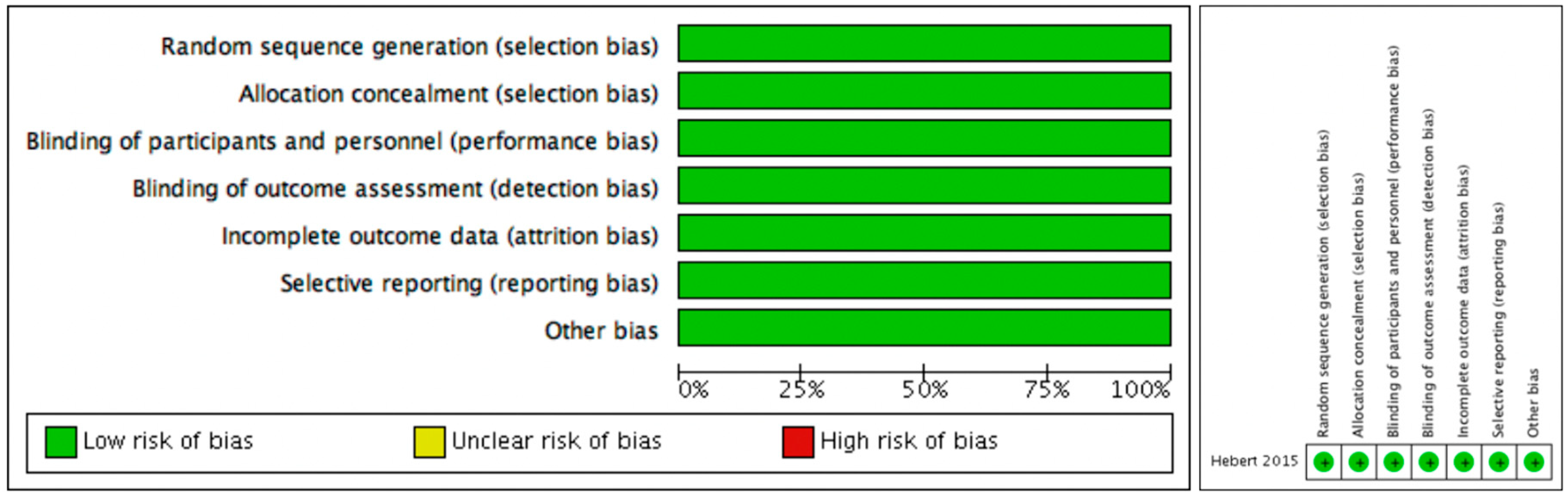
Random sequence generation (selection bias)Low riskA random number generator was used to create a permuted block randomization list with variable block sizes of 4–6.
Allocation concealment (selection bias)Low riskSequentially numbered, opaque envelopes containing the participant’s group assignment were prepared by research staff not affiliated with this trial.
Blinding of participants and personnel (performance bias)Low riskThe envelope was opened after the 2-week postoperative assessment by the treating physical therapist. Group assignments were concealed from participants and outcome assessors.
Blinding of outcome assessment (detection bias)Low riskThe envelope was opened after the 2-week postoperative assessment by the treating physical therapist. Group assignments were concealed from participants and outcome assessors.
Incomplete outcome data (attrition bias)Low riskThere were significant main effects of time (p < 0.01) indicating improvements from baseline in disability, pain, sciatica frequency, sciatica bothersomeness, and LM function (Table 3 and Figure 2).
Selective reporting (reporting bias)Low riskThe results of the intention-to-treat analyses revealed no time by group interactions. There were no statistically significant or clinically important between-group differences in disability, pain, global change, sciatica frequency, sciatica bothersomeness or LM muscle function at 10 weeks or 6 months (Table 3 and Figure 2).
Other biasLow riskNone.
Table 3. Intra-class correlation data.
ReferenceICCIntra-RaterInter-Rater
Wong et al. 20130.990.99–0.98
Liu et al. 20130.84–1.00
Sions et al. 2014 (older) 0.78–0.95 0.74–0.94
Sions et al. 2014 (younger) 0.87–0.970.80–0.95
Djordevic et al. 2014 0.99–1.000.99–1.00
Huang et al. 20160.93–0.99

