Case Report

Managing Wound Healing with a High-Risk Patient: A Case Report

by
Nikolai N. Potekaev
1,
Olga B. Borzykh
2,*,
German V. Medvedev
3,
Marina M. Petrova
2,
Elena I. Karpova
1,
Maria A. Zatolokina
4,
Mustafa Al-Zamil
5,
Olga M. Demina
1,
Ekaterina A. Narodova
2 and
Natalia A. Shnayder
2,6,*
1
Department of Skin Disease and Cosmetology, Pirogov Russian National Research Medical University, 117997 Moscow, Russia
2
Department of Shared Core Facilities “Molecular and Cell Technologies”, Voino-Yasenetsky Krasnoyarsk State Medical University, 660022 Krasnoyarsk, Russia
3
Department of Hand Surgery with Microsurgical Equipment, R. R. Vreden National Medical Research Centre for Traumatology and Orthopedics, 195427 Saint Petersburg, Russia
4
Department of Histology, Embryology, Cytology, Medical Faculty, Kursk State Medical University, 305014 Kursk, Russia
5
Department of Physiotherapy, Faculty of Continuing Medical Education, Peoples’ Friendship University of Russia, 117198 Moscow, Russia
6
Institute of Personalized Psychiatry and Neurology, Shared Core Facilities, V. M. Bekhterev National Medical Research Centre for Psychiatry and Neurology, 192019 Saint Petersburg, Russia
*
Authors to whom correspondence should be addressed.
Submission received: 11 February 2022 / Revised: 23 February 2022 / Accepted: 24 February 2022 / Published: 26 February 2022

Abstract

:
Wound healing is a complex, multi-step process. This process begins immediately after skin damage. The outcome of wound healing depends on the quality of each stage of this process: a normal or pathological scar. Violation of wound healing entails a decrease in the function of scar tissue as well as aesthetic dissatisfaction with the patient. This problem is especially important in aesthetic surgery. Patients who have come for beauty feel frustration, obtaining pathological scars. We have been dealing with the problem of wound healing after plastic surgery for about 10 years. Our approach includes the assessment of the risk of pathological wound healing and the treatment of high-risk patients. The risk assessment includes historical data on wound healing, signs of connective tissue dysfunction (especially patients with connective tissue dysplasia), and genetic polymorphisms of genes responsible for the structure of the components of the extracellular matrix of the skin. In the future, patients with a high risk of pathological scarring can be prescribed treatment after surgery. This article presents a clinical case in which we demonstrate our approach.

1. Introduction

Wound healing is a complex process aimed at maintaining the integrity of the body [1]. Tissue regeneration without scarring is possible only in the fetal period of development; in the postnatal period, healing occurs with scar formation [2]. With normal healing of skin wounds, normotrophic scars are formed that do not protrude above the surface of the skin. Hypertrophic or keloid scars may occur with impaired wound healing [3]. Hypertrophic scars disrupt the functions of the skin, so there is a decrease in the mechanical properties of the skin inherent in the skin without a scar [4]. In addition, a scar is an aesthetic defect that negatively affects the psychological state of the patient.
A special place is occupied by the problem of hypertrophic scars in aesthetic medicine. On one hand, patients often apply for the correction of an already existing hypertrophic scar. On the other hand, some aesthetic procedures can lead to the formation of scars, most often during plastic surgery and dermatosurgery (when removing skin neoplasms). The formation of hypertrophic scars after aesthetic procedures has an extremely negative assessment by the patient, since instead of the aesthetic improvement that the patient requested, an aesthetic defect was obtained.
There are many reasons for the formation of hypertrophic scars, among which external and internal ones can be noted. However, the search for pathogenetic processes and related treatment principles is still ongoing [5]. Currently, the search for information is focused on molecular mechanisms. There are also studies showing the role of the mutation of genes responsible for the extracellular matrix of the skin in the violation of wound healing [6]. The extracellular matrix of the skin plays a huge role in various skin functions including skin healing [7]. Along with other genes, in the violation of wound healing, the role of genes responsible for the synthesis of skin collagen was shown [8]. Another study showed the role of ELN gene mutations in the development of nonsyndromic striae disease [9]. The association of skin healing disorders with mutations of MMP genes has not yet been revealed [10]. Studies on the role of the mutation of genes responsible for remodeling the extracellular matrix are actively ongoing [11].
Monogenic disorders of the extracellular matrix are known as hereditary connective tissue diseases (the most famous being Ehlers-Danlos syndrome, Marfan syndrome, etc.) [12]. There is also a group of multifactorial diseases where various genetic variations of the extracellular matrix genes can presumably play a role, along with other external factors [9]. Often, the manifestations of such pathology appear against the background of involutional changes in the skin, leading to a decrease in synthetic and proliferative cell activity [13]. A unified global approach to the classification of such conditions has not yet been developed: some studies have mentioned mutations in certain genes, while others have mentioned the marfanoid phenotype. In Russia, such connective tissue pathologies are allocated to the group of connective tissue dysplasia (not related to hereditary connective tissue dysplasia). The clinical symptoms of connective tissue dysplasia, along with other symptoms, are multiple hypertrophic or atrophic scars [14].
A unified approach to the diagnosis of connective tissue has also not yet been developed, but Russian recommendations for the management of such patients include the search for clinical symptoms of connective tissue pathology, which includes pathologies of the bone and joint system, cardiovascular, and other systems. The main skin symptoms include hyperextension of the skin, the vulnerability of the skin, and the presence of hypertrophic or atrophic scars.
Currently, various commercial variants of genetic testing have become widespread. In Russia, such tests are available to patients, so sometimes, such patients come to the doctors with the results of these tests. The difficulty of interpreting the tests lies in the fact that they cover a fairly small part of possible mutations. Thus, they cannot fully assess the risk of pathologies. However, when identifying some mutations, the doctor should take this into account in the general calculation of risks.
For 10 years in our practice, we have been working on a comprehensive approach to the management of patients after surgery (most often after blepharoplasty), the removal of skin neoplasms or other skin injuries to minimize the risk of hypertrophic scar formation. This approach includes an assessment of the risk of pathological scarring and principles of the management of patients with a high risk of pathological scarring. The risk of hypertrophic scars is determined based on the patient’s history, the patient’s lifestyle (such as aggravating factors of smoking, frequent UV-insolation). Additionally taken into account is the presence of clinical symptoms of connective tissue dysplasia, genetic polymorphisms of genes responsible for the synthesis of components of the extracellular matrix.
Management of patients with postoperative wounds is possible in various ways: injections of botulinum toxin type A, platelet-rich plasma, silicone-based patch application, and many other methods [15,16]. Our management of patients with healing wounds is aimed at improving patient compliance as it is well-known that high compliance contributes to more effective treatment [17].
Our patient management involves the complex work of surgical and non-surgical methods aimed at improving the quality of the skin (rejuvenation). Thus, after surgery, the patient undergoes a course of rejuvenation injections, while part of the rejuvenation product is used in the area of wound healing. With this approach, we can see a fairly high patient compliance:
It does not require additional actions of the patient at home;
It does not require the purchase of additional medicine as previously planned rejuvenation injections are used;
It does not require additional time; and
The patient is under the doctor’s control in the next 1–2 months.
When planning plastic surgery or removing skin neoplasms, we initially coordinate the timing with annual skin rejuvenation procedures. In this way, the patient does not need to carry out additional therapeutic measures.
In this example, the main points of clinical work according to this approach are presented. In this case report, we describe an example of scar repair following surgery on the arm region.

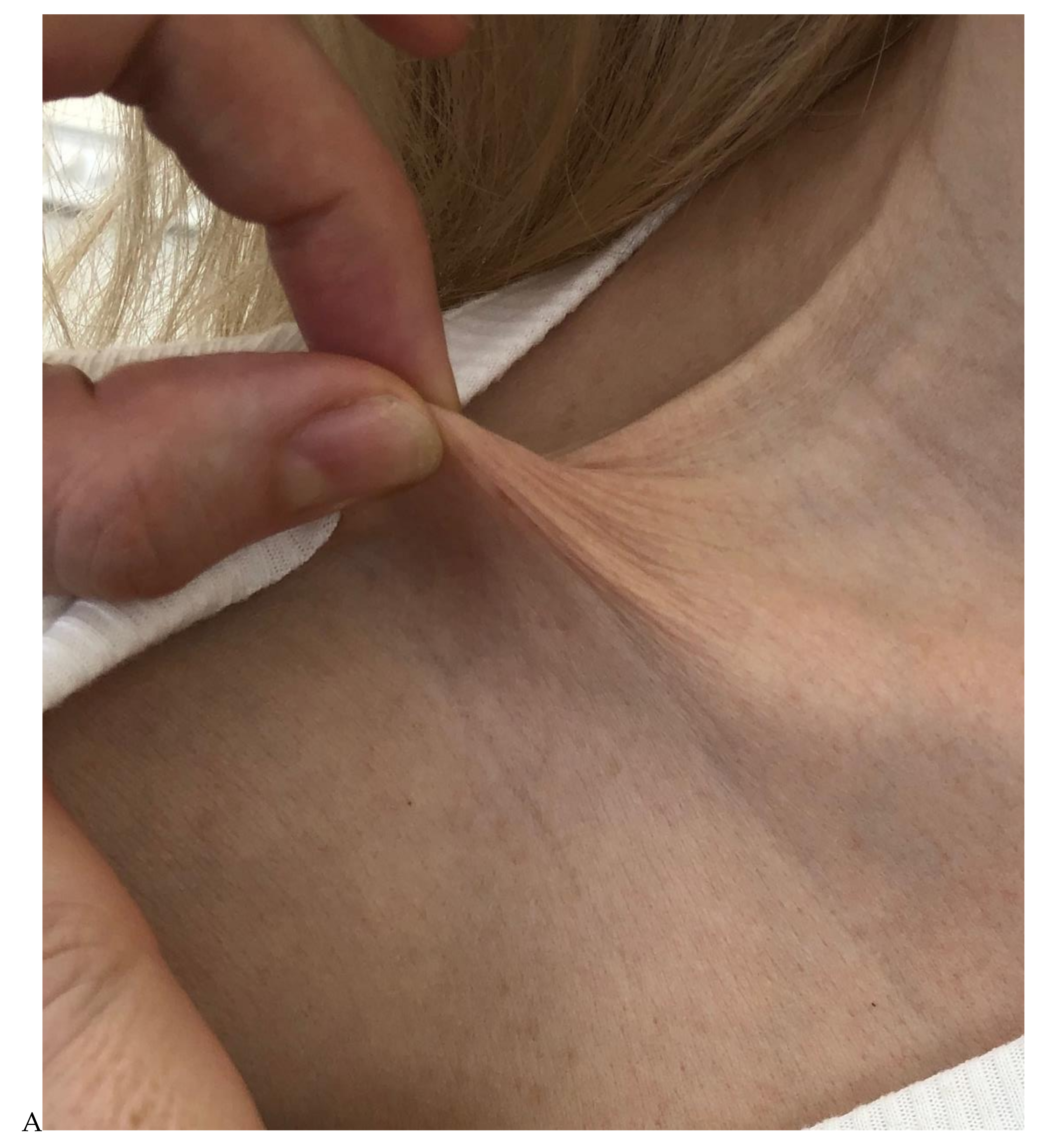
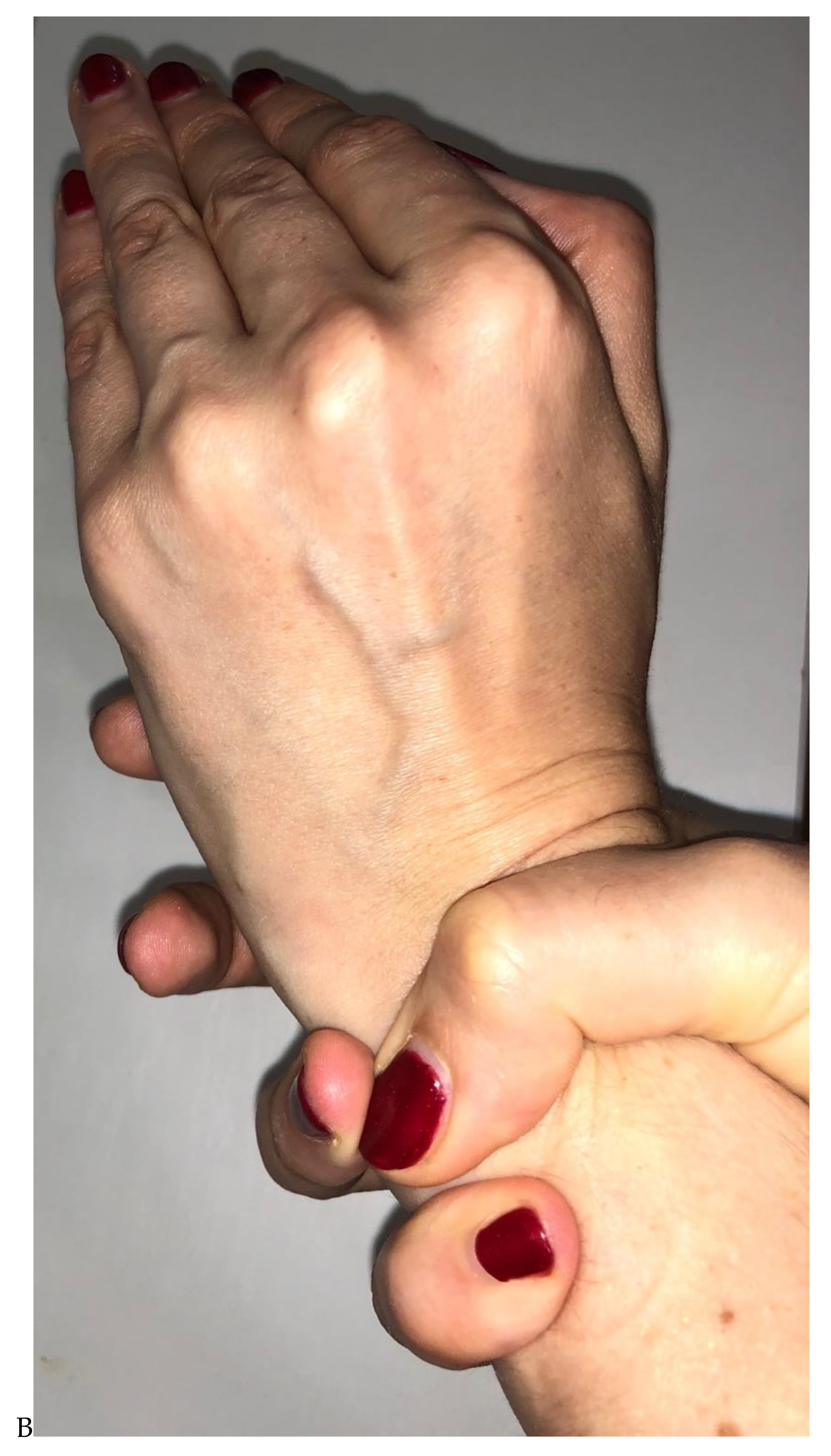
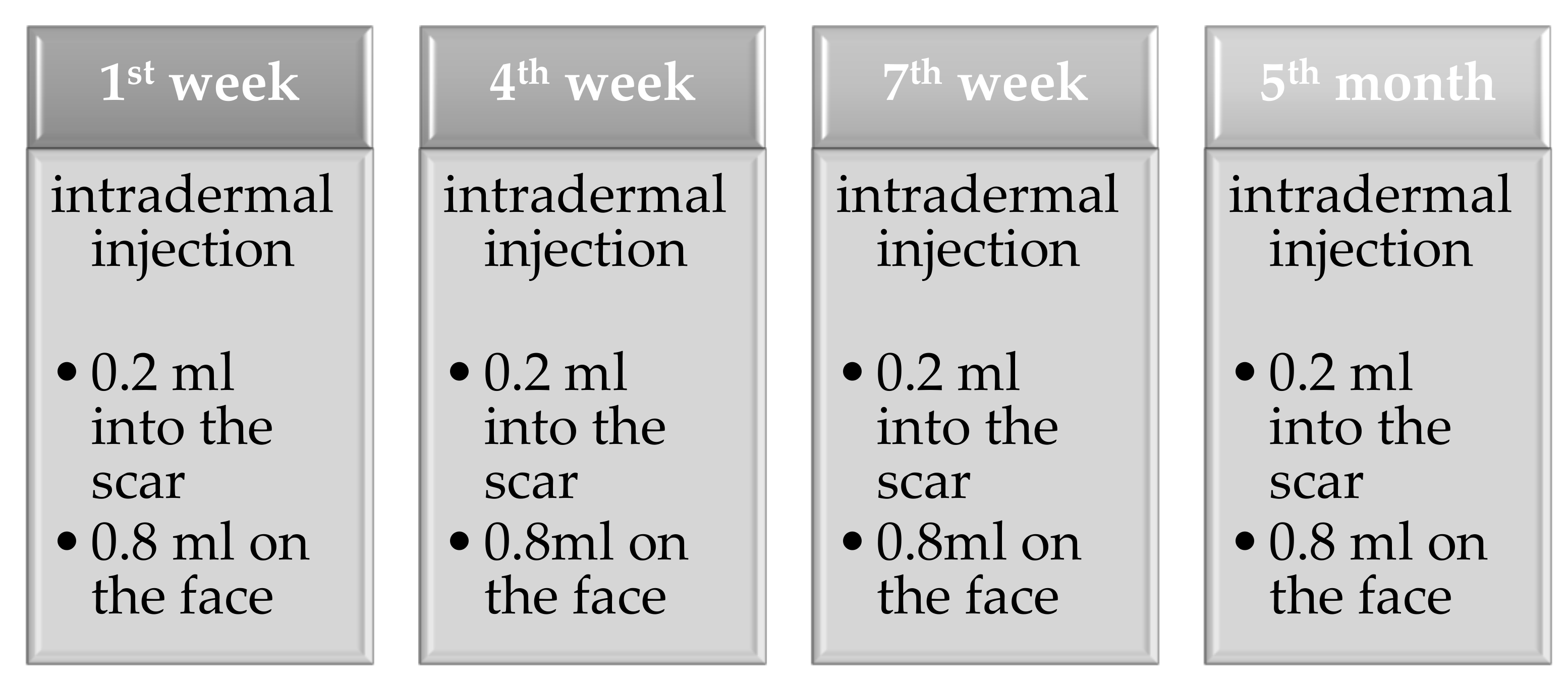
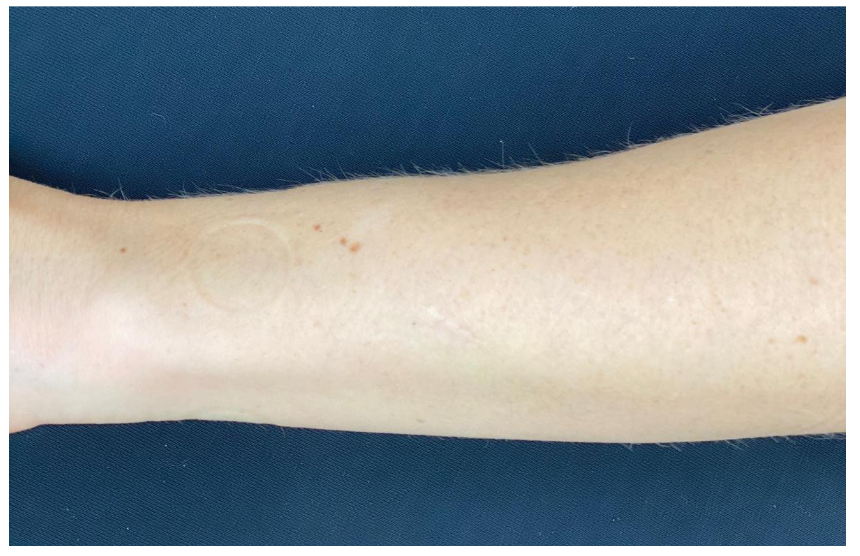
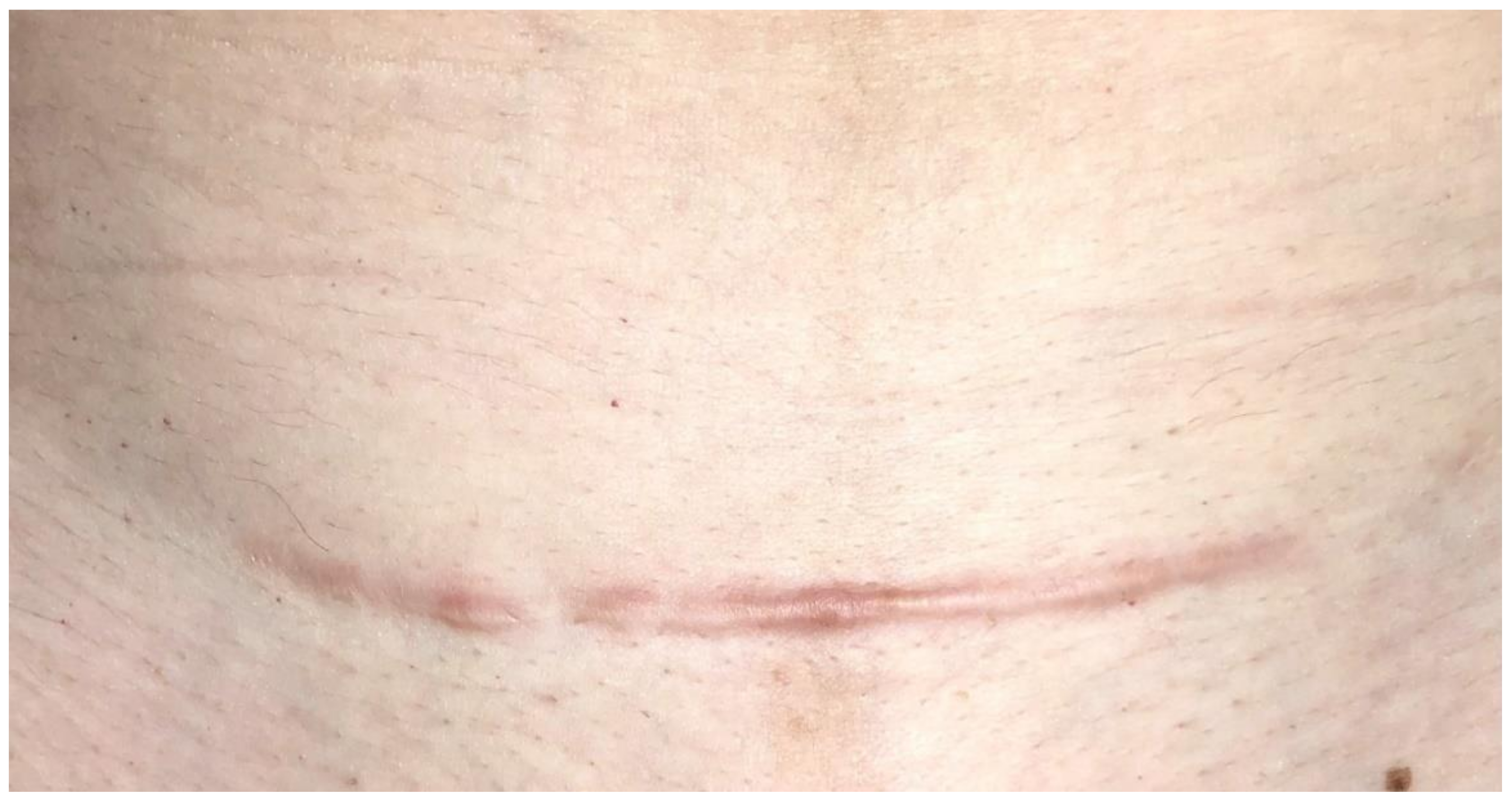
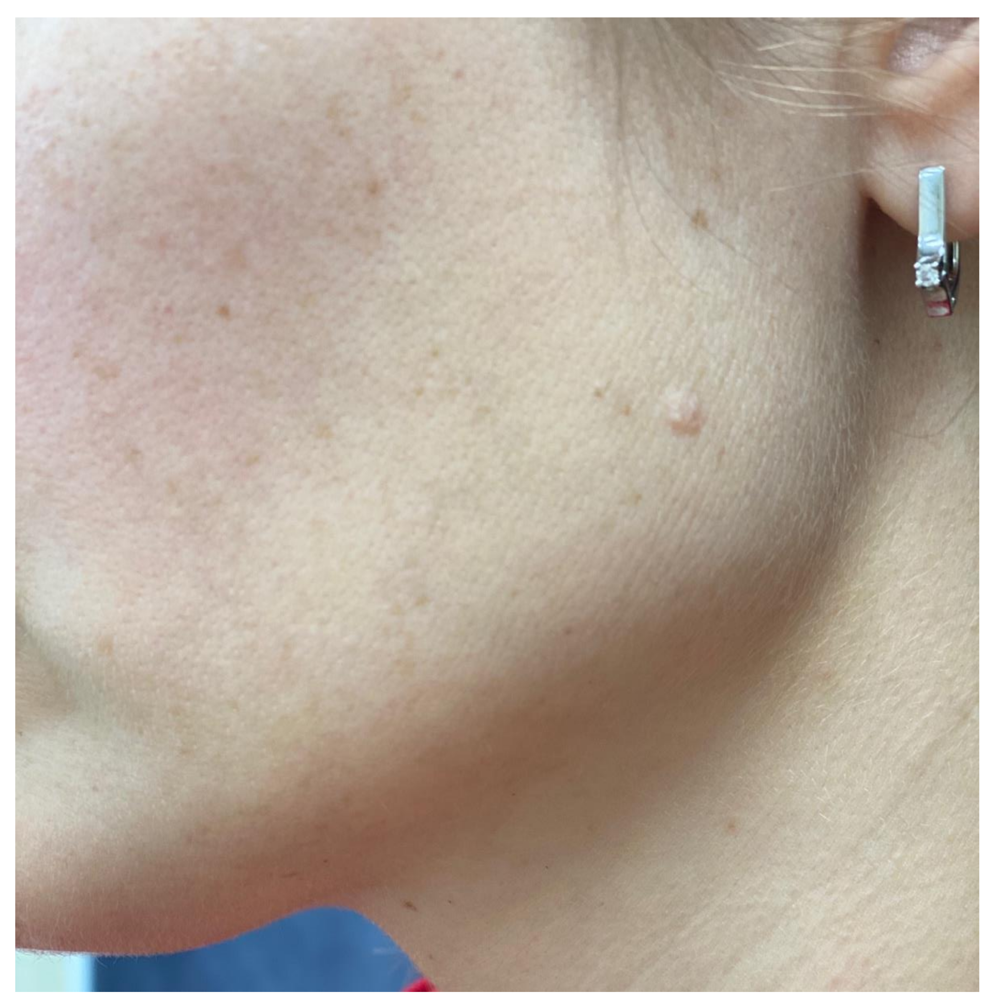
2. Case Presentation

A 40-year-old woman with postoperative 30 mm suture after lipoma removal surgery on the arm presented a strong desire to prevent scar hyperplasia. She had a history of pathological wound healing after cesarean section and removal of skin neoplasms. In addition, there was a divergence of the wound edges under the influence of a mechanical factor after removing the stitches. In the external examination, hypertrophic scar on the remodeling phase were seen in place after cesarean section (18 months ago) as well as several hypertrophic white scars after the removal of skin neoplasms. This patient has some signs related to the clinical symptoms of connective tissue dysplasia: skin hyperextension, arachnodactyly, flat feet, nephroptosis, mitral valve prolapse, and hypertrophic scars (Figure 1).
In the analysis of genetic polymorphisms of connective tissue, SNP was detected in the ELN gene (responsible for elastin synthesis) Rs2071307 (c.1264G > A) (A; G). Other genes involved in extracellular matrix synthesis were medium-population (normal) (Table 1). SNP genes responsible for extracellular matrix remodeling are also present. However, their role in wound healing disorders has not been studied yet.
For the management of the patient, a skin rejuvenation product Teosyal Redensity 1 (Teoxane SA) was selected. This product has a special composition for the synthesis of components of the extracellular matrix. This composition includes hyaluronic acid, α-lipoic acid, glutathione, N-acetyl-l-cystein, l-arginine, l-isoleucine, l-leucine, l-lysine, glycine, l-valine, l-threonine, l-proline, pyridoxine, zinc acetate, and copper sulfate.
In the process of wound healing in the skin, the construction of proteins comes from amino acids and any lack of amino acids in the body will affect the wound healing. In total, four procedures were performed: intradermal injection of the product in the area of the entire scar by 0.2 mL per 1 session (Figure 2). The remainder of the product (0.8 mL) was used to rejuvenate the skin of the face. The interval between the first three procedures was three weeks, the fourth procedure was performed three months after the end of the previous three procedures.
Complete healing of the scar occurred after nine months. A normotrophic scar formed, not protruding above the surface of the skin. The width of the scar in the initial parts was about 0.5 mm, in the middle of the scar (where there was a discrepancy of the postoperative suture) at 2.5 mm (Figure 3). Under the scar area, there was a slight deficiency of subcutaneous fat due to lipoma removal. For comparison, Figure 4, Figure 5 and Figure 6 show other scars on the patient. The scar in Figure 4 was formed after cesarean section (after 26 months). In the area of the scar, areas of hypertrophy are visible, with the elevation of the scar above the skin surface up to 2 mm, and the remodeling stage was not completed. Figure 5 shows a scar after the removal of a neoplasm on the skin of the face after 10 years. Scar tissue protrudes above the skin surface by 2 mm, and the diameter of the scar is 5 mm. Figure 6 shows two scars after the removal of a neoplasm on the skin of the neck. The first (on the lateral side) shows the scar after 12 years. Scar tissue protrudes above the skin surface by 1.5 mm, and the diameter of the scar is 5 mm, with an initial wound diameter of about 3–4 mm. The second scar (on the central part) was improved with a rejuvenation injection (Teosyal Redensity 1) five years ago. It is almost invisible; we could see just hypopigmentation in this place.

3. Discussion and Conclusions

Hypertrophic and keloid scars are a serious problem in aesthetic medicine. Even small scars after medical interventions are difficult for patients to perceive [18]. The problem of wound healing management has not yet been solved despite numerous studies on this topic [19,20,21,22,23,24,25,26,27,28]. From the authors’ point of view, this problem should be investigated from two positions. First, we need to understand which patients are at high risk of scarring disorders. If there are clear criteria, these patients are more likely to need special therapeutic measures aimed at normalizing the wound healing process. Moreover, before procedures aimed at damaging the skin, such patients will need to pay special attention to the possibility of pathological scarring during the consultation. Before performing procedures that damage the skin, patients need to evaluate all the pros and cons. The second aspect that is necessary is the search for effective methods of prevention and the treatment of scars. Currently, many different methods are known including injections of botulinum toxin type A, platelet-rich plasma, silicone-based patch application, and many other methods [15,24].
Despite the large number of existing methods for the treatment of scars, few plastic surgeons in Russia currently provide such recommendations after operations. Unfortunately, we have a situation where plastic surgeons work separately from cosmetologists. Thus, patients with pathological scars arrive at cosmetologists too late. In any case, the lack of a unified approach for managing patients leads to the patients’ disappointment in aesthetic medicine when receiving pathological scars.
This clinical case reflects our approach to the management of patients after surgery. The main idea of this approach is to include the patient after surgery (mostly blepharoplasty) in the general aesthetic treatment. Patients who require blepharoplasty most often need facial rejuvenation. Thus, by including the patient in the process of general rejuvenation, no additional methods are required to correct the scar. The absence of additional treatments, actions, and payment increases the patient’s compliance.
Thus, all patients who have come to our clinic for surgery are first evaluated from the position of the risk of wound healing disorders. Then, patients with a high risk of pathology are treated together by cosmetologists and plastic surgeons using the above-mentioned approach. This method is applicable to different patients during wound healing.

4. Declaration of Patient Consent

The authors certify that they obtained all appropriate patient consent forms. In the form, the patient has given her consent for her images and other clinical information to be reported in the journal. The patient understands that their name and initials will not be published and due efforts will be made to conceal their identity, but anonymity cannot be guaranteed.

Author Contributions

Conceptualization, N.A.S. and O.B.B.; Methodology, E.I.K. and N.A.S.; Software, O.B.B. and O.M.D.; Validation, N.N.P., N.A.S., and M.M.P.; Formal analysis, M.M.P.; Data curation, G.V.M. and M.A.Z.; Writing—original draft preparation, O.B.B.; Writing—review and editing, N.A.S., M.A.-Z., and G.V.M.; Visualization, O.B.B. and E.A.N.; Supervision, N.N.P.; Project administration, N.A.S. All authors have read and agreed to the published version of the manuscript.

Funding

This research received no external funding.

Institutional Review Board Statement

The study was conducted according to the guidelines of the Declaration of Helsinki and approved by the Ethics Committee of the Pirogov Russian National Research Medical University (protocol code 206, date of approval 22 March 2021).

Informed Consent Statement

Informed consent was obtained from all subjects involved in the study. Written informed consent has been obtained from the patient to publish this paper.

Data Availability Statement

Not applicable.

Conflicts of Interest

O.B.B. received fees as an expert for Teoxane SA. The other authors declare no conflict of interest.

References

  1. Mostaço-Guidolin, L.; Rosin, N.L.; Hackett, T.L. Imaging Collagen in Scar Tissue: Developments in Second Harmonic Generation Microscopy for Biomedical Applications. Int. J. Mol. Sci. 2017, 18, 1772. [Google Scholar] [CrossRef]
  2. Ud-Din, S.; Volk, S.W.; Bayat, A. Regenerative healing, scar-free healing and scar formation across the species: Current concepts and future perspectives. Exp. Dermatol. 2014, 23, 615–619. [Google Scholar] [CrossRef] [PubMed]
  3. Van den Broek, L.J.; Limandjaja, G.C.; Niessen, F.B.; Gibbs, S. Human hypertrophic and keloid scar models: Principles, limitations and future challenges from a tissue engineering perspective. Exp. Dermatol. 2014, 23, 382–386. [Google Scholar] [CrossRef] [PubMed] [Green Version]
  4. Kaplani, K.; Koutsi, S.; Armenis, V.; Skondra, F.G.; Karantzelis, N.; Champeris Tsaniras, S.; Taraviras, S. Wound healing related agents: Ongoing research and perspectives. Adv. Drug Deliv. Rev. 2018, 129, 242–253. [Google Scholar] [CrossRef]
  5. Frykberg, R.G.; Banks, J. Challenges in the Treatment of Chronic Wounds. Adv Wound Care 2015, 4, 560–582. [Google Scholar] [CrossRef] [Green Version]
  6. Shih, B.; Bayat, A. Genetics of keloid scarring. Arch. Dermatol. Res. 2010, 302, 319–339. [Google Scholar] [CrossRef] [PubMed]
  7. Huang, C.; Nie, F.; Qin, Z.; Li, B.; Zhao, X. A snapshot of gene expression signatures generated using microarray datasets associated with excessive scarring. Am. J. Dermatopathol. 2013, 35, 64–73. [Google Scholar] [CrossRef] [PubMed]
  8. Xue, M.; Jackson, C.J. Extracellular Matrix Reorganization During Wound Healing and Its Impact on Abnormal Scarring. Adv. Wound Care 2015, 4, 119–136. [Google Scholar] [CrossRef] [Green Version]
  9. Tung, J.Y.; Kiefer, A.K.; Mullins, M.; Francke, U.; Eriksson, N. Genome-wide association analysis implicates elastic microfibrils in the development of nonsyndromic striae disease. J. Investig. Dermatol. 2013, 133, 2628–2631. [Google Scholar] [CrossRef] [PubMed] [Green Version]
  10. Vierkötter, A.; Schikowski, T.; Sugiri, D.; Matsui, M.S.; Krämer, U.; Krutmann, J. MMP-1 and -3 promoter variants are indicative of a common susceptibility for skin and lung aging: Results from a cohort of elderly women (SALIA). J. Investig. Dermatol. 2015, 135, 1268–1274. [Google Scholar] [CrossRef] [PubMed] [Green Version]
  11. Potekaev, N.N.; Borzykh, O.B.; Medvedev, G.V.; Pushkin, D.V.; Petrova, M.M.; Petrov, A.V.; Dmitrenko, D.V.; Karpova, E.I.; Demina, O.M.; Shnayder, N.A. The Role of Extracellular Matrix in Skin Wound Healing. J. Clin. Med. 2021, 10, 5947. [Google Scholar] [CrossRef] [PubMed]
  12. Stone, R.C.; Chen, V.; Burgess, J.; Pannu, S.; Tomic-Canic, M. Genomics of Human Fibrotic Diseases: Disordered Wound Healing Response. Int. J. Mol. Sci. 2020, 21, 8590. [Google Scholar] [CrossRef]
  13. Gensemer, C.; Burks, R.; Kautz, S.; Judge, D.P.; Lavallee, M.; Norris, R.A. Hypermobile Ehlers-Danlos syndromes: Complex phenotypes, challenging diagnoses, and poorly understood causes. Dev. Dyn. 2021, 250, 318–344. [Google Scholar] [CrossRef]
  14. Borzykh, O.B.; Petrova, M.M.; Shnayder, N.A.; Nasyrova, R.F. Problems of implementation of personalized medicine in medical cosmetology in Russia. Sib. Med. Rev. 2021, 2, 12–22. [Google Scholar] [CrossRef]
  15. Shirakami, E.; Yamakawa, S.; Hayashida, K. Strategies to prevent hypertrophic scar formation: A review of therapeutic interventions based on molecular evidence. Burns Trauma 2020, 8, tkz003. [Google Scholar] [CrossRef] [Green Version]
  16. Bi, M.; Sun, P.; Li, D.; Dong, Z.; Chen, Z. Intralesional Injection of Botulinum Toxin Type A Compared with Intralesional Injection of Corticosteroid for the Treatment of Hypertrophic Scar and Keloid: A Systematic Review and Meta-Analysis. Med. Sci. Monit. 2019, 25, 2950–2958. [Google Scholar] [CrossRef] [PubMed]
  17. Mohiuddin, A.K. Patient Compliance: Fact or Fiction? Innov. Pharm. 2019, 10, 3. [Google Scholar] [CrossRef] [PubMed] [Green Version]
  18. Poetschke, J.; Gauglitz, G.G. Current options for the treatment of pathological scarring. JDDG J. Dtsch. Dermatol. Ges. 2016, 14, 467–477. [Google Scholar] [CrossRef] [PubMed]
  19. Huguier, V.; Giot, J.-P.; Simonneau, M.; Levillain, P.; Charreau, S.; Garcia, M.; Jégou, J.-F.; Bodet, C.; Morel, F.; Lecron, J.-C.; et al. M exerts a protective effect against excessive scarring by counteracting the inductive effect of TGFβ1 on fibrosis markers. Sci. Rep. 2019, 9, 2113. [Google Scholar] [CrossRef]
  20. Martino, M.M.; Tortelli, F.; Mochizuki, M.; Traub, S.; Ben-David, D.; Kuhn, G.A.; Müller, R.; Livne, E.; Eming, S.A.; Hubbell, J.A. Engineering the growth factor microenvironment with fibronectin domains to promote wound and bone tissue healing. Sci. Transl. Med. 2011, 3, 100ra189. [Google Scholar] [CrossRef] [PubMed] [Green Version]
  21. Polat, E.; Aksöz, İ.; Arkan, H.; Coşkunpınar, E.; Akbaş, F.; Onaran, İ. Gene expression profiling of Lucilia sericata larvae extraction/secretion-treated skin wounds. Gene 2014, 550, 223–229. [Google Scholar] [CrossRef] [PubMed]
  22. Baumann, L.; Bernstein, E.F.; Weiss, A.S.; Bates, D.; Humphrey, S.; Silberberg, M.; Daniels, R. Clinical Relevance of Elastin in the Structure and Function of Skin. Aesthetic Surg. J. Open Forum 2021, 3, ojab019. [Google Scholar] [CrossRef]
  23. Cohen, B.E.; Geronemus, R.G.; McDaniel, D.H.; Brauer, J.A. The role of elastic fibers in scar formation and treatment. Dermatol. Surg. 2017, 43, 19–24. [Google Scholar] [CrossRef] [PubMed]
  24. Murdock, J.; Sayed, M.S.; Tavakoli, M.; Portaliou, D.M.; Lee, W.W. Safety and efficacy of a growth factor and cytokine-containing topical product in wound healing and incision scar management after upper eyelid blepharoplasty: A prospective split-face study. Clin. Ophthalmol. 2016, 10, 1223–1228. [Google Scholar] [PubMed] [Green Version]
  25. Wang, J.; Liao, Y.; Xia, J.; Wang, Z.; Mo, X.; Feng, J.; He, Y.; Chen, X.; Li, Y.; Lu, F.; et al. Mechanical micronization of lipoaspirates for the treatment of hypertrophic scars. Stem Cell Res. Ther. 2019, 10, 42. [Google Scholar] [CrossRef] [PubMed]
  26. Urciuolo, F.; Casale, C.; Imparato, G.; Netti, P.A. Bioengineered Skin Substitutes: The Role of Extracellular Matrix and Vascularization in the Healing of Deep Wounds. J. Clin. Med. 2019, 8, 2083. [Google Scholar] [CrossRef] [Green Version]
  27. Qiu, S.S.; Dotor, J.; Hontanilla, B. Effect of P144® (Anti-TGF-β) in an “In Vivo” Human Hypertrophic Scar Model in Nude Mice. PLoS ONE 2015, 10, e0144489. [Google Scholar] [CrossRef] [PubMed]
  28. Chen, Q.; Zhao, T.; Xie, X.; Yu, D.; Wu, L.; Yu, W.; Sun, W. MicroRNA-663 regulates the proliferation of fibroblasts in hypertrophic scars via transforming growth factor-β1. Exp. Ther. Med. 2018, 16, 1311–1317. [Google Scholar] [CrossRef] [Green Version]
Figure 1. Some of the phenotypical traits of the patient: hyperextensible skin (above the lateral edge of the clavicle) (A) and arachnodactyly (test of grip of the wrist with one and five fingers) (B).
Cosmetics 09 00028 g001aCosmetics 09 00028 g001b
Figure 2. The general scheme of the treatment.
Cosmetics 09 00028 g002
Figure 3. Normotrophic scar after four procedures (after nine months) on the arm.
Cosmetics 09 00028 g003
Figure 4. Hypertrophic scar after cesarean section (after 26 months).
Cosmetics 09 00028 g004
Figure 5. The scar after removal of a neoplasm on the skin of the face after 10 years.
Cosmetics 09 00028 g005
Figure 6. The scar after removal of a neoplasm on the skin of the neck after 12 years (at the lateral side) and the scar after removal of a neoplasm on the skin of the neck after five years (at the central side).
Cosmetics 09 00028 g006
Table 1. The result of the genetic analysis of genes responsible for the structure of the extracellular matrix.
Gene Protein/Enzyme Encoded by This GeneRSNormalResult
COL1A1Encoding the α1 chain of collagen type IRs1800012G/GG/G
ELNEncoding the elastinRs7787362T/TC/C
HAS1Encoding the hyaluronan synthase 1Rs7248778G/GG/G
MMP1Encoding the matrix metallopeptidase 1Rs1799750-/-G/-
MMP3Encoding the matrix metallopeptidase 3Rs3025058A/AA/A
TIMP1Encoding the tissue inhibitor of metalloproteinase 1Rs4898C/CC/T
Publisher’s Note: MDPI stays neutral with regard to jurisdictional claims in published maps and institutional affiliations.

