Article

Caesalpinia sappan L. Ameliorates Scopolamine-Induced Memory Deficits in Mice via the cAMP/PKA/CREB/BDNF Pathway

1
Faculty of Pharmacy, Universitas Gadjah Mada, Sekip Utara, Yogyakarta 55281, Indonesia
2
Department of Pharmaceutical Biology, Faculty of Pharmacy, Universitas Gadjah Mada, Sekip Utara, Yogyakarta 55281, Indonesia
3
Department of Pharmacology and Clinical Pharmacy, Faculty of Pharmacy, Universitas Gadjah Mada, Sekip Utara, Yogyakarta 55281, Indonesia
*
Author to whom correspondence should be addressed.
Submission received: 22 April 2021 / Revised: 31 May 2021 / Accepted: 8 June 2021 / Published: 18 June 2021

Abstract

:
Memory is an essential aspect of human cognition. A decrease in this aspect is well associated with Alzheimer’s disease (AD). The development of a novel cognitive enhancer (CE) may help overcome AD-related problems. In this study, we evaluated the CE effect of Caesalpinia sappan L. (CS) in memory deficit mice. Administration of its ethanolic extract (250 and 500 mg/kg body weight (BW)) and brazilin (5 and 10 mg/kg BW) ameliorated the scopolamine-amnesic effect, as evidenced by significant decreases (p < 0.01, p < 0.05) in the escape latency time and increases (p < 0.01) in the percentage of time spent in the target quadrant of the Morris water maze test. We also examined the cyclic adenosine monophosphate (cAMP) level, protein kinase A (PKA) activity, and protein expression levels of phosphorylated cAMP response element binding (pCREB) and brain-derived neurotrophic factor (BDNF) in hippocampal tissues to elucidate the underlying molecular mechanism. Results showed that CS wood ethanolic extract and brazilin not only significantly increase (p < 0.01, p < 0.05) cAMP levels and PKA activity but also significantly enhance (p < 0.01, p < 0.05) the expression level of pCREB and BDNF in the hippocampus. These findings indicate that CS activates the cAMP/PKA/CREB/BDNF pathway. Taken together, our results demonstrate that CS is a promising herb that could be developed as a CE agent.

1. Introduction

Dementia is a neurological syndrome characterized by a progressive decline in cognitive function. The most common type of dementia is Alzheimer’s disease (AD) [1,2,3]. The underlying pathophysiology and pathogenesis of AD are complex. In humans, abnormal proteins in the brain, amyloid plaques, and neurofibrillary tangles are common features of the disease and affect central functions, such as memory [4,5,6]. Memory is one of prominent aspects of assessing cognition in AD. Impairments in memory are clinically observed in patients with AD and are linked to dysfunctions of the hippocampus and the prefrontal lobe region [7,8]. Moreover, reductions in cyclic nucleotides (e.g., cyclic adenosine monophosphate (cAMP) and/or cGMP) and neurotrophic proteins (e.g., brain-derived neurotrophic factor (BDNF)) in the brain have also been observed to be positively correlated with AD progression.
Acetylcholinesterase inhibitors (AChEIs) are commonly used as first-line drugs for AD. These compounds act by inhibiting AChE, a hydrolytic enzyme that degrades acetylcholine (ACh) in the brain. ACh is an important cholinergic neurotransmitter for learning and a participant of the memory signaling pathway. AChEI use effectively relieves AD symptoms [9,10,11,12]. However, undesirable outcomes are often observed in the majority of patients that are prescribed this therapy. Consequently, research on safe and effective anti-amnesic drugs as alternative therapies to prevent or treat continues to be a major undertaking.
Several lines of evidence indicate that phosphodiesterase (PDE), the enzyme responsible for the hydrolytic breakdown of cyclic nucleotides (e.g., cAMP/cGMP), is a potential molecular target in AD. Inhibition of PDE activity can increase cyclic nucleotide levels in the brain, which is important to activate brain synaptic protein expression pathways for learning and memory maintenance [13,14]. Vinpocetine and rolipram, two PDE inhibitors currently used to treat AD, have shown great promise in efforts to address the degenerative features of the disease. These drugs remain at the forefront of research on AD drug discovery and development [15,16,17].
Caesalpinia sappan L. (CS) is a leguminous plant that is widely distributed in Indonesia. As a high-potential plant, it is commonly used as a natural coloring agent in cosmetics and the food and beverage industry. CS is also traditionally used in the management of several diseases [18,19]. Scientific evidence indicates that CS and brazilin, its major compound, have broad pharmacological indications. Our previous in vitro study showed that CS, particularly its ethanolic extract, and brazilin effectively inhibited PDE1 activity [20,21]. Thus, we conducted this further study to investigate the cognitive-enhancing (CE) effect of CS wood’s (CSW) ethanolic extract and brazilin on memory assessment in scopolamine-induced memory deficit mice and elucidate the underlying molecular mechanism, especially in terms of the cAMP/protein kinase A (PKA)/cAMP response element binding (CREB)/BDNF pathway. This is our preliminary in vivo study to develop CS as an alternative cognitive enhancer agent.

2. Materials and Methods

2.1. Plant Material

CSW was collected from Bantul, Yogyakarta, and authenticated at the Department of Pharmaceutical Biology, Faculty of Pharmacy, Universitas Gadjah Mada. CSW bark was washed and dried overnight in an oven. The dried bark was cut into small pieces and then ground using a grinding mill.

2.2. Chemicals and Reagents

The chemicals and materials used in this study included brazilin (Chengdu Biopurity, Chengdu, China), carboxymethylcellulose (CMC), formic acid (Sigma Aldrich, St. Louis, MO, USA), hydrochloric acid (HCl), acetone, ethanol, chloroform, TLC silica gel F254 (20 cm × 20 cm), bovine serum albumin (Merck, Darmstadt, Germany), a cAMP enzyme direct immunoassay kit (Sigma Aldrich, USA), a PKA activity kit (Enzo Life Science, Farmingdale, NY, USA), anti-phosphorylated CREB (pCREB), anti-CREB, anti-BDNF, and anti-β-actin (Abclonal, Woburn, MA, USA).

2.3. Drugs

Scopolamine hydrobromide (Sigma Aldrich, MO, USA) was used to induce the memory deficit-animal model, and vinpocetine (Source Naturals®, Scotts Valley, CA, USA) was used as the reference drug. Normal saline (NaCl, 0.9%) was also used.

2.4. Experimental Animals

Seventy 7–8-week-old male Balb/c mice (20–30) were used in this study. The animals were housed in 14 cages under standard laboratory conditions at the Animal House of the Department of Physiology, Faculty of Medicine, Public Health and Nursing, Universitas Gadjah Mada. The mice were kept in an environment with a controlled temperature (25 °C ± 2 °C) and humidity (50% ± 5%) under a normal 12 h/12 h light/dark cycle and were allowed free access to food pellets and water during the experiment. All of the procedures were performed according to animal welfare guidelines.

2.5. CSW Extract Preparation

CSW powder was macerated with 1:5 (w/v) of ethanol for 3 × 24 h and then filtered. The macerate was concentrated using a rotary evaporator at 50 °C, and the solvent residue was air-dried at room temperature (25 °C ± 5 °C).

2.6. TLC-Densitometry Analysis

Quantification of brazilin in the CSW extract (10 mg/mL) was achieved through TLC-densitometry by using serial concentrations of the brazilin standard (1 mg/mL). In this analysis, silica gel F254 was used as the stationary phase and chlorofom:acetone:formic acid (4:2:0.5, v/v) was used as the mobile phase. The brazilin content in the extract was measured at a maximum wavelength of 508 nm. The TLC plate was also visualized under visible and UV light at 254 and 366 nm, respectively.

2.7. Test Solution Preparation

The CSW ethanolic extract, brazilin, and vinpocetine were dissolved in a 1% CMC solution to prepare test solutions with final concentrations of 250, 500, 5, 10, and 4 mg/kg body weight (BW). Scopolamine hydrobromide was dissolved in 0.9% NaCl to obtain a solution of 4 mg/kg BW.

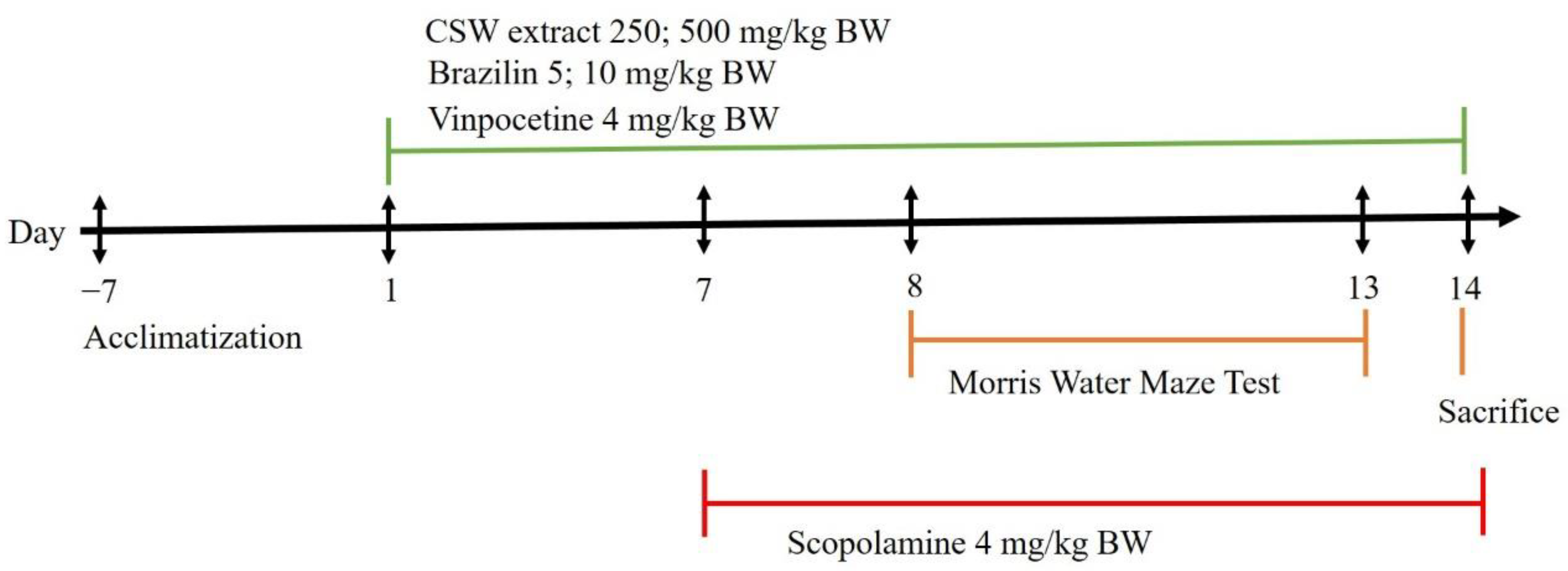
2.8. Experimental Design

The mice were randomly divided into the following seven groups (ten animals per group): (1) the normal group received 0.9% saline intra peritoneal (i.p.); (2) the vehicle group received 1% CMC per oral (p.o.) and scopolamine 4 mg/kg BW (i.p.) as a negative control; (3) the vinpocetine group received vinpocetine 4 mg/kg BW (p.o.) and scopolamine 4 mg/kg BW (i.p.) as a positive control; (4) the CSW 250 group received CSW ethanolic extract 250 mg/kg BW (p.o) and scopolamine 4 mg/kg BW (i.p.); (5) the CSW 500 group received CSW ethanolic extract 500 mg/kg BW (p.o.) and scopolamine 4 mg/kg BW (i.p.); (6) the Brazilin 5 group received brazilin 5 mg/kg BW (p.o.) and scopolamine 4 mg/kg BW (i.p.); and (7) the Brazilin 10 group received brazilin 10 mg/kg BW (p.o.) and scopolamine 4 mg/kg BW (i.p.). CSW ethanolic extract, brazilin, and vinpocetine were administrated once a day (60 min before behavioral test and sacrifice) for 14 consecutive days (day 1 to 14) and scopolamine was injected once a day (30 min before behavioral test and sacrifice) for 7 consecutive days (day 7 to 14). The schedule of treatments is illustrated in Figure 1.

2.9. Morris Water Maze Test

The Morris water maze (MWM) test was conducted in the behavioral testing room of the Department of Physiology, Faculty of Medicine, Public Health and Nursing, Universitas Gadjah Mada. This behavioral test has been effectively used to assess animals’ spatial memory. The MWM apparatus consisted of a circular white tank (diameter, 80 cm; height, 40 cm), a white circular tin platform (diameter, 13 cm; height, 16.5 cm), and a video camera recorder (Prolink, Taiwan) installed above the pool. The tank was filled with water (25 °C ± 2 °C) to a depth of 18–19 cm (0.5 cm below the surface); ±5 L of fresh milk was added to the pool to render the water opaque. The maze was divided equally into four imaginary quadrants (A, B, C, and D) with eight starting points (1–8) and numerous fixed-visual cues in the test room wall. The platform was placed at the center of a selected quadrant and maintained at this location throughout the experiment. The MWM test consisted of an acquisition phase and the probe trial was conducted for six consecutive days. Each mouse received four trials per day for five consecutive days in the acquisition phase with semi-random starting points. Each mouse was allowed to reach the hidden platform within 60 s, and its escape latency time (s) was recorded if its four limbs stayed on top of the platform. The probe trial was performed the day after the last day of the acquisition phase (day 6). In this phase, the platform was removed, and the mice were given time to explore the pool for 60 s. The time spent in the area in which the platform had previously been located (target quadrant) was recorded [22].

2.10. Hippocampus Collection

On the last day of sample administration (day 14), the mice were sacrificed by decapitation under anesthesia. The brains were carefully removed from the skull, and the hippocampus were immediately separated on frozen 0.9% NaCl. Hippocampal tissues (n = 10) from each animal in all of the experimental groups were maintained at −80 °C. We divided the hippocampal tissues for cAMP level analysis (n = 5) and for protein isolation (n = 5).

2.11. cAMP Level Analysis

Hippocampal tissues (25 mg) were homogenized with 10 volumes of 0.1 N HCl and then centrifuged at 600× g for 15 min at room temperature. The supernatant was used for the cAMP level assay using a cAMP enzyme direct immunoassay kit (Sigma Aldrich, USA), according to the manufacturer’s instructions. The cAMP level assay was measured based on the direct ELISA principle that used a polyclonal antibody for cAMP binding from the sample and a secondary antibody for recognition. The intensity of color after the incubation was measured at 405 nm and inversely represented the level of cAMP in the sample. The cAMP level in the hippocampus was expressed as pmol/mL.

2.12. PKA Activity Analysis

PKA activity in the hippocampus was determined using a PKA activity kit (Enzo Life Science, USA), according to the manufacturer’s instructions. The PKA activity assay was measured based on the ELISA principle that utilized a specific synthetic peptide as a phosphospecific substrate antibody for PKA and a polyclonal antibody for the phosphorylated substrate recognition. The color intensity after the incubation was measured at 450 nm and linearly represented the activity of PKA in the sample. The PKA activity was expressed as ng/µg protein.

2.13. Protein Isolation

Hippocampal tissues were homogenized and incubated with a protein extraction solution (Pro-prepTM, Intron Biotechnology, Seongnam, South Korea) containing 1.0 mM PMSF, 1.0 mM EDTA, 1 µM pepstatin A, 1 µM leupeptin, and 0.1 µM aprotinin and then kept in a freezer at −20 °C for 20 min. After incubation, the homogenates were centrifuged at 13,000× g rpm for 5 min at 4 °C. The total protein concentration was measured using a PierceTM protein assay reagent (Thermo Fisher, Waltham, MA, USA). The isolated protein was used for PKA activity and Western blot analysis.

2.14. Western Blot Analysis

The protein homogenates of the samples (20 µg) were separated by using 15% sodium dodecyl sulfate-polyacrylamide gel electrophoresis and transferred to polyvinylidene difluoride (PVDF) membranes (Millipore-ImmobilonTM, Merck) at 400 mA for 2.5 h. After the transfer process, the PVDF membranes were incubated with 0.25% gelatin in Tris-buffered saline with 0.05% Triton-100 (1× NET-gelatin) at 4 °C overnight. After blocking, the membranes were washed four times with a phosphate-buffered saline with 0.01% Tween-20 (PBST) and then incubated overnight with anti-β-actin (1:1000), anti-pCREB (1:1000), anti-CREB (1:1000), and anti-BDNF (1:500) at room temperature. After four washes with PBST, the membranes were incubated with a secondary anti-rabbit IgG:HRP conjugate (Abcam, Cambridge, UK) (1:500) for 1 h at room temperature and then washed once more with PBST. The membranes were reacted with the enhanced chemiluminescence Western blotting substrate (Promega, Madison, WI, USA) for 3 min and then visualized using Geldoc G:BOX Chemi-XRQ (Syngene, Bengaluru, India). Images of the target proteins were analyzed by using ImageJ 1.52a software (Wayne Rasband, National Institutes of Health, Kensington, MD, USA).

2.15. Statistical Analysis

The data were presented as mean ± SEM. Statistical analysis was performed using a one-way ANOVA and the Kruskal–Wallis test followed by the Mann–Whitney, Tukey HSD, and LSD tests for post-hoc analyses. The significance level was determined at p < 0.01 and p < 0.05 by using IBM SPSS Statistics version 21.

3. Results

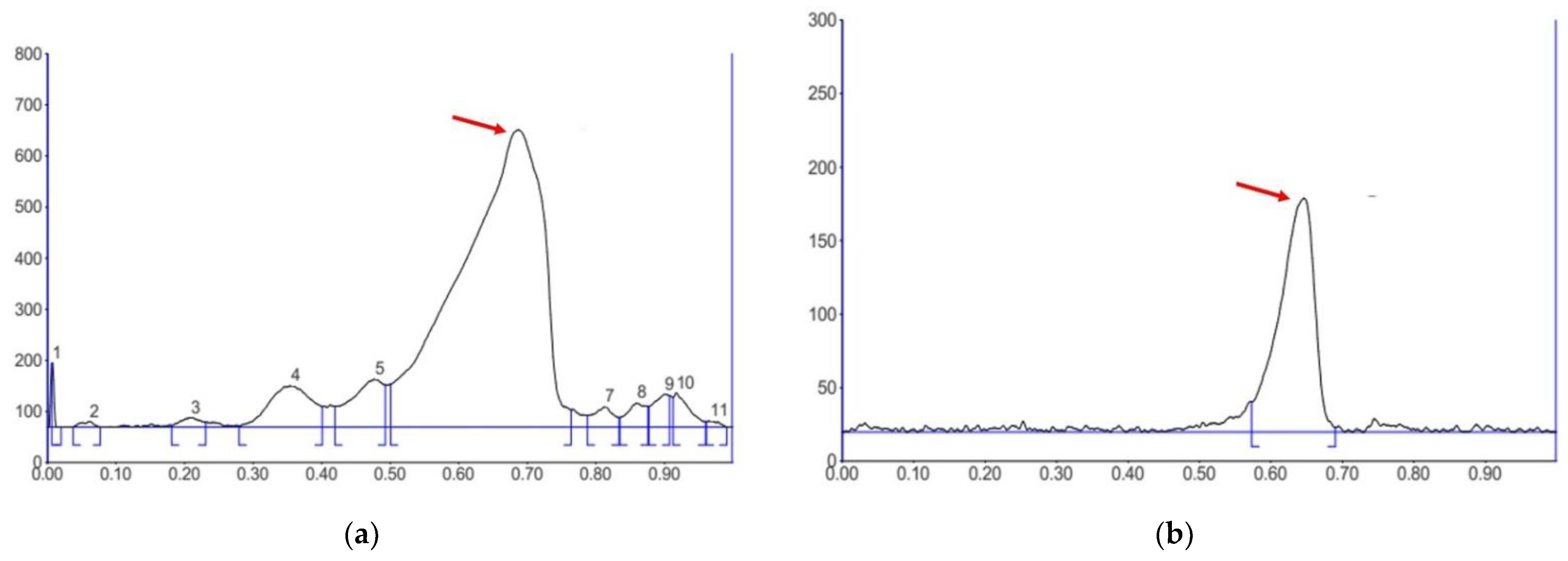
3.1. Determination of Brazilin Content in the CSW Extract

We performed TLC-densitometry to measure the brazilin content in the CSW ethanolic extract for quality control. According to our results, the CSW ethanolic extract contained 16.53% ± 0.2% brazilin. The presence of brazilin in the extract was manifested by a single strong peak with a maximum retention factor (Rf) of 0.69 (Figure 2a), similar to the Rf of the brazilin reference compound (Figure 2b).

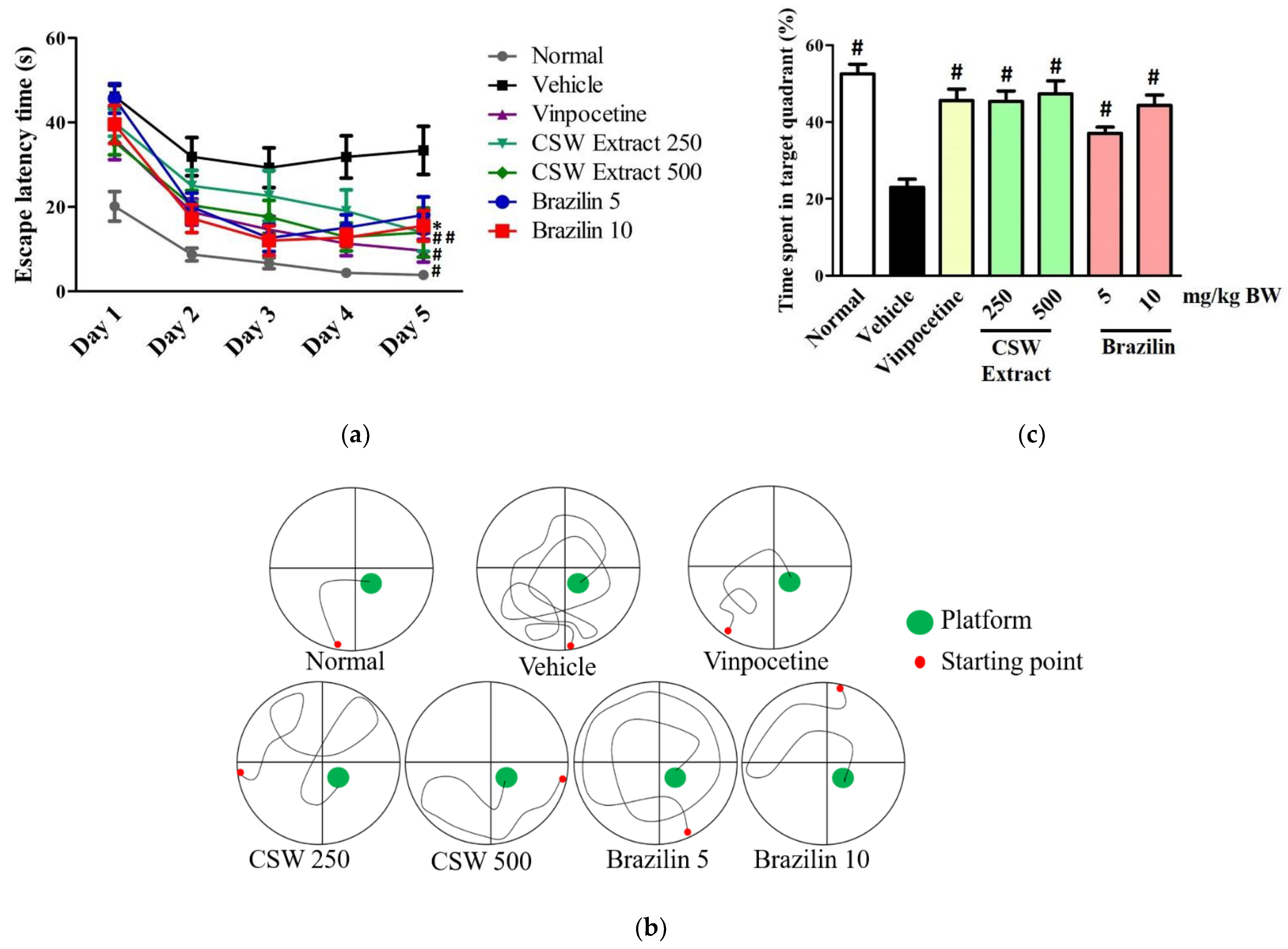
3.2. CSW Extract and Brazilin Improved Spatial Memory in the MWM Test

We performed the MWM test to assess the animals’ spatial memory performance. As shown in Figure 3a, the mean time required by all of the mice, except those in the vehicle group, to find the hidden platform decreased in the acquisition phase from day 1 to day 5. On the last day of the acquisition phase (day 5), the groups that received CSW extract at doses of 250 mg/kg BW (13.88 s ± 4.4 s) and 500 mg/kg BW (13.93 s ± 5.8 s) or brazilin at doses of 5 mg/kg BW (18.08 s ± 4.3 s) and 10 mg/kg BW (15.50 s ± 3.6 s) showed significantly (p < 0.01, p < 0.05) shorter escape latency times compared to those that received the vehicle (33.38 s ± 5.7 s). Interestingly, however, the differences observed compared to the vinpocetine group (9.65 s ± 2.7 s) were not significantly different (p > 0.05). The swim patterns of all of the groups are presented in Figure 3b. Animals with a memory deficit (vehicle group) revealed chaotic and irregular swim patterns compared to those in the normal and treated groups. The probe trial (day 6) results are presented in Figure 3c. The percentage of time that the mice treated with CSW extract at doses of 250 and 500 mg/kg BW or brazilin at doses of 5 and 10 mg/kg BW spent in the target quadrant increased by 45.37% ± 2.7%, 47.33% ± 3.4%, 37.08% ± 1.6%, and 44.33% ± 2.7%, respectively. These data are significantly (p < 0.01) higher than those observed in the vehicle group (23.00% ± 2.1%), but not significantly different (p > 0.05) compared to those observed in the normal (52.50% ± 2.5%) and vinpocetine (45.56% ± 3.0%) groups.

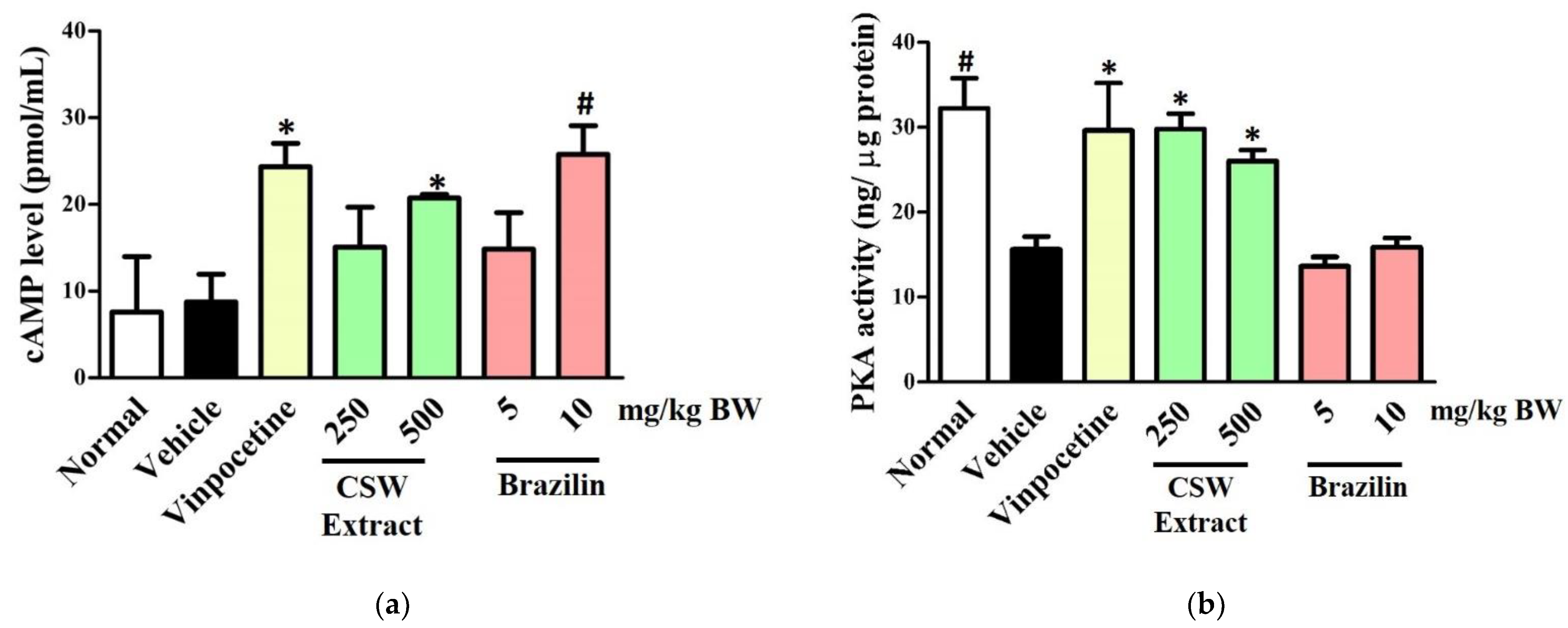
3.3. CSW Extract and Brazilin Increased cAMP Levels and PKA Activity in the Hippocampus

To elucidate the CE mechanisms of CS, we conducted molecular signaling pathway investigations using hippocampal tissue. First, we examined the cAMP levels and PKA activity in the hippocampus of the mice. The results were presented in Figure 4.
Our results in Figure 4a showed that the administration of CSW extract at doses of 250 and 500 mg/kg BW or brazilin at doses of 5 and 10 mg/kg BW could increase the cAMP levels in the hippocampus by 15.09 ± 4.6, 20.76 ± 0.4, 14.85 ± 4.2, and 25.78 ± 3.3 pmol/mL, respectively. The cAMP level in hippocampal tissues of the mice treated with CSW at a dose of 500 mg/kg BW or brazilin at a dose of 10 mg/kg BW significantly increased (p < 0.01, p < 0.05), compared to those of the normal and vehicle (untreated) groups, to 7.58 ± 6.4 and 8.75 ± 3.2 pmol/mL, respectively. In addition, treatment with CSW extract at doses of 250 and 500 mg/kg BW or brazilin at doses of 5 and 10 mg/kg BW increased the PKA activity in the hippocampus (Figure 4b) by 29.78 ± 1.8, 26.01 ± 1.3, 13.63 ± 1.1, and 15.85 ± 1.1 ng/µg protein, respectively. These values are significantly higher (p < 0.05) compared to those observed in the vehicle group (15.60 ± 1.5 ng/µg protein).

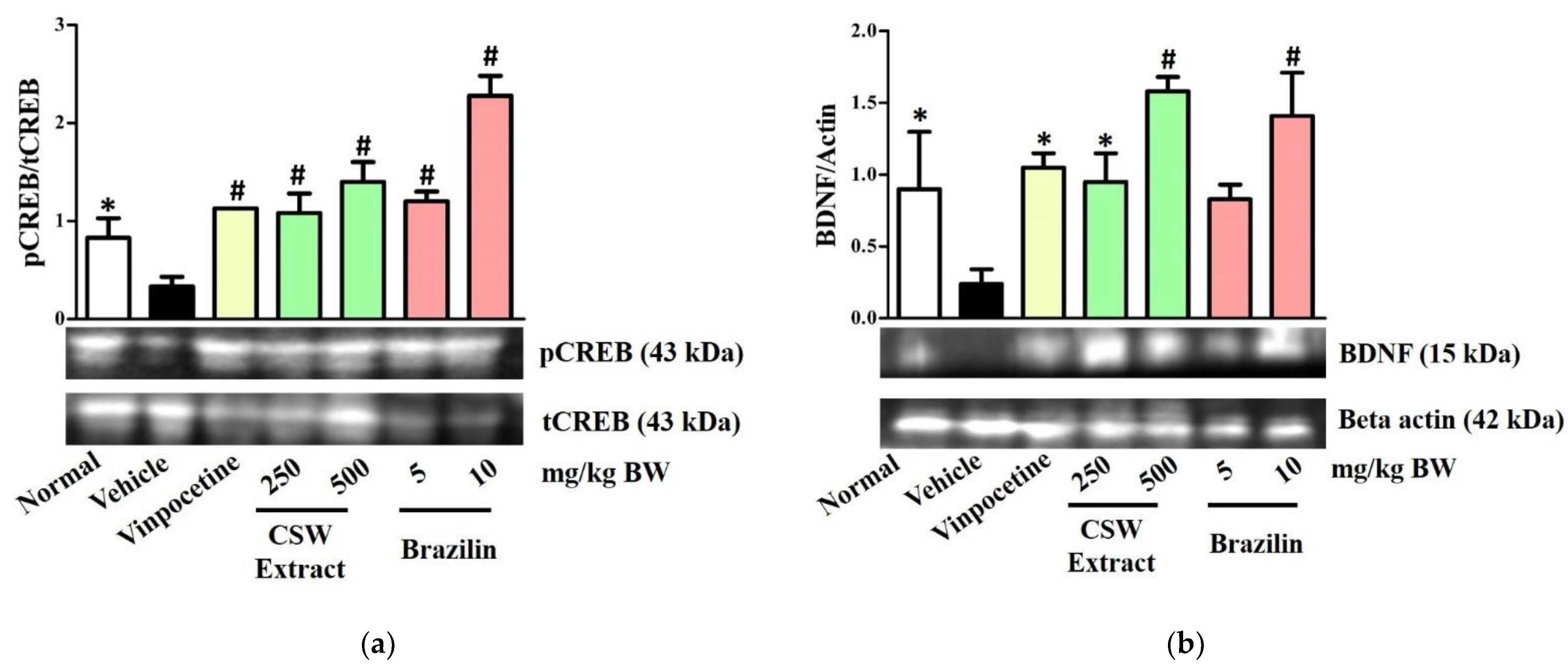
3.4. CSW Extract and Brazilin Increased the Protein Expression Level of pCREB and BDNF in the Hippocampus

Next, we investigated the protein expression level of pCREB and BDNF in the hippocampus using Western blot analysis. According to Figure 5a,b, scopolamine (4 mg/kg BB) administration significantly reduces the pCREB and BDNF expression levels in the hippocampus, as shown by the results of the vehicle group. Treatment with CS ethanolic extract at doses of 250 and 500 mg/kg BW or brazilin at doses of 5 and 10 mg/kg BW significantly increased (p < 0.01, p < 0.05) the protein expression levels of pCREB and BDNF by nearly 3–4-fold compared to those in the vehicle group.

4. Discussion

A CE agent is a substance used to improve the features of cognitive impairment (CI). A CE significantly reduces cognitive problems in many cases of dementia, such as in memory, intelligence, focus, attention, and motivation. CEs are also known as “smart drugs” or “nootropics” that are applied for amnesia amelioration in AD; these compounds exert various effects through heterogeneous mechanisms [17,23,24]. Several lines of evidence indicate that CSW is composed of various biologically active substances. Brazilin, the major compound in CSW, has been reported as a pharmacologically active substance responsible for many of the biological activities of CSW, including its antibacterial, anticancer, antifungal, antioxidant, anti-inflammation, and vasodilator properties [25,26]. We investigated the CE effect of CSW, particularly its ethanolic extract and brazilin, on scopolamine-induced memory deficit mice. As mentioned before, we hypothesized that CSW had a potential CE effect based on our previous study, which showed that CS ethanolic extract and brazilin at concentrations of 100 and 11.45 µg/mL, respectively, effectively inhibit PDE1 activity [20,21]. PDE1 is a member of the group of PDEs responsible for the hydrolysis of phosphodiester bonds during cAMP breakdown. This enzyme is abundantly found in several cognition-related regions of the brain, such as the hippocampus [14,27,28].
The hippocampus, one of the most important regions of the brain, is necessary for processing memory, particularly cognition. Animals with good spatial memory are expected to remember, and spend a long time in, the target quadrant in which the hidden platform was previously located. Spatial memory is a cognitive ability used to access information on position and to navigate to a certain location [29,30]. Spatial memory is classified as short-term memory that is closely associated with the functions of the hippocampus and prefrontal lobe [7,8,31].
In the present study, we evaluated the cAMP levels and PKA activity in the hippocampal tissues of scopolamine-induced memory deficit mice. cAMP is a secondary messenger for several molecular signaling pathways, including cognition maintenance. Decreases in cAMP levels are well associated with CI in many clinical contexts. Consequently, increases in intracellular brain cAMP levels through PDE inhibition is a potential therapeutic target for CI treatment. Vinpocetine and rolipram are two commercially available PDE inhibitors used to treat dementia in AD. Thus, we used vinpocetine as positive control in this study. An adequate level of the brain’s cAMP is required to activate PKA and phosphorylate several transcription factor proteins in the nucleus, such as CREB.
Inhibition of PDE1 activity effectively increased the cAMP level and further increased PKA activity in the hippocampus. PKA is a kinase enzyme that contributes to phosphate transfer during protein phosphorylation, including the CREB protein. This process is a key step in the activation of transcription factor proteins in the nucleus. CREB is a nucleus-localized protein with a crucial role in gene transcription. This protein directly interacts with gene promoters in the nucleus. Phosphorylation of the serine-33 residue of this protein structure leads to a “switch on” mechanism that enhances the expression of several genes, including BDNF. BDNF is closely related to cognition. This neurotrophic protein has been reported to play an essential role in the proliferation, differentiation, and plasticity of synapses to regulate and maintain cognitive function, especially learning and memory. The expression level of BDNF depends on the extent of CREB phosphorylation [32,33,34].
Scopolamine, an anti-muscarinic receptor agent, effectively induces memory disruption by blocking the cholinergic nervous system, thereby mimicking AD symptoms. Scopolamine-induced memory deficit has been widely used as an experimental pharmacological model for anti-dementia drug screening. A number of studies have indicated that scopolamine-induced amnesia occurs through muscarinic-1 (M1) receptor blockage. This blockage inactivates the synaptic ACh-M1 signaling pathway and leads to BDNF downregulation. Moreover, scopolamine is also known to increase the production of endogenous reactive oxygen species and pro-inflammatory cytokines. These conditions lead to severe nervous system damage, especially in the hippocampus and/or prefrontal cortex regions. A decrease in the number of neurons in these brain regions induces spatial memory disturbances and leads to CI-related disease, including AD [35,36]. Interestingly, CS activated the cAMP/PKA/CREB pathway via its PDE1-inhibiting properties to upregulate BDNF in the hippocampus of scopolamine-induced memory deficit mice and exhibited effective ameliorating effects. Brazilin appears to be a promising pharmacologically active compound for cognitive enhancement. Lastly, CS might be one of the potential plants to develop as an alternative CE agent. However, the comprehensive investigation of the molecular mechanism of action and the pharmacological active compound are still required to complete this study.

5. Conclusions

CS ameliorates scopolamine-induced memory deficit in mice. The administration of CSW ethanolic extracts (250 and 500 mg/kg BW) and brazilin (5 and 10 mg/kg BW) effectively improved spatial learning and memory, as evidenced by significant decreases in the escape latency time and increases in the time spent in the target quadrant of the MWM task. The modulation of cAMP level, PKA activity, and pCREB and BDNF expression level was also observed at the molecular level. Thus, CS may be one of the promising plants to develop as an herbal CE agent for cognitive impairment-related diseases through the cAMP/PKA/CREB/BDNF pathway activation in the hippocampus.

Author Contributions

Conceptualization, H.H., Z.I., N.F. and A.N.; methodology, H.H., Z.I., N.F. and A.N.; formal Analysis, H.H.; investigation, H.H.; writing—original draft preparation, H.H.; writing—review and editing, H.H., Z.I., N.F. and A.N.; supervision: Z.I., N.F. and A.N. All authors have read and agreed to the published version of the manuscript.

Funding

The authors thank the Ministry of Education and Culture (KEMDIKBUD) through the Program Magister menuju Doktor untuk Sarjana Unggul (Contract No. 3167/UN1.DITLIT/DIT-LIT/PT/2020) for providing financial support for this research.

Institutional Review Board Statement

The study was conducted according to the guidelines of the Declaration of Helsinki and approved by the Medical and Health Research Ethics Committee (MHREC) of the Faculty of Medicine, Public Health and Nursing, Universitas Gadjah Mada, with reference number KE/FK/0668/EC/2019 on 19 June 2019.

Informed Consent Statement

Not applicable.

Data Availability Statement

Data sharing not applicable.

Acknowledgments

The authors thank Nur Arfian, and Wiwit Ananda Wahyu Setyaningsih, from the Department of Anatomy, Faculty of Medicine, Public Health and Nursing, Universitas Gadjah Mada, for providing the materials for the Western blot analysis support.

Conflicts of Interest

The authors declare no conflict of interest.

References

  1. Rizzi, L.; Rosset, I.; Roriz-cruz, M. Global Epidemiology of Dementia: Alzheimer’s and Vascular Types. BioMed Res. Int. 2014, 2014, 908915. [Google Scholar] [CrossRef]
  2. Richardson, C.; Stephan, B.C.M.; Robinson, L.; Brayne, C.; Matthews, F.E. Two-Decade Change in Prevalence of Cognitive Impairment in the UK. Eur. J. Epidemiol. 2019, 34, 1085–1092. [Google Scholar] [CrossRef] [Green Version]
  3. Li, W.; Sun, L.; Xiao, S. Prevalence, Incidence, Influence Factors and Cognitive Characteristics of Amnestic Mild Cognitive Impairment Among Older Adult: A 1-Year Follow-Up Study in China. Front. Psychiatry 2020, 11, 75. [Google Scholar] [CrossRef]
  4. Deture, M.A.; Dickson, D.W. The Neuropathological Diagnosis of Alzheimer’s Disease. Mol. Neurodegener. 2019, 14, 1–18. [Google Scholar] [CrossRef] [PubMed] [Green Version]
  5. Sun, Q.; Xie, N.; Tang, B.; Li, R.; Shen, Y. Alzheimer’s Disease: From Genetic Variants to the Distinct Pathological Mechanisms. Front. Mol. Neurosci. 2017, 10, 319. [Google Scholar] [CrossRef]
  6. Kent, S.A.; Spires, T.L.; Claire, J. The Physiological Roles of Tau and Aβ: Implications for Alzheimer’s Disease Pathology and Therapeutics. Acta Neuropathol. 2020, 140, 417–447. [Google Scholar] [CrossRef]
  7. Sigurdsson, T.; Duvarci, S. Hippocampal-Prefrontal Interactions in Cognition, Behavior and Psychiatric Disease. Front. Syst. Neurosci. 2016, 9, 190. [Google Scholar] [CrossRef] [Green Version]
  8. Jin, J.; Maren, S. Prefrontal-Hippocampal Interactions in Memory and Emotion. Front. Syst. Neurosci. 2015, 9, 170. [Google Scholar] [CrossRef] [PubMed] [Green Version]
  9. Stanciu, G.D.; Luca, A.; Rusu, R.N.; Bild, V.; Chiriac, S.I.B.; Solcan, C.; Bild, W.; Ababei, D.C. Alzheimer’s Disease Pharmacotherapy in Relation to Cholinergic System Involvement. Biomolecules 2020, 10, 40. [Google Scholar] [CrossRef] [PubMed] [Green Version]
  10. Moss, D.E. Improving Anti-Neurodegenerative Benefits of Acetylcholinesterase Inhibitors in Alzheimer’s Disease: Are Irreversible Inhibitors the Future? Int. J. Mol. Sci. 2020, 21, 3438. [Google Scholar] [CrossRef]
  11. Mehta, M.; Adem, A.; Sabbagh, M. New Acetylcholinesterase Inhibitors for Alzheimer’s Disease. Int. J. Alzheimer’s Dis. 2012, 2012, 728983. [Google Scholar] [CrossRef]
  12. Mufson, E.J.; Counts, S.E.; Perez, S.E.; Ginsberg, S.D. Cholinergic System During the Progression of Alzheimer’s Disease: Therapeutic Implications. Expert Rev. Neurother. 2009, 8, 1703–1718. [Google Scholar] [CrossRef] [Green Version]
  13. Wu, Y.; Li, Z.; Huang, Y.; Wu, D.; Luo, H. Novel Phosphodiesterase Inhibitors for Cognitive Improvement in Alzheimer’s Disease. J. Med. Chem. 2018, 61, 5467–5483. [Google Scholar] [CrossRef]
  14. Conti, M.; Richter, W. Phosphodiesterases and Cyclic Nucleotide Signaling in the CNS. In Cyclic Nucleotide Phosphodiesterases in the Central Nervous System; Brandon, J., West, N.J., Eds.; John Wiley & Sons, Inc.: Hoboken, NJ, USA, 2014; pp. 1–46. [Google Scholar]
  15. Kumar, A.; Sharma, V.; Pal, V.; Kaundal, M. Herbs to Curb Cyclic Nucleotide Phosphodiesterase and Their Potential Role in Alzheimer’s Disease. Mech. Ageing Dev. 2015, 149, 75–87. [Google Scholar] [CrossRef]
  16. Rahimi, R.; Ghiasi, S.; Azimi, H.; Fakhari, S.; Abdollahi, M. Cytokine A Review of the Herbal Phosphodiesterase Inhibitors: Future Perspective of New Drugs. Cytokine 2010, 49, 123–129. [Google Scholar] [CrossRef]
  17. Froestl, W.; Muhs, A.; Pfeifer, A. Cognitive Enhancers (Nootropics) Part 2: Drugs Interacting with Enzymes Update 2014. J. Alzheimer Dis. 2014, 42, 1–68. [Google Scholar] [CrossRef] [Green Version]
  18. Rina, O.; Ibrahim, S.; Dharma, A.; Afrizal, W.C.U.; Widodo, Y.R. Stabilities Natural Colorant of Sappan Wood (Caesalpinia sappan L.) for Food and Beverages in Various pH, Temperature and Matrices of Food. Int. J. ChemTech Res. 2017, 10, 98–103. [Google Scholar]
  19. Rahmawati, F. Kajian Potensi “Wedang Uwuh” Sebagai Minuman Fungsional. In Proceedings of the Seminar Wonderfull Indonesia, Yogyakarta, Indonesia, 3 December 2011; pp. 619–631. [Google Scholar]
  20. Helmi, H.; Fakhrudin, N.; Nurrochmad, A.; Sudarmanto, A.; Ikawati, Z. Caesalpinia sappan L. Wood Is A Potential Source of Natural Phosphodiesterase-1 Inhibitors. Pharmacogn. J. 2020, 12, 1206–1217. [Google Scholar]
  21. Helmi, H.; Fakhrudin, N.; Nurrochmad, A.; Sudarmanto, B.S.A.; Ikawati, Z. In Vitro and In Silico Studies of Secang Wood (Caesalpinia sappan L.) Extracts and Brazilin as Natural Phosphodiesterase-1 (PDE1) Inhibitor for Herbal Cognitive Enhancer Development. Res. J. Pharm. Technol. 2020, 13, 2269–2274. [Google Scholar] [CrossRef]
  22. Yuliani, S.; Partadiredja, G. Turmeric (Curcuma longa L.) Extract May Prevent the Deterioration of Spatial Memory and the Deficit of Estimated Total Number of Hippocampal Pyramidal Cells of Trimethyltin-Exposed Rats. Drug Chem. Toxicol. 2018, 41, 62–71. [Google Scholar]
  23. Tabassum, N.; Rasool, S.; Malik, Z.A.; Ahmad, F. Natural Cognitive Enhancers. J. Pharm. Res. 2012, 5, 153–160. [Google Scholar]
  24. Krishna, G.; Muralidhara, M. Dietary Supplements as Cognitive Enhancers the Role of Flavonoid-Rich Foods and Their Relevance in Age-Related Neurodegeneration. In Bioactive Nutraceuticals and Dietery Supplements in Neurological and Brain Disease: Prevention and Therapy, 1st ed.; Watson, R.R., Preedy, V.R., Eds.; Academic Press Elsevier: Amsterdam, The Netherlands, 2015. [Google Scholar]
  25. Nirmal, N.P.; Panichayupakaranant, P. Antioxidant, Antibacterial and Anti-inflammatory Activities of Standardized Brazilin-Rich Caesalpinia sappan Extract. Pharm. Biol. 2015, 53, 1339–1343. [Google Scholar]
  26. Nirmal, N.P.; Rajput, M.S.; Prasad, R.G.S.V.; Ahmad, M. Brazilin from Caesalpinia sappan Heartwood and its Pharmacological Activities: A review. Asian Pac. J. Trop. Med. 2015, 8, 421–430. [Google Scholar] [CrossRef] [Green Version]
  27. Omori, K.; Kotera, J. Overview of PDEs and Their Regulation. Circl. Res. 2007, 100, 309–327. [Google Scholar] [CrossRef] [PubMed]
  28. Medina, A.E. Therapeutic Utility of Phosphodiesterase Type I Inhibitors in Neurological Conditions. Front. Neurosci. 2011, 5, 2007–2011. [Google Scholar] [CrossRef] [PubMed] [Green Version]
  29. Mccabe, D.P.; Mcdaniel, M.A.; Hambrick, D.Z. The Relationship Between Working Memory Capacity and Executive Functioning: Evidence for A Common Executive Attention Construct. Neuropsychology 2011, 24, 222–243. [Google Scholar] [CrossRef] [PubMed] [Green Version]
  30. Sapiurka, M.E. Spatial Navigation and Memory: The Role of the Hippocampus and Neocortical Structures. Ph.D. Thesis, Unniversity of California, San Diego, CA, USA, 2016. [Google Scholar]
  31. Negrón-oyarzo, I.; Espinoza, N.; Aguilar-Rivera, M.; Fuenzalida, M.; Aboitiz, F.; Fuentealba, P. Coordinated Prefrontal–Hippocampal Activity and Navigation Strategy-Related Prefrontal Firing during Spatial Memory Formation. Proc. Natl. Acad. Sci. USA 2018, 115, 7123–7128. [Google Scholar] [CrossRef] [Green Version]
  32. Alberini, C.M. Transcription Factor in Long-Term Memory and Synaptic Plasticity. Phys. Rev. 2014, 89, 121–145. [Google Scholar] [CrossRef] [PubMed]
  33. Saavedra, A.; Giralt, A.; Arumí, H.; Alberch, J.; Pérez-navarro, E. Regulation of Hippocampal cGMP Levels as a Candidate to Treat Cognitive Deficits in Huntington’s Disease. PLoS ONE. 2013, 8, e73664. [Google Scholar] [CrossRef] [Green Version]
  34. Keravis, T.; Lugnier, C. Cyclic Nucleotide Phosphodiesterase (PDE) Isozymes as Targets of the Intracellular Signaling Network: Benefits of PDE Inhibitors in Various Diseases and Perspectives for Future. Br. J. Pharmacol. 2012, 165, 1288–1305. [Google Scholar] [CrossRef] [Green Version]
  35. Balmus, I.M.; Ciobica, A. Main Plant Extracts’ Active Properties Effective on Scopolamine-Induced Memory Loss. Am. J. Alzheimers. Dis. Other Demen. 2017, 32, 418–428. [Google Scholar] [CrossRef] [PubMed]
  36. Prashar, Y.; Gill, N.S.; Kakkar, S. A Review on Medicinal Plants Affecting Amnesia on Scopolamine Induced Model. Pharmatutor Mag. 2014, 2, 20–28. [Google Scholar]
Figure 1. Treatment schedule.
Scipharm 89 00029 g001
Figure 2. Quantification of brazilin in CSW ethanolic extract using TLC-densitometry analysis. Chromatograms of the CSW ethanolic extract (a) and brazilin standard (b) were obtained at a detection wavelength of 508 nm. A brazilin peak (red arrow) was observed with a maximum retention factor of 0.69. TLC-densitometry was conducted on silica gel F254 as the stationary phase with chloroform:acetone:formic acid (4:2:0.5, v/v) as the mobile phase.
Scipharm 89 00029 g002
Figure 3. Effect of CSW ethanolic extract and brazilin on the spatial memory of mice in the Morris water maze test. (a) Escape latency time in the acquisition phase (days 1–5), (b) representative swim patterns in the maze (day 5), and (c) time spent in the target quadrant in the probe trial (day 6). Data are presented as mean ± SEM (n = 10) and were analyzed using the Kruskal–Wallis test followed by the Mann–Whitney post-hoc test to assess escape latency time and a one-way ANOVA followed by the Tukey HSD post-hoc test to analyze time spent in the target quadrant. * p < 0.05 and # p < 0.01 indicate significant differences compared to the vehicle (untreated) group.
Scipharm 89 00029 g003
Figure 4. Effect of CSW ethanolic extract and brazilin on (a) cAMP levels and (b) PKA activity in the hippocampus. Data are presented as mean ± SEM (n = 3) and were analyzed using a one-way ANOVA followed by the LSD post-hoc test. * p < 0.05 and # p < 0.01 indicate significant differences compared to the vehicle (untreated) group.
Scipharm 89 00029 g004
Figure 5. Effect of CSW ethanolic extract and brazilin on the protein expression level of (a) pCREB and (b) BDNF in the hippocampus. Data are presented as mean ± SEM (n = 3) and were analyzed using a one-way ANOVA followed by the LSD post-hoc test. * p < 0.05 and # p < 0.01 indicate significant differences compared to the vehicle (untreated) group.
Scipharm 89 00029 g005
Publisher’s Note: MDPI stays neutral with regard to jurisdictional claims in published maps and institutional affiliations.

