
Viscous Cervical Environment-on-a-Chip for Selecting High-Quality Sperm from Human Semen

 ,
,  , and
, and 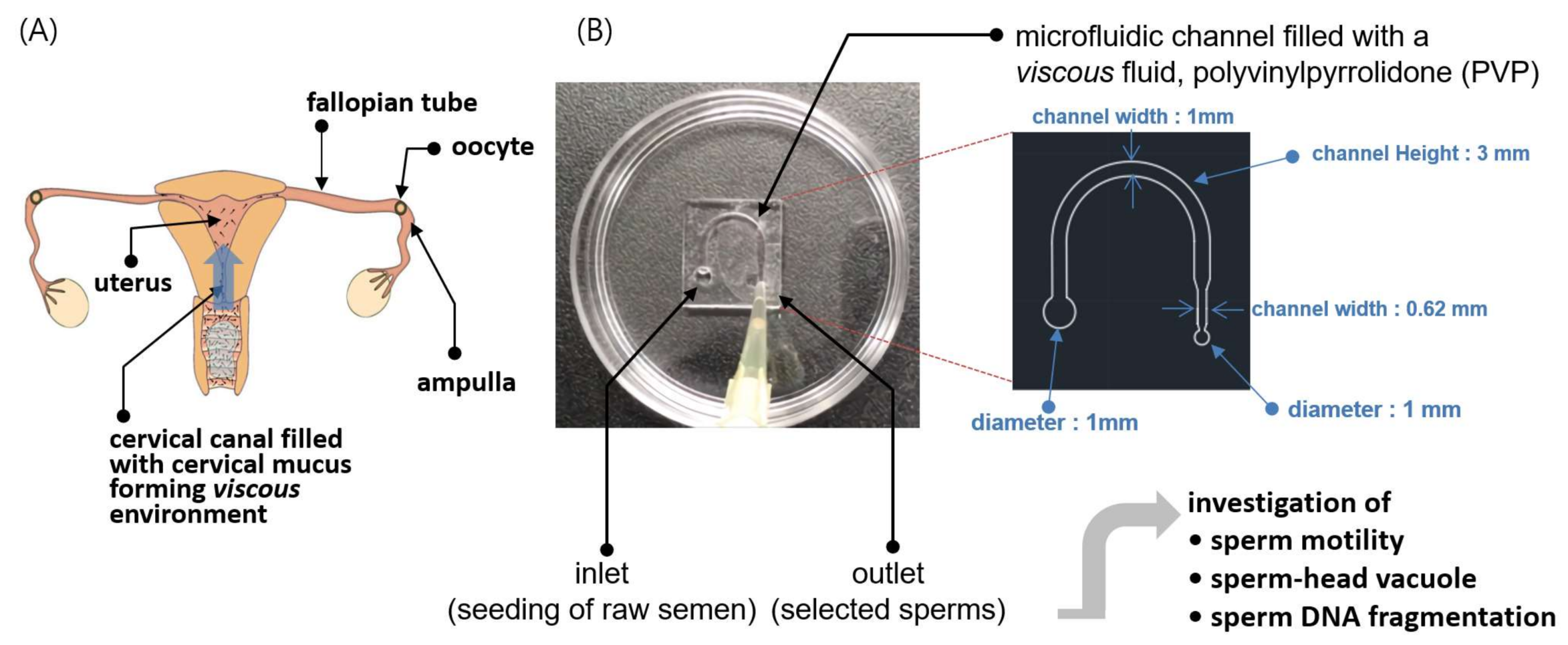
Abstract
:1. Introduction
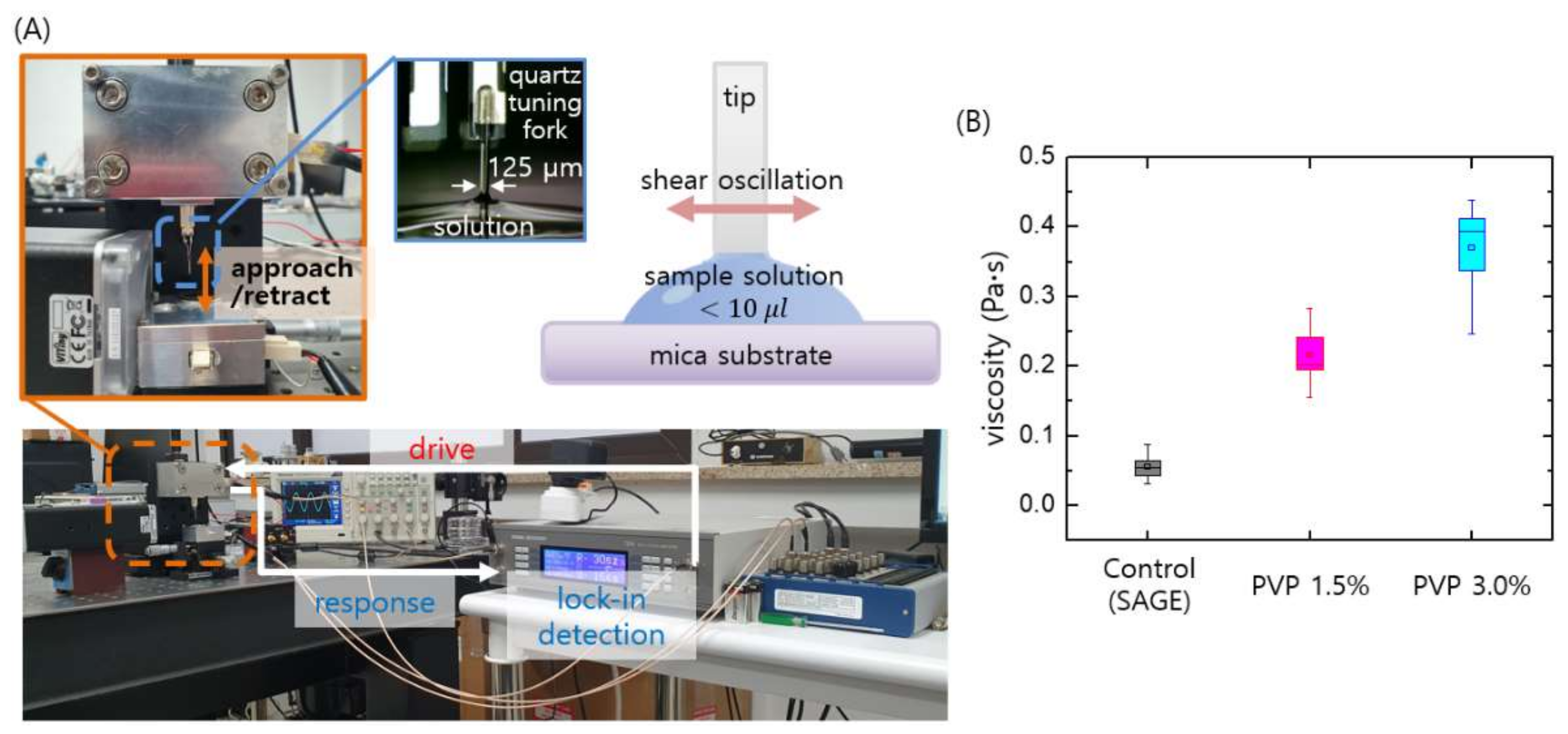
2. Materials and Methods
2.1. Sample Collection and Preparation of Sperm Suspension
2.2. Fabrication of Sperm-Sorting Chip
2.3. Purification of the Sperm Samples and Analyses of Sperm Motilities, Velocities, and Morphologies
2.4. Evaluation of Sperm DNA Fragmentation
2.5. Numerical Simulation of Sperm Dynamics
2.6. Statistical Analysis
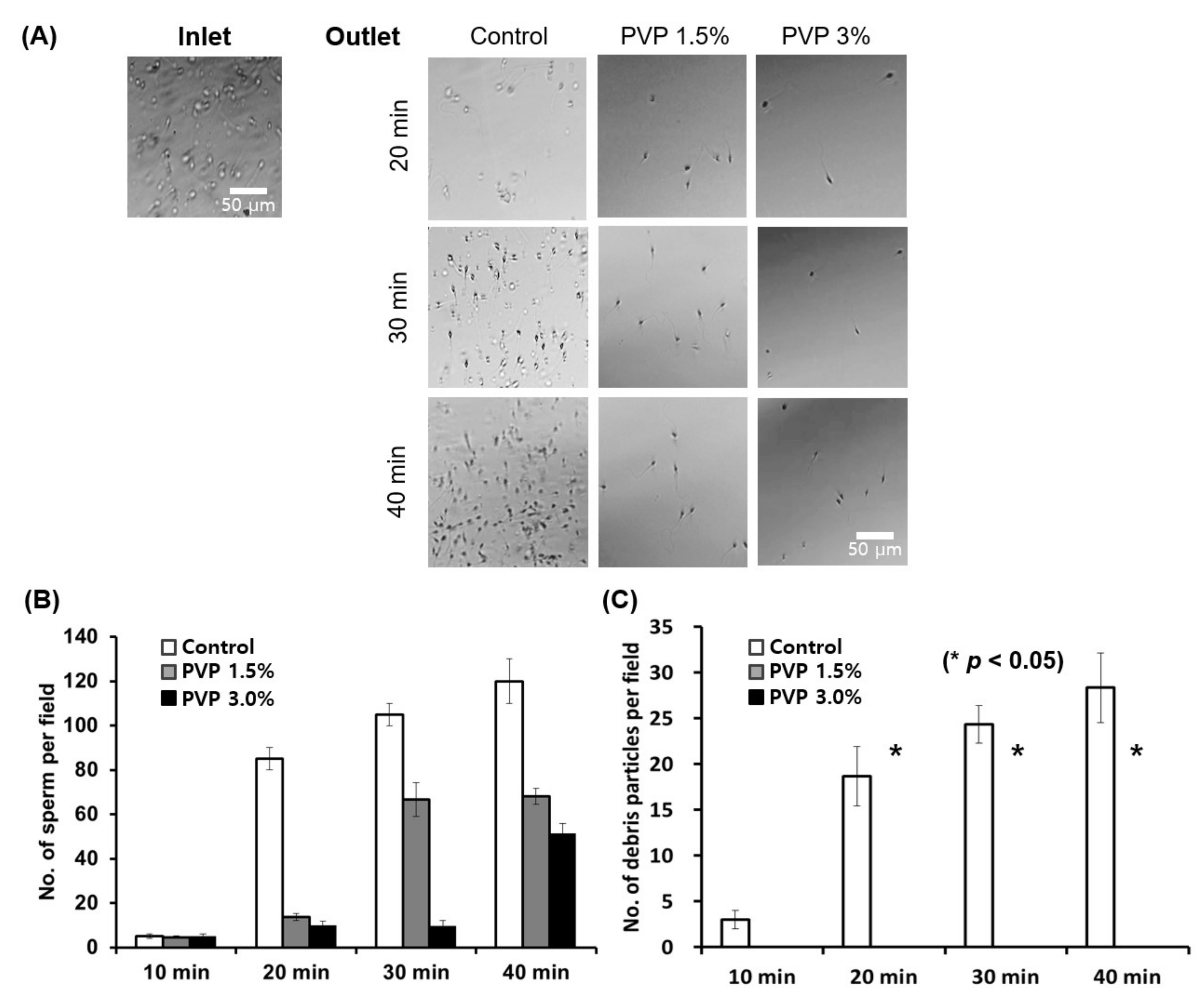
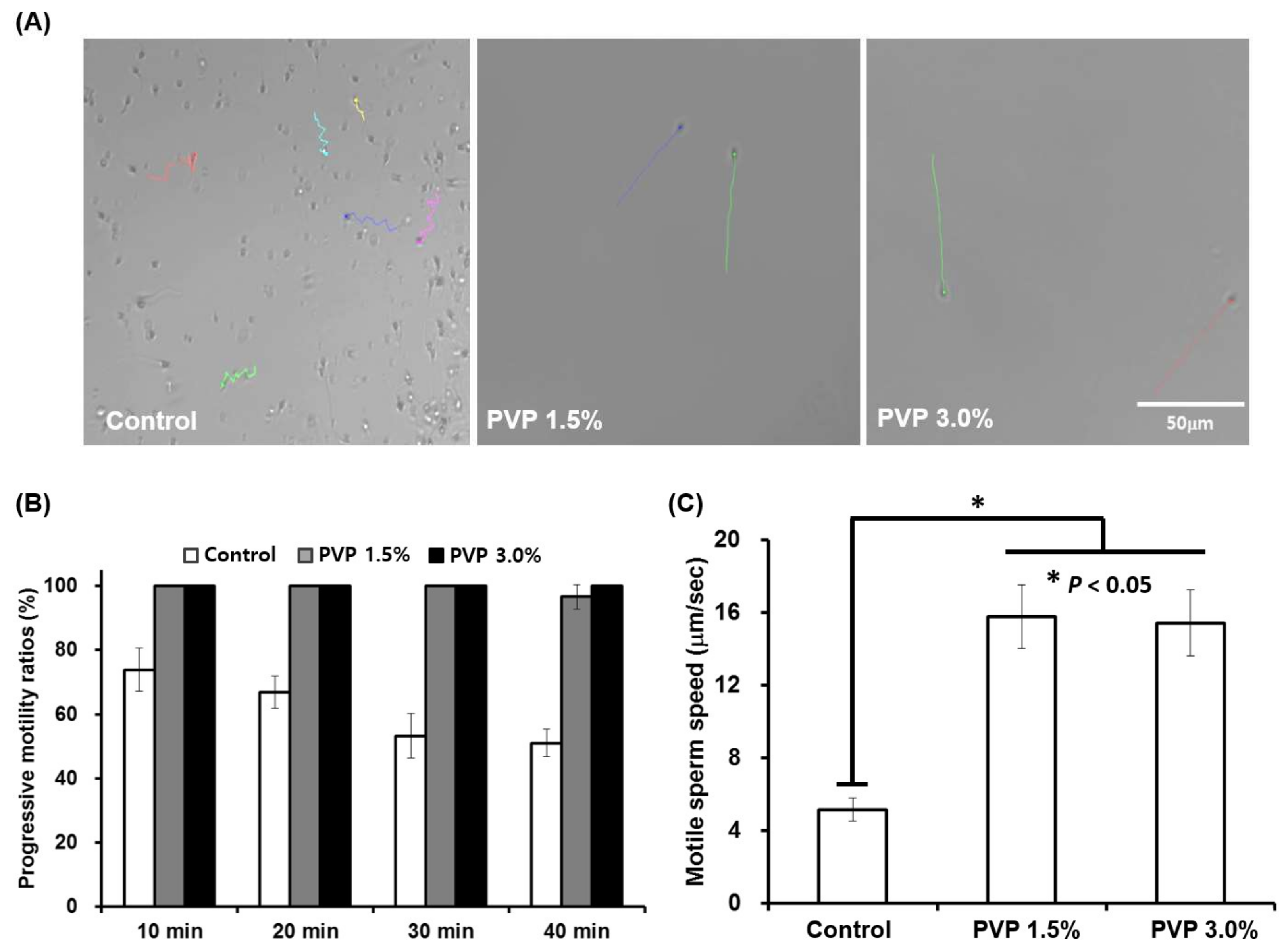
3. Results and Discussion
4. Conclusions
Supplementary Materials
Author Contributions
Funding
Institutional Review Board Statement
Informed Consent Statement
Data Availability Statement
Acknowledgments
Conflicts of Interest
References
- Adamson, G.D.; Tabangin, M.; Macaluso, M.; De Mouzon, J. The number of babies born globally after treatment with the assisted reproductive technologies (ART). Fertil. Steril. 2013, 100, S42. [Google Scholar] [CrossRef]
- Beck-Fruchter, R.; Lavee, M.; Weiss, A.; Geslevich, Y.; Shalev, E. Rescue intracytoplasmic sperm injection: A systematic review. Fertil Steril. 2014, 101, 690–698. [Google Scholar] [CrossRef]
- Komiya, A.; Kato, T.; Kawauchi, Y.; Watanabe, A.; Fuse, H. Clinical Factors Associated with Sperm DNA Fragmentation in Male Patients with Infertility. Sci. World J. 2014, 2014, 1–11. [Google Scholar] [CrossRef] [Green Version]
- Komiya, A.; Watanabe, A.; Kato, T.; Kawauchi, Y.; Fuse, H. Observation of spermatozoa by a high-magnification microscope. Reprod. Med. Biol. 2014, 13, 21–28. [Google Scholar] [CrossRef] [Green Version]
- Chatziparasidou, A.; Christoforidis, N.; Samolada, G.; Nijs, M. Sperm aneuploidy in infertile male patients: A systematic review of the literature. Andrologia 2015, 47, 847–860. [Google Scholar] [CrossRef] [PubMed]
- Utsuno, H.; Oka, K.; Yamamoto, A.; Shiozawa, T. Evaluation of sperm head shape at high magnification revealed correlation of sperm DNA fragmentation with aberrant head ellipticity and angularity. Fertil. Steril. 2013, 99, 1573–1580. [Google Scholar] [CrossRef]
- Sakkas, D. Novel technologies for selecting the best sperm for in vitro fertilization and intracytoplasmic sperm injection. Fertil. Steril. 2013, 99, 1023–1029. [Google Scholar] [CrossRef]
- Sakkas, D.; Ramalingam, M.; Garrido, N.; Barratt, C.L. Sperm selection in natural conception: What can we learn from Mother Nature to improve assisted reproduction outcomes? Hum. Reprod. Update 2015, 21, 711–726. [Google Scholar] [CrossRef] [Green Version]
- Quinn, M.M.; Jalalian, L.; Ribeiro, S.; Ona, K.; Demirci, U.; Cedars, M.I.; Rosen, M.P. Microfluidic sorting selects sperm for clinical use with reduced DNA damage compared to density gradient centrifugation with swim-up in split semen samples. Hum. Reprod. 2018, 33, 1388–1393. [Google Scholar] [CrossRef] [PubMed] [Green Version]
- Smith, G.D.; Takayama, S. Application of microfluidic technologies to human assisted reproduction. Mol. Hum. Reprod. 2017, 23, 257–268. [Google Scholar] [CrossRef] [PubMed] [Green Version]
- Knowlton, S.M.; Sadasivam, M.; Tasoglu, S. Microfluidics for sperm research. Trends Biotechnol. 2015, 33, 221–229. [Google Scholar] [CrossRef] [PubMed]
- Sano, H.; Matsuura, K.; Naruse, K.; Funahashi, H. Application of a microfluidic sperm sorter to the in-vitro fertilization of porcine oocytes reduced the incidence of polyspermic penetration. Theriogenology 2010, 74, 863–870. [Google Scholar] [CrossRef]
- Domingues, R.M.; Silva, M.; Gershovich, P.; Betta, S.; Babo, P.; Caridade, S.G.; Mano, J.F.; Motta, A.; Reis, R.L.; Gomes, M.E. Development of Injectable Hyaluronic Acid/Cellulose Nanocrystals Bionanocomposite Hydrogels for Tissue Engineering Applications. Bioconjug. Chem. 2015, 26, 1571–1581. [Google Scholar] [CrossRef] [PubMed]
- Eamer, L.; Nosrati, R.; Vollmer, M.; Zini, A.; Sinton, D. Microfluidic assessment of swimming media for motility-based sperm selection. Biomicrofluidics 2015, 9, 044113. [Google Scholar] [CrossRef] [Green Version]
- Ivic, A.; Onyeaka, H.; Girling, A.; Brewis, A.I.; Ola, B.; Hammadieh, N.; Papaioannou, S.; Barratt, C.L.R. Critical evaluation of methylcellulose as an alternative medium in sperm migration tests. Hum. Reprod. 2002, 17, 143–149. [Google Scholar] [CrossRef] [PubMed] [Green Version]
- Sanchez-Alvarez, J.; Cano-Corres, R.; Fuentes-Arderiu, X. A Complement for the WHO Laboratory Manual for the Examination and Processing of Human Semen (2010). EJIFCC. 2012, 23, 103–106. [Google Scholar]
- Fisher, H.S.; Giomi, L.; Hoekstra, H.E.; Mahadevan, L. The dynamics of sperm cooperation in a competitive environment. Proc. R. Soc. B Boil. Sci. 2014, 281, 20140296. [Google Scholar] [CrossRef] [Green Version]
- Lee, M.; Kim, B.; Kim, Q.; Hwang, J.; An, S.; Jhe, W. Viscometry of single nanoliter-volume droplets using dynamic force spectroscopy. Phys. Chem. Chem. Phys. 2016, 18, 27684–27690. [Google Scholar] [CrossRef] [PubMed]
- Lee, M.; Jhe, W. General Theory of Amplitude-Modulation Atomic Force Microscopy. Phys. Rev. Lett. 2006, 97, 036104. [Google Scholar] [CrossRef]
- Lee, M.; Jahng, J.; Kim, K.; Jhe, W. Quantitative atomic force measurement with a quartz tuning fork. Appl. Phys. Lett. 2007, 91, 23117. [Google Scholar] [CrossRef]
- Lee, M.; Kim, B.; Kim, J.; Jhe, W. Noncontact friction via capillary shear interaction at nanoscale. Nat. Commun. 2015, 6, 7359. [Google Scholar] [CrossRef] [Green Version]
- Samuel, K.L.; O’Hanlon, D.E.; Harrold, S.; Man, S.T.; Wang, Y.-Y.; Cone, R.; Hanes, J. Rapid transport of large polymeric nanoparticles in fresh undiluted human mucus. Proc. Natl. Acad. Sci. USA 2007, 104, 1482–1487. [Google Scholar]
- Piomboni, P.; Focarelli, R.; Stendardi, A.; Ferramosca, A.; Zara, V. The role of mitochondria in energy production for human sperm motility. Int. J. Androl. 2012, 35, 109–124. [Google Scholar] [CrossRef] [PubMed] [Green Version]
- Suarez, S.S.; Pacey, A. Sperm transport in the female reproductive tract. Hum. Reprod. Update 2006, 12, 23–37. [Google Scholar] [CrossRef] [PubMed] [Green Version]
- Boitrelle, F.; Ferfouri, F.; Petit, J.M.; Segretain, D.; Tourain, C.; Bergere, M.; Bailly, M.; Vialard, F.; Albert, M.; Selva, J. Large human sperm vacuoles observed in motile spermatozoa under high magnification: Nuclear thumbprints linked to failure of chromatin condensation. Hum. Reprod. 2011, 26, 1650–1658. [Google Scholar] [CrossRef] [Green Version]
- Boitrelle, F.; Guthauser, B.; Alter, L.; Bailly, M.; Wainer, R.; Vialard, F.; Albert, M.; Selva, J. The nature of human sperm head vacuoles: A systematic literature review. Basic Clin. Androl. 2013, 23, 3. [Google Scholar] [CrossRef] [PubMed] [Green Version]
- Fekonja, N.; Štrus, J.; Žnidarič, M.T.; Knez, K.; Bokal, E.V.; Verdenik, I.; Virant-Klun, I. Clinical and Structural Features of Sperm Head Vacuoles in Men Included in the In Vitro Fertilization Programme. BioMed Res. Int. 2014, 2014, 1–12. [Google Scholar] [CrossRef]
- Komiya, A.; Kawauchi, Y.; Kato, T.; Watanabe, A.; Tanii, I.; Fuse, H. Sperm Nuclear Vacuoles in relation to Acrosome Reactions and Sperm Motility. Sci. World J. 2014, 2014, 1–8. [Google Scholar] [CrossRef]
- Tandara, M.; Bajic, A.; Tandara, L.; Bilic-Zulle, L.; Sunj, M.; Kozina, V.; Goluža, T.; Jukić, M. Sperm DNA integrity testing: Big halo is a good predictor of embryo quality and pregnancy after conventional IVF. Andrology 2014, 2, 678–686. [Google Scholar] [CrossRef]
- De Vos, A.; Polyzos, N.P.; Verheyen, G.; Tournaye, H. Intracytoplasmic morphologically selected sperm injection (IMSI): A critical and evidence-based review. Basic Clin. Androl. 2013, 23, 10. [Google Scholar] [CrossRef] [Green Version]
- Tung, C.-K.; Hu, L.; Fiore, A.G.; Ardon, F.; Hickman, D.G.; Gilbert, R.O.; Suarez, S.S.; Wu, M. Grooves and flow guide sperm and reject pathogens. Proc. Natl. Acad. Sci. USA 2015, 112, 5431–5436. [Google Scholar] [CrossRef] [Green Version]
- Zaferani, M.; Palermo, G.D.; Abbaspourrad, A. Strictures of a microchannel impose fierce competition to select for highly motile sperm. Sci. Adv. 2019, 5, eaav2111. [Google Scholar] [CrossRef] [PubMed] [Green Version]
- Gai, J.; Nosrati, R.; Neild, A. High DNA integrity sperm selection using surface acoustic waves. Lab Chip 2020, 20, 4262–4272. [Google Scholar] [CrossRef] [PubMed]
- Orihara, H.; Takikawa, Y. Brownian motion in shear flow: Direct observation of anomalous diffusion. Phys. Rev. E 2011, 84, 061120. [Google Scholar] [CrossRef] [Green Version]
- Howse, J.; Jones, R.; Ryan, A.; Gough, T.; Vafabakhsh, R.; Golestanian, R. Self-motile colloidal particles: From directed propulsion to random walk. Phys. Rev. Lett. 2007, 99, 048102. [Google Scholar] [CrossRef] [PubMed] [Green Version]
- Tasoglu, S.; Safaee, H.; Zhang, X.; Kingsley, J.L.; Catalano, P.N.; Gurkan, U.A.; Nureddin, A.; Kayaalp, E.; Anchan, R.M.; Maas, R.L. Exhaustion of racing sperm in nature-mimicking microfluidic channels during sorting. Small 2013, 9, 3374–3384. [Google Scholar] [CrossRef] [Green Version]
- Kirkman-Brown, J.; Smith, D. Sperm motility: Is viscosity fundamental to progress? Mol. Hum. Reprod. 2011, 17, 539–544. [Google Scholar] [CrossRef] [Green Version]
- Rikmenspoel, R. The equation of motion for sperm flagella. Biophys. J. 1978, 23, 177–206. [Google Scholar] [CrossRef] [Green Version]
- Hayashi, S.; Shingyoji, C. Mechanism of flagellar oscillation–bending-induced switching of dynein activity in elastase-treated axonemes of sea urchin sperm. J. Cell Sci. 2008, 121, 2833–2843. [Google Scholar] [CrossRef] [Green Version]
- Yang, Y.; Elgeti, J.; Gompper, G. Cooperation of sperm in two dimensions: Synchronization, attraction, and aggregation through hydrodynamic interactions. Phys. Rev. E 2008, 78, 061903. [Google Scholar] [CrossRef] [Green Version]
- Kato, Y.; Nagao, Y. Effect of polyvinylpyrrolidone on sperm function and early embryonic development following intracytoplasmic sperm injection in human assisted reproduction. Reprod. Med. Biol. 2012, 11, 165–176. [Google Scholar] [CrossRef] [PubMed] [Green Version]


Publisher’s Note: MDPI stays neutral with regard to jurisdictional claims in published maps and institutional affiliations. |
© 2021 by the authors. Licensee MDPI, Basel, Switzerland. This article is an open access article distributed under the terms and conditions of the Creative Commons Attribution (CC BY) license (https://creativecommons.org/licenses/by/4.0/).
Share and Cite
Lee, M.; Park, J.W.; Kim, D.; Kwon, H.; Cho, M.J.; Lee, E.J.; Shin, T.E.; Kim, D.K.; Lee, S.; Byeun, D.G.; et al. Viscous Cervical Environment-on-a-Chip for Selecting High-Quality Sperm from Human Semen. Biomedicines 2021, 9, 1439. https://0-doi-org.brum.beds.ac.uk/10.3390/biomedicines9101439
Lee M, Park JW, Kim D, Kwon H, Cho MJ, Lee EJ, Shin TE, Kim DK, Lee S, Byeun DG, et al. Viscous Cervical Environment-on-a-Chip for Selecting High-Quality Sperm from Human Semen. Biomedicines. 2021; 9(10):1439. https://0-doi-org.brum.beds.ac.uk/10.3390/biomedicines9101439
Chicago/Turabian StyleLee, Manhee, Jin Woo Park, Dongwon Kim, Hyojeong Kwon, Min Jeong Cho, Eun Ji Lee, Tai Eun Shin, Dae Keun Kim, Seungki Lee, Do Gyeung Byeun, and et al. 2021. "Viscous Cervical Environment-on-a-Chip for Selecting High-Quality Sperm from Human Semen" Biomedicines 9, no. 10: 1439. https://0-doi-org.brum.beds.ac.uk/10.3390/biomedicines9101439