Case Report

Selective Bronchial Occlusion for Treatment of a Bronchopleural Fistula in an Extremely Preterm Infant

1
Neonatal Intensive Care Unit, Fondazione IRCCS Ca’ Granda Ospedale Maggiore Policlinico, 20122 Milan, Italy
2
Department of Clinical Sciences and Community Health, Università degli Studi di Milano, 20122 Milan, Italy
3
Neonatal Intensive Care Unit, Fondazione IRCCS Policlinico San Matteo, 27100 Pavia, Italy
*
Author to whom correspondence should be addressed.
Submission received: 19 October 2021 / Revised: 12 December 2021 / Accepted: 16 December 2021 / Published: 20 December 2021
(This article belongs to the Special Issue Neonatal Respiratory Distress Update)

Abstract

:
Neonatal pulmonary air leak commonly occurs as a complication of mechanical ventilation in infants with underlying hyaline membrane disease. They can commonly be managed conservatively or with the application of a chest drain, but some severe cases pose a significant challenge in finding an alternative therapeutic solution. Selective bronchial occlusion represents an unconventional rescue therapy for treating bronchopleural fistula resistant to the standard therapy. A 27-week gestation preterm infant ventilated for respiratory distress syndrome developed tension right-sided pneumothorax. Conventional modalities of treatment were tried and were unsuccessful. Intermittent selective bronchial occlusion with a Fogarty’s catheter and high-frequency oscillatory ventilation resulted in considerable improvement in the infant’s clinical condition and radiographic findings.

1. Introduction

Pulmonary air leak is a possible complication in preterm infants receiving mechanical ventilation for respiratory distress syndrome [1]. It is caused by an over-stretching and subsequent rupture of the alveolar wall leading to an aberrant communication between the alveolar space and a different anatomic site. The main forms are pulmonary interstitial emphysema (PIE), pneumomediastinum, pneumopericardium, and pneumothorax [2]. However, the appropriate management of this condition is not always clearly defined, and various therapeutic strategies have been reported in case of failure of conventional therapy [3].
We report our experience with an extremely preterm infant affected by persistent right bronchopleural fistula treated successfully with selective bronchial occlusion and high-frequency oscillatory ventilation (HFOV).

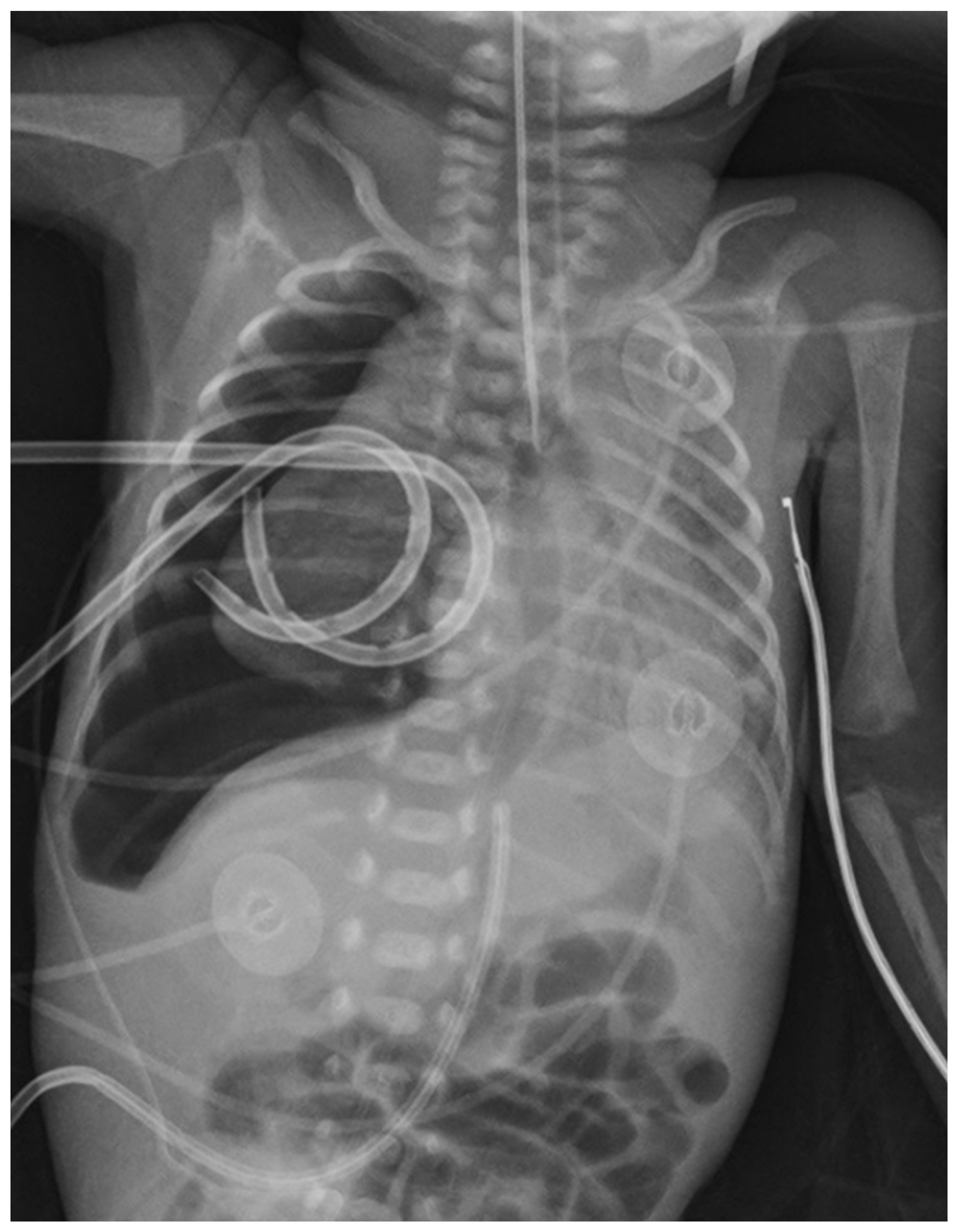
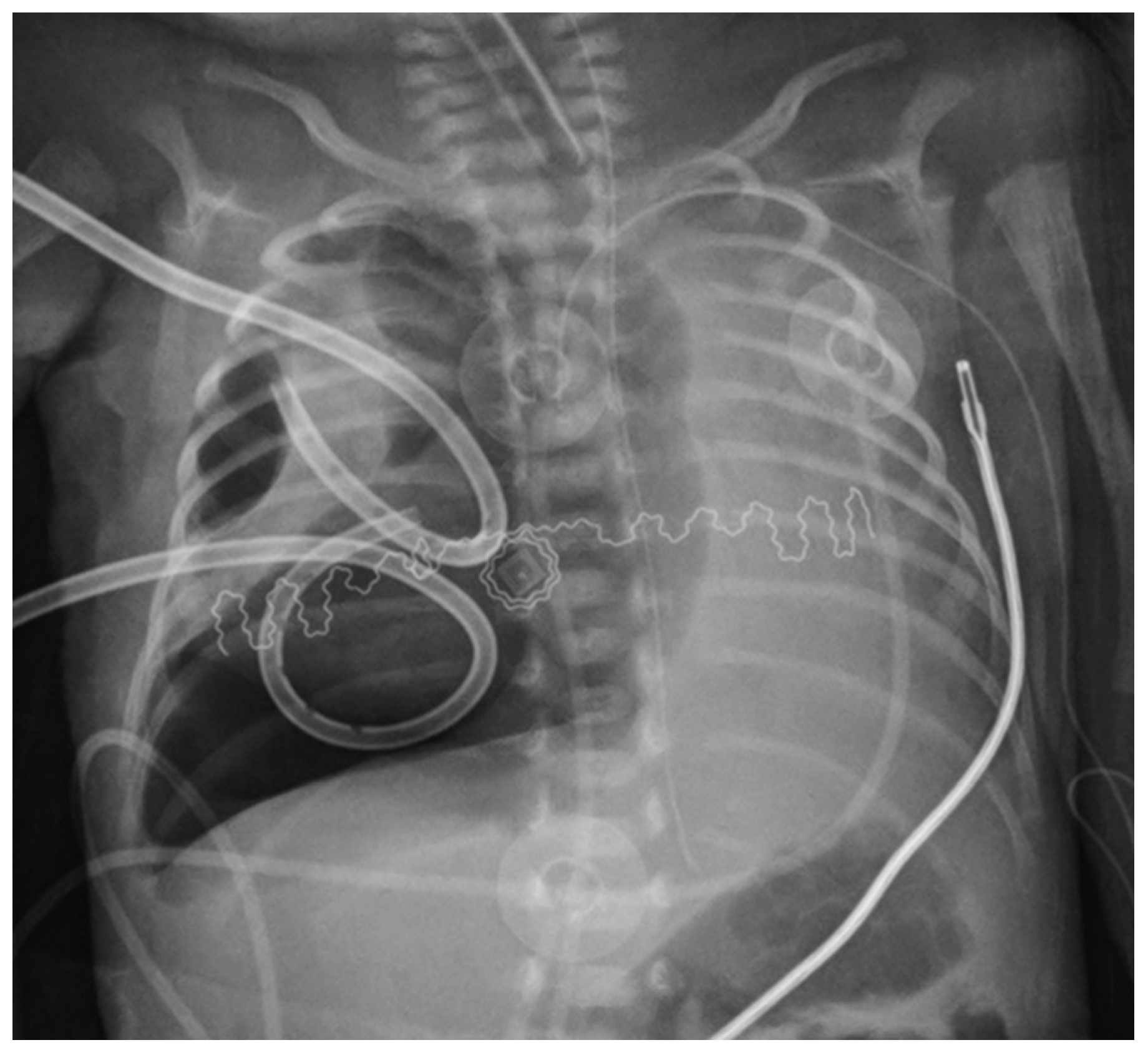
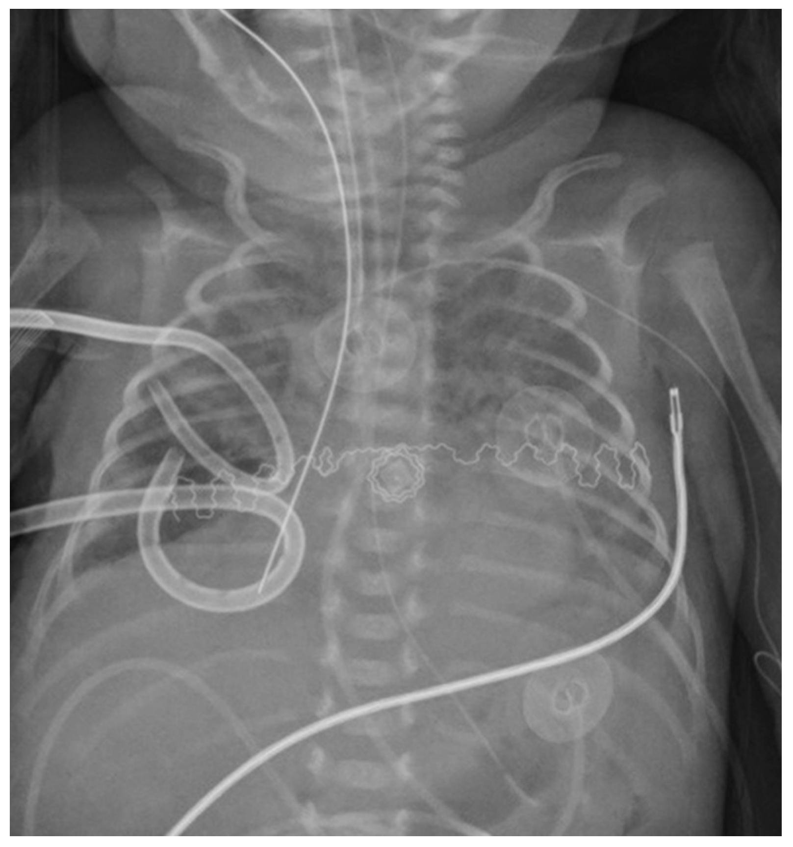
2. Case Presentation

Parents provided written informed consent for the publication of this case report, including images. All information revealing the subject’s identity was avoided, and all information was anonymized. A 600 g preterm male was born at 27 weeks gestation by cesarean section due to intrauterine growth reduction associated with pathological placental flows. The APGAR score was 4 and 8 at 1 and 5 min, respectively. At 4 h, poractant alfa (Curosurf, Chiesi Pharmaceuticals, Parma, Italy) was administered by the less invasive surfactant administration (LISA) technique for a hyaline membrane disease, but a tension right-sided pneumothorax occurred almost immediately (Figure 1).
Stabilization proved difficult with a consistently high flow through the bronchopleural fistula requiring the application of two suction drains at −10 cmH2O. Simultaneously, mechanical ventilation was started (PIP 25, MAP 10, FiO2 0.4) and the infant was positioned in right lateral decubitus.
Despite postural therapy and the maintenance of two suction drains, the respiratory condition remained critical in the following days. Ultrasound and X-ray checks repeatedly documented the reduction of the pneumothorax size. Still, the two drains showed continuous bubbling in the water seal chamber, meaning a persistent bronchopleural leakage. Furthermore, during the patient’s daily care, the drains presented transient obstructions due to their tiny size, leading to the immediate recurrence of the right pneumothorax and clinical destabilization. On day of life (DOL) 16, the infant deteriorated, and radiographs indicated a worsening pneumothorax associated with further hypoxemia (Figure 2).
After obtaining informed parental consent, a 3 French Fogarty’s catheter was entered through the vocal cords to protect the right lung from further injury. The tip was placed in the right main bronchus before intubation with a new endotracheal tube. The catheter was inserted 11 cm from the lips by direct laryngoscopy, approximately 4 cm deeper than the conventional orotracheal tube position. The insertion depth was previously calculated by adding the length of the endotracheal tube, the distance measured from its tip, and the desired position. The Fogarty catheter was spontaneously directed in the right main bronchus using the anatomical alignment with the trachea compared to the left main bronchus [4]. A chest X-Ray confirmed its exact position, and the 5 mm long balloon, located 1 cm from the tip, was inflated with 0.2 mL of air. The flow through the thoracic drains immediately stopped, and HFOV was started (MAP 14 cmH2O, Delta P 27 mmHg, Hz 14, FiO2 1.0). The balloon was deflated for 2 h for every hour inflated to minimize bronchial wall pressure necrosis. The patient tolerated the procedure well. Hypoxemia was rapidly corrected, with FiO2 decreasing up to 0.4 in forty-eight hours. On DOL 17, a chest x-ray showed a substantial reduction of pneumothorax associated with the partial exclusion of the right lower lobe from ventilation and gradual reaeration of the left lung, and then suction was stopped (Figure 3).
On DOL 19, the Fogarty catheter was removed, conventional ventilation was reestablished, and the drains were clamped intermittently and removed on DOL 21. Finally, on DOL 23, the patient was extubated and discharged home at 39 weeks of postmenstrual age in typical room air.

3. Discussion

Pneumothorax represents the most frequent form of pulmonary air leak in preterm infants, with an incidence ranging from 3 to 9% in very low birth weight infants, and is associated with increased mortality and morbidity [3]. Conventional treatment of symptomatic pneumothorax consists of the application of a chest drain and downward lateral positioning of the affected hemithorax associated with low tidal volume ventilation, more easily obtained with HFOV (1.5–2.5 mL/kg) than conventional mechanical ventilation (4–6 mL/kg) [2,4,5]. Selective bronchial intubation of the nonaffected lung and selective bronchial occlusion of the affected lung are two additional treatment options described more often in other forms of neonatal air leak such as PIE, whose evidence is based on single case reports, and small case series [1]. However, the first seems to have a low success rate and an increased risk of complications such as hypoxia associated with reintubation and acquired bronchial stenosis [6,7]. In turn, successful selective bronchial occlusion has been reported in less than 30 cases of premature infants with different occlusion techniques [4,8,9,10,11,12,13,14,15,16]. Characteristics of these patients and the procedure used are summarized in Table 1.
In our case, suction chest drains and postural therapy were immediately applied, but the multiple relapses of pneumothorax associated with periods of hypoxemia exposed the infant to prolonged artificial ventilation at high concentrations of oxygen. This condition represented a challenge in the ventilation strategy due to a self-perpetuated process, which required a further increase in ventilation pressure, which led to greater air escape through the bronchopleural fistula. The situation made it necessary to partially exclude the bronchopleural fistula from ventilation and reduce the tidal volume through the HFOV.
The literature review of our previous experience prompted us to perform selective bronchial occlusion [4]. The catheter was easily placed through the trachea in the right bronchus; the left side may be more difficult. However, the procedure proved safe, no adverse side effects were noted, and there was no recurrence of pneumothorax. In addition, the inflation of the balloon caused a rapid clinical and radiologic reduction of the air in the pleural cavity.

4. Conclusions

In conclusion, selective bronchial occlusion with Fogarty’s catheter insertion remains a rescue therapy. However, it may be important for neonatologists to be aware of this procedure in cases of persistent bronchopleural fistula. In addition, the technical appropriateness of the instrumentation used, the best length of stay of the catheter, and the best strategy for intermittent deflation and inflation of the balloon remain to be explored in order to improve its effectiveness and minimize possible complications.

Author Contributions

G.S.A., M.C., G.R., I.A., S.G. (Silvia Gulden), G.C., V.C., S.G. (Stefano Ghirardello) and F.M. contributed to the study conception and design; G.S.A., G.R., G.C., S.G. (Silvia Gulden) and S.G. (Stefano Ghirardello) wrote the first draft of the manuscript. All authors have read and agreed to the published version of the manuscript.

Funding

The authors declare that public or private funding was not used to prepare the data or the manuscript.

Institutional Review Board Statement

The present case report was carried out in accordance with the principles of good clinical practice and the World Medical Association Helsinki Declaration, as well as the national legislative and administrative provisions in force. The local Ethics Committee approved the case report (Milan Area 2, Italy) with approval number 878_2021, after evaluation of the: 1. cover letter; 2. Information on the processing of patients’ data (including photographic and video supports) according to Article 13 of Regulation no. 2016/679/EU and the national privacy legislation in force (with relative expression of the consent); and 3. Draft of the manuscript.

Informed Consent Statement

Parents gave written informed consent for publication of the details of their medical case and any accompanying images.

Data Availability Statement

The data supporting this study’s findings are not publicly available because they contain information that could compromise participants’ privacy, but are available from the corresponding author.

Acknowledgments

The authors would like to thank all of the nurses and neonatologists of the neonatal intensive care unit team of the Fondazione IRCCS Ca’ Granda Ospedale Maggiore Policlinico.

Conflicts of Interest

The authors have no conflict of interest to declare.

References

  1. Andreou, A.; Papouli, M.; Netskos, D.; Papadopoulou, F.; Rossiou, E. One-sided high-frequency oscillatory ventilation in the management of an acquired neonatal lobar emphysema. A case report and review. J. Perinatol. 2001, 21, 61–64. [Google Scholar] [CrossRef] [Green Version]
  2. Bruschettini, M.; Romantsik, O.; Zappettini, S.; O’Donnell, C.P.; Calevo, M.G. Needle aspiration versus intercostal tube drainage for pneumothorax in the newborn. Cochrane Database Syst Rev. 2019, 2, CD011724. [Google Scholar] [CrossRef] [PubMed]
  3. Kitsommart, R.; Martins, B.; Bottino, M.N. Expectant management of pneumothorax in preterm infants receiving assisted ventilation: Report of 4 cases and review of the literature. Respir Care 2012, 57, 789–793. [Google Scholar] [CrossRef] [PubMed]
  4. Mosca, F.; Lattanzio, M.; Colnaghi, M.; Pugliese, S. Bronchopleural fistula: Successful selective bronchial occlusion with a Fogarty’s catheter in a preterm infant. Acta Paediatr. 1995, 84, 1079–1082. [Google Scholar] [CrossRef] [PubMed]
  5. Aurilia, C.; Ricci, C.; Tana, M.; Tirone, C.; Lio, A.; Gambacorta, A.; Paladini, A.; Vento, G. Management of pneumothorax in hemodynamically stable preterm infants using high frequency oscillatory ventilation: Report of five cases. Ital. J. Pediatr. 2017, 43, 114. [Google Scholar] [CrossRef] [PubMed]
  6. Goussard, P.; Morrison, J.; Bekker, A.; Fourie, B. Acquired neonatal bronchial stenosis after selective intubation: Successful managed with balloon dilatation. Clin. Case Rep. 2019, 7, 917–919. [Google Scholar] [CrossRef] [PubMed] [Green Version]
  7. Glenski, J.A.; Thibeault, D.W.; Hall, F.K.; Hall, R.T.; Germann, D.R. Selective bronchial intubation in infants with lobar emphysema. Am. J. Perinatol. 1986, 3, 199–204. [Google Scholar] [CrossRef] [PubMed]
  8. Hathorn, C.; Armitage, N.; Wensley, D.; Seear, M. Bronchial balloon occlusion in children with complex pulmonary air leaks. Arch. Dis. Child. 2013, 98, 136–140. [Google Scholar] [CrossRef] [PubMed]
  9. Rastogi, S.; Gupta, A.; Wung, J.-T.; Berdon, W.E. Treatment of giant pulmonary interstitial emphysema by ipsilateral bronchial occlusion with a swan-ganz catheter. Pediatr. Radiol. 2007, 37, 1130–1134. [Google Scholar] [CrossRef] [PubMed]
  10. Al-Alaiyan, S.; Katan, A. Pulmonary interstitial emphysema treated by selective bronchial obstruction and high-frequency oscillatory ventilation. Am. J. Perinatol. 1994, 11, 433–435. [Google Scholar] [CrossRef] [PubMed]
  11. Al Jishi, N.; Dyer, D.; Sharief, N.; Al-Alaiyan, S. Selective bronchial occlusion for treatment of bullous interstitial emphysema and bronchopleural fistula. J. Pediatr. Surg. 1994, 29, 1545–1547. [Google Scholar] [CrossRef]
  12. Weintraub, Z.; Oliven, A. Successful resolution of unilateral pulmonary interstitial emphysema in a premature infant by selective bronchial balloon catheterization. J. Pediatr. Surg. 1988, 23, 1005–1006. [Google Scholar] [CrossRef]
  13. Lewis, S.; Pelausa, E.; Ojah, C.; Paes, B. Pulmonary interstitial emphysema: Selective bronchial occlusion with a swan-ganz catheter. Arch. Dis. Child. 1988, 63, 313–315. [Google Scholar] [CrossRef] [PubMed] [Green Version]
  14. DeWitte, D.B.; Batton, D.G.; Prudent, L.; Maisels, M.J. Endotracheal tube modification for therapy of right-sided pulmonary interstitial emphysema. Clin. Pediatr. 1986, 25, 626–628. [Google Scholar] [CrossRef] [PubMed]
  15. Auerbach, D.A.; Blackmon, L.R.; Filston, H.C.; Merten, D.F.; Kirks, D.R. Localized pulmonary interstitial emphysema: Treatment by bronchial occlusion. Am. J. Perinatol. 1983, 1, 52–57. [Google Scholar] [CrossRef] [PubMed]
  16. Mathew, O.P.; Thach, B.T. Selective bronchial obstruction for treatment of bullous interstitial emphysema. J. Pediatr. 1980, 96, 475–477. [Google Scholar] [CrossRef]
Figure 1. X-ray on the first day of life. Tension right-sided pneumothorax with the leftward shift of the mediastinum after surfactant administration.
Children 08 01208 g001
Figure 2. X-ray in 16th day of life. Recurrence of pneumothorax despite two suction chest drains.
Children 08 01208 g002
Figure 3. X-ray on the 17th day of life, 24 h after the occlusion. The Fogarty’s catheter in the right bronchus reduces pneumothorax and partial collapse of the right lower lobe.
Children 08 01208 g003
Table 1. Overview of successful selective bronchial occlusion cases reported in the literature in preterm infants.
AuthorGA (Weeks)Weight (g)Air LeakPositionUnsuccessful TreatmentHFOInstrumentation UsedInsertionOcclusion TypeLength
Present study27600PTXRightChest drainage and postural therapyYesFogarty’s catheter 3 FR, inflated 0.2 mLDirect laryngoscopyInflated 1 h/deflated 2 h72 h
Al alaiyan301500PIERightPostural therapy and chest drainageYesBalloon catheter inflated 0.3 mLDirect laryngoscopyDeflated for 5 min each hour48 h
Alijishi281240PIERightPostural therapy, chest drainage, and selective left bronchial intubationNoBalloon catheter inflated 0.35 mLDirect laryngoscopyDeflated for 5 min each hour60 h
Auerbach301620PIELeftChest drainage and selective right bronchial intubationNoFogarty’s catheter 4 FRBronchoscopyContinuous48 h
321250PIELeftNANoFogarty’s catheter 3 FRBronchoscopyInflated 4 h/deflated 4 h96 h
Dewitte291340PIERightPostural therapyNAET 2.5 mm modified *Direct laryngoscopyContinuous72 h
281120PIERightChest drainageNAET 2.5 mm modified *Direct laryngoscopyContinuous48 h
Hathorn332390PIERightNANABalloon catheter 4 FRDirect laryngoscopyContinuous96 h
271200PIELeftNANABalloon catheter 4 FRBronchoscopy with Fluoroscopic guidanceContinuous7 days
Lewis26760PIERightPostural therapy, chest drainage, and selective left bronchial intubationNoSwan-Ganz catheter 5 FRDirect laryngoscopyDeflated 5 min each hour72 h
Mathew29950PIE and PTXRightChest drainage and selective left bronchial intubationNoUmbilical catheter modified °,
inflated 0.7 mL
Direct laryngoscopyContinuous48 + 72 h Ψ
Mosca261000PIE and PTXRightPostural therapy and chest drainageNoFogarty’s catheter 5 FR, inflated 0.3 mLDirect laryngoscopyDeflated 5 min each hour26 h
Weintraub281200PIERightPostural therapyNoBalloon catheter 4 FR, inflated 0.75 mLDirect laryngoscopyContinuousNA
Rastoigi ⊥25–32600–170012 PIE/2 PTXNAPostural therapy3 yes/11 noSwan-Ganz catheter 5 FR,
inflated 0.6 mL
Direct laryngoscopyEvery hour at the hour24 h after the PIE resolution
PIE = pulmonary interstitial emphysema; PTX = pneumothorax; ET = endotracheal tube; NA = not available; FR = French (1 Fr = 1/3 of mm). * An adapted ET positioned with the apex in the right bronchus, with the distal opening occluded and a hole along the radiopaque line starting at 1 cm and ending 2 cm from the distal end. ° An adapted umbilical catheter attached to a handmade latex esophageal pressure balloon, length 1.25 cm, width 1 cm. Ψ The first occlusion lasted 48 h, followed by a second occlusion of 72 h for a recurrence of PIE. ⊥ A case series reporting 14 cases of successful selective bronchial occlusion in premature babies, the history of each infant is not available. Gestational age and weight ranges are reported.
Publisher’s Note: MDPI stays neutral with regard to jurisdictional claims in published maps and institutional affiliations.

