
Novel Lung Growth Strategy with Biological Therapy Targeting Airway Remodeling in Childhood Bronchial Asthma


1
Department of Pediatric Acute Diseases, Okayama University Academic Field of Medicine, Dentistry, and Pharmaceutical Sciences, Okayama 700-8558, Japan
2
Okayama University School of Medicine, Okayama 700-8558, Japan
3
Department of Pediatrics, Okayama University Academic Field of Medicine, Dentistry, and Pharmaceutical Sciences, Okayama 700-8558, Japan
*
Author to whom correspondence should be addressed.
Children 2022, 9(8), 1253; https://0-doi-org.brum.beds.ac.uk/10.3390/children9081253
Submission received: 7 August 2022
/
Revised: 16 August 2022
/
Accepted: 18 August 2022
/
Published: 19 August 2022
(This article belongs to the Special Issue Asthma and Its Impact in Adolescents)
Abstract
:Anti-inflammatory therapy, centered on inhaled steroids, suppresses airway inflammation in asthma, reduces asthma mortality and hospitalization rates, and achieves clinical remission in many pediatric patients. However, the spontaneous remission rate of childhood asthma in adulthood is not high, and airway inflammation and airway remodeling persist after remission of asthma symptoms. Childhood asthma impairs normal lung maturation, interferes with peak lung function in adolescence, reduces lung function in adulthood, and increases the risk of developing chronic obstructive pulmonary disease (COPD). Early suppression of airway inflammation in childhood and prevention of asthma exacerbations may improve lung maturation, leading to good lung function and prevention of adult COPD. Biological drugs that target T-helper 2 (Th2) cytokines are used in patients with severe pediatric asthma to reduce exacerbations and airway inflammation and improve respiratory function. They may also suppress airway remodeling in childhood and prevent respiratory deterioration in adulthood, reducing the risk of COPD and improving long-term prognosis. No studies have demonstrated a suppressive effect on airway remodeling in childhood severe asthma, and further clinical trials using airway imaging analysis are needed to ascertain the inhibitory effect of biological drugs on airway remodeling in severe childhood asthma. In this review, we describe the natural prognosis of lung function in childhood asthma and the risk of developing adult COPD, the pathophysiology of allergic airway inflammation and airway remodeling via Th2 cytokines, and the inhibitory effect of biological drugs on airway remodeling in childhood asthma.
1. Introduction
1.1. Childhood Asthma
Bronchial asthma is a chronic respiratory disease characterized by airway obstruction and hyperresponsiveness [1], with inflammation of the airways, resulting from exposure to allergens or other environmental irritants, causing bronchoconstriction, wheezing, and shortness of breath. The global prevalence of childhood asthma is estimated to be 5–10%, with significant differences between countries [2]. The prevalence of childhood asthma is reported to be 8.4% in the United States [3] and 9.4% in the European Union [4].
Although the severity of asthma varies, most childhood asthma is controlled using low-to-medium doses of inhaled corticosteroids (ICS), with or without additional controller drugs. However, despite treatment with high doses of ICS, and long acting β2 agonists or oral corticosteroids, some patients with severe asthma exhibit persistent asthma symptoms, frequent deterioration, and decreased lung function. Uncontrollable persistent asthma occurs in 5–7% of children with asthma, with geographical differences, and approximately 1 in 100–150 children has severe asthma [5,6,7]. Asthma in childhood causes allergic inflammation in the airways and airway remodeling, leading to persistent airflow obstruction and decreased respiratory function [8]. Children with persistent asthma exhibit a high frequency of asthma attacks, pulmonary dysfunction, poor quality of life, risk of medication-related side effects, and constitute a high medical burden for treatment and emergency consultations. A computerized search was performed using PubMed, combining the terms (asthma OR COPD) AND (trajectory OR lung development OR lung growth) AND (baby OR child OR pediatrics) with an English language filter, to identify studies on the lung growth trajectory in children with asthma, published until 30 June 2022. Furthermore, references within the included articles were scanned for other relevant papers. We excluded articles on lung growth in other chronic lung diseases were excluded.
1.2. Natural Prognosis of Lung Function in Childhood Asthma
Approximately 80% of children with bronchial asthma develop this condition by the age of 3 years, but most of these patients remit after puberty and exhibit remission of respiratory symptoms [9]. Hence, bronchial asthma is considered to be a disease with a good prognosis [10]. However, a large-scale study has reported a low proportion of children with asthma who achieve “complete remission” without asthma symptoms or inhaled corticosteroid use, normal lung function, and no bronchial hyperreactivity [11], suggesting that asthma severity at a young age is directly related to that in adolescence and adulthood. In a study of patients with moderate-to-severe pediatric asthma, 15% were in remission, 22% had intermittent asthma, and the rest had persistent asthma [12]. In a cohort study in Melbourne, Australia, approximately half of the 7-year-old patients with pediatric asthma were not in remission at 50 years of age, and only 15% of 7-year-old patients with severe asthma were in remission at 50 years of age [13]. Many cohort studies investigating lung function in childhood asthma from childhood to adolescence have reported that persistent wheezing was associated with decreased lung function development in adolescence [14,15,16]. In one cohort study, lung function was followed in children between the ages of 9 and 26 and 25% of the children had persistent or recurrent wheezing from childhood, with consistently low lung function being observed in these patients [17]. In the Childhood Asthma Management Program (CAMP) study, approximately one-third of participants presented with decreased lung function, and 5–10% of children with asthma experienced severe asthma in adulthood [18]. These studies indicate that asthma that develops in childhood may persist into adulthood, and patients with persistent asthma in adulthood already have decreased lung function from childhood. Furthermore, childhood asthma is strongly associated with respiratory depression and long-term prognosis in adulthood, and some patients develop COPD with irreversible airflow limitation [19].
1.3. Decreased Respiratory Function in Childhood and Onset of COPD in Adulthood
COPD is a disease with a high global incidence and mortality. COPD is caused by abnormalities in the airways and alveoli, characterized by persistent respiratory symptoms and irreversible airflow limitation [20]. COPD typically presents with small airway obstruction in the early stages and progresses slowly with diminished lung function. The onset of COPD is affected by genetic and environmental factors, with exposure to harmful particles and gases (particularly tobacco) constituting the main risk factor [21]. However, smoking is not the sole factor in the development of COPD, as only 10% of smokers develop COPD and 25–50% of COPD patients are non-smokers [22,23].
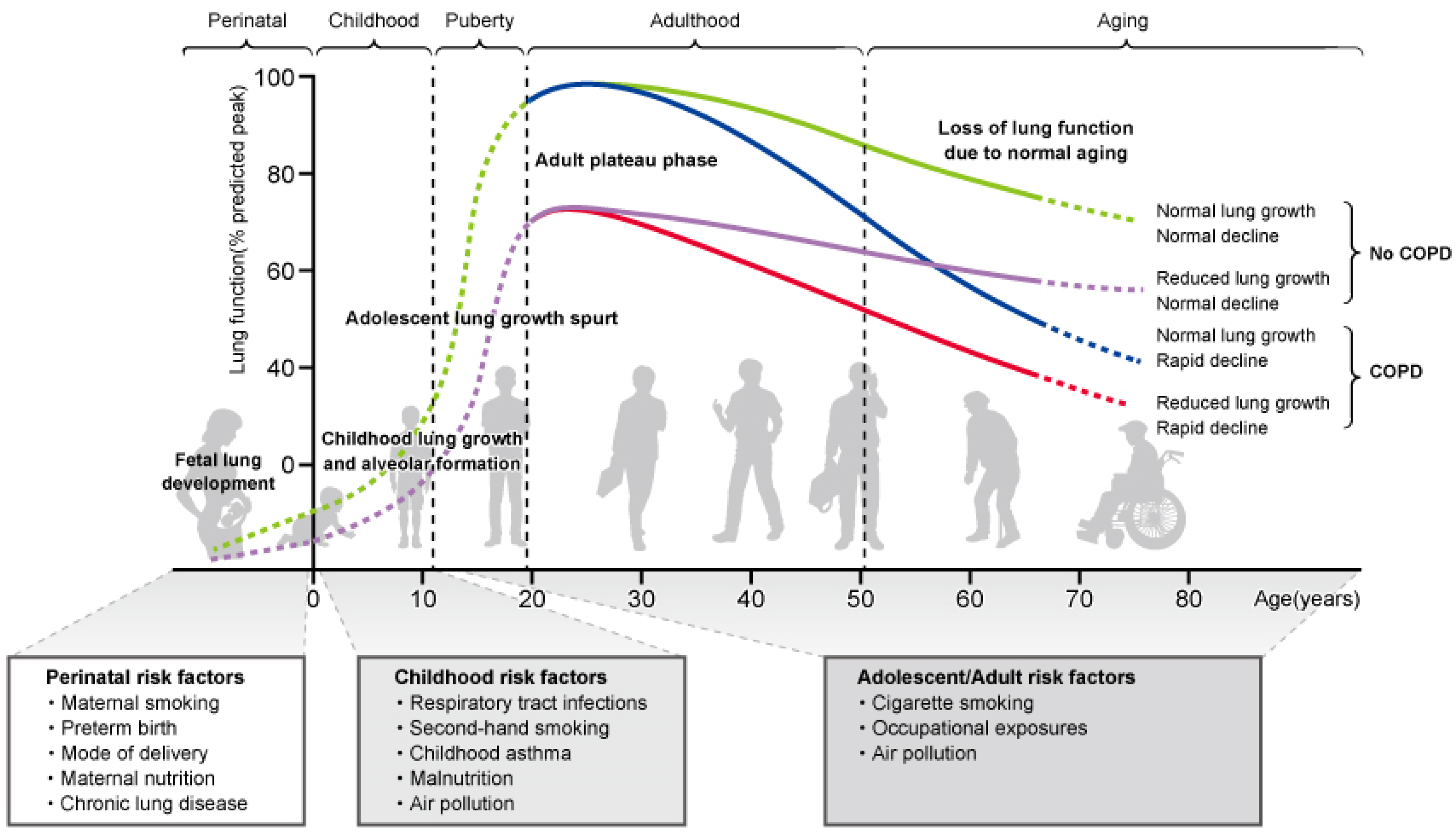
Patients with COPD already have diminished lung function in childhood, and the onset of COPD in adulthood may result from diminished peak respiratory function that emerges in adolescence [24]. Low forced expiratory volume in one second (FEV1) levels in adolescence increase COPD morbidity and mortality [25], and decreased lung function at 7 years of age is a risk factor for developing both asthma and COPD in adulthood [26]. A study that followed the trajectory of lung function from childhood to adulthood reported that children with persistently low lung function had severe and recurrent wheezing associated with exposure to tobacco smoke [27]. In one pediatric cohort study, patients with rapid decline in lung function in adulthood and those with low lung function in adolescence were more likely to develop COPD compared to participants with a normal rate of decline in lung function [28]. These studies indicate that the onset of COPD involves smoking in adulthood as well as decreased lung function in childhood [29,30,31] (Figure 1).
1.4. Lung Development in Childhood
Lung development is a continuous lifelong process that begins in the womb and continues from birth to adolescence and early adulthood, and is referred to as the lung function trajectory. The lungs develop by 20 years of age, resulting in increased lung size and number of alveoli and structural complexity of the lung [32,33]. Lung function plateaus between 20 and 25 years and then gradually declines with age. There are sex differences in lung development, with maximal lung function achieved in women at 15–20 years of age and in men at 20–25 years of age [34]. Lung growth in childhood is critical for achieving peak lung function in adulthood.
However, lung development in childhood is affected by various factors, including genetic predisposition, maternal smoking, preterm birth, neonatal chronic lung disease, passive smoking, childhood asthma, childhood respiratory infections, poor nutrition, and air pollution [19,31,35,36]. Impairment in lung function development due to these factors can lead to inadequate peak lung function in adolescence and predispose to COPD in adulthood. Case-control studies of former adult smokers and current smokers reported that a history of childhood pneumonia and/or asthma was associated with the onset or exacerbation of COPD [37]. A large study reported that risk factors for COPD included asthma in parents, exposure to smoking in the prenatal period and childhood, preterm birth, low birth weight, lower respiratory tract infections, recurrent asthma, childhood asthma, and second-hand smoking [38]. Reports indicate that childhood wheezing and decreased lung function due to parental smoking are associated with a high risk of developing COPD in adulthood [8,39].
1.5. Impaired Lung Growth, and Increased Risk of Developing COPD, Due to Childhood Asthma
Childhood asthma is a cause of impaired lung growth in childhood, and is one of the risk factors for COPD, similar to smoking [30,40,41]. In a retrospective study in adults, 19% of patients with COPD had a history of childhood asthma, and the risk associated with childhood asthma was equivalent to smoking 62 packs year [42]. A study reported that adults aged 50 years with a history of severe asthma between the ages of 6 and 7 years had a 32-fold higher risk of COPD compared to those without such a history [30]. In addition, a study of adult patients with asthma and COPD identified airflow obstruction and decreased lung volume in adolescence as strongly involved in the onset of COPD in adulthood [43]. A prospective cohort study of children aged 10–15 years reported that childhood asthma was associated with an increased risk of COPD [40], and another pediatric cohort study showed that children with childhood asthma were more likely to be treated or hospitalized with COPD during adulthood [44]. In the CAMP cohort study, 11% of patients with pediatric asthma met COPD criteria based on spirometry in adolescence, and more patients with a low growth pattern of lung function developed COPD compared to those with a normal growth pattern [45]. Various longitudinal studies on long-term lung function trajectories for childhood asthma have reported that childhood asthma impairs lung function maturation, and subsequent decline in lung function in adolescence and adulthood can lead to COPD [17,30,46,47,48,49,50,51]. In this regard, early treatment strategies tailored to the severity of childhood asthma may improve pulmonary dysfunction and reduce the risk of developing COPD in the future.
2. Airway Inflammation and Remodeling with T2-High Asthma in Children
2.1. T2-High Asthma in Childhood Asthma
Two major end types of bronchial asthma are currently recognized, based on the type of underlying airway immune inflammation, defined as type 2 (T2)-high asthma and T2-low asthma [52]. T2-high asthma encompasses more than 50% of the endotypes of asthma, and most patients with pediatric asthma have T2-high asthma. T2-low asthma is more common in adults than in children. T2-high asthma includes allergic non-eosinophil, non-allergic eosinophil, and mixed granulocytic phenotypes. It is characterized by increased peripheral blood eosinophil counts, exhaled nitric oxide fractions, and allergen-specific immunoglobulin E (IgE) levels [53].
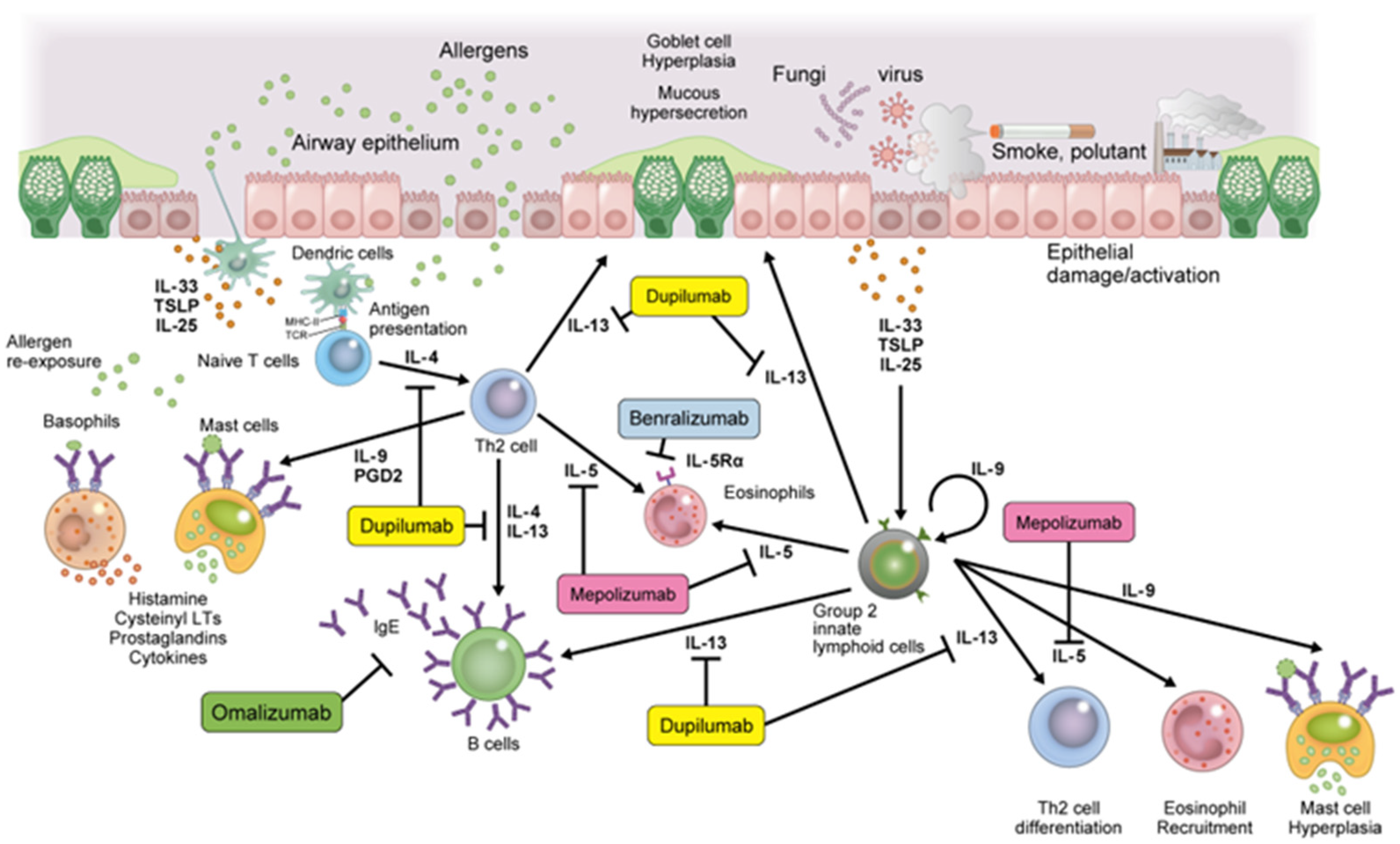
T2-high asthma involves T helper 2 (Th2) cells, eosinophils/basophils, and group 2 innate lymphoid cells (ILC2), in conjunction with epithelial cell-derived cytokines, such as interleukin (IL)-25, IL-33, and thymic stromal lymphopoietin (TSLP), leading to allergic sensitization and airway inflammation sustained by IgE, IL-4, IL-5, and IL-13 [54]. IgE is mediated by Th2 cells and produced by plasma cells via the release of IL-4 and IL-13 cytokines. IgE binds to the high-affinity IgE receptor (FcεR1) expressed on mast cells, basophils, dendritic cells, airway smooth muscle cells, and eosinophils. Cross-linking with allergens releases pro-inflammatory mediators, such as histamine, prostaglandin D2, leukotriene C4, tryptase, and chimase, causing bronchoconstriction and airway inflammation [55,56]. IL-5 is produced by mast cells, Th2 cells, and ILC2 cells and binds to the IL-5 receptor (IL-5R). IL-5 mobilizes eosinophils from the bone marrow and is involved in eosinophil differentiation, proliferation, and activation in the respiratory tract. Induction of eosinophil inflammation promotes airway remodeling, subepithelial thickening, goblet cell metaplasia, and changes in mucin composition in sputum [57]. IL-4 and IL-13 are produced by mast cells, Th2 cells, and ILC-2 cells. These cytokines bind to the type 2 receptor complex (IL-4Rα/IL-13Rα1) on airway epithelium, smooth muscle cells, eosinophils, and mast cells [58]. IL-4 induces T cell differentiation into Th2 lymphocytes, induces B cell class switching, and upregulates IgE synthesis [59]. IL-13 directly affects airway contraction, increases airway hypersensitivity and mucus production, stimulates periostin release from airway epithelial cells, and plays a key role in airway remodeling [60] (Figure 2).
2.2. Th2 Cytokines and Airway Remodeling
In a subset of patients with asthma, variable airway restriction progresses to persistent airway restriction or fixed airway obstruction, leading to persistent asthma symptoms, worsening of lung function, airway dilation, and poor response to bronchodilators [30]. Airway restriction in asthma is associated with bronchoconstriction, airway edema, mucus secretion, airway hyperresponsiveness, as well as irreversible pathological structural changes in the airways, called airway remodeling [61,62]. Airway remodeling tends to worsen in parallel with increasing asthma severity, but it is also observed in patients with mild asthma [63]. In addition, airway remodeling has been observed in preschoolers and school-aged patients with asthma, indicating that airway remodeling may have already occurred early during asthma onset [64,65]. Indeed, exposure to asthma triggers results in a rapid increase in airway remodeling parameters [66].
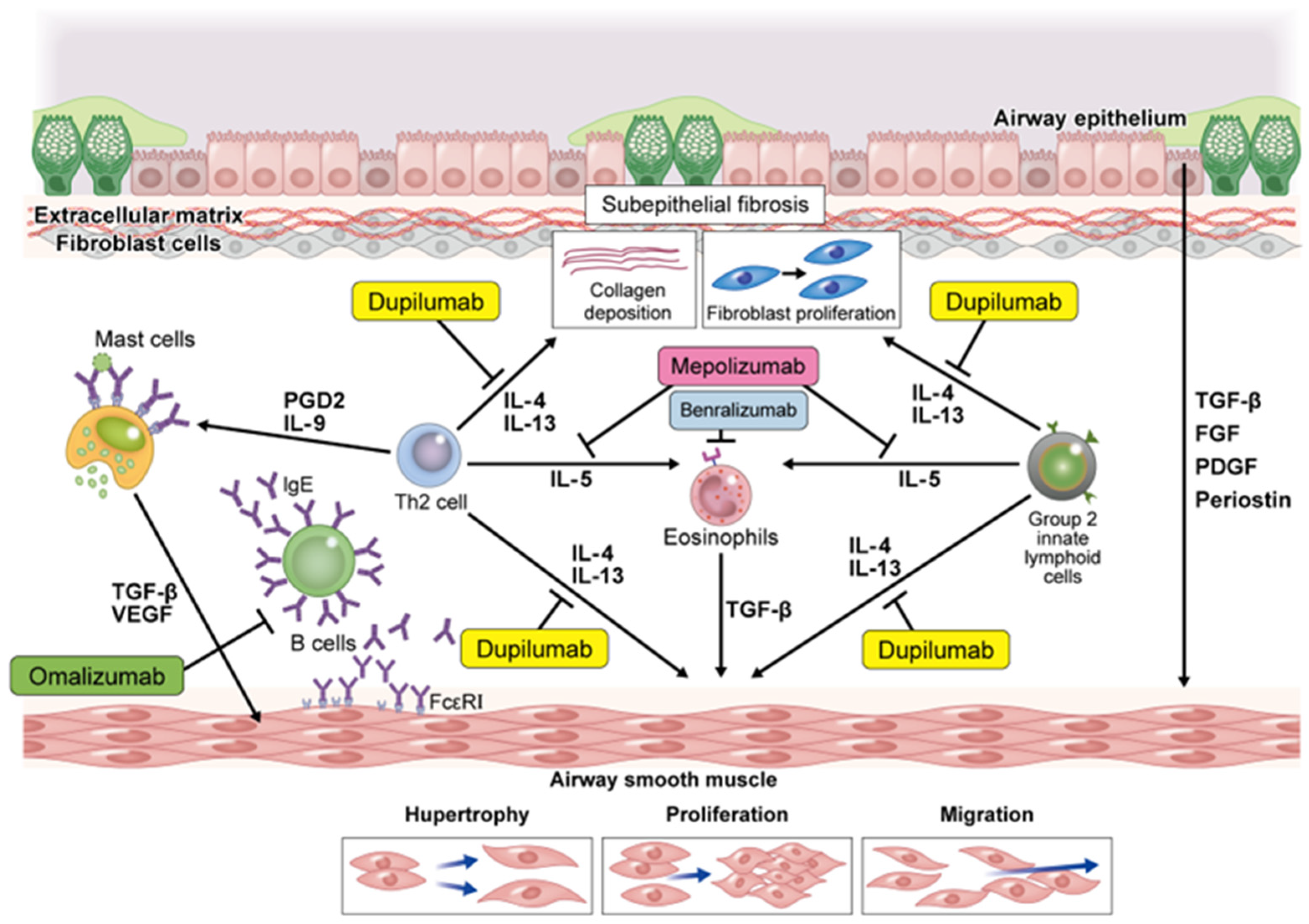
Pathological changes in airway remodeling include loss of airway epithelial cells, goblet cell hyperplasia, myofibroblast proliferation, airway smooth muscle cell hyperplasia, submembrane tissue fibrosis, and increased angiogenesis [67]. One cause of pathological changes in the airways is Th2 inflammation caused by crosstalk between different cell types in the airway wall and submucosa [68]. Th2 inflammation-related cytokines, such as IgE, IL-4, IL-5, and TSLP, can release chemokines and pro-inflammatory and/or fibrosis-promoting cytokines [69,70,71,72,73]. Downstream factors of these Th2 inflammatory mediators include platelet-derived growth factor, transforming growth factor β (TGF-β), and fibroblast growth factor, which promote the production of extracellular matrix (ECM) proteins that contribute to the pathogenesis of airway remodeling [74,75]. ECM proteins promote the proliferation of airway smooth muscle cells and remodeling in vivo [76]. In particular, TGF-β is considered to be an important factor in the airway remodeling of asthma. Subepithelial fibrosis is a major factor in airway remodeling, and TGF-β1 production by immunocompetent cells in asthmatics induces epithelial-mesenchymal transformation (EMT), which transforms airway epithelial cells into mesenchymal cells [74,77,78,79]. In addition, TGF-β is activated in response to the mechanical environment of the airways, such as bronchoconstriction and increased airway wall rigidity, leading to EMT, ECM protein production by fibroblasts, and airway smooth muscle cell proliferation [80] (Figure 3).
3. Efficacy of Biological Drugs in Childhood Asthma and Effects on Airway Remodeling
The underlying endotype of the pathology of childhood asthma was identified as Th2-high asthma by surrogate markers of airway T2 inflammation. As a result, biological drugs targeting T2 inflammatory mediators have been developed, enabling personalized treatment strategies for severe childhood asthma [81,82,83]. Long-term uncontrolled airway inflammation due to childhood asthma promotes airway remodeling, leading to serious and long-term impairment of lung function [62]. Considering the immunomodulatory effects of these biologics, inhibitory effects may be exerted on airway remodeling. Currently, the biologics approved for the treatment of severe asthma in children are omalizumab, mepolizumab, benralizumab, and dupilumab. The use of these biologics may suppress asthma symptoms and improve the natural course of childhood asthma by suppressing airway remodeling.
3.1. Omalizumab
Omalizumab is a humanized anti-IgE monoclonal antibody that binds to the Fcε3 segment, which is the constant region of free IgE. It prevents free IgE binding to IgE high affinity receptors (FcεR1) receptors on the surface of mast cells, basophils, and dendritic cells, and reduces the downstream allergic inflammatory cascade [84]. It also reduces free IgE circulating in the reticular endothelial system, resulting in a further reduction in allergic reactions [85]. In addition, omalizumab reduces the expression of FcεRI in basophils, mast cells, and dendritic cells, and suppresses the release of Th2 cytokines [86]. Omalizumab was approved by the U.S. Food and Drug Administration (FDA) and European Medicines Agency (EMA) in 2003 as an additional treatment for moderate-to-severe allergic asthma in children and adults aged 6 years and older [87,88].
In a randomized controlled trial of children aged 6–12 years with moderate-to-severe allergic asthma, the omalizumab group exhibited decreased asthma exacerbations, hospitalization rates, and doses of oral corticosteroids; and improved lung function and quality of life [89,90,91]. Omalizumab has also been reported to suppress asthma attacks in spring and autumn. A multicenter study of children and adolescents, aged 6–20 years, with allergic persistent asthma reported that asthma attacks in spring and autumn were reduced in the omalizumab group relative to the placebo group [92]. A study of 478 pediatric patients with severe asthma demonstrated that omalizumab reduced the rate of asthma attacks in the fall and enhanced the peripheral blood mononuclear cell IFN-α response to rhinovirus in vitro [93,94].
3.2. Mepolizumab
Mepolizumab is a mouse humanized immunoglobulin G1 monoclonal antibody that selectively binds to circulating IL-5 and prevents the binding of IL-5 to IL-5R on eosinophils and reduces eosinophil activation [95]. It is currently approved by the FDA and EMA as an additional maintenance therapy option in patients with severe eosinophilic asthma over 6 years of age [96]. Clinical trials investigated the efficacy of mepolizumab in patients with eosinophilic asthma aged 12 years and older and reported reduced asthma exacerbations, emergency visits, and hospitalization rates, as well as improvements in FEV1 and quality of life [97,98]. A 52-week open-label extension study reported a long-term improvement in asthma exacerbations and reduction in oral corticosteroid doses [99]. A long-term extended safety and efficacy study in patients with asthma reported a decrease in the asthma exacerbation rate and blood eosinophil counts alongside improved asthma control [100]. In a European multicenter study, children aged 6–11 years with severe eosinophilic asthma were treated with mepolizumab for 52 weeks, reducing decreased blood eosinophil counts and the frequency of asthma attacks [101,102]. Various studies have reported on the efficacy rates of this treatment for eosinophilic asthma [103,104].
3.3. Benralizumab
In contrast to mepolizumab, which binds to IL-5 itself, benralizumab is a humanized monoclonal antibody that binds to the α-subunit of IL-5R (IL-5Rα) on the surface of eosinophils and basophils. Benralizumab blocks the binding of IL-5 to its receptors and inhibits eosinophil differentiation and maturation in the bone marrow. In addition, this antibody enhances binding to the FcγRIIIA receptor on natural killer cells, accelerating apoptosis of circulating and tissue-existing eosinophils through antibody-dependent cellular cytotoxicity [105]. In a large multicenter phase III study in patients including adults and adolescents, benralizumab treatment in patients with severe eosinophilic asthma resulted in improvements in asthma symptoms, respiratory function, and quality of life, as well as a reduction in annual asthma exacerbations and doses of oral steroids [106,107,108]. Benralizumab was approved by the FDA and EMA in 2017 as additional maintenance therapy for patients with severe eosinophilic asthma over the age of 12 years. Benralizumab is approved for use in children over 12 years of age in the United States, but is only approved for use in adults in Europe. Further research on the efficacy and safety of benralizumab in the pediatric population under 12 years is warranted.
3.4. Dupilumab
Dupilumab is a humanized monoclonal antibody that targets the IL-4 receptor alpha chain (IL-4Rα) and inhibits signal transduction mediated by IL-4 and IL-13 by binding to the receptor [109]. Dupilumab was approved by the FDA and EMA as additional maintenance therapy for patients with moderate-to-severe asthma over the age of 12 years with elevated blood eosinophils and/or fractional exhaled nitric oxide (FeNO) [110].
A study of patients aged 12 years and older, with moderate-to-severe asthma, investigated the efficacy of dupilumab. Administration of dupilumab decreased the rate of exacerbation of asthma and improved lung function compared with placebo [111,112]. Greater efficacy was observed in patients with high baseline blood eosinophil counts and high FeNO. Post-hoc analysis of subgroups of adolescents aged 12–17 years revealed improved lung function and reduced asthma exacerbations [113]. In a study of glucocorticoid-dependent patients with severe asthma over 12 years of age, a 24-week administration of dupilumab reduced oral glucocorticoid use, reduced asthma exacerbations, and improved lung function, compared to the placebo group [114]. In addition, in a study of children aged 6 to 12 years with persistent asthma, additional doses of dupilumab reduced asthma exacerbations and improved lung function and asthma control compared to placebo [115]. Further research on the efficacy and safety of dupilumab in the pediatric population under 12 years is warranted.
4. Inhibitory Effect of Airway Remodeling by Biological Drugs
The most reliable way to assess structural changes in airway remodeling is pathological examination of biopsied bronchial or lung tissue [116]. Intrabronchial biopsy using a bronchoscope is performed for the purpose of differential diagnosis or evaluation of the effect of asthma treatment. Although it is possible to determine the thickening of submucosal tissue and changes in smooth muscle mass mainly in the proximal central airway, it is difficult to collect tissue in the peripheral small airways. Transbronchial biopsy is possible to collect peripheral airway tissue, but it is highly invasive. Both tests are difficult to perform, especially in pediatric patients, and can cause bleeding and pneumothorax complications.
As a non-invasive examination to evaluate the airway structure, an evaluation utilizing recent advances in image analysis technology has been reported [117,118,119]. In particular, airway morphology analysis using computed tomography (CT) can objectively quantify the degree of structural change in the bronchi [120,121,122]. Changes in airway structure obtained by this analysis correlate with airflow restriction and pathological change [123,124,125]. Furthermore, analysis using high-resolution CT enables analysis of bronchial trees, structural analysis of the distal airway, and area measurement of the airway wall cross section, making it possible to evaluate the degree of airway remodeling more accurately [126,127,128,129,130]. However, CT airway analysis using three-dimensional (3D) images has been used mainly in adult studies to date [119,129,131], and 3D-CT airway analysis was considered to be difficult in children due to their small size. A previous study using two-dimensional CT image analysis has been reported in pediatric asthma [132], but recent advances in CT equipment and image analysis software are enabling CT analysis of airway structures in children [133]. However, the airway structure obtained by CT includes not only irreversible tissue changes, but also reversible changes in the airway mucosa. In addition, there is a risk of radiation exposure, and there are restrictions in terms of practicality and ethics.
Biological agents currently used for childhood asthma targeting Th2-high asthma may affect airway remodeling, and CT airway analysis, as well as pathological assessment of biopsied airway mucosa, is revealing their effects (Table 1). Three studies using biopsied bronchial tissue before and after omalizumab treatment in adult patients with severe asthma showed a significant reduction in bronchial reticular basement membrane thickness, airway smooth muscles, and fibronectin deposition [134,135,136]. Furthermore, three studies using CT airway analysis gave similar assessments, reporting that omalizumab treatment in adult patients with severe asthma significantly reduced airway wall thickness and area [137,138,139]. The improving effect on airway remodeling has been confirmed not only with omalizumab, but also with other biological drugs. CT airway analysis before and after administration of mepolizumab for severe asthma in adults showed a significant decrease in the airway wall area after treatment [140]. It was also reported that benralizumab administration reduced airway smooth muscle mass by 29% in adults with severe asthma [141]. There are no reports of animal model or human studies on the effect of dupilumab on improving airway remodeling. Unfortunately, all the above studies have been conducted in adults with severe asthma, and no studies have demonstrated a suppressive effect on airway remodeling in childhood severe asthma. An ongoing Italian clinical trial is conducting CT airway analysis using a deep learning model prospectively in children with severe asthma to develop a prognostic scoring (ClinicalTrials.gov number, NCT05140889). Further investigation is needed to determine whether biological treatment from childhood prevents the progression of airway remodeling.
5. Conclusions
In childhood asthma, chronic airway inflammation impairs normal lung growth, even after remission of respiratory symptoms. The inability to obtain peak lung function in adolescence affects lung function in adulthood, which leads to a risk of developing COPD. Therefore, early suppression of airway inflammation in childhood asthma and prevention of asthma exacerbations may improve lung maturation, leading to good lung function and prevention of COPD in adulthood. As airway inflammation due to T2-high asthma in childhood causes persistent pathological airway remodeling with asthma exacerbations, administration of biologics targeting Th2 cytokines may suppress the progression of airway remodeling and prevent pulmonary dysfunction in adulthood. It is necessary to conduct clinical trials using minimally invasive evaluation methods, such as CT airway analysis, to ascertain the inhibitory effect of biological drugs on airway remodeling in severe childhood asthma.
Author Contributions
Conceptualization, M.T. and M.I.; writing—original draft preparation, M.T.; writing—review and editing, M.I. and H.T.; supervision, H.T.; project administration, H.T. All authors have read and agreed to the published version of the manuscript.
Funding
This research received no external funding.
Institutional Review Board Statement
Not applicable.
Informed Consent Statement
Not applicable.
Data Availability Statement
Not applicable.
Conflicts of Interest
The authors declare no conflict of interest.
References
- Papi, A.; Brightling, C.; Pedersen, S.E.; Reddel, H.K. Asthma. Lancet 2018, 391, 783–800. [Google Scholar] [CrossRef]
- Dharmage, S.C.; Perret, J.L.; Custovic, A. Epidemiology of Asthma in Children and Adults. Front. Pediatr. 2019, 7, 246. [Google Scholar] [CrossRef]
- Zahran, H.S.; Bailey, C.M.; Damon, S.A.; Garbe, P.L.; Breysse, P.N. Vital Signs: Asthma in Children—United States, 2001–2016. MMWR Morb. Mortal. Wkly. Rep. 2018, 67, 149–155. [Google Scholar] [CrossRef]
- Gibson, G.J.; Loddenkemper, R.; Lundbäck, B.; Sibille, Y. Respiratory health and disease in Europe: The new European Lung White Book. Eur. Respir. J. 2013, 42, 559–563. [Google Scholar] [CrossRef]
- Guilbert, T.W.; Bacharier, L.B.; Fitzpatrick, A.M. Severe asthma in children. J. Allergy Clin. Immunol. Pract. 2014, 2, 489–500. [Google Scholar] [CrossRef]
- Fleming, L.; Murray, C.; Bansal, A.T.; Hashimoto, S.; Bisgaard, H.; Bush, A.; Frey, U.; Hedlin, G.; Singer, F.; van Aalderen, W.M.; et al. The burden of severe asthma in childhood and adolescence: Results from the paediatric U-BIOPRED cohorts. Eur. Respir. J. 2015, 46, 1322–1333. [Google Scholar] [CrossRef]
- Pijnenburg, M.W.; Fleming, L. Advances in understanding and reducing the burden of severe asthma in children. Lancet Respir. Med. 2020, 8, 1032–1044. [Google Scholar] [CrossRef]
- Bui, D.S.; Lodge, C.J.; Burgess, J.A.; Lowe, A.J.; Perret, J.; Bui, M.Q.; Bowatte, G.; Gurrin, L.; Johns, D.P.; Thompson, B.R.; et al. Childhood predictors of lung function trajectories and future COPD risk: A prospective cohort study from the first to the sixth decade of life. Lancet Respir. Med. 2018, 6, 535–544. [Google Scholar] [CrossRef]
- Tsuge, M.; Ikeda, M.; Matsumoto, N.; Yorifuji, T.; Tsukahara, H. Current Insights into Atopic March. Children 2021, 8, 1067. [Google Scholar] [CrossRef]
- Tantisira, K.G.; Colvin, R.; Tonascia, J.; Strunk, R.C.; Weiss, S.T.; Fuhlbrigge, A.L. Airway responsiveness in mild to moderate childhood asthma: Sex influences on the natural history. Am. J. Respir. Crit. Care Med. 2008, 178, 325–331. [Google Scholar] [CrossRef]
- Vonk, J.M.; Postma, D.S.; Boezen, H.M.; Grol, M.H.; Schouten, J.P.; Koëter, G.H.; Gerritsen, J. Childhood factors associated with asthma remission after 30 year follow up. Thorax 2004, 59, 925–929. [Google Scholar] [CrossRef]
- Limb, S.L.; Brown, K.C.; Wood, R.A.; Wise, R.A.; Eggleston, P.A.; Tonascia, J.; Hamilton, R.G.; Adkinson, N.F., Jr. Adult asthma severity in individuals with a history of childhood asthma. J. Allergy Clin. Immunol. 2005, 115, 61–66. [Google Scholar] [CrossRef]
- Tai, A.; Tran, H.; Roberts, M.; Clarke, N.; Gibson, A.M.; Vidmar, S.; Wilson, J.; Robertson, C.F. Outcomes of childhood asthma to the age of 50 years. J. Allergy Clin. Immunol. 2014, 133, 1572–1578.e1573. [Google Scholar] [CrossRef]
- Belgrave, D.C.; Buchan, I.; Bishop, C.; Lowe, L.; Simpson, A.; Custovic, A. Trajectories of lung function during childhood. Am. J. Respir. Crit. Care Med. 2014, 189, 1101–1109. [Google Scholar] [CrossRef]
- Lodge, C.J.; Lowe, A.J.; Allen, K.J.; Zaloumis, S.; Gurrin, L.C.; Matheson, M.C.; Axelrad, C.; Welsh, L.; Bennett, C.M.; Hopper, J.; et al. Childhood wheeze phenotypes show less than expected growth in FEV1 across adolescence. Am. J. Respir. Crit. Care Med. 2014, 189, 1351–1358. [Google Scholar] [CrossRef]
- Hallberg, J.; Thunqvist, P.; Schultz, E.S.; Kull, I.; Bottai, M.; Merritt, A.S.; Chiesa, F.; Gustafsson, P.M.; Melén, E. Asthma phenotypes and lung function up to 16 years of age-the BAMSE cohort. Allergy 2015, 70, 667–673. [Google Scholar] [CrossRef]
- Sears, M.R.; Greene, J.M.; Willan, A.R.; Wiecek, E.M.; Taylor, D.R.; Flannery, E.M.; Cowan, J.O.; Herbison, G.P.; Silva, P.A.; Poulton, R. A longitudinal, population-based, cohort study of childhood asthma followed to adulthood. N. Engl. J. Med. 2003, 349, 1414–1422. [Google Scholar] [CrossRef]
- Covar, R.A.; Spahn, J.D.; Murphy, J.R.; Szefler, S.J. Progression of asthma measured by lung function in the childhood asthma management program. Am. J. Respir. Crit. Care Med. 2004, 170, 234–241. [Google Scholar] [CrossRef]
- Postma, D.S.; Bush, A.; van den Berge, M. Risk factors and early origins of chronic obstructive pulmonary disease. Lancet 2015, 385, 899–909. [Google Scholar] [CrossRef]
- Halpin, D.M.G.; Criner, G.J.; Papi, A.; Singh, D.; Anzueto, A.; Martinez, F.J.; Agusti, A.A.; Vogelmeier, C.F. Global Initiative for the Diagnosis, Management, and Prevention of Chronic Obstructive Lung Disease. The 2020 GOLD Science Committee Report on COVID-19 and Chronic Obstructive Pulmonary Disease. Am. J. Respir. Crit. Care Med. 2021, 203, 24–36. [Google Scholar] [CrossRef]
- Taniguchi, A.; Tsuge, M.; Miyahara, N.; Tsukahara, H. Reactive Oxygen Species and Antioxidative Defense in Chronic Obstructive Pulmonary Disease. Antioxidants 2021, 10, 1537. [Google Scholar] [CrossRef] [PubMed]
- Lamprecht, B.; McBurnie, M.A.; Vollmer, W.M.; Gudmundsson, G.; Welte, T.; Nizankowska-Mogilnicka, E.; Studnicka, M.; Bateman, E.; Anto, J.M.; Burney, P.; et al. COPD in never smokers: Results from the population-based burden of obstructive lung disease study. Chest 2011, 139, 752–763. [Google Scholar] [CrossRef] [PubMed]
- Salvi, S.S.; Barnes, P.J. Chronic obstructive pulmonary disease in non-smokers. Lancet 2009, 374, 733–743. [Google Scholar] [CrossRef]
- Lange, P.; Celli, B.; Agustí, A.; Boje Jensen, G.; Divo, M.; Faner, R.; Guerra, S.; Marott, J.L.; Martinez, F.D.; Martinez-Camblor, P.; et al. Lung-Function Trajectories Leading to Chronic Obstructive Pulmonary Disease. N. Engl. J. Med. 2015, 373, 111–122. [Google Scholar] [CrossRef]
- Vasquez, M.M.; Zhou, M.; Hu, C.; Martinez, F.D.; Guerra, S. Low Lung Function in Young Adult Life Is Associated with Early Mortality. Am. J. Respir. Crit. Care Med. 2017, 195, 1399–1401. [Google Scholar] [CrossRef] [PubMed]
- Bui, D.S.; Burgess, J.A.; Lowe, A.J.; Perret, J.L.; Lodge, C.J.; Bui, M.; Morrison, S.; Thompson, B.R.; Thomas, P.S.; Giles, G.G.; et al. Childhood Lung Function Predicts Adult Chronic Obstructive Pulmonary Disease and Asthma-Chronic Obstructive Pulmonary Disease Overlap Syndrome. Am. J. Respir. Crit. Care Med. 2017, 196, 39–46. [Google Scholar] [CrossRef] [PubMed]
- Belgrave, D.C.M.; Granell, R.; Turner, S.W.; Curtin, J.A.; Buchan, I.E.; Le Souëf, P.N.; Simpson, A.; Henderson, A.J.; Custovic, A. Lung function trajectories from pre-school age to adulthood and their associations with early life factors: A retrospective analysis of three population-based birth cohort studies. Lancet Respir. Med. 2018, 6, 526–534. [Google Scholar] [CrossRef]
- Berry, C.E.; Billheimer, D.; Jenkins, I.C.; Lu, Z.J.; Stern, D.A.; Gerald, L.B.; Carr, T.F.; Guerra, S.; Morgan, W.J.; Wright, A.L.; et al. A Distinct Low Lung Function Trajectory from Childhood to the Fourth Decade of Life. Am. J. Respir. Crit. Care Med. 2016, 194, 607–612. [Google Scholar] [CrossRef]
- Johannessen, A.; Bakke, P.S.; Hardie, J.A.; Eagan, T.M. Association of exposure to environmental tobacco smoke in childhood with chronic obstructive pulmonary disease and respiratory symptoms in adults. Respirology 2012, 17, 499–505. [Google Scholar] [CrossRef]
- Tai, A.; Tran, H.; Roberts, M.; Clarke, N.; Wilson, J.; Robertson, C.F. The association between childhood asthma and adult chronic obstructive pulmonary disease. Thorax 2014, 69, 805–810. [Google Scholar] [CrossRef]
- Martinez, F.D. Early-Life Origins of Chronic Obstructive Pulmonary Disease. N. Engl. J. Med. 2016, 375, 871–878. [Google Scholar] [CrossRef] [PubMed]
- Narayanan, M.; Owers-Bradley, J.; Beardsmore, C.S.; Mada, M.; Ball, I.; Garipov, R.; Panesar, K.S.; Kuehni, C.E.; Spycher, B.D.; Williams, S.E.; et al. Alveolarization continues during childhood and adolescence: New evidence from helium-3 magnetic resonance. Am. J. Respir. Crit. Care Med. 2012, 185, 186–191. [Google Scholar] [CrossRef] [PubMed]
- Mullassery, D.; Smith, N.P. Lung development. Semin. Pediatr. Surg. 2015, 24, 152–155. [Google Scholar] [CrossRef] [PubMed]
- Wang, X.; Mensinga, T.T.; Schouten, J.P.; Rijcken, B.; Weiss, S.T. Determinants of maximally attained level of pulmonary function. Am. J. Respir. Crit. Care Med. 2004, 169, 941–949. [Google Scholar] [CrossRef]
- Allinson, J.P.; Hardy, R.; Donaldson, G.C.; Shaheen, S.O.; Kuh, D.; Wedzicha, J.A. Combined Impact of Smoking and Early-Life Exposures on Adult Lung Function Trajectories. Am. J. Respir. Crit. Care Med. 2017, 196, 1021–1030. [Google Scholar] [CrossRef]
- Perret, J.L.; Walters, H.; Johns, D.; Gurrin, L.; Burgess, J.; Lowe, A.; Thompson, B.; Markos, J.; Morrison, S.; Thomas, P.; et al. Mother’s smoking and complex lung function of offspring in middle age: A cohort study from childhood. Respirology 2016, 21, 911–919. [Google Scholar] [CrossRef]
- Hayden, L.P.; Hobbs, B.D.; Cohen, R.T.; Wise, R.A.; Checkley, W.; Crapo, J.D.; Hersh, C.P. Childhood pneumonia increases risk for chronic obstructive pulmonary disease: The COPDGene study. Respir. Res. 2015, 16, 115. [Google Scholar] [CrossRef]
- Savran, O.; Ulrik, C.S. Early life insults as determinants of chronic obstructive pulmonary disease in adult life. Int. J. Chron. Obstruct. Pulmon. Dis. 2018, 13, 683–693. [Google Scholar] [CrossRef]
- Duan, P.; Wang, Y.; Lin, R.; Zeng, Y.; Chen, C.; Yang, L.; Yue, M.; Zhong, S.; Wang, Y.; Zhang, Q. Impact of early life exposures on COPD in adulthood: A systematic review and meta-analysis. Respirology 2021, 26, 1131–1151. [Google Scholar] [CrossRef]
- Tagiyeva, N.; Devereux, G.; Fielding, S.; Turner, S.; Douglas, G. Outcomes of Childhood Asthma and Wheezy Bronchitis. A 50-Year Cohort Study. Am. J. Respir. Crit. Care Med. 2016, 193, 23–30. [Google Scholar] [CrossRef]
- Ali, K.M. Childhood asthma as a risk factor for adult chronic obstructive pulmonary disease: A systematic review and meta-analysis. Expert Rev. Respir. Med. 2022, 16, 461–467. [Google Scholar] [CrossRef] [PubMed]
- Shirtcliffe, P.; Marsh, S.; Travers, J.; Weatherall, M.; Beasley, R. Childhood asthma and GOLD-defined chronic obstructive pulmonary disease. Intern. Med. J. 2012, 42, 83–88. [Google Scholar] [CrossRef] [PubMed]
- Agusti, A.; Faner, R. Lung function trajectories in health and disease. Lancet Respir. Med. 2019, 7, 358–364. [Google Scholar] [CrossRef]
- Bisgaard, H.; Nørgaard, S.; Sevelsted, A.; Chawes, B.L.; Stokholm, J.; Mortensen, E.L.; Ulrik, C.S.; Bønnelykke, K. Asthma-like symptoms in young children increase the risk of COPD. J. Allergy Clin. Immunol. 2021, 147, 569–576.e569. [Google Scholar] [CrossRef]
- McGeachie, M.J.; Yates, K.P.; Zhou, X.; Guo, F.; Sternberg, A.L.; Van Natta, M.L.; Wise, R.A.; Szefler, S.J.; Sharma, S.; Kho, A.T.; et al. Patterns of Growth and Decline in Lung Function in Persistent Childhood Asthma. N. Engl. J. Med. 2016, 374, 1842–1852. [Google Scholar] [CrossRef]
- Phelan, P.D.; Robertson, C.F.; Olinsky, A. The Melbourne Asthma Study: 1964–1999. J. Allergy Clin. Immunol. 2002, 109, 189–194. [Google Scholar] [CrossRef]
- James, A.L.; Palmer, L.J.; Kicic, E.; Maxwell, P.S.; Lagan, S.E.; Ryan, G.F.; Musk, A.W. Decline in lung function in the Busselton Health Study: The effects of asthma and cigarette smoking. Am. J. Respir. Crit. Care Med. 2005, 171, 109–114. [Google Scholar] [CrossRef]
- Stern, D.A.; Morgan, W.J.; Wright, A.L.; Guerra, S.; Martinez, F.D. Poor airway function in early infancy and lung function by age 22 years: A non-selective longitudinal cohort study. Lancet 2007, 370, 758–764. [Google Scholar] [CrossRef]
- Marossy, A.E.; Strachan, D.P.; Rudnicka, A.R.; Anderson, H.R. Childhood chest illness and the rate of decline of adult lung function between ages 35 and 45 years. Am. J. Respir. Crit. Care Med. 2007, 175, 355–359. [Google Scholar] [CrossRef]
- Marcon, A.; Locatelli, F.; Keidel, D.; Beckmeyer-Borowko, A.B.; Cerveri, I.; Dharmage, S.C.; Fuertes, E.; Garcia-Aymerich, J.; Heinrich, J.; Imboden, M.; et al. Airway responsiveness to methacholine and incidence of COPD: An international prospective cohort study. Thorax 2018, 73, 825–832. [Google Scholar] [CrossRef]
- Bui, D.S.; Walters, H.E.; Burgess, J.A.; Perret, J.L.; Bui, M.Q.; Bowatte, G.; Lowe, A.J.; Russell, M.A.; Thompson, B.R.; Hamilton, G.S.; et al. Childhood Respiratory Risk Factor Profiles and Middle-Age Lung Function: A Prospective Cohort Study from the First to Sixth Decade. Ann. Am. Thorac. Soc. 2018, 15, 1057–1066. [Google Scholar] [CrossRef] [PubMed]
- Licari, A.; Castagnoli, R.; Brambilla, I.; Marseglia, A.; Tosca, M.A.; Marseglia, G.L.; Ciprandi, G. Asthma Endotyping and Biomarkers in Childhood Asthma. Pediatr. Allergy Immunol. Pulmonol. 2018, 31, 44–55. [Google Scholar] [CrossRef] [PubMed]
- Diamant, Z.; Vijverberg, S.; Alving, K.; Bakirtas, A.; Bjermer, L.; Custovic, A.; Dahlen, S.E.; Gaga, M.; Gerth van Wijk, R.; Giacco, S.D.; et al. Toward clinically applicable biomarkers for asthma: An EAACI position paper. Allergy 2019, 74, 1835–1851. [Google Scholar] [CrossRef] [PubMed]
- Cevhertas, L.; Ogulur, I.; Maurer, D.J.; Burla, D.; Ding, M.; Jansen, K.; Koch, J.; Liu, C.; Ma, S.; Mitamura, Y.; et al. Advances and recent developments in asthma in 2020. Allergy 2020, 75, 3124–3146. [Google Scholar] [CrossRef]
- Pepper, A.N.; Renz, H.; Casale, T.B.; Garn, H. Biologic Therapy and Novel Molecular Targets of Severe Asthma. J. Allergy Clin. Immunol. Pract. 2017, 5, 909–916. [Google Scholar] [CrossRef]
- Stokes, J.R.; Casale, T.B. Characterization of asthma endotypes: Implications for therapy. Ann. Allergy Asthma Immunol. 2016, 117, 121–125. [Google Scholar] [CrossRef]
- Saglani, S.; Lloyd, C.M. Eosinophils in the pathogenesis of paediatric severe asthma. Curr. Opin. Allergy Clin. Immunol. 2014, 14, 143–148. [Google Scholar] [CrossRef]
- LaPorte, S.L.; Juo, Z.S.; Vaclavikova, J.; Colf, L.A.; Qi, X.; Heller, N.M.; Keegan, A.D.; Garcia, K.C. Molecular and structural basis of cytokine receptor pleiotropy in the interleukin-4/13 system. Cell 2008, 132, 259–272. [Google Scholar] [CrossRef]
- Kau, A.L.; Korenblat, P.E. Anti-interleukin 4 and 13 for asthma treatment in the era of endotypes. Curr. Opin. Allergy Clin. Immunol. 2014, 14, 570–575. [Google Scholar] [CrossRef]
- Saco, T.V.; Pepper, A.; Casale, T.B. Uses of biologics in allergic diseases: What to choose and when. Ann. Allergy Asthma Immunol. 2018, 120, 357–366. [Google Scholar] [CrossRef]
- Halwani, R.; Al-Muhsen, S.; Hamid, Q. Airway remodeling in asthma. Curr. Opin. Pharmacol. 2010, 10, 236–245. [Google Scholar] [CrossRef]
- Boulet, L.P. Airway remodeling in asthma: Update on mechanisms and therapeutic approaches. Curr. Opin. Pulm. Med. 2018, 24, 56–62. [Google Scholar] [CrossRef]
- Wilson, S.J.; Rigden, H.M.; Ward, J.A.; Laviolette, M.; Jarjour, N.N.; Djukanović, R. The relationship between eosinophilia and airway remodelling in mild asthma. Clin. Exp. Allergy 2013, 43, 1342–1350. [Google Scholar] [CrossRef]
- O’Reilly, R.; Ullmann, N.; Irving, S.; Bossley, C.J.; Sonnappa, S.; Zhu, J.; Oates, T.; Banya, W.; Jeffery, P.K.; Bush, A.; et al. Increased airway smooth muscle in preschool wheezers who have asthma at school age. J. Allergy Clin. Immunol. 2013, 131, 1024–1032, 1032.e1001-1016. [Google Scholar] [CrossRef]
- Lezmi, G.; Gosset, P.; Deschildre, A.; Abou-Taam, R.; Mahut, B.; Beydon, N.; de Blic, J. Airway Remodeling in Preschool Children with Severe Recurrent Wheeze. Am. J. Respir. Crit. Care Med. 2015, 192, 164–171. [Google Scholar] [CrossRef]
- Grainge, C.L.; Lau, L.C.; Ward, J.A.; Dulay, V.; Lahiff, G.; Wilson, S.; Holgate, S.; Davies, D.E.; Howarth, P.H. Effect of bronchoconstriction on airway remodeling in asthma. N. Engl. J. Med. 2011, 364, 2006–2015. [Google Scholar] [CrossRef]
- Holgate, S.T. Epithelium dysfunction in asthma. J. Allergy Clin. Immunol. 2007, 120, 1233–1244, quiz 1245-1236. [Google Scholar] [CrossRef]
- Doherty, T.; Broide, D. Cytokines and growth factors in airway remodeling in asthma. Curr. Opin. Immunol. 2007, 19, 676–680. [Google Scholar] [CrossRef]
- Batra, V.; Musani, A.I.; Hastie, A.T.; Khurana, S.; Carpenter, K.A.; Zangrilli, J.G.; Peters, S.P. Bronchoalveolar lavage fluid concentrations of transforming growth factor (TGF)-beta1, TGF-beta2, interleukin (IL)-4 and IL-13 after segmental allergen challenge and their effects on alpha-smooth muscle actin and collagen III synthesis by primary human lung fibroblasts. Clin. Exp. Allergy 2004, 34, 437–444. [Google Scholar] [CrossRef]
- Tanaka, H.; Komai, M.; Nagao, K.; Ishizaki, M.; Kajiwara, D.; Takatsu, K.; Delespesse, G.; Nagai, H. Role of interleukin-5 and eosinophils in allergen-induced airway remodeling in mice. Am. J. Respir. Cell Mol. Biol. 2004, 31, 62–68. [Google Scholar] [CrossRef]
- Malavia, N.K.; Mih, J.D.; Raub, C.B.; Dinh, B.T.; George, S.C. IL-13 induces a bronchial epithelial phenotype that is profibrotic. Respir. Res. 2008, 9, 27. [Google Scholar] [CrossRef]
- Redhu, N.S.; Shan, L.; Al-Subait, D.; Ashdown, H.L.; Movassagh, H.; Lamkhioued, B.; Gounni, A.S. IgE induces proliferation in human airway smooth muscle cells: Role of MAPK and STAT3 pathways. Allergy Asthma Clin. Immunol. 2013, 9, 41. [Google Scholar] [CrossRef]
- Cao, L.; Liu, F.; Liu, Y.; Liu, T.; Wu, J.; Zhao, J.; Wang, J.; Li, S.; Xu, J.; Dong, L. TSLP promotes asthmatic airway remodeling via p38-STAT3 signaling pathway in human lung fibroblast. Exp. Lung Res. 2018, 44, 288–301. [Google Scholar] [CrossRef]
- Halwani, R.; Al-Muhsen, S.; Al-Jahdali, H.; Hamid, Q. Role of transforming growth factor-β in airway remodeling in asthma. Am. J. Respir. Cell Mol. Biol. 2011, 44, 127–133. [Google Scholar] [CrossRef]
- Kardas, G.; Daszyńska-Kardas, A.; Marynowski, M.; Brząkalska, O.; Kuna, P.; Panek, M. Role of Platelet-Derived Growth Factor (PDGF) in Asthma as an Immunoregulatory Factor Mediating Airway Remodeling and Possible Pharmacological Target. Front. Pharmacol. 2020, 11, 47. [Google Scholar] [CrossRef]
- Johnson, P.R.; Burgess, J.K.; Underwood, P.A.; Au, W.; Poniris, M.H.; Tamm, M.; Ge, Q.; Roth, M.; Black, J.L. Extracellular matrix proteins modulate asthmatic airway smooth muscle cell proliferation via an autocrine mechanism. J. Allergy Clin. Immunol. 2004, 113, 690–696. [Google Scholar] [CrossRef]
- Tatler, A.L.; John, A.E.; Jolly, L.; Habgood, A.; Porte, J.; Brightling, C.; Knox, A.J.; Pang, L.; Sheppard, D.; Huang, X.; et al. Integrin αvβ5-mediated TGF-β activation by airway smooth muscle cells in asthma. J. Immunol. 2011, 187, 6094–6107. [Google Scholar] [CrossRef]
- Pain, M.; Bermudez, O.; Lacoste, P.; Royer, P.J.; Botturi, K.; Tissot, A.; Brouard, S.; Eickelberg, O.; Magnan, A. Tissue remodelling in chronic bronchial diseases: From the epithelial to mesenchymal phenotype. Eur. Respir. Rev. 2014, 23, 118–130. [Google Scholar] [CrossRef]
- Sohal, S.S.; Ward, C.; Walters, E.H. Importance of epithelial mesenchymal transition (EMT) in COPD and asthma. Thorax 2014, 69, 768. [Google Scholar] [CrossRef]
- Shkumatov, A.; Thompson, M.; Choi, K.M.; Sicard, D.; Baek, K.; Kim, D.H.; Tschumperlin, D.J.; Prakash, Y.S.; Kong, H. Matrix stiffness-modulated proliferation and secretory function of the airway smooth muscle cells. Am. J. Physiol. Lung Cell Mol. Physiol. 2015, 308, L1125–L1135. [Google Scholar] [CrossRef]
- Licari, A.; Manti, S.; Marseglia, A.; De Filippo, M.; De Sando, E.; Foiadelli, T.; Marseglia, G.L. Biologics in Children with Allergic Diseases. Curr. Pediatr. Rev. 2020, 16, 140–147. [Google Scholar] [CrossRef] [PubMed]
- Abrams, E.M.; Becker, A.B.; Szefler, S.J. Current State and Future of Biologic Therapies in the Treatment of Asthma in Children. Pediatr. Allergy Immunol. Pulmonol. 2018, 31, 119–131. [Google Scholar] [CrossRef] [PubMed]
- Porcaro, F.; Cutrera, R.; Pajno, G.B. Options of immunotherapeutic treatments for children with asthma. Expert Rev. Respir. Med. 2019, 13, 937–949. [Google Scholar] [CrossRef] [PubMed]
- Kawakami, T.; Blank, U. From IgE to Omalizumab. J. Immunol 2016, 197, 4187–4192. [Google Scholar] [CrossRef]
- Walsh, G.M. An update on biologic-based therapy in asthma. Immunotherapy 2013, 5, 1255–1264. [Google Scholar] [CrossRef]
- Licari, A.; Castagnoli, R.; Panfili, E.; Marseglia, A.; Brambilla, I.; Marseglia, G.L. An Update on Anti-IgE Therapy in Pediatric Respiratory Diseases. Curr. Respir. Med. Rev. 2017, 13, 22–29. [Google Scholar] [CrossRef]
- Berger, W.; Gupta, N.; McAlary, M.; Fowler-Taylor, A. Evaluation of long-term safety of the anti-IgE antibody, omalizumab, in children with allergic asthma. Ann. Allergy Asthma Immunol. 2003, 91, 182–188. [Google Scholar] [CrossRef]
- Chipps, B.E.; Lanier, B.; Milgrom, H.; Deschildre, A.; Hedlin, G.; Szefler, S.J.; Kattan, M.; Kianifard, F.; Ortiz, B.; Haselkorn, T.; et al. Omalizumab in children with uncontrolled allergic asthma: Review of clinical trial and real-world experience. J. Allergy Clin. Immunol. 2017, 139, 1431–1444. [Google Scholar] [CrossRef]
- Brodlie, M.; McKean, M.C.; Moss, S.; Spencer, D.A. The oral corticosteroid-sparing effect of omalizumab in children with severe asthma. Arch. Dis. Child. 2012, 97, 604–609. [Google Scholar] [CrossRef]
- Deschildre, A.; Marguet, C.; Salleron, J.; Pin, I.; Rittié, J.L.; Derelle, J.; Taam, R.A.; Fayon, M.; Brouard, J.; Dubus, J.C.; et al. Add-on omalizumab in children with severe allergic asthma: A 1-year real life survey. Eur. Respir. J. 2013, 42, 1224–1233. [Google Scholar] [CrossRef]
- Busse, W.W.; Humbert, M.; Haselkorn, T.; Ortiz, B.; Trzaskoma, B.L.; Stephenson, P.; Garcia Conde, L.; Kianifard, F.; Holgate, S.T. Effect of omalizumab on lung function and eosinophil levels in adolescents with moderate-to-severe allergic asthma. Ann. Allergy Asthma Immunol. 2020, 124, 190–196. [Google Scholar] [CrossRef] [PubMed]
- Busse, W.W.; Morgan, W.J.; Gergen, P.J.; Mitchell, H.E.; Gern, J.E.; Liu, A.H.; Gruchalla, R.S.; Kattan, M.; Teach, S.J.; Pongracic, J.A.; et al. Randomized trial of omalizumab (anti-IgE) for asthma in inner-city children. N. Engl. J. Med. 2011, 364, 1005–1015. [Google Scholar] [CrossRef] [PubMed]
- Teach, S.J.; Gill, M.A.; Togias, A.; Sorkness, C.A.; Arbes, S.J., Jr.; Calatroni, A.; Wildfire, J.J.; Gergen, P.J.; Cohen, R.T.; Pongracic, J.A.; et al. Preseasonal treatment with either omalizumab or an inhaled corticosteroid boost to prevent fall asthma exacerbations. J. Allergy Clin. Immunol. 2015, 136, 1476–1485. [Google Scholar] [CrossRef] [PubMed]
- Esquivel, A.; Busse, W.W.; Calatroni, A.; Togias, A.G.; Grindle, K.G.; Bochkov, Y.A.; Gruchalla, R.S.; Kattan, M.; Kercsmar, C.M.; Khurana Hershey, G.; et al. Effects of Omalizumab on Rhinovirus Infections, Illnesses, and Exacerbations of Asthma. Am. J. Respir. Crit. Care Med. 2017, 196, 985–992. [Google Scholar] [CrossRef]
- Emma, R.; Morjaria, J.B.; Fuochi, V.; Polosa, R.; Caruso, M. Mepolizumab in the management of severe eosinophilic asthma in adults: Current evidence and practical experience. Ther. Adv. Respir. Dis. 2018, 12, 1753466618808490. [Google Scholar] [CrossRef]
- Licari, A.; Manti, S.; Castagnoli, R.; Parisi, G.F.; Salpietro, C.; Leonardi, S.; Marseglia, G.L. Targeted Therapy for Severe Asthma in Children and Adolescents: Current and Future Perspectives. Paediatr. Drugs 2019, 21, 215–237. [Google Scholar] [CrossRef]
- Pavord, I.D.; Korn, S.; Howarth, P.; Bleecker, E.R.; Buhl, R.; Keene, O.N.; Ortega, H.; Chanez, P. Mepolizumab for severe eosinophilic asthma (DREAM): A multicentre, double-blind, placebo-controlled trial. Lancet 2012, 380, 651–659. [Google Scholar] [CrossRef]
- Ortega, H.G.; Liu, M.C.; Pavord, I.D.; Brusselle, G.G.; FitzGerald, J.M.; Chetta, A.; Humbert, M.; Katz, L.E.; Keene, O.N.; Yancey, S.W.; et al. Mepolizumab treatment in patients with severe eosinophilic asthma. N. Engl. J. Med. 2014, 371, 1198–1207. [Google Scholar] [CrossRef]
- Lugogo, N.; Domingo, C.; Chanez, P.; Leigh, R.; Gilson, M.J.; Price, R.G.; Yancey, S.W.; Ortega, H.G. Long-term Efficacy and Safety of Mepolizumab in Patients With Severe Eosinophilic Asthma: A Multi-center, Open-label, Phase IIIb Study. Clin. Ther. 2016, 38, 2058–2070.e1. [Google Scholar] [CrossRef]
- Khatri, S.; Moore, W.; Gibson, P.G.; Leigh, R.; Bourdin, A.; Maspero, J.; Barros, M.; Buhl, R.; Howarth, P.; Albers, F.C.; et al. Assessment of the long-term safety of mepolizumab and durability of clinical response in patients with severe eosinophilic asthma. J. Allergy Clin. Immunol. 2019, 143, 1742–1751.e1747. [Google Scholar] [CrossRef]
- Gupta, A.; Ikeda, M.; Geng, B.; Azmi, J.; Price, R.G.; Bradford, E.S.; Yancey, S.W.; Steinfeld, J. Long-term safety and pharmacodynamics of mepolizumab in children with severe asthma with an eosinophilic phenotype. J. Allergy Clin. Immunol. 2019, 144, 1336–1342.e1337. [Google Scholar] [CrossRef] [PubMed]
- Gupta, A.; Pouliquen, I.; Austin, D.; Price, R.G.; Kempsford, R.; Steinfeld, J.; Bradford, E.S.; Yancey, S.W. Subcutaneous mepolizumab in children aged 6 to 11 years with severe eosinophilic asthma. Pediatr. Pulmonol. 2019, 54, 1957–1967. [Google Scholar] [CrossRef] [PubMed]
- Harrison, T.; Canonica, G.W.; Chupp, G.; Lee, J.; Schleich, F.; Welte, T.; Valero, A.; Gemzoe, K.; Maxwell, A.; Joksaite, S.; et al. Real-world mepolizumab in the prospective severe asthma REALITI-A study: Initial analysis. Eur. Respir. J. 2020, 56. [Google Scholar] [CrossRef] [PubMed]
- Sposato, B.; Camiciottoli, G.; Bacci, E.; Scalese, M.; Carpagnano, G.E.; Pelaia, C.; Santus, P.; Maniscalco, M.; Masieri, S.; Corsico, A.; et al. Mepolizumab effectiveness on small airway obstruction, corticosteroid sparing and maintenance therapy step-down in real life. Pulm. Pharmacol. Ther. 2020, 61, 101899. [Google Scholar] [CrossRef] [PubMed]
- Kolbeck, R.; Kozhich, A.; Koike, M.; Peng, L.; Andersson, C.K.; Damschroder, M.M.; Reed, J.L.; Woods, R.; Dall’acqua, W.W.; Stephens, G.L.; et al. MEDI-563, a humanized anti-IL-5 receptor alpha mAb with enhanced antibody-dependent cell-mediated cytotoxicity function. J. Allergy Clin. Immunol. 2010, 125, 1344–1353.e1342. [Google Scholar] [CrossRef]
- Bleecker, E.R.; FitzGerald, J.M.; Chanez, P.; Papi, A.; Weinstein, S.F.; Barker, P.; Sproule, S.; Gilmartin, G.; Aurivillius, M.; Werkström, V.; et al. Efficacy and safety of benralizumab for patients with severe asthma uncontrolled with high-dosage inhaled corticosteroids and long-acting β(2)-agonists (SIROCCO): A randomised, multicentre, placebo-controlled phase 3 trial. Lancet 2016, 388, 2115–2127. [Google Scholar] [CrossRef]
- FitzGerald, J.M.; Bleecker, E.R.; Nair, P.; Korn, S.; Ohta, K.; Lommatzsch, M.; Ferguson, G.T.; Busse, W.W.; Barker, P.; Sproule, S.; et al. Benralizumab, an anti-interleukin-5 receptor α monoclonal antibody, as add-on treatment for patients with severe, uncontrolled, eosinophilic asthma (CALIMA): A randomised, double-blind, placebo-controlled phase 3 trial. Lancet 2016, 388, 2128–2141. [Google Scholar] [CrossRef]
- Nair, P.; Wenzel, S.; Rabe, K.F.; Bourdin, A.; Lugogo, N.L.; Kuna, P.; Barker, P.; Sproule, S.; Ponnarambil, S.; Goldman, M. Oral Glucocorticoid-Sparing Effect of Benralizumab in Severe Asthma. N. Engl. J. Med. 2017, 376, 2448–2458. [Google Scholar] [CrossRef]
- Le Floc’h, A.; Allinne, J.; Nagashima, K.; Scott, G.; Birchard, D.; Asrat, S.; Bai, Y.; Lim, W.K.; Martin, J.; Huang, T.; et al. Dual blockade of IL-4 and IL-13 with dupilumab, an IL-4Rα antibody, is required to broadly inhibit type 2 inflammation. Allergy 2020, 75, 1188–1204. [Google Scholar] [CrossRef]
- Wenzel, S.; Castro, M.; Corren, J.; Maspero, J.; Wang, L.; Zhang, B.; Pirozzi, G.; Sutherland, E.R.; Evans, R.R.; Joish, V.N.; et al. Dupilumab efficacy and safety in adults with uncontrolled persistent asthma despite use of medium-to-high-dose inhaled corticosteroids plus a long-acting β2 agonist: A randomised double-blind placebo-controlled pivotal phase 2b dose-ranging trial. Lancet 2016, 388, 31–44. [Google Scholar] [CrossRef]
- Castro, M.; Corren, J.; Pavord, I.D.; Maspero, J.; Wenzel, S.; Rabe, K.F.; Busse, W.W.; Ford, L.; Sher, L.; FitzGerald, J.M.; et al. Dupilumab Efficacy and Safety in Moderate-to-Severe Uncontrolled Asthma. N. Engl. J. Med. 2018, 378, 2486–2496. [Google Scholar] [CrossRef] [PubMed]
- Castro, M.; Rabe, K.F.; Corren, J.; Pavord, I.D.; Katelaris, C.H.; Tohda, Y.; Zhang, B.; Rice, M.S.; Maroni, J.; Rowe, P.; et al. Dupilumab improves lung function in patients with uncontrolled, moderate-to-severe asthma. ERJ Open Res. 2020, 6. [Google Scholar] [CrossRef] [PubMed]
- Busse, W.W.; Maspero, J.F.; Rabe, K.F.; Papi, A.; Wenzel, S.E.; Ford, L.B.; Pavord, I.D.; Zhang, B.; Staudinger, H.; Pirozzi, G.; et al. Liberty Asthma QUEST: Phase 3 Randomized, Double-Blind, Placebo-Controlled, Parallel-Group Study to Evaluate Dupilumab Efficacy/Safety in Patients with Uncontrolled, Moderate-to-Severe Asthma. Adv. Ther. 2018, 35, 737–748. [Google Scholar] [CrossRef]
- Rabe, K.F.; Nair, P.; Brusselle, G.; Maspero, J.F.; Castro, M.; Sher, L.; Zhu, H.; Hamilton, J.D.; Swanson, B.N.; Khan, A.; et al. Efficacy and Safety of Dupilumab in Glucocorticoid-Dependent Severe Asthma. N. Engl. J. Med. 2018, 378, 2475–2485. [Google Scholar] [CrossRef]
- Bacharier, L.B.; Maspero, J.F.; Katelaris, C.H.; Fiocchi, A.G.; Gagnon, R.; de Mir, I.; Jain, N.; Sher, L.D.; Mao, X.; Liu, D.; et al. Dupilumab in Children with Uncontrolled Moderate-to-Severe Asthma. N. Engl. J. Med. 2021, 385, 2230–2240. [Google Scholar] [CrossRef] [PubMed]
- Saglani, S.; Lloyd, C.M. Novel concepts in airway inflammation and remodelling in asthma. Eur. Respir. J. 2015, 46, 1796–1804. [Google Scholar] [CrossRef]
- Fuso, L.; Macis, G.; Condoluci, C.; Sbarra, M.; Contu, C.; Conte, E.G.; Angeletti, G.; Montuschi, P. Impulse oscillometry and nitrogen washout test in the assessment of small airway dysfunction in asthma: Correlation with quantitative computed tomography. J. Asthma 2019, 56, 323–331. [Google Scholar] [CrossRef]
- Mostaço-Guidolin, L.B.; Osei, E.T.; Ullah, J.; Hajimohammadi, S.; Fouadi, M.; Li, X.; Li, V.; Shaheen, F.; Yang, C.X.; Chu, F.; et al. Defective Fibrillar Collagen Organization by Fibroblasts Contributes to Airway Remodeling in Asthma. Am. J. Respir. Crit. Care Med. 2019, 200, 431–443. [Google Scholar] [CrossRef]
- Zhang, X.; Xia, T.; Lai, Z.; Zhang, Q.; Guan, Y.; Zhong, N. Uncontrolled asthma phenotypes defined from parameters using quantitative CT analysis. Eur. Radiol. 2019, 29, 2848–2858. [Google Scholar] [CrossRef]
- Shimizu, K.; Hasegawa, M.; Makita, H.; Nasuhara, Y.; Konno, S.; Nishimura, M. Comparison of airway remodelling assessed by computed tomography in asthma and COPD. Respir. Med. 2011, 105, 1275–1283. [Google Scholar] [CrossRef]
- Gorska, K.; Korczynski, P.; Mierzejewski, M.; Kosciuch, J.; Zukowska, M.; Maskey-Warzechowska, M.; Krenke, R. Comparison of endobronchial ultrasound and high resolution computed tomography as tools for airway wall imaging in asthma and chronic obstructive pulmonary disease. Respir. Med. 2016, 117, 131–138. [Google Scholar] [CrossRef] [PubMed]
- Trivedi, A.; Hall, C.; Hoffman, E.A.; Woods, J.C.; Gierada, D.S.; Castro, M. Using imaging as a biomarker for asthma. J. Allergy Clin. Immunol. 2017, 139, 1–10. [Google Scholar] [CrossRef] [PubMed]
- Gono, H.; Fujimoto, K.; Kawakami, S.; Kubo, K. Evaluation of airway wall thickness and air trapping by HRCT in asymptomatic asthma. Eur. Respir. J. 2003, 22, 965–971. [Google Scholar] [CrossRef] [PubMed]
- Hoshino, M.; Matsuoka, S.; Handa, H.; Miyazawa, T.; Yagihashi, K. Correlation between airflow limitation and airway dimensions assessed by multidetector CT in asthma. Respir. Med. 2010, 104, 794–800. [Google Scholar] [CrossRef]
- Kasahara, K.; Shiba, K.; Ozawa, T.; Okuda, K.; Adachi, M. Correlation between the bronchial subepithelial layer and whole airway wall thickness in patients with asthma. Thorax 2002, 57, 242–246. [Google Scholar] [CrossRef] [PubMed]
- Nishimoto, K.; Karayama, M.; Inui, N.; Mori, K.; Kono, M.; Hozumi, H.; Suzuki, Y.; Furuhashi, K.; Enomoto, N.; Fujisawa, T.; et al. Relationship between fraction of exhaled nitric oxide and airway morphology assessed by three-dimensional CT analysis in asthma. Sci. Rep. 2017, 7, 10187. [Google Scholar] [CrossRef] [PubMed]
- Jiang, D.; Wang, Z.; Yu, N.; Shen, C.; Deng, L.; Guo, Y. Airway Remodeling in Asthma: Evaluation in 5 Consecutive Bronchial Generations by Using High-Resolution Computed Tomography. Respir. Care 2018, 63, 1399–1406. [Google Scholar] [CrossRef]
- Jiang, D.; Wang, Z.; Shen, C.; Jin, C.; Yu, N.; Wang, J.; Yin, N.; Guo, Y. Small airway dysfunction may be an indicator of early asthma: Findings from high-resolution CT. Ann. Allergy Asthma Immunol. 2019, 122, 498–501. [Google Scholar] [CrossRef]
- Gupta, S.; Hartley, R.; Khan, U.T.; Singapuri, A.; Hargadon, B.; Monteiro, W.; Pavord, I.D.; Sousa, A.R.; Marshall, R.P.; Subramanian, D.; et al. Quantitative computed tomography-derived clusters: Redefining airway remodeling in asthmatic patients. J. Allergy Clin. Immunol. 2014, 133, 729–738.e718. [Google Scholar] [CrossRef]
- Shim, S.S.; Schiebler, M.L.; Evans, M.D.; Jarjour, N.; Sorkness, R.L.; Denlinger, L.C.; Rodriguez, A.; Wenzel, S.; Hoffman, E.A.; Lin, C.L.; et al. Lumen area change (Delta Lumen) between inspiratory and expiratory multidetector computed tomography as a measure of severe outcomes in asthmatic patients. J. Allergy Clin. Immunol. 2018, 142, 1773–1780.e1779. [Google Scholar] [CrossRef]
- Berair, R.; Hartley, R.; Mistry, V.; Sheshadri, A.; Gupta, S.; Singapuri, A.; Gonem, S.; Marshall, R.P.; Sousa, A.R.; Shikotra, A.; et al. Associations in asthma between quantitative computed tomography and bronchial biopsy-derived airway remodelling. Eur. Respir. J. 2017, 49, 1601507. [Google Scholar] [CrossRef] [PubMed]
- Marchac, V.; Emond, S.; Mamou-Mani, T.; Le Bihan-Benjamin, C.; Le Bourgeois, M.; De Blic, J.; Scheinmann, P.; Brunelle, F. Thoracic CT in pediatric patients with difficult-to-treat asthma. AJR Am. J. Roentgenol 2002, 179, 1245–1252. [Google Scholar] [CrossRef] [PubMed]
- Tsuge, M.; Ikeda, M.; Kondo, Y.; Tsukahara, H. Severe pediatric asthma with a poor response to omalizumab: A report of three cases and three-dimensional bronchial wall analysis. J. Int. Med. Res. 2022, 50, 3000605211070492. [Google Scholar] [CrossRef] [PubMed]
- Riccio, A.M.; Dal Negro, R.W.; Micheletto, C.; De Ferrari, L.; Folli, C.; Chiappori, A.; Canonica, G.W. Omalizumab modulates bronchial reticular basement membrane thickness and eosinophil infiltration in severe persistent allergic asthma patients. Int. J. Immunopathol. Pharmacol. 2012, 25, 475–484. [Google Scholar] [CrossRef]
- Riccio, A.M.; Mauri, P.; De Ferrari, L.; Rossi, R.; Di Silvestre, D.; Benazzi, L.; Chiappori, A.; Dal Negro, R.W.; Micheletto, C.; Canonica, G.W. Galectin-3: An early predictive biomarker of modulation of airway remodeling in patients with severe asthma treated with omalizumab for 36 months. Clin. Transl. Allergy 2017, 7, 6. [Google Scholar] [CrossRef]
- Zastrzeżyńska, W.; Przybyszowski, M.; Bazan-Socha, S.; Gawlewicz-Mroczka, A.; Sadowski, P.; Okoń, K.; Jakieła, B.; Plutecka, H.; Ćmiel, A.; Sładek, K.; et al. Omalizumab may decrease the thickness of the reticular basement membrane and fibronectin deposit in the bronchial mucosa of severe allergic asthmatics. J. Asthma 2020, 57, 468–477. [Google Scholar] [CrossRef]
- Hoshino, M.; Ohtawa, J. Effects of adding omalizumab, an anti-immunoglobulin E antibody, on airway wall thickening in asthma. Respiration 2012, 83, 520–528. [Google Scholar] [CrossRef]
- Tajiri, T.; Niimi, A.; Matsumoto, H.; Ito, I.; Oguma, T.; Otsuka, K.; Takeda, T.; Nakaji, H.; Inoue, H.; Iwata, T.; et al. Comprehensive efficacy of omalizumab for severe refractory asthma: A time-series observational study. Ann. Allergy Asthma Immunol. 2014, 113, 470–475.e2. [Google Scholar] [CrossRef]
- Przybyszowski, M.; Paciorek, K.; Zastrzeżyńska, W.; Gawlewicz-Mroczka, A.; Trojan-Królikowska, A.; Orłowska, A.; Soja, J.; Pawlik, W.; Sładek, K. Influence of omalizumab therapy on airway remodeling assessed with high-resolution computed tomography (HRCT) in severe allergic asthma patients. Adv. Respir. Med. 2018; online ahead of print. [Google Scholar] [CrossRef]
- Haldar, P.; Brightling, C.E.; Hargadon, B.; Gupta, S.; Monteiro, W.; Sousa, A.; Marshall, R.P.; Bradding, P.; Green, R.H.; Wardlaw, A.J.; et al. Mepolizumab and exacerbations of refractory eosinophilic asthma. N. Engl. J. Med. 2009, 360, 973–984. [Google Scholar] [CrossRef]
- Chachi, L.; Diver, S.; Kaul, H.; Rebelatto, M.C.; Boutrin, A.; Nisa, P.; Newbold, P.; Brightling, C. Computational modelling prediction and clinical validation of impact of benralizumab on airway smooth muscle mass in asthma. Eur. Respir. J. 2019, 54. [Google Scholar] [CrossRef]
Figure 1.
Patterns of growth and decline in lung function and risk factors leading to COPD.

Figure 2.
Th2 allergen responses in the asthmatic airway and suppression effect of biological drugs in childhood asthma.
Figure 2.
Th2 allergen responses in the asthmatic airway and suppression effect of biological drugs in childhood asthma.

Figure 3.
Induction of Th2 cytokines for airway remodeling and suppression by biological drugs.


Table 1.
Summary of biological drugs in vivo effects on airway remodeling.
| Drug | Year | Analysis | Study | Ref. |
|---|---|---|---|---|
| Omalizumab | 2012 | Bronchial biopsies | 11 adult severely allergic asthmatics, 12-month treatment
| [134] |
| Omalizumab | 2012 | CT airway analysis | 14 adult severely allergic asthmatics, 16-week treatment
| [137] |
| Omalizumab | 2014 | CT airway analysis | 26 adult severely allergic asthmatics, 48-week treatment
| [138] |
| Omalizumab | 2017 | Bronchial biopsies | 8 adult severely allergic asthmatics, 36-month treatment
| [135] |
| Omalizumab | 2018 | CT airway analysis | 12 adult severely allergic asthmatics, >4-month treatment
| [139] |
| Omalizumab | 2020 | Bronchial biopsies | 13 adult severely allergic asthmatics, >12-month treatment
| [136] |
| Mepolizumab | 2009 | CT airway analysis | 29 adult severely allergic asthmatics, 12-month treatment
| [140] |
| Benralizumab | 2019 | Bronchial biopsies | 15 adult severely allergic asthmatics, 12-month treatment
| [141] |
| Dupilumab | No study on in vivo effects on airway remodeling |
Publisher’s Note: MDPI stays neutral with regard to jurisdictional claims in published maps and institutional affiliations. |
© 2022 by the authors. Licensee MDPI, Basel, Switzerland. This article is an open access article distributed under the terms and conditions of the Creative Commons Attribution (CC BY) license (https://creativecommons.org/licenses/by/4.0/).
Share and Cite
MDPI and ACS Style
Tsuge, M.; Ikeda, M.; Tsukahara, H. Novel Lung Growth Strategy with Biological Therapy Targeting Airway Remodeling in Childhood Bronchial Asthma. Children 2022, 9, 1253. https://0-doi-org.brum.beds.ac.uk/10.3390/children9081253
AMA Style
Tsuge M, Ikeda M, Tsukahara H. Novel Lung Growth Strategy with Biological Therapy Targeting Airway Remodeling in Childhood Bronchial Asthma. Children. 2022; 9(8):1253. https://0-doi-org.brum.beds.ac.uk/10.3390/children9081253
Chicago/Turabian StyleTsuge, Mitsuru, Masanori Ikeda, and Hirokazu Tsukahara. 2022. "Novel Lung Growth Strategy with Biological Therapy Targeting Airway Remodeling in Childhood Bronchial Asthma" Children 9, no. 8: 1253. https://0-doi-org.brum.beds.ac.uk/10.3390/children9081253
Note that from the first issue of 2016, this journal uses article numbers instead of page numbers. See further details here.