Article

Factors Influencing the Accuracy of Freehand Implant Placement: A Prospective Clinical Study

1
Center of Dentistry, Dr. Schnutenhaus MVZ GmbH, 78247 Hilzingen, Germany
2
Department of Prosthetic Dentistry, Center of Dentistry, Ulm University, 89081 Ulm, Germany
*
Author to whom correspondence should be addressed.
Submission received: 22 March 2021 / Revised: 14 April 2021 / Accepted: 7 May 2021 / Published: 10 May 2021

Abstract

:
(1) Background: The objective of implant prosthetic restoration is to ensure the best possible rehabilitation of function and esthetics. Optimal positioning of the implant with regard to the bone availability, surrounding soft tissue, and prosthetic sustainability should be strived for during implant placement. The factors influencing freehand implant placement and the accuracy achieved with this procedure are investigated in this prospective clinical study. (2) Methods: Implants were placed in the single-tooth edentulous sites of the premolar and molar areas in 52 patients. Three-dimensional (3D)-planning was performed virtually prior to the freehand implant operation, and the desired position of the implant was provided to the surgeon. (3) Results: The deviations between the planned and the actually achieved position with freehand implant placement showed the following mean values and standard deviations: angle 8.7 ± 4.8°, 3D deviation at the implant shoulder 1.62 ± 0.87 mm, mesiodistal deviation 0.87 ± 0.75 mm, buccolingual deviation 0.70 ± 0.66 mm, and apiocoronal deviation 0.95 ± 0.61 mm. The type of jaw had a significant influence on accuracy. Major deviations were observed in the lower jaw. Furthermore, the timing of implant placement influenced the mesiodistal deviation and angular deviation; (4) Conclusions: Freehand implant placement demonstrated a higher level of deviation between the planned and actually achieved implant positions. In particular, the ranges showed a large spread. From a prosthetic point of view, there may be complications during the restoration of the prosthetic crown if the implant exit point is not optimally located or if the implants show a high angular deviation.

1. Introduction

The loss of teeth leads to restrictions in chewing function and often in esthetics. Implant prosthetic restorations are intended to remedy these limitations. To achieve this, the implants must be placed in the correct position to ensure a durable and predictable prosthetic restoration. With three-dimensional (3D) prosthetic-based planning, the available bone can be optimally utilized. Such planning can be implemented using computer-assisted surgical procedures. Template-guided placement has been established as a static procedure, whereas real-time navigation is a dynamic process [1]. Freehand implant placement is still used in the conventional method. Implantation must avoid damage to adjacent anatomical structures in addition to achieving an optimal implant position according to prosthetic principles. Guided implant placement can be helpful in complex cases. Studies have shown that guided procedures can improve clinical outcomes, especially in the aesthetic zone [2]. If immediate restorations are planned and will be placed during implant placement, this is difficult to perform with the freehand method. Such restorations can be implemented predictably with a digital protocol [3]. The surgeon’s experience and the anatomical condition have been assessed as factors influencing the accuracy of a freehand procedure [4].
Implant transfer accuracy and its influencing factors in conventional and computer-assisted implant placement methods have already been the subject of a large number of published studies and reviews. Comparisons of template-guided implant placements with conventional freehand implant placements showed significantly more precise results in favor of the guided procedures [5,6,7,8]. Tahmaseb et al. examined 20 clinical studies with regard to the accuracy of statically guided implants. As a result of the meta-analysis, deviations at the shoulder of the implant were calculated to be 1.2 mm at the mean and 1.4 mm at the apex. The angular deviation of the implant axis was 3.5° on average [9]. In a randomized clinical study by Varga et al., the deviations resulting from freehand implant placements and the various implant placements with the aid of drilling templates were compared. They found an average angular deviation of 7.03° in a freehand procedure with a range of 0.7–21.3°. For sleeve-guided template systems, a mean angular deviation of 3.04° and a range of 0.4–6.3° was observed [10]. Large deviations between the prosthetic-based planned implant position and the actual position achieved can have a direct influence on the prosthetic care ability of the implants, particularly, in the case of screw-retained single implants as used in this study. Therefore, the deviation at the implant shoulder and angle deviation are decisive parameters from a prosthetic point of view. Consideration of the deviations at the apex, on the other hand, indicates anatomical situations and thus, surgical risk factors.
The aim of this prospective clinical study was to determine the accuracy of freehand implant placement after 3D planning performed by the surgeon. In addition, the factors that affect the accuracy of freehand implant placement were investigated. The hypothesis of this study was that both the jaw and the location of the implant influence the accuracy of implant placement. Further, we hypothesized that accuracy shows no dependence on the time interval of implant placement after tooth extraction.

2. Materials and Methods

2.1. Patients

A total of 52 patients were included in this prospective clinical study. Patients were recruited following registration of the study with the WHO (DRKS-ID: DRKS00009628) and approval by the responsible ethics committee (Ethics Committee of Ulm University, Application No.: 41/14, approved on 6 May 2014). Implants were placed during the period from February 2016 to April 2020. Patients’ data were collected in the practice of Dr. SiS in Hilzingen (cooperating partner of the Clinic for Dental Prosthetics, Ulm University Clinic). In this study, only single-tooth edentulous sites in the premolar and molar areas were included. The timing of implant placement included early implant placement (Type 3: 3–6 months post-extraction) and late implant placement (Type 4: >6 months post-extraction) as well [11].

2.2. Inclusion Criteria

To participate in the study, patients had to meet the following criteria:
Inclusion criteria:
  • Submission of written informed consent;
  • Restoration of a missing tooth using an implant;
  • Restoration of single-tooth edentulous sites in the premolar or molar region;
  • Sufficient existing bone. The implant had to be surrounded by 1.5 mm of bone on all sides after evaluation of the CBCT;
  • Planned prosthetic restoration with occlusal screw-retained hybrid-abutment crowns;
  • Healing of the extraction socket for at least 3 months.
Exclusion criteria:
  • Patients under 18 years or without any legal capacity;
  • Use of an implant placement template is not possible (restricted mouth opening);
  • Heavy smoker (>10 cigarettes/day);
  • known systematic conditions to alter bone metabolism or affect wound healing (e.g., bisphosphonates);
  • Pregnant women;
  • Alcohol and/or drug abuse;
  • Patients with infectious diseases such as hepatitis, HIV, or AIDS;
  • Patients with uncontrolled severe diabetes mellitus. The long-term glucose parameter HbA1c value was required to be below 6.7%.

2.3. Planning

After obtaining written consent from the patients, cone-beam computed tomography (CBCT) (Gendex CB500, Gendex Dental Systems, Des Plaines, IL, USA) was performed. The CBCT scans were performed in a slightly open mouth position to display the rows of teeth without overlap. The image was acquired with a constant 0.2 voxel resolution. For implant planning, an alginate impression of the respective jaw was made to fabricate a diagnostic plaster model followed by a prosthetic wax-up which was optically scanned (3Shape Scanner D 700, 3Shape A/S, Copenhagen, Denmark) for each patient. The 3D implant planning was carried out using the implant planning software, SMOP (Swiss-Meda Operations-Planning, Swiss-meda, Zurich, Switzerland). In this software, the CBCT data were superimposed with the data records of the planning and the digitized initial situation. This superimposition was performed semi-automatically in the program. The optimum implant position was then planned virtually. The bone volume, the soft tissue situation, and the prosthetic planning objective were all taken into account. All planning steps and subsequent implant placement were performed by the same experienced practitioner (SiS).

2.4. Implant Placement

After planning, implant placement was performed using a freehand procedure. All surgical procedures were performed under local anesthesia. After elevating a minimal mucoperiosteal flap, the implant bed was prepared according to the manufacturer’s protocol. Transgingival healing implants (iSy, Camlog, Wimsheim, Germany) were used. During implant placement, the planning was displayed on a monitor, and the surgeon was briefed to position the implant according to this plan as much as possible.

2.5. Registration of the Implant Position

All implants were restored with screw-retained hybrid abutment crowns. For the prosthetic restoration, the clinical situation was recorded 6–12 weeks after implant placement with the help of an individual spoon, using a transfer cap that was attached to the insertion post and additional silicone impression material (imprint quick, 3M Espe, Seefeld, Germany). All impressions were made by the practitioner. After disinfection, the impression was poured to fabricate a plaster model by a dental technician. The impression post was then supplemented with a screwed-on analogous implant, and the impression was digitized (3Shape Scanner D 700, 3Shape A/S, Copenhagen, Denmark).

2.6. Alignment of Datasets

Overlay datasets were created using the Geomagic Studio program (version 9, Geomagic, NC, USA). All data were consecutively analyzed in terms of location and time by an investigator, regardless of their generation. Data records of the digitized implant impressions were exported as an interface file in STL format. The latter represents the clinically achieved implant position. The digitized gypsum planning model, which was aligned to the CBCT data in the planning program (SMOP), as well as the truncated cones representing the planned implant position, were exported from the planning program (SMOP) and served as the reference dataset.
Datasets were reduced to a defined structure, the unchangeable hard tooth substance, in order to exclude errors due to soft tissue changes or deviating implant positions. The alignment quality was assessed on the basis of the Root Mean Square value (RMS, the root of the mean value of the squares of all deviations).
For the planned analysis of the distance and angle deviations, the use of auxiliary constructs was necessary, which reflected the exact planned position of the implant and the clinically achieved implant position. It was created with the help of the Surfacer 10.6 program (Imageware, Ann Arbor, MI, USA) using simple geometric shapes. The auxiliary constructs were adapted to the respective implant lengths and diameters and then loaded into the Geomagic Studio program for assignment. In this way, it could be ensured that the axis endpoints and axis deviation of the implant positions could be determined in a standardized manner for further analysis. This methodology has already been extensively used and described by Schnutenhaus et al. [12].
The assigned auxiliary constructs, which reproduced key data of the 3D information of the planned and clinically achieved implant position, were loaded into the Surfacer 10.6 Imageware program for further analysis.

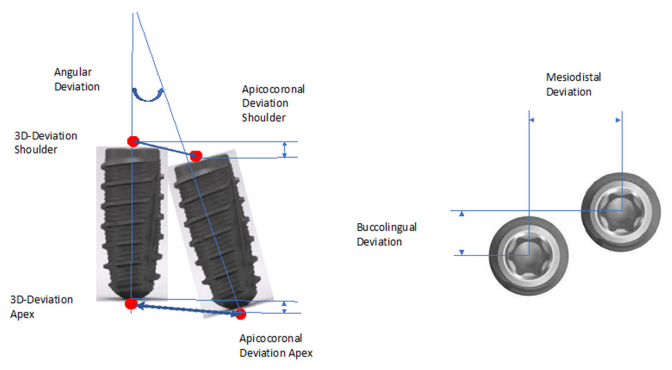
2.7. Analysis of the Implant Position

The metric analysis included the following measurements (Figure 1). The deviations between the planned and the actual implant position were measured. Length measurements were taken from the center of the implant at the shoulder and at the apex.
  • Three-dimensional (3D) deviation.
  • Two-dimensional deviations in apicocoronal direction (height deviation) and mesiodistal and buccolingual direction.
Axis deviation: The measurement method was based on the principle of Tahmaseb et al. [13] to enable better comparability with current and future studies.

2.8. Statistical Analysis

The mean values, standard errors, and 95% confidence intervals were given for the variables. After testing for normal distribution, statistical testing according to the Kruskal-Wallis test was performed for values that were not normally distributed. Given the exploratory nature of this study, all statistical results must be interpreted as hypothesis-generating and not confirmatory. All statistical tests had a significance level of α = 0.05 (two-sided test). No adjustments were made for the multiple tests. Statistical analysis was performed using IBM SPSS Statistics Version 26.0 (IBM Corp. Released 2019, Armonk, NY, USA).

3. Results

Description of the Study Population

Of the 52 patients, 34 were women and 18 were men. The average age was 50.3 years (range, 22–75 years). The distribution of tooth regions is shown in Table 1.
The lengths and diameters of the placed implants are listed in Table 2.
The assignment and overlay of the data records were performed using clearly identifiable hard tooth substances. When assigning the interface data records from the planned implant position and the clinically achieved implant position, the mean RMS error was 85.6 µm (SD 25.4).
The evaluation data of all 52 patients are shown in Table 3.
The evaluation of all implants showed a mean angular deviation of 8.7° and a 3D deviation of 1.62 mm at the shoulder. The distribution of the directional deviations at the prosthetically relevant exit point showed that 59.6% of the implants were placed considerably distal from the planned position. In the buccolingual direction, 53.8% of the implants were placed further lingual, and in the apicocoronal direction, 55.8% of the implants were placed higher than planned.
Data were analyzed with regard to implant placement regions. The results for the upper and lower jaws (Table 4) were compared. Another analysis examined the differences in the premolar and molar regions (Table 5). An evaluation of the sides (right/left) was also performed (Table 6). Another factor investigated was whether the timing of implant placement (early (Type 3)/late (Type 4)) had an influence on the variables (Table 7).
There were no significant differences in the premolar vs. molar and right side vs. left side factors. However, the jaws had a significant impact on the accuracy of the mesiodistal deviation. A higher number of deviations was observed in the lower jaw.
The timing of implant placement also had a significant influence on accuracy. Early implant placements showed higher deviations in both the angle and mesiodistal deviation than were found with late implant placement.

4. Discussion

An in vitro study by Jorba-Garcia et al. demonstrated that the freehand method was significantly less accurate compared to a dynamically guided method [14]. This particularly affected the angular deviation, which averaged 1.6° for dynamic navigation and 9.7° for freehand navigation. Similar values were found in the in vitro study by Chen et al., where the angular deviation at the implant shoulder was 4.45° ± 1.97° with the guided procedure. In contrast, the angular deviation in the freehand procedure was 9.26° ± 3.62° [15]. These values were significantly different. The limits of freehand implantation are clearly marked from these studies. This is an indication that computer-assisted procedures should be preferred for specific indications.
In a cadaver study using the freehand method, deviations at the implant shoulder averaged 1.43 mm (range: 0.65–2.31 mm), 2.20 mm at the apex (range: 1.01–4.02 mm) with an angular deviation of 6.78° (range: 3.08–14.98°) [16]. Another cadaver investigation was conducted by Noharet et al. [17]. A comparison of the in vitro studies with the clinical studies shows that an “implant surgery” can be carried out much more precisely on a model or on the cadaver.
In a clinical study by Vercruyssen et al., significantly higher values were found [8]. Various template-guided methods were examined in comparison with the freehand method. With the freehand method, there were deviations on the implant shoulder averaging 2.77 mm (range: 0.33–8.34 mm), 2.91 mm at the apex (range 0.53–7.37 mm), and an angular deviation of 9.92° (range: 1.45–27.76°). Varga et al., in their clinical study of freehand implant bed preparation, found deviations at the implant shoulder averaging 1.82 mm (range: 0.56–5.38 mm), 2.43 mm at the apex (range 0.54–4.83 mm), and an angular deviation of 7.03° (range: 0.71–21.30°) [10]. Further clinical studies constantly showed significant differences in all parameters of accuracy and a large range between the minimum and maximum values [6,10,18,19]. In particular, a high number of deviations in the angular dimensions have been found in clinical studies. For example, Aydemir and Arisan found a mean deviation of 10.04° with a range of 2.19–20.42° when using the freehand method [20]. All studies show sufficiently accurate values in the mean of the deviations. However, they all confirm a high degree of scatter in the maximum values. These maximum values are worth considering from both a surgical and a prosthetic point of view. Therefore, a determination of the influencing factors appears helpful. In the present study, tooth-delimited single-tooth edentulous sites were restored with implants. Smitkarn et al. examined the accuracy of implant placement in single-tooth edentulous sites. With respect to angular deviation, they achieved a mean value of 6.9° with a range of 0.5–16.9°. The values for the lateral and 3D deviation were within the range of the present study (mesiodistal mean value: 0.6 mm (range 0.03–1.67 mm), buccolingual mean value: 0.5 mm (range 0.00–2.14 mm), apicocoronal mean value 1.0 mm (range 0.03–3.95 mm), and 3D deviation mean value of 1.5 mm (range 0.39–4.03 mm)) [21]. In contrast to the studies mentioned above, Efstathiou et al. showed only minor axial deviations in freehand implantation [22]. However, they performed their measurements two-dimensionally after evaluation of panoramic radiographs. On the one hand, this could be an indication of the influence of the measurement procedure. On the other hand, this could also be a confirmation that the surgeons’ skills in spatially correct implant positioning are the decisive influencing factor.
The values observed in this study, as well as the present study, elucidate that the ranges, in particular, have a decisive influence on prosthetic sustainability in supposedly simple situations. The patients who exhibited high deviation values in the present study were subsequently traced back. It was found that these patients, in particular, had restricted mouth openings or demonstrated little cooperation during the operation. This was observed in addition to confirming the hypothesis that accuracy is dependent on the anatomical situation and the region of implantation. The accuracy that can be achieved with freehand implant placement depends significantly on the surgeon’s experience. In an in vitro study, the freehand method performed by an experienced implant surgeon showed angular deviations with an average of 6.69° compared to an inexperienced surgeon (12.66°) [14]. Even with the use of a drilling template, the experience of a surgeon had a significant influence on the outcome [23].
Comparing the present study with template-guided implant placement minimizes the surgeon’s experience and individual skills as influencing factors. In a study of 122 implants, the mean values of the deviations at the shoulder were 1.2 mm (95% CI 0.1–3.8 mm), 1.8 mm at the apex (95% CI 0.35–0.1 mm), and an angular deviation of 4.8° (95% CI 0.02–14.6°) [24]. In a modified template design with two sleeve guides, the mean values at the shoulder were 0.52 mm (95% CI 0.37–0.67 mm), 0.82 mm at the apex (0.56–1.08 mm) and the angular deviation was 2.8° (2.18–3.51°) [25].
In a study by Vercruyssen et al., there was no difference in patient satisfaction and postoperative pain following implant placement when the free hand and template-guided procedures were compared [26]. However, with regard to postoperative pain and swelling, Pozzi et al. found significantly higher values with the freehand method than with template-guided implant placement [27]. It is known that the presence of an adjacent tooth and the quadrant and the location of the implant site influences the direction and angulation deviation of the implant position [4,28]. This is partially confirmed in the present study. In addition, the influence of the implantation time was found here as a further influencing factor. One explanation could be the degree of mineralization of the newly formed bone and thus its density.
In the present study, only implantations without additional bone augmentation were included. Under the aspect that the implant position can be modified by the possibility of bone augmentation, new influencing factors may arise [29].
Sun et al. showed that the location of implantation had a significant influence on the accuracy of the 3D deviation and angular deviation [19]. This enabled the achievement of higher levels of accuracy in the upper jaw and anterior regions. Implant location exhibited a significant influence on accuracy in an in vitro study by Abduo and Lau. In a comparison between an implant placed in the central incisor area and the first molar area, a lower level of accuracy was measured in molar area for horizontal deviation at the implant shoulder and at the apex of the distal implant [30]. In our study, the hypothesis that the location of the implant has an influence on accuracy could not be confirmed. Arisan et al. presented the limits of freehand implant placement when restoring edentulous jaws. When assessing the implant position using the freehand method, 54.65% of the implants were classified as inadequately positioned in comparison to a mucosa-supported drilling template, where this value was reduced to 15.78% [31].
The ultimate influence of the implant placement procedure, freehand or computer-assisted implant placement, has hardly been worked out so far. In a case series, Baldi et al. could not find any differences in the restoration of maxillary anterior teeth with these two different procedures [32]. They see the advantages of the digital approach in the fact that a consistent prosthetic workflow can be maintained, for example with long-term provisional restorations fabricated in advance.
It has been demonstrated that successful implantation does not depend on the surgical procedure [27]. After an observation period of 3 years, Bernard et al. found no significant differences between template-guided and freehand placement of implants [33]. Pozzi et al. could not determine any differences between freehand and template-guided implant placements in a one-year follow-up study. Among other variables, they examined the implant survival rate, complications, and peri-implant bone loss [27]. The meta-analysis by Yogui and coworkers confirms the statement that there are no significant differences in marginal bone loss, mechanical and biological complications, and implant survival rate between these two methods [34]. However, it could be determined that the conventional surgical method showed significantly higher levels of discomfort in patients, such as pain and swelling than in computer-assisted procedures [35]. This is also reflected in the patient-related outcome measures. The majority of patients prefer a computer-assisted approach as found by Sancho-Puchades et al. in their randomized clinical trial [36].

5. Conclusions

Freehand implant placement is a proven procedure. Studies have shown that implant survival rates do not depend on the surgical method. In the present study, it was shown that the optimal implant position could not be adequately achieved in every case using the freehand method. If there is a significant discrepancy between the achieved position and the optimal planned implant position in terms of angular deviation and implant emergence point, a predictable restoration is questionable from a prosthetic point of view. Within the limitations of the study, it has been shown that larger deviations are possible in difficult anatomical conditions or less accessibility to the surgical site. The use of computer-assisted surgical procedures can, therefore, be an alternative to minimize such deviations and can be recommended from the perspective of prosthetic rehabilitation.

Author Contributions

S.S., C.E., and H.R. contributed to the conception and design of the study, data analysis, and interpretation, and drafted the manuscript; S.S. and M.W. contributed to data acquisition, analysis, and interpretation of the data and critically revised the manuscript and drafted the manuscript; S.S. and H.R. contributed to the statistical analysis, data analysis, and data interpretation; C.E., H.R., and R.G.L. critically revised the manuscript and drafted the manuscript. All authors gave final approval and agreed to be accountable for all aspects of the work. All authors have read and agreed to the published version of the manuscript.

Funding

This study was supported by the Oral Reconstruction Foundation (formerly the Camlog Foundation), Grant No. CF 41.506. A major part of the research was self-funded by the authors and their institutions. The study design, data collection, and data analysis, as well as the writing of the manuscript, were carried out completely independently by the authors.

Institutional Review Board Statement

The study was conducted according to the guidelines of the Declaration of Helsinki and approved by the Ethics Committee of Ulm University (Application No. 41/14 approved on 6 May 2014).

Informed Consent Statement

Informed consent was obtained from all subjects involved in the study.

Data Availability Statement

The complete documentation of all patients enrolled in this study belongs to the authors and is available only upon reasonable request.

Conflicts of Interest

The authors declare no conflict of interest. The funders had no role in the design of the study; in the collection, analyses, or interpretation of data; in the writing of the manuscript, or in the decision to publish the results.

References

  1. Vercruyssen, M.; Fortin, T.; Widmann, G.; Jacobs, R.; Quirynen, M. Different techniques of static/dynamic guided implant surgery: Modalities and indications. Periodontol 2000 2014, 66, 214–227. [Google Scholar] [CrossRef] [PubMed]
  2. D’Haese, J.; Ackhurst, J.; Wismeijer, D.; De Bruyn, H.; Tahmaseb, A. Current state of the art of computer-guided implant surgery. Periodontol 2000 2017, 73, 121–133. [Google Scholar] [CrossRef] [PubMed]
  3. Cattoni, F.; Chirico, L.; Merlone, A.; Manacorda, M.; Vinci, R.; Gherlone, E.F. Digital Smile Designed Computer-Aided Surgery versus Traditional Workflow in “All on Four” Rehabilitations: A Randomized Clinical Trial with 4-Years Follow-Up. Int. J. Environ. Res. Public Health 2021, 18, 3449. [Google Scholar] [CrossRef] [PubMed]
  4. Choi, W.; Nguyen, B.C.; Doan, A.; Girod, S.; Gaudilliere, B.; Gaudilliere, D. Freehand Versus Guided Surgery: Factors Influencing Accuracy of Dental Implant Placement. Implant Dent. 2017, 26, 500–509. [Google Scholar] [CrossRef]
  5. Behneke, A.; Burwinkel, M.; Behneke, N. Factors influencing transfer accuracy of cone beam CT-derived template-based implant placement. Clin. Oral Implant Res. 2012, 23, 416–423. [Google Scholar] [CrossRef]
  6. Nickenig, H.J.; Wichmann, M.; Hamel, J.; Schlegel, K.A.; Eitner, S. Evaluation of the difference in accuracy between implant placement by virtual planning data and surgical guide templates versus the conventional free-hand method-a combined in vivo-in vitro technique using cone-beam CT (Part II). J. Craniomaxillofac. Surg. 2010, 38, 488–493. [Google Scholar] [CrossRef]
  7. Vercruyssen, M.; Coucke, W.; Naert, I.; Jacobs, R.; Teughels, W.; Quirynen, M. Depth and lateral deviations in guided implant surgery: An RCT comparing guided surgery with mental navigation or the use of a pilot-drill template. Clin. Oral Implant Res. 2015, 26, 1315–1320. [Google Scholar] [CrossRef]
  8. Vercruyssen, M.; Cox, C.; Coucke, W.; Naert, I.; Jacobs, R.; Quirynen, M. A randomized clinical trial comparing guided implant surgery (bone- or mucosa-supported) with mental navigation or the use of a pilot-drill template. J. Clin. Periodontol. 2014, 41, 717–723. [Google Scholar] [CrossRef]
  9. Tahmaseb, A.; Wu, V.; Wismeijer, D.; Coucke, W.; Evans, C. The accuracy of static computer-aided implant surgery: A systematic review and meta-analysis. Clin. Oral Implant Res. 2018, 29 (Suppl. 16), 416–435. [Google Scholar] [CrossRef] [Green Version]
  10. Varga, E., Jr.; Antal, M.; Major, L.; Kiscsatari, R.; Braunitzer, G.; Piffko, J. Guidance means accuracy: A randomized clinical trial on freehand versus guided dental implantation. Clin. Oral Implant Res. 2020, 31, 417–430. [Google Scholar] [CrossRef] [Green Version]
  11. Gallucci, G.O.; Hamilton, A.; Zhou, W.; Buser, D.; Chen, S. Implant placement and loading protocols in partially edentulous patients: A systematic review. Clin. Oral Implant Res. 2018, 29 (Suppl. 16), 106–134. [Google Scholar] [CrossRef] [Green Version]
  12. Schnutenhaus, S.; Edelmann, C.; Rudolph, H.; Luthardt, R.G. Retrospective study to determine the accuracy of template-guided implant placement using a novel nonradiologic evaluation method. Oral Surg. Oral Med. Oral Pathol. Oral Radiol. 2016, 121, e72–e79. [Google Scholar] [CrossRef]
  13. Tahmaseb, A.; Wismeijer, D.; Coucke, W.; Derksen, W. Computer technology applications in surgical implant dentistry: A systematic review. Int. J. Oral Maxillofac. Implant 2014, 29, 25–42. [Google Scholar] [CrossRef] [Green Version]
  14. Jorba-Garcia, A.; Figueiredo, R.; Gonzalez-Barnadas, A.; Camps-Font, O.; Valmaseda-Castellon, E. Accuracy and the role of experience in dynamic computer guided dental implant surgery: An in-vitro study. Med. Oral Patol. Oral Cir. Bucal 2019, 24, e76–e83. [Google Scholar] [CrossRef]
  15. Chen, C.K.; Yuh, D.Y.; Huang, R.Y.; Fu, E.; Tsai, C.F.; Chiang, C.Y. Accuracy of Implant Placement with a Navigation System, a Laboratory Guide, and Freehand Drilling. Int. J. Oral Maxillofac. Implant 2018, 33, 1213–1218. [Google Scholar] [CrossRef]
  16. Chen, Z.; Li, J.; Sinjab, K.; Mendonca, G.; Yu, H.; Wang, H.L. Accuracy of flapless immediate implant placement in anterior maxilla using computer-assisted versus freehand surgery: A cadaver study. Clin. Oral Implant Res. 2018, 29, 1186–1194. [Google Scholar] [CrossRef]
  17. Noharet, R.; Pettersson, A.; Bourgeois, D. Accuracy of implant placement in the posterior maxilla as related to 2 types of surgical guides: A pilot study in the human cadaver. J. Prosthet. Dent. 2014, 112, 526–532. [Google Scholar] [CrossRef]
  18. Schneider, D.; Sancho-Puchades, M.; Mir-Mari, J.; Muhlemann, S.; Jung, R.; Hammerle, C. A Randomized Controlled Clinical Trial Comparing Conventional and Computer-Assisted Implant Planning and Placement in Partially Edentulous Patients. Part 4: Accuracy of Implant Placement. Int. J. Periodont. Restor. Dent. 2019, 39, e111–e122. [Google Scholar] [CrossRef]
  19. Sun, T.M.; Lee, H.E.; Lan, T.H. Comparing Accuracy of Implant Installation with a Navigation System (NS), a Laboratory Guide (LG), NS with LG, and Freehand Drilling. Int. J. Environ. Res. Public Health 2020, 17, 2107. [Google Scholar] [CrossRef] [Green Version]
  20. Aydemir, C.A.; Arisan, V. Accuracy of dental implant placement via dynamic navigation or the freehand method: A split-mouth randomized controlled clinical trial. Clin. Oral Implant Res. 2020, 31, 255–263. [Google Scholar] [CrossRef]
  21. Smitkarn, P.; Subbalekha, K.; Mattheos, N.; Pimkhaokham, A. The accuracy of single-tooth implants placed using fully digital-guided surgery and freehand implant surgery. J. Clin. Periodontol. 2019, 46, 949–957. [Google Scholar] [CrossRef] [PubMed]
  22. Efstathiou, A.; Machtei, E.E.; Zigdon-Giladi, H.; Gutmacher, Z.; Horwitz, J. The effect of a surgeon’s position on the axial inclination of dental implants placed freehand: A single-blind study. Quintessence Int. 2021. [Google Scholar] [CrossRef]
  23. Marei, H.F.; Abdel-Hady, A.; Al-Khalifa, K.; Al-Mahalawy, H. Influence of surgeon experience on the accuracy of implant placement via a partially computer-guided surgical protocol. Int. J. Oral Maxillofac. Implant 2019, 34, 1177–1183. [Google Scholar] [CrossRef] [PubMed]
  24. Schnutenhaus, S.; Edelmann, C.; Rudolph, H.; Dreyhaupt, J.; Luthardt, R.G. 3D accuracy of implant positions in template-guided implant placement as a function of the remaining teeth and the surgical procedure: A retrospective study. Clin. Oral Investig. 2018, 22, 2363–2372. [Google Scholar] [CrossRef] [PubMed]
  25. Schnutenhaus, S.; von Koenigsmarck, V.; Blender, S.; Ambrosius, L.; Luthardt, R.G.; Rudolph, H. Precision of sleeveless 3D drill guides for insertion of one-piece ceramic implants: A prospective clinical trial. Int. J. Comput. Dent. 2018, 21, 97–105. [Google Scholar] [PubMed]
  26. Vercruyssen, M.; De Laat, A.; Coucke, W.; Quirynen, M. An RCT comparing patient-centred outcome variables of guided surgery (bone or mucosa supported) with conventional implant placement. J. Clin. Periodontol. 2014, 41, 724–732. [Google Scholar] [CrossRef] [PubMed]
  27. Pozzi, A.; Tallarico, M.; Marchetti, M.; Scarfo, B.; Esposito, M. Computer-guided versus free-hand placement of immediately loaded dental implants: 1-year post-loading results of a multicentre randomised controlled trial. Eur. J. Oral Implantol. 2014, 7, 229–242. [Google Scholar]
  28. Tang, T.; Huang, Z.; Liao, L.; Gu, X.; Zhang, J.; Zhang, X. Factors that Influence Direction Deviation in Freehand Implant Placement. J. Prosthodont. 2019, 28, 511–518. [Google Scholar] [CrossRef]
  29. Sicilia, A.; Botticelli, D.; Working, G. Computer-guided implant therapy and soft- and hard-tissue aspects. The Third EAO Consensus Conference 2012. Clin. Oral Implant Res. 2012, 23 (Suppl. 6), 157–161. [Google Scholar] [CrossRef]
  30. Abduo, J.; Lau, D. Accuracy of static computer-assisted implant placement in anterior and posterior sites by clinicians new to implant dentistry: In vitro comparison of fully guided, pilot-guided, and freehand protocols. Int. J. Implant Dent. 2020, 6, 10. [Google Scholar] [CrossRef] [Green Version]
  31. Arisan, V.; Karabuda, C.Z.; Mumcu, E.; Ozdemir, T. Implant positioning errors in freehand and computer-aided placement methods: A single-blind clinical comparative study. Int. J. Oral Maxillofac. Implant 2013, 28, 190–204. [Google Scholar] [CrossRef] [Green Version]
  32. Baldi, D.; Colombo, J.; Motta, F.; Motta, F.M.; Zillio, A.; Scotti, N. Digital vs. Freehand Anterior Single-Tooth Implant Restoration. BioMed Res. Int. 2020, 2020, 4012127. [Google Scholar] [CrossRef]
  33. Bernard, L.; Vercruyssen, M.; Duyck, J.; Jacobs, R.; Teughels, W.; Quirynen, M. A randomized controlled clinical trial comparing guided with nonguided implant placement: A 3-year follow-up of implant-centered outcomes. J. Prosthet. Dent. 2019, 121, 904–910. [Google Scholar] [CrossRef]
  34. Yogui, F.C.; Verri, F.R.; de Luna Gomes, J.M.; Lemos, C.A.A.; Cruz, R.S.; Pellizzer, E.P. Comparison between computer-guided and freehand dental implant placement surgery: A systematic review and meta-analysis. Int. J. Oral Maxillofac. Surg. 2021, 50, 242–250. [Google Scholar] [CrossRef]
  35. Ozden Yuce, M.; Gunbay, T.; Guniz Baksi, B.; Comlekoglu, M.; Mert, A. Clinical benefits and effectiveness of static computer-aided implant surgery compared with conventional freehand method for single-tooth implant placement. J. Stomatol. Oral Maxillofac. Surg. 2020, 121, 534–538. [Google Scholar] [CrossRef]
  36. Sancho-Puchades, M.; Alfaro, F.H.; Naenni, N.; Jung, R.; Hammerle, C.; Schneider, D. A Randomized Controlled Clinical Trial Comparing Conventional And Computer-Assisted Implant Planning and Placement in Partially Edentulous Patients. Part 2: Patient Related Outcome Measures. Int. J. Periodont. Restor. Dent. 2019, 39, e99–e110. [Google Scholar] [CrossRef]
Figure 1. Representation of the measuring distances and the angular deviation between the planned and actual achieved implant position.
Dentistry 09 00054 g001
Table 1. Distribution of the implants according to regions and tooth groups (premolars or molars).
Right (N = 22)Left (N = 30)
Upper jaw (N = 20)Premolars: 6
Molars: 1
Premolars: 8
Molars: 5
Lower jaw (N = 32)Premolars: 3
Molars: 12
Premolars: 7
Molars: 10
Table 2. Distribution of implant lengths and diameters (mm).
Lengths/Diameters3.84.45.0
9596
1111118
13110
Table 3. Angular deviation (°) and deviations at the shoulder and at the apex (mm) in the total group with 52 implants.
MeanStandard DeviationMinimumMaximum
AngleDeviation8.704.81.822.3
Shoulder3D deviation 1.620.870.133.57
Mesiodistal deviation0.870.750.003.13
Buccolingual
deviation
0.700.660.002.96
Apicocoronal deviation0.950.610.002.32
Apex3D deviation 2.681.520.487.12
Mesiodistal deviation1.931.510.096.51
Buccolingual
deviation
1.101.030.154.36
Apicocoronal deviation1.050.720.412.82
Table 4. Deviations between the planned and achieved implant position according to the jaws. Upper jaw (N = 20) lower jaw (N = 32). Angle variance (°). Linear deviation (mm). Kruskal–Wallis test with a significance level of p < 0.05.
Upper JawLower Jaw
MeanSE (95% CI)MeanSE (95% CI)Sig. Level
AngleDeviation6.980.89 (5.11–8.84)9.770.90 (7.94–11.60)0.050
ShoulderMesiodistal deviation0.540.13 (0.27–0.82)1.070.14 (0.80–1.35)0.011 *
Buccolingual
Deviation
0.710.14 (0.41–1.01)0.690.12 (0.44–0.94)0.742
Apicocoronal Deviation0.970.14 (0.67–1.27)0.940.10 (0.72–1.15)0.903
ApexMesiodistal deviation1.360.27 (0.81–1.92)2.280.28 (1.70–2.85)0.038 *
Buccolingual
deviation
1.270.22 (0.80–1.74)0.990.19 (0.61–1.37)0.140
Apicocoronal deviation1.010.16 (0.67–1.34)1.080.13 (0.82–1.35)0.645
* indicates statistical significance.
Table 5. Deviations between the planned and achieved implant position depending on the tooth region. Premolar (N = 24) Molar (N = 28). Angle variance (°). Linear deviation (mm). Kruskal–Wallis test with a significance level of p < 0.05.
PremolarMolar
MeanSE (95% CI)MeanSE (95% CI)Sig. Level
AngleDeviation7.430.76 (5.86–8.99)9.781.04 (7.66–11.91)0.163
ShoulderMesiodistal deviation0.680.12 (0.44–0.92)1.030.16 (0.70–1.36)0.169
Buccolingual
deviation
0.700.13 (0.43–0.96)0.700.13 (0.43–0.97)0.963
Apicocoronal deviation1.020.12 (0.77–1.28)0.890.12 (0.65–1.12)0.354
ApexMesiodistal deviation1.470.25 (0.97–1.98)2.310.31 (1.67–2.95)0.077
Buccolingual
deviation
1.300.21 (0.87–1.73)0.930.19 (0.53–1.32)0.100
Apicocoronal deviation1.040.13 (0.76–1.31)1.070.15 (0.76–1.37)0.833
Table 6. Deviations between the planned and achieved implant position depending on the sides. Right side (N = 24) Left side (N = 28). Angle variance (°). Linear deviation (mm). Kruskal–Wallis test with a significance level of p < 0.05.
Right SideLeft Side
MeanSE (95% CI)MeanSE (95% CI)Sig. Level
AngleDeviation8.781.08 (6.54–11.02)8.620.85 (6.87–10.37)0.854
ShoulderMesiodistal deviation0.770.15 (0.45–1.09)0.950.14 (0.37–1.24)0.364
Buccolingual
deviation
0.680.12 (0.44–0.92)0.710.14 (0.43–1.00)0.755
Apicocoronal deviation1.040.10 (0.83–1.25)0.870.13 (0.61–1.14)0.088
ApexMesiodistal deviation1.790.32 (1.14–2.45)2.040.28 (1.46–2.62)0.474
Buccolingual
deviation
1.190.25 (0.67–1.71)1.020.16 (0.69–1.34)0.941
Apicocoronal deviation1.120.12 (0.88–1.37)0.990.16 (0.67–1.31)0.169
Table 7. Deviations between the planned and achieved implant position depending on the timing of implant placement. Early implantation (N = 21) Late implantation (N = 31). Angle variance (°). Linear deviation (mm). Kruskal–Wallis test with a significance level of p < 0.05.
Early Implant PlacementLate Implant Placement
MeanSE (95% CI)MeanSE (95% CI)Sig. Level
AngleDeviation11.031.05 (8.85–13.22)7.110.76 (5.55–8.67)0.005 *
ShoulderMesiodistal deviation1.060.15 (0.76–1.36)0.740.14 (0.45–1.03)0.050
Buccolingual
deviation
0.860.18 (0.49–1.24)0.590.09 (0.40–1.24)0.275
Apicocoronal Deviation1.080.14 (0.79–1.37)0.861.04 (0.65–1.07)0.182
ApexMesiodistal deviation2.620.30 (1.99–3.24)1.460.26 (0.63–1.98)0.003 *
Buccolingual
deviation
1.410.29 (0.80–2.01)0.890.13 (0.62–1.16)0.309
Apicocoronal deviation1.270.18 (090–1.64)0.910.11 (0.68–1.14)0.081
* indicates statistical significance.
Publisher’s Note: MDPI stays neutral with regard to jurisdictional claims in published maps and institutional affiliations.

