Article

A Colorimetric Sensor Enabled with Heterogeneous Nanozymes with Phosphatase-like Activity for the Residue Analysis of Methyl Parathion

College of Chemistry and Materials Engineering, Beijing Technology and Business University, Beijing 100048, China
*
Author to whom correspondence should be addressed.
Submission received: 20 June 2023 / Revised: 28 July 2023 / Accepted: 31 July 2023 / Published: 7 August 2023
(This article belongs to the Special Issue Food Contaminant Detection, Analysis and Risk Assessment)

Abstract

:
In this study, a colorimetric sensor was developed for the detection of organophosphorus pesticides (OPs) using a heterogeneous nanozyme with phosphatase-like activity. Herein, this heterogeneous nanozyme (Au-pCeO2) was obtained by the modification of gold nanoparticles on porous cerium oxide nanorods, resulting in synergistic hydrolysis performance for OPs. Taking methyl parathion (MP) as the target pesticide, the catalytic performance and mechanism of Au-pCeO2 were investigated. Based on the phosphatase-like Au-pCeO2, a dual-mode colorimetric sensor for MP was put forward by the analysis of the hydrolysis product via a UV-visible spectrophotometer and a smartphone. Under optimum conditions, this dual-mode strategy can be used for the on-site analysis of MP with concentrations of 5 to 200 μM. Additionally, it can be applied for MP detection in pear and lettuce samples with recoveries ranging from 85.27% to 115.87% and relative standard deviations (RSDs) not exceeding 6.20%, which can provide a simple and convenient method for OP detection in agricultural products.

Graphical Abstract

1. Introduction

Pesticides are a group of chemicals used to regulate plant growth and prevent pests, diseases, and weeds, which are the main chemical hazards to the quality and safety of agricultural products [1,2,3,4]. One of the main categories is organophosphorus pesticides (OPs), which are organic compounds containing phosphorus, mainly phosphate or thiophosphate, and are widely used in the growth of crops because of their broad spectrum and high efficiency [5,6,7]. When OPs are applied to crops, they will inevitably accumulate in the environment and in organisms. Studies show that these residues can accumulate through the food chain and inhibit the activity of cholinesterase in the body, thus causing symptoms in the central nervous system and posing a threat to human health [8,9,10]. Up until now, OPs have been reported as the main pollutants in water and plants [11,12]. Therefore, it is of great significance to detect OP residues for human safety and environmental protection.
In recent years, various methods based on gas chromatography [13], chromatography–mass spectrometry, and other confirmatory techniques have been applied for OP detection [14,15]. Although accurate and sensitive, the above methods are time-consuming and require specialist personnel to operate, which cannot meet the demand for rapid detection. As an important complement, biosensors have the advantage of being fast, field detectable, and easy to miniaturize, making rapid and on-site analysis for OPs possible [16,17,18]. Currently, various biosensors, including colorimetric [19], fluorescent immunoassay [20], and electrochemical biosensors [18,21], have demonstrated excellent analytical performance in the detection of OPs. However, most biosensors are based on the reaction between biological enzymes and OPs, such as acetylcholinesterase or organophosphorus hydrolases [22,23]. Despite their high specificity, these biological enzymes are expensive, difficult to prepare, and have poor stability [24,25,26]. Therefore, it is necessary to develop biomimetic recognition elements to broaden the practical application of biosensors.
Recently, with the development of nanotechnology, nanomaterials have opened new possibilities for wide application and marketization [27]. Due to their high stability, ease of synthesis, low cost, and high enzyme-like activity, nanozymes have a wide range of applications in environmental and food safety. In recent years, nanozymes have been used in electrochemical or optical sensors to analyze OP residues by exploiting their unique phosphatase [28] or peroxidase [29,30] activity. Although they have high sensitivity, peroxidase-like nanozymes commonly require the involvement of a substrate, such as H2O2, which may have autolysis and high toxicity [31]. Meanwhile, phosphatase-like nanozymes can hydrolyze OPs into small molecules with low toxicity directly, making catalysis simpler and more convenient. Recently, various phosphatase-like nanozymes, such as cerium oxide (CeO2) [32], porous hydroxy zirconium oxide (ZrOX-OH) [33], and zeolitic imidazolate frameworks (ZIFs) [34], have been explored. Among them, CeO2 nanozymes, including nanospheres, nanorods, and nanocubes, are inexpensive and environmentally friendly and have been regarded as prospective artificial phosphatases owing to the presence of Ce3+ and Ce4+ species on their surface [32,33]. However, the low specific surface area and weak adsorption ability may limit their catalytic performance. To address these issues, researchers have adopted the hybridization strategy to obtain higher catalytic activity [35,36,37]. Meanwhile, noble metals such as gold, platinum, and palladium are getting a lot of attention. As favorable catalysts, they can exhibit multienzyme activity, such as peroxidase-like activity and oxidase-like activity [26,29,38]. Therefore, the modification of noble metals on the CeO2 nanozyme may have a synergistic catalytic effect on the hydrolysis of OPs.
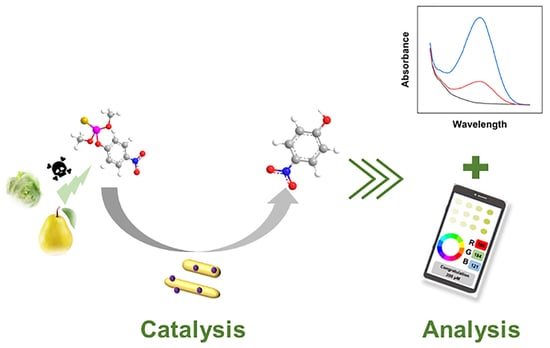
Inspired by this, we developed a heterogeneous nanozyme with phosphatase-like activity. Based on the hydrothermal method, the CeO2 nanorods (denoted as CeO2) were synthesized. After the calcination treatment, the porous CeO2 (denoted as pCeO2) nanozyme was obtained with higher phosphatase-like activity. In order to further improve the catalytic performance, gold nanoparticles (AuNPs) were modified on the substrate of pCeO2 to form the Au-pCeO2 nanozyme. Taking methyl parathion (MP) as the model pesticide, the catalytic performance and mechanism of Au-pCeO2 were studied by measuring the hydrolysis product (i.e., p-nitrophenol, p-NP). Due to the high hydrolysis performance, we constructed a Au-pCeO2 nanozyme-enabled colorimetric sensor. This dual-mode colorimetric sensor could be applied for the on-site detection of MP residues via a UV-visible spectrophotometer (UV-Vis) and a smartphone, which can provide a rapid and reliable approach for the detection of OP residues in agricultural products (Figure 1).

2. Materials and Methods

2.1. Reagents and Instruments

Cerium nitrate hexahydrate (Ce(NO3)3·6H2O) and carbendazim were obtained from Shanghai Aladdin Bio-Chem Technology Co., Ltd. (Shanghai, China). Sodium hydroxide, ethanol, and ethylene glycol (EG) were obtained from Beijing MREDA Technology Co., Ltd. Tris-HCl solution was obtained from Shanghai Sangon Biotech Co., Ltd. (Shanghai, China). MP, paraoxon, phosphoramidite, and monocrotophos were obtained from Alta Scientific Co., Ltd. (Tianjin, China). All other chemicals and reagents used were of analytical grade.
X-ray diffraction (XRD) patterns were obtained using an X’Pert Pro XRD device from XRD-6100 (SHIMADZU, Kyoto, Japan). High-resolution transmission electron microscopy (HRTEM) images were recorded on a Tecnai G2 F30 S-TWIN (FEI, Hillsboro, OR, USA). Raman spectroscopy was obtained with a laser Raman spectrometer (Horiba Jobin Yvon, Kyoto, Japan). To describe the surface electronic states and compositions, a Thermo Fisher Scientific K-Alpha+ X-ray photoelectron spectrometer (XPS) was used.

2.2. Synthesis of Nanozymes

A total of 1.736 g of Ce(NO3)3·6H2O and 19.2 g of NaOH were dissolved in 10 and 70 mL of deionized water, respectively, and then mixed and stirred in a glass vial for 30 min. After that, the mixture was transferred to an automatic temperature-controlled electric furnace and heated in a reaction kettle at 100 °C for 24 h. The obtained materials were cooled naturally to room temperature and then washed with 30 mL water and ethanol alternately by centrifugation until the pH was near neutral. After that, the prepared CeO2 was calcined at 400 °C for 1 h to obtain pCeO2 [35]. A total of 50 mg of pCeO2 powder was then dispersed in 25 mL of EG and sonicated for 30 min to make it homogeneous. A total of 250 μL of NaOH (1 M) in EG and 0.3 mL of chloroauric acid were added and then heated and stored in an oil bath at 140 °C for 3 h [36]. The reaction solution was cooled naturally to room temperature, and the precipitate was washed three times by centrifugation with ultrapure water. After drying in a vacuum oven at 75 °C and grinding to powder with a mortar and pestle, the Au-pCeO2 nanozymes were obtained and stored at room temperature.

2.3. Detection of MP

A total of 10 mg of Au-pCeO2 was first added into a 0.8 mL Tris buffer solution (pH 9.0, 10 mM). After the addition of 200 μL of MP solution with the known concentration, the mixture was reacted at 75 °C for 5 h in a metal bath. Then, the tube was centrifuged for 8 min at 13,523 rcf. The supernatant was then filtered through a 0.22 μm filter membrane and added to the centrifuge tube. Then, 200 μL of the supernatant was transferred into a 96-well plate for UV-Vis detection in a wavelength range of 300–500 nm. The absorbance was recorded at a wavelength of 400 nm. To realize the OP detection by a smartphone, the imaging of the samples was analyzed with the image analysis app “Color Picker” by reading the color information (RGB value) within each well.

2.4. Real Sample Analysis

Pears and lettuces were selected for the real sample analysis. They were purchased from a local supermarket in Beijing. Firstly, these samples were crushed in a blender and then weighed at 1.00 g (accurate to 0.01 g) into a 50 mL centrifuge tube. After that, the known concentration of MP was added to the samples for 0.5 h at room temperature. To extract MP from samples, 5 mL of acetonitrile and 1 g of NaCl were added and vortexed for 5 min. After that, the tube was centrifuged at 2348 rcf for 10 min. To reduce the matrix interference, 25 mg of primary secondary amine (PSA) and 25 mg of octadecyl silane (C18) were added to 1 mL of the supernatant and then vortexed for 3 min. After centrifugation for 10 min (2348 rcf), the obtained supernatant was filtered through a 0.22 μm filter membrane and collected as the extraction solution. The above solution was then blown dry with nitrogen and redissolved with Tris buffer (pH 9.0, 10 mM) for further analysis.

3. Results and Discussions

3.1. Comparison of CeO2, pCeO2, and Au-CeO2 Nanozymes

First of all, the morphology and chemical structure of three nanozymes were compared in this study. As shown in the insert of Figure 2a, the color of CeO2 is dark yellow, and pCeO2 is light yellow. After the addition of the precursor solution and the reduction process, the obtained nanozyme appears black-purple, which is due to the formation of AuNPs on the surface of pCeO2. From the TEM images in Figure S1a, our CeO2 nanozyme appeared to have a rod-like structure. After the high-temperature calcination, the nanozyme had a porous rod-like morphology, indicating the successful preparation of pCeO2 (Figure S1b,c). As shown in Figure S2, the length and width of pCeO2 were 42.91 ± 2.82 nm and 7.04 ± 0.17 nm, respectively.
After that, we analyzed the chemical structure of the nanozymes. The XRD patterns of the CeO2, pCeO2, and Au-pCeO2 nanozymes are shown in Figure 2a. The diffraction peaks of CeO2 were present at 2θ of 28.7°, 32.9°, 47.5°, and 54.5°, which is consistent with the reported literature [37]. After calcination, the peak pattern of pCeO2 was sharper than that of the CeO2 nanorods. After the modification of AuNPs, a weak diffraction peak appeared at 38.4°, indicating the successful preparation of the Au-pCeO2 nanozyme [39]. Moreover, our Au-pCeO2 nanozyme still had a similar crystal structure compared to pCeO2, so our nanozymes have the two features of AuNPs and pCeO2, which could provide the chance for the synergistic effect of heterogeneous nanozymes.
The Raman spectra were used to analyze the structural arrangement of the nanozymes. As shown in Figure 2b, these three nanozymes exhibited a prominent vibration at 455 cm−1, which was caused by the strong Raman-active F2g vibrational mode typical of the CeO2 fluorite structure. Moreover, the peaks at this position were relatively similar for all three nanozymes, indicating the integrality of a rod-like structure. Due to the existence of a defect-induced (D) mode [40], CeO2 and pCeO2 showed a weak peak at 600 cm−1. After the loading with AuNPs, there was a significant change in the peak here, which also indicated AuNPs doping into the CeO2 lattice. It could be further concluded that the AuNPs were present on our prepared Au-pCeO2 nanozyme.
Using MP as the model substrate, the phosphatase-like catalytical performance of CeO2, pCeO2, and Au-pCeO2 for OPs was assessed by the yield of p-NP. A nanozyme with phosphatase-like activity can catalyze the generation of p-NP from MP. According to the p-NP standard curve (Figure S3), we can calculate the yield of p-NP. Here, three parallel experiments were set up. As shown in Figure 2c, the yields obtained for CeO2-, pCeO2-, and Au-pCeO2-catalyzed MP were 19.98% ± 1.25%, 27.16% ± 1.96%, and 45.65% ± 4.93%, respectively. Compared to CeO2, the catalytic performance of pCeO2 was higher, which was due to the surface defects and higher Ce4+ species [41,42]. After the hybridization of AuNPs, the activity of the nanozyme effectively increased, reaching levels twice as high as those of pCeO2. To reveal the catalytic mechanism, the distribution of Ce species on CeO2, pCeO2, and Au-pCeO2 was investigated by chemical valence state analysis (Figure S4a–d). According to Figure S4h and Figure 2d, the proportion of Ce4+ species increased after the calcination treatment for CeO2. This indicates that Ce3+ was shifted to Ce4+ in pCeO2 due to high-temperature (400 °C) calcination, which can increase the hydrolysis ability of the nanozyme [30]. In the Au 4f XPS spectra of Au-pCeO2, double peaks appeared and were assigned to Au 4f7/2 and 4f5/2. The peaks at 83.46 and 87.13 eV could be assigned to Au (Figure S4g). Although the modification of AuNPs can lead to a decrease in Ce3+/Ce4+, the higher catalytic performance of Au-pCeO2 for MP may be attributed to the synergistic catalytic ability of two heterogeneous catalysts.

3.2. Optimization and Characterization of Au-CeO2 Nanozymes

Based on the above, we designed a series of Au-CeO2 nanozymes with different proportions of AuNPs by modulating the addition amount of the precursor solution (50 mM HAuCl4). In three parallel sets of experiments (Figure 3a), with the addition of HAuCl4 solution, the yield of p-NP of the obtained nanozyme increased. However, the catalytic performance of the obtained Au-CeO2 nanozyme became lower as the volume continued to increase. We inferred that was because more AuNPs could aggregate on the surface of pCeO2, which could occupy the active sites of the nanozymes. When the addition volume of HAuCl4 was 0.3 mL, the obtained Au-pCeO2 nanozyme showed higher and more stable catalytic performance with a 44.24% ± 2.62% yield of p-NP. Thus, the optimal addition volume of HAuCl4 was set at 0.3 mL.
After that, the morphology of the optimal Au-pCeO2 nanozyme was characterized using TEM and HRTEM (Figure 3b,c). It is obvious that AuNPs with a size of 10.52 ± 0.30 nm (Figure S2c) had a favorable distribution on the surface of the Au-pCeO2 nanozyme, and pCeO2 still retained a rod-like structure with a clearly visible porous structure. Subsequently, the elemental composition of the Au-CeO2 nanozyme was further confirmed using EDS mapping analysis. As shown in Figure 3d, Au, Ce, and O elements are clearly visible, corresponding to the composition of the Au-CeO2 nanozyme and indicating the successful modification of AuNPs on the surface of pCeO2.

3.3. Catalytic Conditions of Au-pCeO2 Nanozyme

To improve the performance of the assay, the influence of various factors on catalytic activity was studied, including reaction temperature, reaction time, buffer pH, and the amount of Au-pCeO2. The optimum catalytic conditions were investigated by calculating the p-NP yields of MP (200 μM) under different conditions. Firstly, we assessed the influence of reaction temperatures (35, 45, 55, 65, 75, and 85 °C) on the hydrolysis performance of the Au-CeO2 nanozyme (10 mg) with a reaction time of 1 h. As shown in Figure 4a, the reaction temperature had a significant effect on the production of p-NP. With the increase in temperature, the yield of p-NP increased. After the temperature reached 75 °C, the yield of p-NP increased the most. Therefore, we selected 75 °C as the optimum reaction temperature. Then, the yields of p-NP under different reaction time (0.5, 1, 2, 3, 4, 5, and 6 h) were compared at 75 °C. As shown in Figure 4b, a higher p-NP yield was obtained with a reaction time of 5 h. Thus, the optimal reaction time was set to 5 h.
After that, we investigated the effect of pH (6, 7, 8, 9, and 10) on the activity of Au-pCeO2 (10 mg) under the reaction condition of 75 °C for 5 h. From Figure 4c, the absorbance at 400 nm increased when the pH value of Tris buffer tended to be alkaline. This takes place because the absorption peak of p-NP would shift to 310 nm under acid conditions, leading to a weak absorption peak [43]. When the pH value of the buffer solution was 9, the peak of p-NP was the highest. Thus, the optimum pH value of the buffer solution was 9. Finally, the amount of Au-pCeO2 nanozyme was studied under the above optimized conditions. As shown in Figure 4d, upon increasing the amount of Au-pCeO2 from 0 to 10 mg, more p-NP was produced by the hydrolysis action. While the yield of p-NP tends to decrease keeping increasing the amount of Au-pCeO2. We inferred that too many nanozymes had poor dispersion in the reaction system, so the optimal amount of Au-pCeO2 was 10 mg.

3.4. Detection Performance for MP

Under optimal conditions, we developed a Au-pCeO2 nanozyme-based colorimetric sensor and applied it for MP analysis via UV-Vis and a smartphone. First of all, the UV-Vis method was used to investigate the detection performance of our sensor. When the MP concentration was in the range of 5 to 200 μM, the absorbance at 400 nm (A400 nm) had a linear relationship with the concentration of MP (Figure 5a). As shown in Figure 5b, the standard curve was fitted with the MP concentration as the horizontal coordinate and A400 as the vertical coordinate. The linear equation was y = 0.0029x + 0.0821, with a high correlation coefficient (R2 = 0.9984), where y represented A400 and x represented the MP concentration. On the basis of the equation of detection limit (LOD) = 3 Sb/m (Sb is the standard deviation of UV-Vis in the blank experiment and m is the slope of the calibration curve) [33], the LOD of this method is 0.5 μM, which exhibits comparable analytical performance compared to the reported phosphatase-like nanozyme-enabled sensors (Table S1).
In addition, the analytical performance of this sensor was also evaluated on a smartphone, which can be used for the on-site analysis of MP without complex equipment or instruments. By taking a picture of the reaction solution in a 96-well plate, the R/B value was recorded by the “Color Picker” app (Figure 5c and Figure S5). As shown in Figure 5d, the G/B value was also linearly related to the concentration of MP, with a linear equation of y = 0.0025x + 1.0515 (where y is the G/B value and x is the MP concentration) and a correlation coefficient of R2 = 0.9981. Based on these two analytical methods, the dual-model strategy proposed in this study can provide an accurate and reliable method for the on-site detection of MP.

3.5. Selectivity, Interference, Stability, and Recoverability

To investigate the selectivity of the prepared sensor, we compared the hydrolysis ability of Au-pCeO2 for methyl paraoxon, carbendazim, phosphamidon, and monocrotophos with the same concentration of MP (100 μM). As shown in Figure 6a, our nanozyme exhibited high p-NP yields for MP (24.16% ± 4.06%) and methyl paraoxon (65.46% ± 4.07%), while other pesticides produced almost no p-NP, indicating its excellent selectivity for these two OPs. The higher yield of p-NP for catalytic paraoxon may be due to its P=O bond, resulting in its higher toxicity, which is more easily hydrolyzed by the phosphatase [44].
In addition, the anti-interference study of our sensor was evaluated by testing the hydrolysis product (i.e., p-NP) of MP (100 μM) along with different coexisting substances (K+, Na+, and Ca2+ at 20 mM and ascorbic acid, glucose, and glycine at 10 mM). As shown in Figure 6b, the interference was negligible for most of the co-existing substances, except for ascorbic acid. The addition of ascorbic acid may change the original pH of the buffer, which is significantly different from the optimized conditions, thus affecting the yield of p-NP. It is worth noting that the amount of ascorbic acid in most agricultural products is less than 10 mM. Hence, our sensor has good anti-interference performance, making real sample analysis possible.
After that, the storage stability of our Au-pCeO2 nanozyme was investigated by testing the yield of p-NP for MP (100 μM) in five days at room temperature. As can be seen from Figure 6c, Au-pCeO2 retained 89% of the activity after five days compared to the original nanozyme, which demonstrates the satisfactory storage stability of the synthesized Au-pCeO2.
Recoverability is also an important indicator for catalysts in practical applications. For the recoverability study, our nanozyme was used for the hydrolysis of MP (100 μM) under optimal conditions, and then the used Au-pCeO2 was washed three times and dried. The recyclable Au-pCeO2 was then sonicated and dispersed into the buffer for the next five rounds of reaction. As shown in Figure 6d, the yield of p-NP was not less than 17.3% ± 2.08%, indicating our Au-pCeO2 nanozyme can be reused many times, offering economic benefits and convenience.

3.6. Real Sample Analysis

In this study, pears and lettuces were selected as the real samples. Before the analysis, no MP was detected in these samples, allowing them to be further used as blank samples for the recovery experiment. Herein, three spiking levels (25, 50, and 100 μM) were set in the blank samples. The sample pretreatment method was performed in Section 2.4. According to the UV-Vis analysis, the recoveries in the pear samples ranged from 87.47% to 104.83%, with relative standard deviations (RSDs) ranging from 1.69% to 2.69% (n = 3), while the recoveries in the lettuce samples ranged from 93.33% to 103.12%, with RSDs of 1.21% to 2.15% (n = 3) (Table 1). To verify the availability of our dual-mode strategy, the recovery experiment was also assessed by the RGB analysis, which also exhibited similar recoveries ranging from 85.27% to 115.87%, with RSDs of 1.96% to 6.20% for these two samples, demonstrating the favorable availability of our dual-mode strategy for MP detection in agricultural products.

4. Conclusions

In this study, a heterogeneous nanozyme with phosphatase-like activity was developed through the modification of AuNPs on the surface of a pCeO2 structure. Owing to the synergistic catalytic mechanism of these two kinds of nanostructures, our Au-pCeO2 nanozyme appeared to have favorable phosphatase-like activity, which can be used as a biomimetic hydrolase for the degradation of OPs. Based on this nanozyme, we proposed a dual-mode colorimetric sensor for the on-site detection of MP via UV-Vis and a smartphone. Under optimum conditions, this sensor showed a favorable linear relationship with the concentration of MP ranging from 5 μM to 200 µM. In addition, this dual-mode analytical strategy was successfully applied for MP detection in pear and lettuce samples with satisfying recoveries and low RSDs. This dual-mode assay has excellent recoverability, high stability, and good selectivity and does not need complicated equipment or professionals, which can provide a reliable and simple analytical method for OP residue determination in agricultural products.

Supplementary Materials

The following supporting information can be downloaded at: https://0-www-mdpi-com.brum.beds.ac.uk/article/10.3390/foods12152980/s1, Figure S1: TEM images of (a) CeO2, (b,c) pCeO2 at different magnifications; Figure S2: (a) Length and (b) width of pCeO2. (c) Size of AuNPs of Au-pCeO2 nanozyme; Figure S3: A standard curve of p-NP concentration versus absorbance change. Figure S4: XPS spectra of the (a) Ce and (b) O of CeO2, (c) Ce and (d) O of pCeO2, and (e) Ce, (f) O, and (g) Au of Au-pCeO2. (h) Proportions of Ce3+ and Ce4+ species in three nanozymes, which are simulated from XPS data; Figure S5: Digital photo of the hydrolysis product of different concentrations of MP under the action of Au-pCeO2 nanozyme; Table S1: Comparison of various phosphatase-like nanozymes for OP determination [28,32,33,34].

Author Contributions

F.Z.: Conceptualization, supervision, funding acquisition, project administration, and writing—review and editing. M.L.: Methodology, validation, formal analysis, investigation, and writing of the original draft. L.W.: Methodology, validation, and formal analysis. M.W.: Supervision and writing—review and editing. All authors have read and agreed to the published version of the manuscript.

Funding

This research was supported by the National Natural Science Foundation of China (grant number 32202160) and the Research Foundation for Youth Scholars of Beijing Technology and Business University (grant number QNJJ2022-27).

Data Availability Statement

The data used to support the findings of this study can be made available by the corresponding author upon request.

Conflicts of Interest

The authors declare no conflict of interest.

References

  1. Bhattu, M.; Verma, M.; Kathuria, D. Recent advancements in the detection of organophosphate pesticides: A review. Anal. Methods 2021, 13, 4390–4428. [Google Scholar] [CrossRef] [PubMed]
  2. Sidhu, G.K.; Singh, S.; Kumar, V.; Dhanjal, D.S.; Datta, S.; Singh, J. Toxicity, monitoring and biodegradation of organophosphate pesticides: A review. Crit. Rev. Environ. Sci. Technol. 2019, 49, 1135–1187. [Google Scholar] [CrossRef]
  3. Veloo, K.V.; Ibrahim, N.A.S. Analytical Extraction Methods and Sorbents’ Development for Simultaneous Determination of Organophosphorus Pesticides’ Residues in Food and Water Samples: A Review. Molecules 2021, 26, 5495. [Google Scholar] [CrossRef]
  4. Wang, L.; Li, M.; Li, B.; Wang, M.; Zhao, H.; Zhao, F. Electrochemical Sensor Based on Laser-Induced Graphene for Carbendazim Detection in Water. Foods 2023, 12, 2277. [Google Scholar] [CrossRef]
  5. Fu, H.; Tan, P.; Wang, R.; Li, S.; Liu, H.; Yang, Y.; Wu, Z. Advances in organophosphorus pesticides pollution: Current status and challenges in ecotoxicological, sustainable agriculture, and degradation strategies. J. Hazard. Mater. 2022, 424 Pt B, 127494. [Google Scholar] [CrossRef]
  6. Zhao, F.; Wu, J.; Ying, Y.; She, Y.; Wang, J.; Ping, J. Carbon nanomaterial-enabled pesticide biosensors: Design strategy, biosensing mechanism, and practical application. Trends Anal. Chem. 2018, 106, 62–83. [Google Scholar] [CrossRef]
  7. Zhao, F.; Wang, L.; Li, M.; Wang, M.; Liu, G.; Ping, J. Nanozyme-based biosensor for organophosphorus pesticide monitoring: Functional design, biosensing strategy, and detection application. Trends Anal. Chem. 2023, 165, 117152. [Google Scholar] [CrossRef]
  8. Alvarez-Alvarez, I.; Andrade, R.J. Organophosphate pesticides: Another silent liver hazard? Liver Int. 2023, 43, 268–270. [Google Scholar] [CrossRef] [PubMed]
  9. Salazar-Flores, J.; Lomeli-Martinez, S.M.; Ceja-Galvez, H.R.; Torres-Jasso, J.H.; Torres-Reyes, L.A.; Torres-Sanchez, E.D. Impacts of Pesticides on Oral Cavity Health and Ecosystems: A Review. Int. J. Environ. Res. Public Health 2022, 19, 11257. [Google Scholar] [CrossRef]
  10. Fatunsin, O.T.; Oyeyiola, A.O.; Moshood, M.O.; Akanbi, L.M.; Fadahunsi, D.E. Dietary risk assessment of organophosphate and carbamate pesticide residues in commonly eaten food crops. Sci. Afr. 2020, 8, 2468. [Google Scholar] [CrossRef]
  11. Mwevura, H.; Kylin, H.; Vogt, T.; Bouwman, H. Dynamics of organochlorine and organophosphate pesticide residues in soil, water, and sediment from the Rufiji River Delta, Tanzania. Reg. Stud. Mar. Sci. 2021, 41, 101607. [Google Scholar] [CrossRef]
  12. Zhao, F.; Yao, Y.; Li, X.; Lan, L.; Jiang, C.; Ping, J. Metallic Transition Metal Dichalcogenide Nanosheets as an Effective and Biocompatible Transducer for Electrochemical Detection of Pesticide. Anal. Chem. 2018, 90, 11658–11664. [Google Scholar] [CrossRef]
  13. Valenzuela, E.F.; André, L.C.; Menezes, H.C.; Cardeal, Z.L.; Llorent Mart Nez, E.J. Development of a Simple Integrative Carbon Nanomaterial Microextraction Method with GC-MS for Assessing Pesticide Residues in Apples. J. Chem. 2023, 2023, 5561490. [Google Scholar] [CrossRef]
  14. Arjmand, M.; Saghafifar, H.; Alijanianzadeh, M.; Soltanolkotabi, M. A sensitive tapered-fiber optic biosensor for the label-free detection of organophosphate pesticides. Sens. Actuators B Chem. 2017, 249, 523–532. [Google Scholar] [CrossRef]
  15. Song, D.; Li, Y.; Lu, X.; Sun, M.; Liu, H.; Yu, G.; Gao, F. Palladium-copper nanowires-based biosensor for the ultrasensitive detection of organophosphate pesticides. Anal. Chim. Acta 2017, 982, 168–175. [Google Scholar] [CrossRef]
  16. Rodrigues, A.C.M.; Barbieri, M.V.; Chino, M.; Manco, G.; Febbraio, F. A FRET Approach to Detect Paraoxon among Organophosphate Pesticides Using a Fluorescent Biosensor. Sensors 2022, 22, 561. [Google Scholar] [CrossRef] [PubMed]
  17. Lu, X.; Li, Y.; Tao, L.; Song, D.; Wang, Y.; Li, Y.; Gao, F. Amorphous metal boride as a novel platform for acetylcholinesterase biosensor development and detection of organophosphate pesticides. Nanotechnology 2019, 30, 055501. [Google Scholar] [CrossRef]
  18. Hu, H.; Yang, L. Development of enzymatic electrochemical biosensors for organophosphorus pesticide detection. J. Environ. Sci. Health B. 2021, 56, 168–180. [Google Scholar] [CrossRef] [PubMed]
  19. Kaur, J.; Bandyopadhyay, D.; Singh, P.K. A simple and convenient choline oxidase inhibition based colorimetric biosensor for detection of organophosphorus class of pesticides. J. Mol. Liq. 2022, 347, 118258. [Google Scholar] [CrossRef]
  20. Wang, T.; Liu, Y. A lanthanide-based ratiometric fluorescent biosensor for the enzyme-free detection of organophosphorus pesticides. Anal. Methods 2021, 13, 2005–2010. [Google Scholar] [CrossRef]
  21. Zhao, G.; Zhou, B.; Wang, X.; Shen, J.; Zhao, B. Detection of organophosphorus pesticides by nanogold/mercaptomethamidophos multi-residue electrochemical biosensor. Food Chem. 2021, 354, 129511. [Google Scholar] [CrossRef] [PubMed]
  22. Nguyen, H.H.; Lee, S.H.; Lee, U.J.; Fermin, C.D.; Kim, M. Immobilized Enzymes in Biosensor Applications. Materials 2019, 12, 121. [Google Scholar] [CrossRef] [PubMed] [Green Version]
  23. Zhao, F.; He, J.; Li, X.; Bai, Y.; Ying, Y.; Ping, J. Smart plant-wearable biosensor for in-situ pesticide analysis. Biosens. Bioelectron. 2020, 170, 112636. [Google Scholar] [CrossRef] [PubMed]
  24. Arsawiset, S.; Sansenya, S.; Teepoo, S. Nanozymes paper-based analytical device for the detection of organophosphate pesticides in fruits and vegetables. Anal. Chim. Acta 2023, 1267, 341377. [Google Scholar] [CrossRef]
  25. Ge, J.; Yang, L.; Li, Z.; Wan, Y.; Mao, D.; Deng, R.; Zhou, Q.; Yang, Y.; Tan, W. A colorimetric smartphone-based platform for pesticides detection using Fe-N/C single-atom nanozyme as oxidase mimetics. J. Hazard. Mater. 2022, 436, 129199. [Google Scholar] [CrossRef]
  26. Naveen Prasad, S.; Bansal, V.; Ramanathan, R. Detection of pesticides using nanozymes: Trends, challenges and outlook. Trends Anal. Chem. 2021, 144, 116429. [Google Scholar] [CrossRef]
  27. Zhang, C.; Jiang, C.; Lan, L.; Ping, J.; Ye, Z.; Ying, Y. Nanomaterial-based biosensors for agro-product safety. Trends Anal. Chem. 2021, 143, 116369. [Google Scholar] [CrossRef]
  28. Gai, P.; Pu, L.; Wang, C.; Zhu, D.; Li, F. CeO2@NC nanozyme with robust dephosphorylation ability of phosphotriester: A simple colorimetric assay for rapid and selective detection of paraoxon. Biosens. Bioelectron. 2023, 220, 114841. [Google Scholar] [CrossRef]
  29. He, L.; Jiang, Z.W.; Li, W.; Li, C.M.; Huang, C.Z.; Li, Y.F. In Situ Synthesis of Gold Nanoparticles/Metal-Organic Gels Hybrids with Excellent Peroxidase-Like Activity for Sensitive Chemiluminescence Detection of Organophosphorus Pesticides. ACS Appl. Mater. Interfaces 2018, 10, 28868–28876. [Google Scholar] [CrossRef]
  30. Xu, H.; Guo, J.; Yang, L.; Gao, Z.; Song, Y.Y. Construction of Peroxidase-like Metal-Organic Frameworks in TiO2 Nanochannels: Robust Free-Standing Membranes for Diverse Target Sensing. Anal. Chem. 2021, 93, 9486–9494. [Google Scholar] [CrossRef]
  31. Wu, J.; Yang, Q.; Li, Q.; Li, H.; Li, F. Two-Dimensional MnO2 Nanozyme-Mediated Homogeneous Electrochemical Detection of Organophosphate Pesticides without the Interference of H2O2 and Color. Anal. Chem. 2021, 93, 4084–4091. [Google Scholar] [CrossRef] [PubMed]
  32. Wei, J.; Yang, L.; Luo, M.; Wang, Y.; Li, P. Nanozyme-assisted technique for dual mode detection of organophosphorus pesticide. Ecotoxicol. Environ. Saf. 2019, 179, 17–23. [Google Scholar] [CrossRef] [PubMed]
  33. Chang, J.; Yu, L.; Hou, T.; Hu, R.; Li, F. Direct and Specific Detection of Glyphosate Using a Phosphatase-like Nanozyme-Mediated Chemiluminescence Strategy. Anal. Chem. 2023, 95, 4479–4485. [Google Scholar] [CrossRef]
  34. Fu, T.; Xu, C.; Guo, R.; Lin, C.; Huang, Y.; Tang, Y.; Wang, H.; Zhou, Q.; Lin, Y. Zeolitic Imidazolate Framework-90 Nanoparticles as Nanozymes to Mimic Organophosphorus Hydrolase. ACS Appl. Nano Mater. 2021, 4, 3345–3350. [Google Scholar] [CrossRef]
  35. Chen, J.; Wanyan, Y.; Zeng, J.; Fang, H.; Li, Z.; Dong, Y.; Qin, R.; Wu, C.; Liu, D.; Wang, M.; et al. Surface Engineering Protocol To Obtain an Atomically Dispersed Pt/CeO2 Catalyst with High Activity and Stability for CO Oxidation. ACS Sustain. Chem. Eng. 2018, 6, 14054–14062. [Google Scholar] [CrossRef]
  36. Liu, X.; Wang, X.; Qi, C.; Han, Q.; Xiao, W.; Cai, S.; Wang, C.; Yang, R. Sensitive colorimetric detection of ascorbic acid using Pt/CeO2 nanocomposites as peroxidase mimics. Appl. Surf. Sci. 2019, 479, 532–539. [Google Scholar] [CrossRef]
  37. Lv, W.; Yuan, X.; Yan, C.; Ma, Q.; Wang, B.; Du, J.; Zheng, B.; Xiao, D. Dual-readout performance of Eu3+-doped nanoceria as a phosphatase mimic for degradation and detection of organophosphate. Anal. Methods 2021, 13, 4747–4755. [Google Scholar] [CrossRef]
  38. Zhao, F.; Yao, Y.; Jiang, C.; Shao, Y.; Barcelo, D.; Ying, Y.; Ping, J. Self-reduction bimetallic nanoparticles on ultrathin MXene nanosheets as functional platform for pesticide sensing. J. Hazard. Mater. 2020, 384, 121358. [Google Scholar] [CrossRef] [PubMed]
  39. Gia-Thien Ho, T.; Do, B.L.; Pham, B.V.; Nguyen, T.T.V.; Phan, H.P.; Nguyen, H.B.; Vo, P.P.T.; Tri, N. High-efficiency reduction of p-nitrophenol on green-synthesized gold nanoparticles decorated on ceria nanorods. RSC Adv. 2022, 12, 25753–25763. [Google Scholar] [CrossRef]
  40. Wu, K.; Zhou, L.; Jia, C.-J.; Sun, L.-D.; Yan, C.-H. Pt-embedded-CeO2 hollow spheres for enhancing CO oxidation performance. Mater. Chem. Front. 2017, 1, 1754–1763. [Google Scholar] [CrossRef]
  41. Liu, H.; Liu, J. Self-limited Phosphatase-mimicking CeO2 Nanozymes. ChemNanoMat 2020, 6, 947–952. [Google Scholar] [CrossRef]
  42. Yao, T.; Tian, Z.; Zhang, Y.; Qu, Y. Phosphatase-like Activity of Porous Nanorods of CeO2 for the Highly Stabilized Dephosphorylation under Interferences. ACS Appl. Mater. Interfaces 2019, 11, 195–201. [Google Scholar] [CrossRef]
  43. Wei, J.; Yang, Y.; Dong, J.; Wang, S.; Li, P. Fluorometric determination of pesticides and organophosphates using nanoceria as a phosphatase mimic and an inner filter effect on carbon nanodots. Microchim. Acta 2019, 186, 66. [Google Scholar] [CrossRef]
  44. Liu, X.Y.; Chen, F.F.; Li, C.X.; Luo, X.J.; Chen, Q.; Bai, Y.P.; Xu, J.H. Improved efficiency of a novel methyl parathion hydrolase using consensus approach. Enzyme Microb. Technol. 2016, 93–94, 11–17. [Google Scholar] [CrossRef]
Figure 1. Schematic illustration of the dual-mode colorimetric sensor for MP detection based on Au-pCeO2 nanozyme in agricultural products.
Foods 12 02980 g001
Figure 2. (a) XRD patterns of CeO2, pCeO2, and Au-pCeO2; the insert shows the digital photos of the prepared nanozymes. (b) Raman spectrogram of CeO2, pCeO2, and Au-pCeO2. (c) Yield of p-NP of CeO2, pCeO2, and Au-pCeO2 for MP (200 mM). (d) Proportions of Ce3+ and Ce4+ species simulated from XPS data.
Foods 12 02980 g002
Figure 3. Optimization and characterization of Au-pCeO2 nanozyme. (a) Various p-NP yields of nanozymes at different HAuCl4 addition levels. (b) TEM and (c) HRTEM images of the optimal Au-pCeO2 nanozyme. (d) EDS spectra of the Au-pCeO2 nanozyme.
Foods 12 02980 g003
Figure 4. Optimization of reaction conditions of Au-pCeO2 nanozyme for the hydrolysis of 200 μM MP. (a) Reaction temperature, (b) reaction time, (c) pH value of Tris buffer, and (d) amount of Au-pCeO2 for the catalytic performance.
Foods 12 02980 g004
Figure 5. Au-pCeO2 nanozyme-based colorimetric sensor for MP detection. (a) UV-Vis spectra of various concentrations of MP (0–200 μM). (b) The corresponding calibration curve at 400 nm with UV-Vis spectra. (c) Photograph of the smartphone analysis of various concentrations of MP (0–200 μM). (d) The corresponding calibration curve with the G/B value on a smartphone.
Foods 12 02980 g005
Figure 6. (a) Selectivity for various pesticides with the same concentration (100 μM); (b) interference resistance in the presence of coexistence substances (K+, Na+, and Ca2+ at 20 mM and ascorbic acid, glucose, and glycine at 10 mM); (c) storage stability in five days; (d) reproducibility study under five cycles of the proposed colorimetric sensor.
Foods 12 02980 g006
Table 1. Recovery study of MP in pear and lettuce samples by UV-Vis and RGB analysis (n = 3).
MethodSamplesAdded
(µM)
Found
(µM)
Recovery
(%)
RSD
(%, n = 3)
UV-VisPear2526.21 ± 0.71104.832.69
5049.22 ± 0.8398.451.69
10087.47 ± 1.7687.472.01
Lettuce2525.78 ± 0.56103.122.15
5046.67 ± 0.8093.331.72
10095.92 ± 1.1695.921.21
RGBPear2528.97 ± 0.57115.871.96
5042.63 ± 1.6085.273.76
10094.40 ± 4.8594.405.14
Lettuce2523.67 ± 0.8594.673.59
5044.07 ± 2.7388.136.20
10093.23 ± 2.3193.232.47
Disclaimer/Publisher’s Note: The statements, opinions and data contained in all publications are solely those of the individual author(s) and contributor(s) and not of MDPI and/or the editor(s). MDPI and/or the editor(s) disclaim responsibility for any injury to people or property resulting from any ideas, methods, instructions or products referred to in the content.