Article

Evidence for Anti-Inflammatory Activity of Isoliquiritigenin, 18β Glycyrrhetinic Acid, Ursolic Acid, and the Traditional Chinese Medicine Plants Glycyrrhiza glabra and Eriobotrya japonica, at the Molecular Level

Institute of Pharmacy and Molecular Biotechnology, Heidelberg University, Im Neuenheimer Feld 364, Heidelberg 69120, Germany
*
Author to whom correspondence should be addressed.
Submission received: 1 April 2019 / Revised: 5 May 2019 / Accepted: 8 May 2019 / Published: 10 May 2019

Abstract

:
Background: We investigated the effect of root extracts from the traditional Chinese medicine (TCM) plants Glycyrrhiza glabra L., Paeonia lactiflora Pall., and the leaf extract of Eriobotrya japonica (Thunb.) Lindl., and their six major secondary metabolites, glycyrrhizic acid, 18β glycyrrhetinic acid, liquiritigenin, isoliquiritigenin, paeoniflorin, and ursolic acid, on lipopolysaccharide (LPS)-induced NF-κB expression and NF-κB-regulated pro-inflammatory factors in murine macrophage RAW 264.7 cells. Methods: The cytotoxicity of the substances was determined using the 3-(4,5-dimethylthiazol-2-yl)-2,5-diphenyltetrazolium bromide (MTT) method. RAW 264.7 cells were treated with LPS (1 μg/mL) or LPS plus single substances; the gene expression levels of NF-κB subunits (RelA, RelB, c-Rel, NF-κB1, and NF-κB2), and of ICAM-1, TNF-α, iNOS, and COX-2 were measured employing real-time PCR; nitric oxide (NO) production by the cells was quantified with the Griess assay; nuclear translocation of NF-κB was visualized by immunofluorescence microscopy with NF-κB (p65) staining. Results: All the substances showed moderate cytotoxicity against RAW 264.7 cells except paeoniflorin with an IC50 above 1000 μM. Glycyrrhiza glabra extract and Eriobotrya japonica extract, as well as 18β glycyrrhetinic acid and isoliquiritigenin at low concentrations, inhibited NO production in a dose-dependent manner. LPS upregulated gene expressions of NF-κB subunits and of ICAM-1, TNF-α, iNOS, and COX-2 within 8 h, which could be decreased by 18β glycyrrhetinic acid, isoliquiritigenin and ursolic acid similarly to the anti-inflammatory drug dexamethasone. NF-κB translocation from cytoplasm to nucleus was observed after LPS stimulation for 2 h and was attenuated by extracts of Glycyrrhiza glabra and Eriobotrya japonica, as well as by 18β glycyrrhetinic acid, isoliquiritigenin, and ursolic acid. Conclusions: 18β glycyrrhetinic acid, isoliquiritigenin, and ursolic acid inhibited the gene expressions of ICAM-1, TNF-α, COX-2, and iNOS, partly through inhibiting NF-κB expression and attenuating NF-κB nuclear translocation. These substances showed anti-inflammatory activity. Further studies are needed to elucidate the exact mechanisms and to assess their usefulness in therapy.

1. Introduction

The nuclear transcription factor NF-κB plays a key role in inflammatory response. NF-κB is composed of homo- or heterodimers of Rel protein family members (RelA (p65), RelB, Rel, NF-κB1 (p105/p50), and NF-κB2 (p100/p52)) [1]. In the cytoplasm, NF-κB is complexed with its inhibitory protein IκB. Upon various extracellular signals, such as lipopolysaccharide (LPS) from Gram-negative bacteria, stress, cytokines (TNF-α, IL-1), and others [2,3], IκB is phosphorylated by activated IκB kinase (IKK) and ubiquitinated. Then, the free NF-κB moves to the nucleus where it binds to various promotors and regulates the expression of corresponding genes [2,4], such as cytokines (TNF-α, IL-6, etc.), cell adhesion molecules (ICAM-1, VCAM-1, etc.), transcription factors (c-Rel, IκBα, p105, etc.), stress response genes (angiotensin II, COX-2), and enzymes (collagenase 1, iNOS), and so on [3,5]. These proteins play important roles in inflammatory response, cell survival, proliferation and adhesion, cancer, and the immune system.
Dysregulated NF-κB is an important player in the pathophysiology and progression of several diseases [6]. Tumor necrosis factor-α (TNF-α), mainly produced in macrophages through LPS-mediated NF-κB activation [7,8], is associated with various inflammatory conditions such as sepsis, rheumatoid arthritis, inflammatory bowel disease, and so on [9,10]. Cyclooxygenase is a key catalytic enzyme in arachidonic acid metabolism to prostanoids (thromboxanes, prostacyclins and prostaglandins) [11]. NF-κB activation by inflammatory stimuli such as bacterial LPS, cytokines (TNF-α, IL-1, etc.), or others can cause a rapid increase in COX-2 expression [12,13,14,15]. The increased COXs and prostaglandins may promote the development and progression of some diseases such as cancer, pain, inflammation, Alzheimer’s disease, and others [16,17]. The cell adhesion molecule family member ICAM-1 [18] is widely expressed in various cells and undergoes fast activation and increase by pro-inflammatory cytokines such as interleukin-1, TNF-α, interferon-γ, or LPS, via NF-κB [18,19,20]. Elevated ICAM-1 is associated with inflammatory syndromes, inflammatory conditions, cancer, and vascular disease development [20,21,22]. Inducible nitric oxide synthase (iNOS) is the main enzyme that produces nitric oxide (NO) from L-arginine under physiological conditions [23]. iNOS is usually activated by NF-κB and signal transducer and activator of transcription 1 (STAT-1α) under immunological or inflammatory challenge [24,25]. The free radical NO plays important roles in the cardiovascular, pulmonary, nervous, and immune systems [26,27,28]. Over-production of NO can cause toxic effects, tissue damage, and other pathological changes [29,30].
The non-steroidal anti-inflammatory drugs (NSAIDs) salicylic acid and aspirin inhibit cyclooxygenases. They also modulate NF-κB activation [14,31]. However, NSAIDs may cause adverse effects in the gastrointestinal tract and in the kidneys [32], and several selective developed COX-2 inhibitors showed some cardiovascular risks [33].
Many plant secondary metabolites (PSMs) exhibit anti-inflammatory activities [34], which can explain the use of many medicinal plants to treat pain and inflammation [35,36]. The anti-inflammatory activities of three extracts from traditional Chinese medicine (TCM) plants (the root extracts of Glycyrrhiza glabra (Ge), Paeonia lactiflora (mixed with Paeonia veitchii) (Pe), and the leaf extract of Eriobotrya japonica (Ue)) and six of their major secondary metabolites (the triterpenes glycyrrhizic acid (ga) and 18β glycyrrhetinic acid (18ga), the flavonoids liquiritigenin (liq) and isoliquiritigenin (iso) from G. glabra, monoterpene paeoniflorin (pae) from P. lactiflora, and triterpenoid ursolic acid (urs) from E. japonica) have been studied. The plant extracts and the structures of the PSMs were previously introduced [37]. We measured the cytotoxicity of the nine substances (plant extracts and PSMs) to illustrate the safe use of these substances as potential drugs. We measured their effects on LPS-induced inflammatory responses including nitric oxide production, gene expressions of NF-κB subunits and pro-inflammatory factors TNF-α, ICAM-1, COX-2, iNOS, and NF-κB and nuclear translocation in RAW 264.7 cells, in order to elucidate the molecular mechanisms underlying their anti-inflammatory activities. These results help to evaluate these substances to be developed into anti-inflammatory drugs.

2. Materials and Methods

2.1. Plant Materials and Chemicals

The origins of the plant materials (Glycyrrhiza glabra, Paeonia lactiflora, and Eriobotrya japonica) and their extraction processes were previously described [37]. Lipopolysaccharide (LPS) from Escherichia coli O55:B5, 3-(4,5-dimethylthiazol-2-yl)-2,5-diphenyltetrazolium bromide (MTT), doxorubicin, sodium nitrite, sulfanilamide, N-1-napthylethylenediamine dihydrochloride (NED), and dexamethasone were obtained from Sigma-Aldrich (Darmstadt, Germany). NF-κB p65 polyclonal antibody and Goat anti-Rabbit IgG (H+L) highly cross-adsorbed secondary antibody, Alexa Fluor 488, were purchased from ThermoFisher Scientific (Karlsruhe, Germany). Glycyrrhizic acid, 18β glycyrrhetinic acid, isoliquiritigenin, liquiritigenin, paeoniflorin, and ursolic acid came from Baoji Herbest Bio-Tech (Baoji, China).

2.2. Cell Culture and Cell Viability Assay with MTT

The murine macrophage cell line RAW 264.7 was a gift from PD Dr. Volker Lohmann (Department of Infectious Diseases, Molecular Virology, Centre for Integrative Infectious Disease Research (CIID), Heidelberg University, Heidelberg, Germany). The cell culture and MTT method were the same as in the previous publication [38].

2.3. Griess Assay

Nitric oxide has a very short half-life and in seconds it can be oxidized into other radicals such as nitrite (NO2), nitrate (NO3), nitrogen dioxide (NO2), dinitrogen trioxide (N2O3), and peroxynitrite (ONOO), etc., in vivo. The total production of these radicals is referred to as nitric oxide (NO) and can be evaluated by measuring the stable end products nitrite and nitrate (nitrites) by a Griess assay [39,40]. The Griess assay was carried out following the protocol of Griess Reagent System (Promega). 1% sulfanilamide solution was prepared in 5% phosphoric acid, and 0.1% N-(1-naphthyl) ethylenediamine dihydrochloride (NED) solution in water. All the solutions were protected from light and stored at 4 °C. Before use, the sulfanilamide solution and NED solution were allowed to equilibrate to room temperature (ca. 15 min).
Before the substance testing, a pre-experiment was performed: cells in six T25 flasks were treated with different concentrations of LPS (0.01 μg/mL, 0.025 μg/mL, 0.05 μg/mL, 0.1 μg/mL, 0.5 μg/mL, and 1 μg/mL) respectively and the NO production in each flask was analyzed at 1 h, 2 h, 3 h, 4 h, 5 h, 6 h, 7 h, 8 h, 9 h, 10 h, 11 h, 12 h, 22 h and 24 h with Griess reagents to obtain the curve of nitrite amount vs. time and the optimal dose of LPS.
RAW 264.7 cells in T25 flasks were treated with 1 μg/mL (this concentration value was set based on the pre-experiment) LPS or LPS plus various non-toxic doses of single substances for 24 h. A concentration gradient of sodium nitrite (100, 50, 25, 12.5, 6.25, 3.13 and 1.56 μM in 100 µL media) was prepared in a 96-well plate to produce a nitrite standard curve. 100 µL of the supernatant taken from the cell flasks was added into the 96-well plate in triplicate. Then, 50 µL of the sulfanilamide solution was added to all experimental samples, including the nitrite standard series, and was left to react for 5–10 min in darkness at room temperature. Afterwards, 50 µL of the NED solution was dispensed to all the samples and incubated for 5–10 min in darkness at room temperature. The absorption was measured at 550 nm with Tecan Nano Quant infinite M200 PRO Plate Reader (Tecan, Männedorf, Switzerland). The nitric oxide synthase inhibitor L-NG-nitroarginine methyl ester (L-NAME) [41,42,43] was used as a positive control.

2.4. NF-κB-Related Gene Expression Analysis by Real-Time PCR

RAW 264.7 cells were treated with 1 μg/mL LPS for 0 h, 1 h, 2 h, 3 h, 4 h, 6 h, and 8 h. The gene expressions of NF-κB1, NF-κB2, RelA, RelB, Rel, ICAM-1, TNF-α, iNOS, and COX-2 were measured by real-time PCR. According to the expression results, the time points for the expression peaks of various genes were chosen in order to test whether the substances could inhibit the gene expressions. Namely, substances were added along with LPS into the cell media. Dexamethasone, which inhibited NF-κB and TNF-α transcription, and ICAM-1 expression via NF-κB [7,44], was used as a positive control. The protocol for real-time PCR with SYBR Green I dye followed the previous publication [37]. Primers are provided in Table 1.

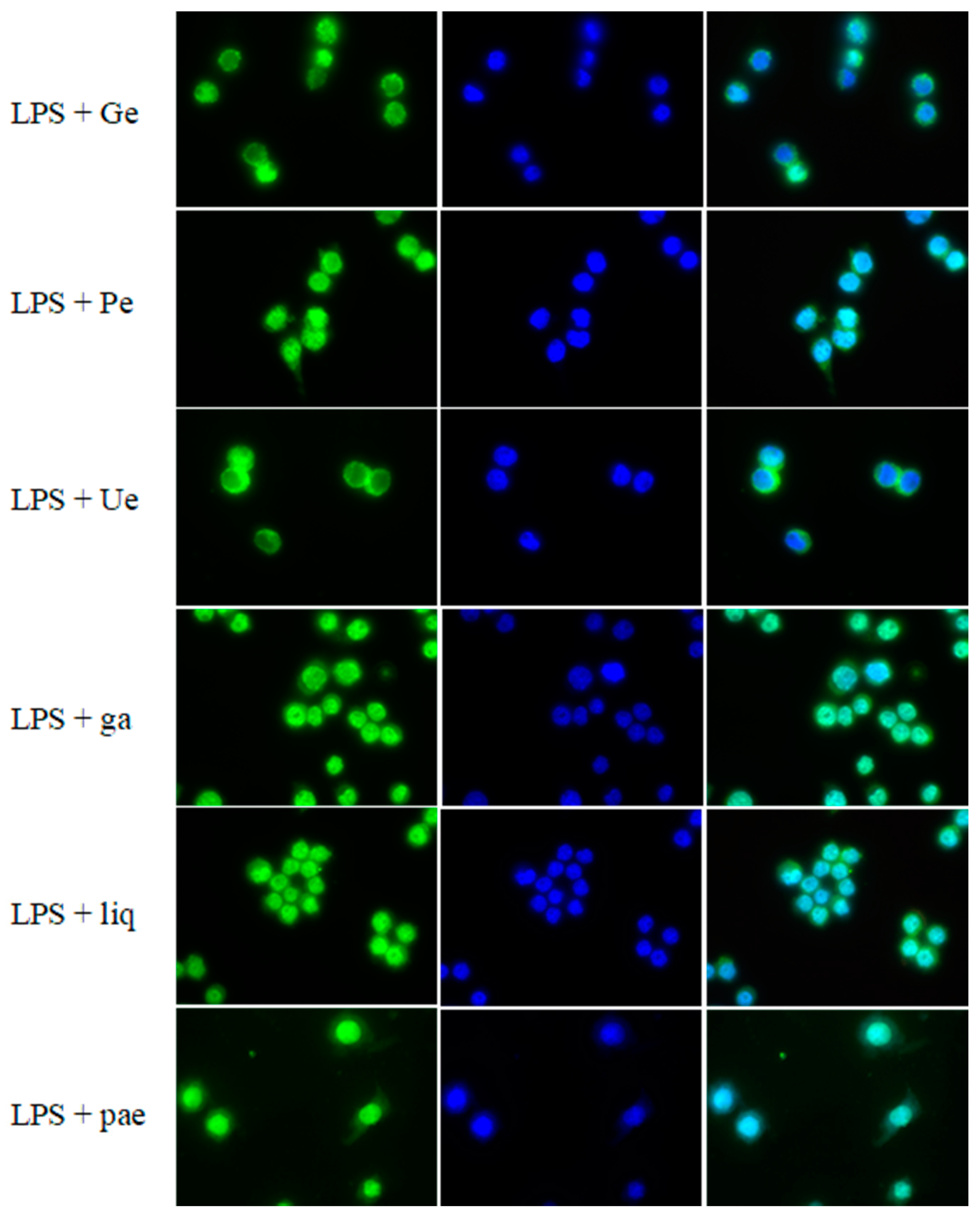
2.5. Imaging of NF-κB Nuclear Translocation

RAW 264.7 cells were grown on round glass coverslips (Thermo Scientific, Braunschweig, Germany) in a 6-well plate. They were treated with LPS (1 μg/mL) or LPS plus a single drug (30 μg/mL Ge, 10 μg/mL Pe, 30 μg/mL Ue, 10 μM ga, 20 μM 18ga, 50 μM liq, 4 μM iso, 20 μM pae and 10 μM urs) for 2 h. Dexamethasone (10 μM) was used as a positive control. Then, the cells were washed twice with cold PBS, and fixed with 4% paraformaldehyde for 15–20 min, followed by cold PBS-washing. Then, the cells were blocked with 4% skimmed milk dissolved in PBS for 1 h. After PBS-washing, cells were incubated with NF-κB (p65) polyclonal antibody (1:400) (ThermoFisher Scientific, Fisher Scientific GmbH, Schwerte, Germany) overnight at 4 °C. Followed by a 1 h incubation with Goat anti-Rabbit IgG (H+L) highly cross-adsorbed secondary antibody (1:1000) (ThermoFisher Scientific, Fisher Scientific GmbH, Schwerte, Germany) at room temperature. Afterwards, DAPI was used to stain the cell nucleus at room temperature for 5 min. Then, the coverslips were visualized by fluorescence microscopy (BZ-9000, Keyence, Keyence Deutschland GmbH, Neu-Isenburg, Germany).

2.6. Statistical Analysis

Results were expressed as mean ± SD from at least three independent experiments. Data analysis was performed with SigmaPlot® 11.0 (Systat Software, San Jose, CA, USA) for the cytotoxicity assay and GraphPad Prism 6 (Graphpad Software, San Diego, CA, USA) for the gene expression experiments. Statistical significance was evaluated employing one-way ANOVA and when p < 0.05, the difference was regarded as significant (* p < 0.05; ** p < 0.01; *** p < 0.001; **** p < 0.0001).

3. Results

3.1. Cytotoxicity of Individual Substances in RAW 264.7 Cells

The cytotoxicity of the nine substances in RAW 264.7 cells at 24 h was examined using an MTT assay. As Table 2 shows, doxorubicin was very toxic, pae was not toxic with an IC50 value above 1000 μM, and the other substances showed moderate toxicity with IC50 values between 20 and 263 μM (or μg/mL).

3.2. Nitrite Level after LPS Stimulation in RAW 264.7 Cells

RAW 264.7 cells were treated with different concentrations of LPS (1 μg/mL, 0.5 μg/mL, 0.1 μg/mL, 0.05 μg/mL, 0.025 μg/mL, and 0.01 μg/mL) and the nitrite level was determined at 1 h, 2 h, 3 h, 4 h, 5 h, 6 h, 7 h, 8 h, 9 h, 10 h, 11 h, 12 h, 22 h and 24 h. Nitrite production was time-dependent and LPS dose-dependent (Figure 1). 1 μg/mL LPS induced the highest concentration of nitrite, and the nitrite level peaked at 22 h and decreased afterwards. The cells didn’t look good and most died at 24 h. For the other experiments, 1 μg/mL LPS was used as a stimulant and the nitrite level was determined after 24 h treatment.

3.3. Effect of Single Substances on Nitrite Production in RAW 264.7 Cells

Three non-toxic concentrations of single substances (below IC10) were chosen to be added along with LPS (1 μg/mL) to RAW 264.7 cells in T25 flasks for 24 h. Cells without treatment were used as the control group. The nitric oxide synthase inhibitor L-NAME was used as a positive control. As shown in Figure 2, 1 μg/mL LPS induced a 15- to 30-fold increase in the nitrite amount, and L-NAME, Ge, Ue, 18ga, and iso inhibited nitrite production in a dose-dependent manner.

3.4. Effect of Single Substances on the Gene Expressions of NF-κB Subunits and Pro-Inflammatory Factors in RAW 264.7 Cells

RAW 264.7 cells were treated with LPS (1 μg/mL) or LPS plus a single substance for 1, 2, 3, 4, 6, and 8 h. Gene expressions of NF-κB subunits NF-κB1, NF-κB2, RelA, RelB, and Rel, and NF-κB-regulated genes ICAM-1, TNF-α, iNOS, and COX-2, were analyzed by real-time PCR. Non-treated cells were set as the control group (expression was 1.00). As shown in Figure 3a, LPS induced an expression increase in all the genes within 8 h. RelA (p65), RelB, and Rel peaked at 2 h, TNF-α and iNOS peaked at 3 h, NF-κB1 (p50), NF-κB2, ICAM-1, and COX-2 peaked at 4 h.
According to the NO inhibition results, the PSMs 18ga and iso were selected to test their effect on the gene expressions of the NF-κB subunits and pro-inflammatory factors. Besides, urs inhibited NO production at IC10 (data not shown), so urs (IC10 = 10 μM) was also selected. A non-cytotoxic concentration of the single substances 18ga (20 μM), iso (4 μM), and urs (10 μM), was added along with LPS to RAW 264.7 cells for 2 h to test RelA, RelB, Rel, and NF-κB2 expressions; for 3 h to test ICAM-1, TNF-α, and iNOS expressions; for 4 h to test NF-κB1; and 6 h to test COX-2 expressions (Figure 3b). Dexamethasone (10 μM) was used as a positive control in each case. Dexamethasone and the three PSMs all inhibited the tested gene expressions to some degree.

3.5. Effect of Single Substances on the Nuclear Translocation of NF-κB

The same non-toxic concentrations of 18ga, iso, and urs (20 μM 18ga, 4 μM iso, and 10 μM urs) as those in the NF-κB-related gene experiment, and other substances (30 μg/mL Ge, 10 μg/mL Pe, 30 μg/mL Ue, 10 μM ga, 50 μM liq, 20 μM pae), were selected to examine their effect on the nuclear translocation of NF-κB. In the inactive state, NF-κB is bound to the inhibitory protein IκB and stays in the cytoplasm. When activated by LPS, NF-κB dissociates from IκB and translocates to the nucleus. As seen in Figure 4, NF-κB was located in the cytoplasm in the control group, and after activation by LPS, NF-κB translocated to the nucleus. The PSMs 18ga, iso, and urs and plant extracts Ge and Ue could inhibit NF-κB nuclear translocation.

4. Discussion

Macrophages are one of the first cells in the host innate immune system to recognize and detect infectious sources such as LPS [45] and major producers of pro-inflammatory cytokines [46].
A number of medicinal plants have been used or are still in use to treat inflammations [34,35]. Some studies have reported that Eriobotrya japonica extract, ursolic acid, glycyrrhizic acid, 18β glycyrrhetinic acid, isoliquiritigenin, and paeoniflorin exhibited anti-inflammatory effects, partly through NF-κB and other pathways [47,48,49,50,51,52,53], but few studies have reported this for G. glabra extract, P. lactiflora extract, and liquiritigenin. Few studies have revealed the time-course changes of LPS-induced pro-inflammatory factors in detail. In Figure 1, we can see clearly how nitrite level changed during 24 h and the influence of different LPS concentrations. Below 0.1 μg/mL, the effect of LPS was not apparent. Before 8 h, the nitrite level hardly changed, but the expression of NF-κB subunits and down-stream genes (e.g., iNOS) was already activated and reached peaks within 8 h. This confirms the regulating role of NF-κB and iNOS on NO production.
In the time-course study of NF-κB subunits and NF-κB-regulated gene expressions, all the genes except COX-2 showed a bell-like curve. They reached their expression peaks within 8 h (Figure 3a). The expression of NF-κB1 (p50), NF-κB2, RelA (p65), and Rel increased up to four-fold at 2 h, NF-κB1 (p50) peaked at 4 h with a near 10-fold increase. The increases in NF-κB-regulated gene (ICAM-1, TNF-α, iNOS, and COX-2) expressions were apparent at 3 h. When we started our experiments, we stimulated the cells for 12 and 24 h with LPS, however, most of the cells were dead after that time, which might be due to the high sensitivity of the cell line. As a consequence, we selected 2 h and 4 h for NF-κB1 to test the effect of PSMs on NF-κB subunit expressions, and 3 and 6 h to test the effect of PSMs on ICAM-1, TNF-α, iNOS, and COX-2 expressions. As expected, the three PSMs 18ga, iso, and urs inhibited all the genes in a similar way to the positive control dexamethasone (Figure 3b), confirming the anti-inflammatory effect of these substances.
The positive control L-NAME inhibited NO production, but did not always have a significant inhibition effect in all experiments, which might be due to experimental conditions. Similarly, except in the gene expression of ICAM-1 and COX-2 groups, the positive control dexamethasone did have effects.
Isoliquiritigenin and ursolic acid blocked NF-κB translocation (Figure 4), which agrees with some reports that these PSMs can disturb NF-κB translocation. In other cell lines, these PSMs were involved with the inhibition of IκBα degradation NF-κB (p65) phosphorylation, as well as ERK and JNK pathways [47,54,55]. 18β glycyrrhetinic acid was reported to exert an anti-inflammatory effect by inhibiting phosphoinositide-3-kinase (PI3K) [53], glucocorticoid receptor activation [56], and JNK and NF-κB pathways [50,57]. Here, 18ga partly attenuated NF-κB translocation (Figure 4), probably via IκBα inhibition, which needs to be confirmed by more experiments. Besides, Ge and Ue partly blocked NF-κB translocation, while other substances did not, which more or less agrees with the NO inhibition results. As p38 mitogen-activated protein kinase (MAPK) was suggested to play a crucial role in regulating LPS-induced NF-κB and activator protein-1 (AP-1) activation as well as iNOS and COX-2 protein expressions [58], it will be interesting to elucidate concrete mechanisms underlining our results. Therefore, further studies are needed.
From a pharmacodynamic point of view, all the six PSMs showed low bioavailability [59,60,61,62,63,64,65]. However, various methods can be used to improve their bioavailability, for example, rectal or nasal administration, or co-administration with fatty acids and sodium deoxycholate/phospholipid-mixed nanomicelles of glycyrrhizic acid [59,60]. Ursolic acid nanoparticles and phospholipid complex could also enhance their bioavailability [66,67,68]. Besides, the phospholipid complex of ursolic acid also exhibited hepatoprotective effect enhancement [68], and a proniosomal gel of ursolic acid showed excellent anti-inflammatory activity [69]. Glycyrrhetinic acid-related nanoparticles were developed to deliver the drug [70,71]. A phosphate prodrug of isoliquiritigenin was developed and showed good stability and solubility [72]; co-crystals of isoliquiritigenin enhanced bioavailability and pharmacokinetic performance [73]; isoliquiritigenin-loaded F127/P123 polymeric micelles showed a strengthened effect [74]. With the help of biotechnological methods, these substances have some potential to be developed as effective drugs.

5. Conclusions

Extracts of Glycyrrhiza glabra and Eriobotrya japonica, 18β glycyrrhetinic acid, and isoliquiritigenin at low concentrations inhibited LPS-induced nitric oxide release in RAW 264.7 cells. 18β glycyrrhetinic acid, isoliquiritigenin, and ursolic acid inhibited the expression of ICAM-1, TNF-α, COX-2, and iNOS, partly through the inhibition of NF-κB expression and the attenuation of NF-κB nuclear translocation. Extracts of Glycyrrhiza glabra and Eriobotrya japonica also inhibited NF-κB nuclear translocation. These results support the use of Glycyrrhiza glabra and Eriobotrya japonica and their PSMs in TCM and phytotherapy to treat inflammations.

Author Contributions

Conceptualization, J.-X.Z. and M.W.; writing—original draft preparation, J.-X.Z.; review and editing, M.W.; supervision, M.W.

Funding

This research received no external funding.

Acknowledgments

We acknowledge financial support by Deutsche Forschungsgemeinschaft within the funding programme Open Access Publishing, by the Baden-Württemberg Ministry of Science, Research and the Arts and by Ruprecht-Karls-Universität Heidelberg.

Conflicts of Interest

The authors declare no conflict of interest.

References

  1. Hayden, M.S.; Ghosh, S. Signaling to NF-κB. Genes Dev. 2004, 18, 2195–2224. [Google Scholar] [CrossRef] [PubMed]
  2. Ghosh, S.; May, M.J.; Kopp, E.B. NF-κB and rel proteins: Evolutionarily conserved mediators of immune responses. Ann. Rev. Immunol. 1998, 16, 225–260. [Google Scholar] [CrossRef] [PubMed]
  3. Pahl, H. Activators and target genes of Rel/NF-κB transcription factors. Oncogene 1999, 18, 6853–6866. [Google Scholar] [CrossRef] [PubMed] [Green Version]
  4. Baeuerle, P.A.; Henkel, T. Function and activation of NF-κB in the immune system. Ann. Rev. Immunol. 1994, 12, 141–179. [Google Scholar] [CrossRef] [PubMed]
  5. Yang, Y.; Wu, J.; Wang, J. A database and functional annotation of NF-κB target genes. Int. J. Clin. Exp. Med. 2016, 9, 7986–7995. [Google Scholar]
  6. Kumar, A.; Takada, Y.; Boriek, A.M.; Aggarwal, B.B. Nuclear factor-kappaB: Its role in health and disease. J. Mol. Med. 2004, 82, 434–448. [Google Scholar] [CrossRef]
  7. Collart, M.A.; Baeuerle, P.; Vassalli, P. Regulation of tumor necrosis factor alpha transcription in macrophages: Involvement of four kappa B-like motifs and of constitutive and inducible forms of NF-kappa B. Mol. Cell. Biol. 1990, 10, 1498–1506. [Google Scholar] [CrossRef] [PubMed]
  8. Shakhov, A.N.; Collart, M.A.; Vassalli, P.; Nedospasov, S.A.; Jongeneel, C.V. Kappa B-type enhancers are involved in lipopolysaccharide-mediated transcriptional activation of the tumor necrosis factor alpha gene in primary macrophages. J. Exp. Med. 1990, 171, 35–47. [Google Scholar] [CrossRef] [Green Version]
  9. Popa, C.; Netea, M.G.; van Riel, P.L.; van der Meer, J.W.; Stalenhoef, A.F. The role of TNF-alpha in chronic inflammatory conditions, intermediary metabolism, and cardiovascular risk. J. Lipid Res. 2007, 48, 751–762. [Google Scholar] [CrossRef]
  10. Parameswaran, N.; Patial, S. Tumor Necrosis Factor-α signaling in macrophages. Crit. Rev. Eukaryot. Gene Expr. 2010, 20, 87–103. [Google Scholar] [CrossRef]
  11. O’Neill, G.P.; Ford-Hutchinson, A.W. Expression of mRNA for cyclooxygenase-1 and cyclooxygenase-2 in human tissues. FEBS Lett. 1993, 330, 156–160. [Google Scholar] [CrossRef]
  12. Yamamoto, K.; Arakawa, T.; Ueda, N.; Yamamoto, S. Transcriptional roles of nuclear factor kappa B and nuclear factor-interleukin-6 in the tumor necrosis factor alpha-dependent induction of cyclooxygenase-2 in MC3T3-E1 cells. J. Biol. Chem. 1995, 270, 31315–31320. [Google Scholar] [CrossRef]
  13. Vane, J.R.; Bakhle, Y.S.; Botting, R.M. Cyclooxygenases 1 and 2. Ann. Rev. Pharmacol. Toxicol. 1998, 38, 97–120. [Google Scholar] [CrossRef]
  14. Kaltschmidt, B.; Linker, R.A.; Deng, J.; Kaltschmidt, C. Cyclooxygenase-2 is a neuronal target gene of NF-κB. BMC Mol. Biol. 2002, 3, 16. [Google Scholar] [CrossRef]
  15. Ackerman IV, W.E.; Summerfield, T.L.S.; Vandre, D.D.; Robinson, J.M.; Kniss, D.A. Nuclear Factor-kappa B regulates inducible prostaglandin E Synthase expression in human amnion mesenchymal cells. Biol. Reprod. 2008, 78, 68–76. [Google Scholar] [CrossRef]
  16. Harris, S.G.; Padilla, J.; Koumas, L.; Ray, D.; Phipps, R.P. Prostaglandins as modulators of immunity. Trends Immunol. 2002, 23, 144–150. [Google Scholar] [CrossRef]
  17. Chandrasekharan, N.V.; Simmons, D.L. The cyclooxygenases. Genome Biol. 2004, 5, 241. [Google Scholar] [CrossRef]
  18. Imhof, B.A.; Dunon, D. Leukocyte migration and adhesion. Adv. Immunol. 1995, 58, 345–416. [Google Scholar]
  19. Elgavish, A. Effects of Escherichia coli and E. coli lipopolysaccharides on the function of human ureteral epithelial cells cultured in serum-free medium. Infect Immun. 1993, 61, 3304–3312. [Google Scholar]
  20. Meager, A. Cytokine regulation of cellular adhesion molecule expression in infammation. Cytokine Growth Factor Rev. 1999, 10, 27–39. [Google Scholar] [CrossRef]
  21. Golias, C.; Tsoutsi, E.; Matziridis, A.; Makridis, P.; Batistatou, A.; Charalabopoulos, K. Leukocyte and endothelial cell adhesion molecules in inflammation focusing on inflammatory heart disease. In Vivo 2007, 21, 757–769. [Google Scholar]
  22. Frank, P.G.; Lisanti, M.P. ICAM-1: Role in inflammation and in the regulation of vascular permeability. Am. J. Physiol. Heart Circ. Physiol. 2008, 295, 926–927. [Google Scholar] [CrossRef]
  23. Hill, B.G.; Dranka, B.P.; Bailey, S.M.; Lancaster, J.R., Jr.; Darley-Usmar, V.M. What part of NO don’t you understand? Some answers to the cardinal questions in nitric oxide biology. J. Biol. Chem. 2010, 285, 19699–19704. [Google Scholar] [CrossRef]
  24. Nathan, C.; Xie, Q. Regulation of biosynthesis of nitric oxide. J. Biol. Chem. 1994, 269, 13725–13728. [Google Scholar]
  25. Kleinert, H.; Pautz, A.; Linker, K.; Schwarz, P.M. Regulation of the expression of inducible nitric oxide synthase. Eur. J. Pharmacol. 2004, 500, 255–266. [Google Scholar] [CrossRef]
  26. Moncada, S.; Palmer, R.M.; Higgs, E.A. Nitric oxide: Physiology, pathophysiology, and pharmacology. Pharmacol. Rev. 1991, 43, 109–142. [Google Scholar]
  27. Moncada, S.; Higgs, E.A. The L-arginine-nitric oxide pathway. N. Engl. J. Med. 1993, 329, 2002–2012. [Google Scholar] [CrossRef]
  28. Coleman, J.W. Nitric oxide in immunity and inflammation. Int. Immunopharmacol. 2001, 1, 1397–1406. [Google Scholar] [CrossRef]
  29. Laskin, J.D.; Heck, D.E.; Laskin, D.L. Multifunctional role of nitric oxide in inflammation. Trends Endocrinol. Metab. 1994, 5, 377–382. [Google Scholar] [CrossRef]
  30. Wu, C. Nitric oxide and inflammation. Curr. Med. Chem. Anti-Inflamm. Anti-Allergy Agents 2004, 3, 217–222. [Google Scholar] [CrossRef]
  31. Kopp, E.; Ghosh, S. Inhibition of NF-κB by sodium salicylate and aspirin. Science 1994, 265, 956–959. [Google Scholar] [CrossRef]
  32. Gaddi, A.; Cicero, A.F.G.; Pedro, E.J. Clinical perspectives of anti-inflammatory therapy in the elderly: The lipoxigenase (LOX)/cycloxigenase (COX) inhibition concept. Arch. Gerontol. Geriatr. 2004, 38, 201–212. [Google Scholar] [CrossRef]
  33. Mendes, R.T.; Stanczyk, C.P.; Sordi, R.; Otuki, M.F.; dos Santos, F.A.; Fernandes, D. Selective inhibition of cyclooxygenase-2: Risks and benefits. Rev. Bras. Reumatol. 2012, 52, 767–782. [Google Scholar] [CrossRef]
  34. Pushpangadan, P.; Ijinu, T.P.; George, V. Plant based anti-inflammatory secondary metabolites. Ann. Phytomed. 2015, 4, 17–36. [Google Scholar]
  35. Van Wyk, B.E.; Wink, M. Medicinal Plants of the World, 2nd ed.; Briza: Pretoria, South Africa, 2017. [Google Scholar]
  36. Van Wyk, B.E.; Wink, M. Phytomedicines, Herbal Drugs and Poisons; University of Chicago Press: Chicago, IL, USA, 2015. [Google Scholar]
  37. Zhou, J.; Wink, M. Reversal of multidrug resistance in human colon cancer and human leukemia cells by three plant extracts and their major secondary metabolites. Medicines 2018, 5, 123. [Google Scholar] [CrossRef]
  38. Zhou, J.; Braun, M.S.; Wetterauer, P.; Wetterauer, B.; Wink, M. Antioxidant, cytotoxic, and antimicrobial activities of Glycyrrhiza glabra L., Paeonia lactiflora Pall., and Eriobotrya japonica (Thunb.) Lindl. extracts. Medicines 2019, 6, 43. [Google Scholar] [CrossRef]
  39. MacMicking, J.; Xie, Q.; Nathan, C. Nitric oxide and macrophage function. Ann. Rev. Immunol. 1997, 15, 323–350. [Google Scholar] [CrossRef]
  40. Sun, J.; Zhang, X.; Broderick, M.; Fein, H. Measurement of nitric oxide production in biological systems by using griess reaction assay. Sensors 2003, 3, 276–284. [Google Scholar] [CrossRef]
  41. Rees, D.D.; Palmer, R.M.; Schulz, R.; Hodson, H.F.; Moncada, S. Characterization of three inhibitors of endothelial nitric oxide synthase in vitro and in vivo. Br. J. Pharmacol. 1990, 101, 746–752. [Google Scholar] [CrossRef] [Green Version]
  42. Borowicz, K.K.; Starownik, R.; Kleinrok, Z.; Czuczwar, S.J. The influence of L-NG-nitroarginine methyl ester, an inhibitor of nitric oxide synthase, upon the anticonvulsive activity of conventional antiepileptic drugs against maximal electroshock in mice. J. Neural Transm. 1998, 105, 1–12. [Google Scholar] [CrossRef]
  43. Rawls, S.M.; Tallarida, R.J.; Gray, A.M.; Geller, E.B.; Adler, M.W. L-NAME (N-Nitro-L-Arginine Methyl Ester), a nitric-oxide synthase inhibitor, and WIN 55212-2 [4,5-dihydro-2-methyl-4(4-morpholinylmethyl)-1-(1-naphthalenyl-carbonyl)-6H-pyrrolo[3,2,1ij]quinolin-6-one], a cannabinoid agonist, interact to evoke synergistic hypothermia. J. Pharmacol. Exp. Ther. 2004, 308, 780–786. [Google Scholar] [CrossRef]
  44. Beutler, B.; Cerami, A. Cachectin and tumour necrosis factor as two sides of the same biological coin. Nature 1986, 320, 584–588. [Google Scholar] [CrossRef]
  45. Zhang, G.; Ghosh, S. Molecular mechanisms of NF-κB activation induced by bacterial lipopolysaccharide through Toll-like receptors. J. Endotoxin Res. 2000, 6, 453–457. [Google Scholar] [CrossRef]
  46. Thiyagarajan, P.; Chandrasekaran, C.V.; Deepak, H.B.; Agarwal, A. Modulation of lipopolysaccharide-induced pro-inflammatory mediators by an extract of Glycyrrhiza glabra and its phytoconstituents. Inflammopharmacology 2011, 19, 235–241. [Google Scholar] [CrossRef]
  47. Shishodia, S.; Majumdar, S.; Banerjee, S.; Aggarwal, B.B. Ursolic acid inhibits nuclear factor-kappa B activation induced by carcinogenic agents through suppression of Ikappa B alpha kinase and p65 phosphorylation: Correlation with down-regulation of cyclooxygenase 2, matrix metalloproteinase 9, and cyclin D1. Cancer Res. 2003, 63, 4375–4383. [Google Scholar]
  48. Kim, J.Y.; Park, S.J.; Yun, K.J.; Cho, Y.W.; Park, H.J.; Lee, K.T. Isoliquiritigenin isolated from the roots of Glycyrrhiza uralensis inhibits LPS-induced iNOS and COX-2 expression via the attenuation of NF-kappaB in RAW 264.7 macrophages. Eur. J. Pharmacol. 2008, 584, 175–184. [Google Scholar] [CrossRef]
  49. Lee, C.; Wu, S.; Chen, J.; Li, C.; Lo, H.; Cheng, W.; Lin, J.; Chang, Y.; Hsiang, C.; Ho, T. Eriobotrya japonica leaf and its triterpenes inhibited lipopolysaccharide-induced cytokines and inducible enzyme production via the Nuclear Factor-κB signaling pathway in lung epithelial cells. Am. J. Chin. Med. 2008, 36, 1185–1198. [Google Scholar] [CrossRef]
  50. Chang, Y.L.; Chen, C.L.; Kuo, C.L.; Chen, B.C.; You, J.S. Glycyrrhetinic acid inhibits ICAM-1 expression via blocking JNK and NF-kappaB pathways in TNF-alpha-activated endothelial cells. Acta Pharmacol. Sin. 2010, 31, 546–553. [Google Scholar] [CrossRef]
  51. Uto, T.; Suangkaew, N.; Morinaga, O.; Kariyazono, H.; Oiso, S.; Shoyama, Y. Eriobotryae Folium extract suppresses LPS-induced iNOS and COX-2 expression by inhibition of NF-κB and MAPK activation in murine macrophages. Am. J. Chin. Med. 2010, 38, 985–994. [Google Scholar] [CrossRef]
  52. Jin, L.; Zhang, L.; Xie, K.; Ye, Y.; Feng, L. Paeoniflorin suppresses the expression of intercellular adhesion molecule-1 (ICAM-1) in endotoxintreated human monocytic cells. Br. J. Pharmacol. 2011, 164, 694–703. [Google Scholar] [CrossRef]
  53. Wang, C.; Kao, T.; Lo, W.; Yen, G. Glycyrrhizic Acid and 18β glycyrrhetinic acid acid modulate lipopolysaccharide-induced inflammatory response by suppression of NF-κB through PI3K p110δ and p110γ Inhibitions. J. Agric. Food Chem. 2011, 59, 7726–7733. [Google Scholar] [CrossRef]
  54. Kwon, H.M.; Choi, Y.J.; Choi, J.S.; Kang, S.W.; Bae, J.Y.; Kang, I.J.; Jun, J.G.; Lee, S.S.; Lim, S.S.; Kang, Y.H. Blockade of cytokine-induced endothelial cell adhesion molecule expression by licorice isoliquiritigenin through NF-kappaB signal disruption. Exp. Biol. Med. 2007, 232, 235–245. [Google Scholar]
  55. Ma, X.; Zhang, Y.; Wang, Z.; Shen, Y.; Zhang, M.; Nie, Q.; Hou, Y.; Bai, G. Ursolic acid, a natural nutraceutical agent, targets caspase3 and alleviates inflammation-associated downstream signal transduction. Mol. Nutr. Food Res. 2017, 61. [Google Scholar] [CrossRef]
  56. Kao, T.; Shyu, M.; Yen, G. Glycyrrhizic acid and 18β-glycyrrhetinic acid inhibit inflammation via PI3K/Akt/GSK3β signaling and glucocorticoid receptor activation. J. Agric. Food Chem. 2010, 58, 8623–8629. [Google Scholar] [CrossRef]
  57. Chen, H.J.; Kang, S.P.; Lee, I.J.; Lin, Y.L. Glycyrrhetinic acid suppressed NF-κB activation in TNF-α-induced hepatocytes. J. Agric. Food Chem. 2014, 62, 618–625. [Google Scholar] [CrossRef]
  58. Chen, B.; Chen, Y.; Lin, W. Involvement of p38 mitogen-activated protein kinase in lipopolysaccharide-induced iNOS and COX-2 expression in J774 macrophages. Immunology 1999, 97, 124–129. [Google Scholar] [CrossRef]
  59. Sasaki, K.; Yonebayashi, S.; Yoshida, M.; Shimizu, K.; Aotsuka, T.; Takayama, K. Improvement in the bioavailability of poorly absorbed glycyrrhizin via various non-vascular administration routes in rats. Int. J. Pharm. 2003, 265, 95–102. [Google Scholar] [CrossRef]
  60. Jin, S.; Fu, S.; Han, J.; Jin, S.; Lv, Q.; Lu, Y.; Qi, J.; Wu, W.; Yuan, H. Improvement of oral bioavailability of glycyrrhizin by sodium deoxycholate/phospholipid-mixed nanomicelles. J. Drug Target. 2012, 20, 615–622. [Google Scholar] [CrossRef]
  61. Takeda, S.; Ishthara, K.; Wakui, Y.; Amagaya, S.; Maruno, M.; Akao, T.; Kobashi, K. Bioavailability study of glycyrrhetic acid after oral administration of glycyrrhizin in rats; relevance to the intestinal bacterial hydrolysis. J. Pharm. Pharmacol. 1996, 48, 902–905. [Google Scholar] [CrossRef]
  62. Kang, H.E.; Cho, Y.K.; Jung, H.Y.; Choi, K.Y.; Sohn, S.I.; Baek, S.R.; Lee, M.G. Pharmacokinetics and first-pass effects of liquiritigenin in rats: Low bioavailability is primarily due to extensive gastrointestinal first-pass effect. Xenobiotica 2009, 39, 465–475. [Google Scholar] [CrossRef]
  63. Dai, J.Y.; Yang, J.L.; Li, C. Transport and metabolism of flavonoids from Chinese herbal remedy Xiaochaihu-tang across human intestinal Caco-2 cell monolayers. Acta Pharmacol. Sin. 2008, 29, 1086–1093. [Google Scholar] [CrossRef]
  64. Takeda, S.; Isono, T.; Wakui, Y.; Matsuzaki, Y.; Sasaki, H.; Amagaya, S.; Maruno, M. Absorption and excretion of paeoniflorin in rats. J. Pharm. Pharmacol. 1995, 7, 1036–1040. [Google Scholar] [CrossRef]
  65. Hirsh, S.; Huber, L.; Zhang, P.; Stein, R.; Joyal, S. A single ascending dose, initial clinical pharmacokinetic and safety study of ursolic acid in healthy adult volunteers. Nutrition 2014, 28, 1044.6. [Google Scholar]
  66. Yang, L.; Sun, Z.; Zu, Y.; Zhao, C.; Sun, X.; Zhang, Z.; Zhang, L. Physicochemical properties and oral bioavailability of ursolic acid nanoparticles using supercritical anti-solvent (SAS) process. Food Chem. 2012, 132, 319–325. [Google Scholar] [CrossRef]
  67. Ge, Z.; Du, X.; Huang, X.; Qiao, B. Enhanced oral bioavailability of ursolic acid nanoparticles via antisolvent precipitation with TPGS1000 as a stabilizer. J. Drug Deliv. Sci. Technol. 2015, 29, 210–217. [Google Scholar] [CrossRef]
  68. Biswas, S.; Mukherjee, P.K.; Harwansh, R.K.; Bannerjee, S.; Bhattacharjee, P. Enhanced bioavailability and hepatoprotectivity of optimized ursolic acid-phospholipid complex. Drug Dev. Ind. Pharm. 2019, 45. [Google Scholar] [CrossRef]
  69. Garg, M.; Garg, P. Encapsulation of bio active compound ursolic acid as proniosomes and its evaluation. Asian J. Pharm. 2013, 7, 158–162. [Google Scholar] [CrossRef]
  70. Tian, Q.; Wang, W.; He, X.; Zhu, X.; Huang, W.; Zhang, C.; Yuan, Z.; Chen, X. Glycyrrhetinic acid-modified nanoparticles for drug delivery: Preparation and characterization. Chin. Sci. Bull. 2009, 54, 3121–3126. [Google Scholar] [CrossRef]
  71. Wu, F.; Li, X.; Jiang, B.; Yan, J.; Zhang, Z.; Qin, J.; Yu, W.; Gao, Z. Glycyrrhetinic acid functionalized nanoparticles for drug delivery to liver cancer. J. Biomed. Nanotechnol. 2018, 14, 1837–1852. [Google Scholar] [CrossRef]
  72. Boyapelly, K.; Bonin, M.; Traboulsi, H.; Cloutier, A.; Phaneuf, S.C.; Fortin, D.; Cantin, A.M.; Richter, M.V.; Marsault, E. synthesis and characterization of a phosphate prodrug of isoliquiritigenin. J. Nat. Prod. 2017, 80, 879–886. [Google Scholar] [CrossRef]
  73. Xu, J.; Huang, Y.; Ruan, S.; Chi, Z.; Qin, K.; Cai, B.; Cai, T. Cocrystals of isoliquiritigenin with enhanced pharmacokinetic performance. CrystEngComm 2016, 18, 8776–8786. [Google Scholar] [CrossRef]
  74. Xie, Y.; Wang, Q.; Adu-Frimpong, M.; Liu, J.; Zhang, K.; Xu, X.; Yu, J. Preparation and evaluation of Isoliquiritigenin-loaded F127/P123 polymeric micelles. Drug Dev. Ind. Pharm. 2019, 45. [Google Scholar] [CrossRef]
Figure 1. The nitrite concentration (µM) tested at different times with stimulation of different lipopolysaccharide (LPS) doses. Different colors correspond to the different concentrations of LPS (from 0.01 μg/mL to 1 μg/mL).
Medicines 06 00055 g001
Figure 2. Nitrite levels (µM) in the supernatant of RAW 264.7 cells after treatment with LPS and LPS plus PSMs. The non-treated group was defined as the control group. The results are expressed as the mean ± SD from at least three independent experiments. When p < 0.05, the difference was regarded as significant (* p < 0.05; ** p < 0.01; *** p < 0.001; **** p < 0.0001).
Medicines 06 00055 g002
Figure 3. LPS induced NF-κB subunit and NF-κB-regulated gene expression changes. (a) NF-κB subunit (NF-κB1, NF-κB2, RelA, RelB, and Rel) and NF-κB-regulated gene (ICAM-1, TNF-α, iNOS, and COX-2) expression changes after 1 μg/mL LPS stimulation for different times. (b) Effect of dex, iso, 18ga, or urs on NF-κB subunit and NF-κB-regulated gene expressions at the appointed times. The non-treated group was set as the control group (1.00). The results are expressed as the mean ± SD from at least three independent experiments. When p < 0.05, the difference was regarded as significant (* p < 0.05; ** p < 0.01; *** p < 0.001; **** p < 0.0001).
Medicines 06 00055 g003aMedicines 06 00055 g003b
Figure 4. Effect of PSMs on the LPS-induced nuclear translocation of NF-κB. RAW 264.7 cells were treated with LPS or LPS plus plant extracts and PSMs for 2 h. Immunofluorescence and fluorescence microscopy were used to analyze NF-κB translocation. In the first column, the green color was due to the immunofluorescence of an NF-κB secondary antibody; the middle column of blue color shows DAPI-dyed nuclei; and the last column was a merge of the first two columns. When NF-κB moved to the nucleus, the merged nuclei showed a bright white color. The results are expressed as the mean ± SD from at least three independent experiments. (The plotting scale is showed in the control group with p65 staining).
Medicines 06 00055 g004aMedicines 06 00055 g004b
Table 1. qPCR primers for NF-κB-related genes.
GeneSequenceAmplicon
RPS201 *Forward: CCCGAAGTGGCGATTCACC77 nt
Reverse: TCCGCACAAACCTTCTCCAG
NF-κB1Forward: AAGGCAAAGCGAATCCAAA72 nt
Reverse: GAAGGCCTTGAATGAAATCG
NF-κB2Forward: GTAATCACTGGGCAGACAAGG95 nt
Reverse: AAGTGGAGGGCGGAGTCT
RelAForward: TCACCAAGGATCCACCTCA87 nt
Reverse: GGCAGAGGTCAGCCTCATAG
RelBForward: TGTTCAAAACGCCACCCTAC70 nt
Reverse: CGCTGCAAGAACACATTGAC
c-Rel #Forward: AGACTGCGACCTCAATGTGG117 nt
Reverse: GCACGGTTGTCATAAATTGGGTT
ICAM-1Forward: TGGAAGCTGTTTGAGCTGAG67 nt
Reverse: TGCCACAGTTCTCAAAGCAC
TNF-αForward: TTGTCTTAATAACGCTGATTTGGT61 nt
Reverse: GGGAGCAGAGGTTCAGTGAT
iNOSForward: CAGACTGGATTTGGCTGGTC68 nt
Reverse: CAACATCTCCTGGTGGAACA
COX-2 +Forward: TGAGCAACTATTCCAAACCAGC74 nt
Reverse: GCACGTAGTCTTCGATCACTATC
* Primer RPS201 was the reference gene, and its sequences were from PrimerBank MGH-PGA with ID: 146135021c1. https://pga.mgh.harvard.edu/cgi-bin/primerbank/new_displayDetail2.cgi?primerID=146135021c1. # Primer c-Rel sequences were from PrimerBank MGH-PGA with ID: 112181203c3. https://pga.mgh.harvard.edu/cgi-bin/primerbank/new_displayDetail2.cgi?primerID=112181203c3. + Primer COX-2 sequences were from PrimerBank MGH-PGA with ID: 31981525a1. https://pga.mgh.harvard.edu/cgi-bin/primerbank/new_displayDetail2.cgi?primerID=31981525a1. All the primers were synthesized by Eurofins Genomics (Ebersberg, Germany).
Table 2. Cytotoxicity (IC50 values *) of individual plant extracts and PSMs in RAW 264.7 cells.
SubstancesRAW 264.7SubstancesRAW 264.7
Dox #0.26 ± 0.0518ga90.19 ± 2.05
Ge #94.11 ± 4.88liq262.84 ± 26.88
Pe #67.08 ± 5.31iso42.56 ± 5.70
Ue #60.53 ± 4.03pae1034.91 ± 883.85
ga163.95 ± 74.98urs23.40 ± 1.42
* Units for Ge, Pe, and Ue are μg/mL; others are μM; Dox = doxorubicin; Ge = G. glabra extract; Pe = P. lactiflora extract; Ue = E. japonica extract; ga = glycyrrhizic acid; 18ga = 18β glycyrrhetinic acid; liq = liquiritigenin; iso = isoliquiritigenin; pae = paeoniflorin; urs = ursolic acid. # Values for Dox, Ge, Pe, and Ue were previously stated [38].

