Recent Advances of Polyaniline-Based Biomaterials for Phototherapeutic Treatments of Tumors and Bacterial Infections

 ,
,  , , and
Abstract
:1. Introduction
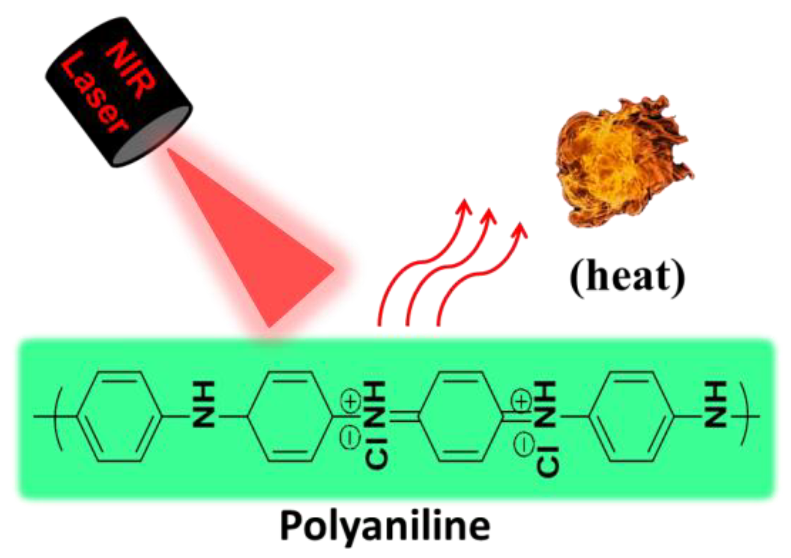
2. Chemical Structure and Stability of PANI
3. PANI-Based Biomaterials for Tumor Ablation
3.1. Synthesis of PANI PTAs
3.1.1. Chemical Polymerization
3.1.2. Enzyme-Catalyzed Polymerization
3.2. Multifucntional PANI-Based Materials
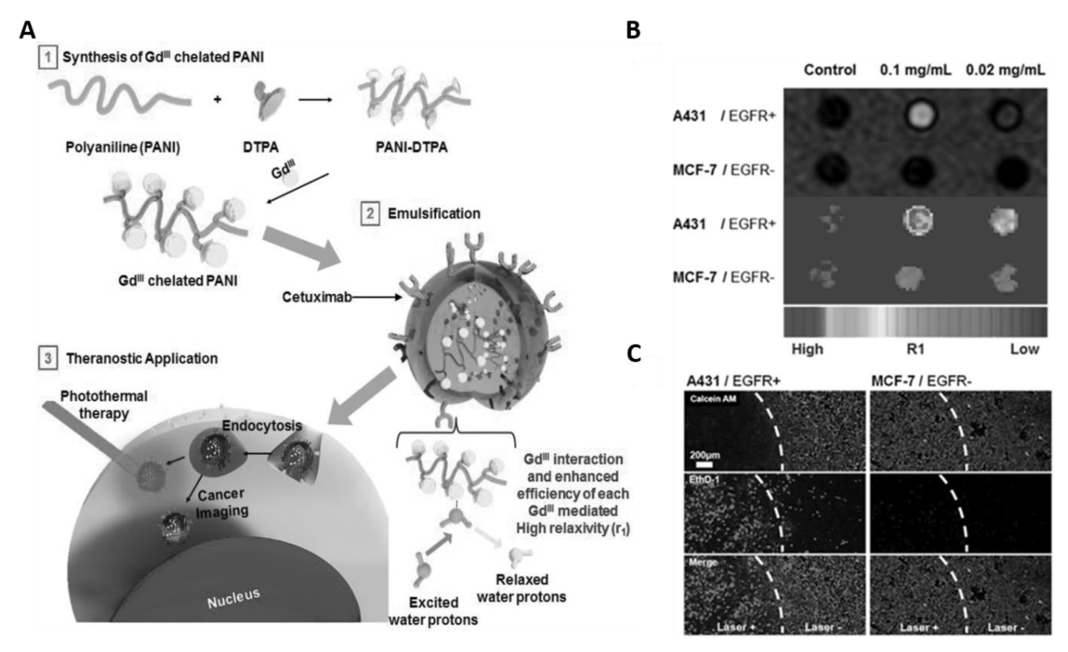
3.2.1. PANI Nanoparticles with MRI Imaging and PTT
3.2.2. PANI Nanoparticles with PAI Imaging and PTT
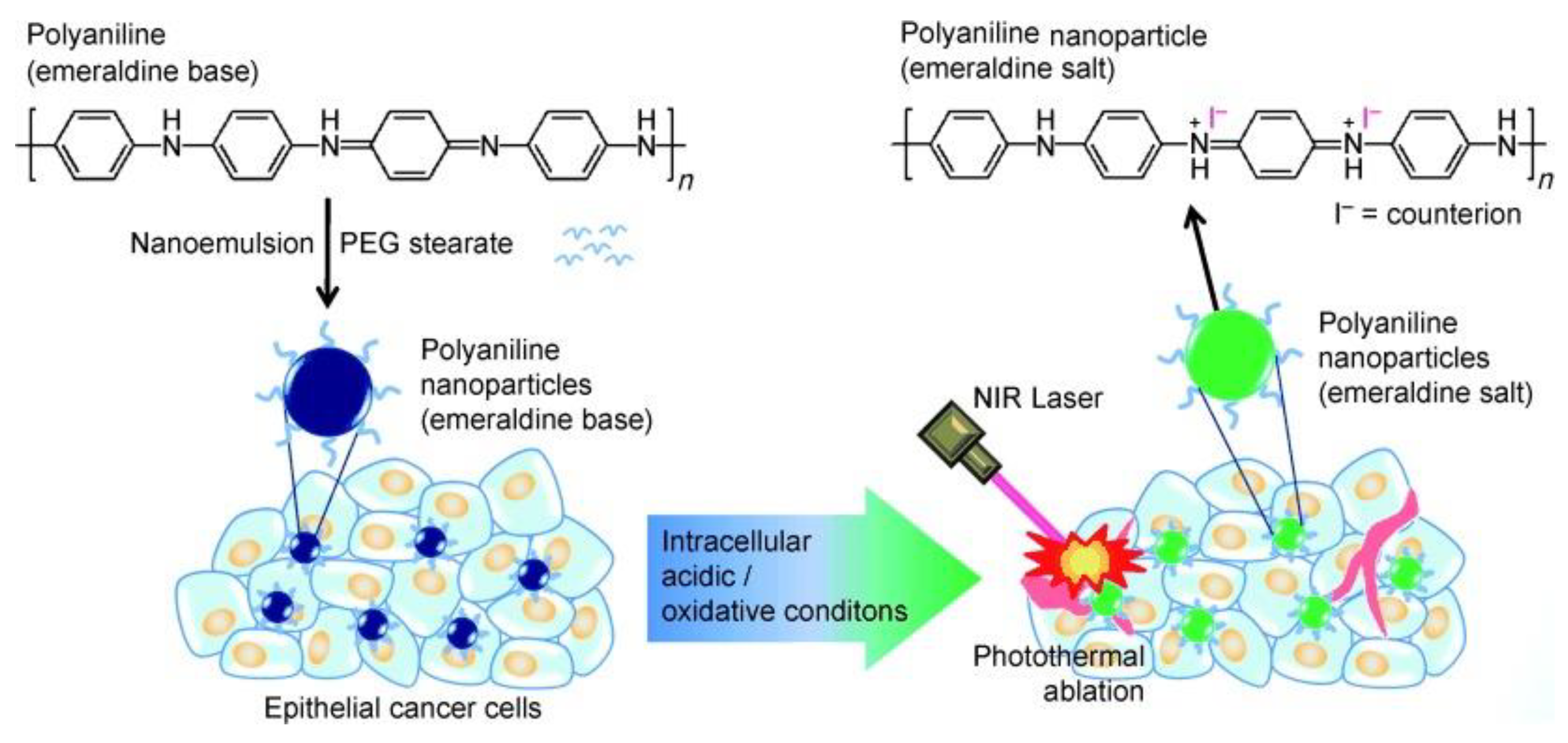
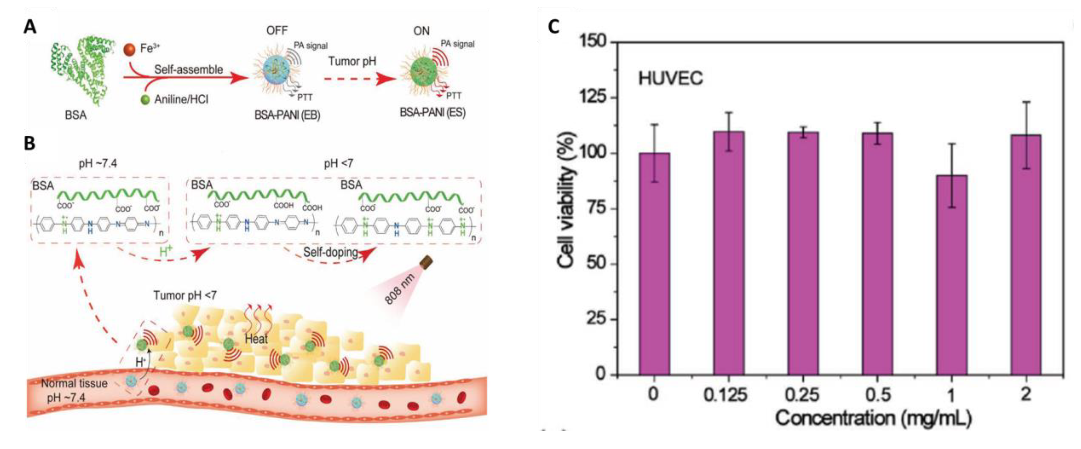
3.3. Self-Doping of PANI
3.4. Combination Therapy of Cancer Using PANI-Based Materials
3.4.1. PANI Nanocomposites with Chemotherapy and PTT
3.4.2. PANI Nanocomposites with RT and PTT
3.4.3. PANI Nanocomposites with PDT and PTT
4. PANI-Based Biomaterials for Eradication of Bacterial Infections
4.1. Self-Doping of PANI
4.2. Bacteria-Trageting and Multifucntinal PANI-Based Materials
5. Summary and Future Perspectives
Author Contributions
Funding
Conflicts of Interest
References
- Van der Zee, J. Heating the patient: A promising approach? Ann. Oncol. 2002, 13, 1173–1184. [Google Scholar] [CrossRef]
- Hildebrandt, B.; Wust, P.; Ahlers, O.; Dieing, A.; Sreenivasa, G.; Kerner, T.; Felix, R.; Riess, H. The cellular and molecular basis of hyperthermia. Crit. Rev. Oncol. Hemat. 2002, 43, 33–56. [Google Scholar] [CrossRef]
- Ibelli, T.; Templeton, S.; Levi-Polyachenko, N. Progress on utilizing hyperthermia for mitigating bacterial infections. Int. J. Hyperther. 2018, 34, 144–156. [Google Scholar] [CrossRef] [PubMed]
- Schmidt, K.L.; Simon, E. Thermotherapy of pain, trauma, and inflammatory and degenerative rheumatic diseases. In Thermotherapy for Neoplasia, Inflammation, and Pain; Kosaka, M., Sugahara, T., Schmidt, K.L., Simon, E., Eds.; Springer Japan: Tokyo, Japan, 2001; pp. 527–539. [Google Scholar]
- DeNardo, G.L.; DeNardo, S.J. Update: Turning the heat on cancer. Cancer Biother. Radiopharm. 2008, 23, 671–680. [Google Scholar] [CrossRef] [PubMed] [Green Version]
- Wust, P.; Hildebrandt, B.; Sreenivasa, G.; Rau, B.; Gellermann, J.; Riess, H.; Felix, R.; Schlag, P.M. Hyperthermia in combined treatment of cancer. Lancet Oncol. 2002, 3, 487–497. [Google Scholar] [CrossRef]
- Hegyi, G.; Szigeti, G.P.; Szasz, A. Hyperthermia versus Oncothermia: Cellular Effects in Complementary Cancer Therapy. Evid.-Based Complement. Altern. Med. 2013, 2013, 672873. [Google Scholar] [CrossRef] [PubMed]
- Ashikbayeva, Z.; Tosi, D.; Balmassov, D.; Schena, E.; Saccomandi, P.; Inglezakis, V. Application of Nanoparticles and Nanomaterials in Thermal Ablation Therapy of Cancer. Nanomaterials (Basel) 2019, 9, 1195. [Google Scholar] [CrossRef] [PubMed] [Green Version]
- Khot, M.I.; Andrew, H.; Svavarsdottir, H.S.; Armstrong, G.; Quyn, A.J.; Jayne, D.G. A Review on the Scope of Photothermal Therapy–Based Nanomedicines in Preclinical Models of Colorectal Cancer. Clin. Colorectal Cancer 2019, 18, e200–e209. [Google Scholar] [CrossRef]
- Shanmugam, V.; Selvakumar, S.; Yeh, C.S. Near-infrared light-responsive nanomaterials in cancer therapeutics. Chem. Soc. Rev. 2014, 43, 6254–6287. [Google Scholar] [CrossRef] [Green Version]
- Cheng, L.; Wang, C.; Feng, L.Z.; Yang, K.; Liu, Z. Functional Nanomaterials for Phototherapies of Cancer. Chem. Rev. 2014, 114, 10869–10939. [Google Scholar] [CrossRef]
- Bao, Z.H.; Liu, X.R.; Liu, Y.D.; Liu, H.Z.; Zhao, K. Near-infrared light-responsive inorganic nanomaterials for photothermal therapy. Asian J. Pharm. Sci. 2016, 11, 349–364. [Google Scholar] [CrossRef] [Green Version]
- Hu, J.-J.; Cheng, Y.-J.; Zhang, X.-Z. Recent advances in nanomaterials for enhanced photothermal therapy of tumors. Nanoscale 2018, 10, 22657–22672. [Google Scholar] [CrossRef] [PubMed]
- Yang, J.; Choi, J.; Bang, D.; Kim, E.; Lim, E.K.; Park, H.; Suh, J.S.; Lee, K.; Yoo, K.H.; Kim, E.K.; et al. Convertible Organic Nanoparticles for Near-Infrared Photothermal Ablation of Cancer Cells. Angew. Chem. Int. Ed. 2011, 50, 441–444. [Google Scholar] [CrossRef] [PubMed]
- Cheng, L.; Yang, K.; Chen, Q.; Liu, Z. Organic Stealth Nanoparticles for Highly Effective in Vivo Near-Infrared Photothermal Therapy of Cancer. ACS Nano 2012, 6, 5605–5613. [Google Scholar] [CrossRef] [PubMed]
- Chen, M.; Fang, X.L.; Tang, S.H.; Zheng, N.F. Polypyrrole nanoparticles for high-performance in vivo near-infrared photothermal cancer therapy. Chem. Commun. 2012, 48, 8934–8936. [Google Scholar] [CrossRef]
- Chen, C.; Tang, W.; Jiang, D.W.; Yang, G.L.; Wang, X.L.; Zhou, L.N.; Zhang, W.A.; Wang, P. Hyaluronic acid conjugated polydopamine functionalized mesoporous silica nanoparticles for synergistic targeted chemo-photothermal therapy. Nanoscale 2019, 11, 11012–11024. [Google Scholar] [CrossRef]
- Jiang, Y.Y.; Cui, D.; Fang, Y.; Zhen, X.; Upputuri, P.K.; Pramanik, M.; Ding, D.; Pu, K.Y. Amphiphilic semiconducting polymer as multifunctional nanocarrier for fluorescence/photoacoustic imaging guided chemo-photothermal therapy. Biomaterials 2017, 145, 168–177. [Google Scholar] [CrossRef]
- MacLaughlin, C.M.; Ding, L.; Jin, C.; Cao, P.; Siddiqui, I.; Hwang, D.M.; Chen, J.; Wilson, B.C.; Zheng, G.; Hedley, D.W. Porphysome nanoparticles for enhanced photothermal therapy in a patient-derived orthotopic pancreas xenograft cancer model: A pilot study. J. Biomed. Opt. 2016, 21, 84002. [Google Scholar] [CrossRef] [Green Version]
- Xu, L.; Cheng, L.; Wang, C.; Peng, R.; Liu, Z. Conjugated polymers for photothermal therapy of cancer. Polym. Chem. 2014, 5, 1573–1580. [Google Scholar] [CrossRef]
- Wang, J.; Qiu, J. A review of organic nanomaterials in photothermal cancer therapy. Cancer Res. Front. 2016, 2, 67. [Google Scholar] [CrossRef]
- Jung, H.S.; Verwilst, P.; Sharma, A.; Shin, J.; Sessler, J.L.; Kim, J.S. Organic molecule-based photothermal agents: An expanding photothermal therapy universe. Chem. Soc. Rev. 2018, 47, 2280–2297. [Google Scholar] [CrossRef] [PubMed]
- Zhou, Y.; Hu, Y.; Sun, W.; Zhou, B.; Zhu, J.; Peng, C.; Shen, M.; Shi, X. Polyaniline-loaded γ-polyglutamic acid nanogels as a platform for photoacoustic imaging-guided tumor photothermal therapy. Nanoscale 2017, 9, 12746–12754. [Google Scholar] [CrossRef] [PubMed]
- Zare, E.N.; Makvandi, P.; Ashtari, B.; Rossi, F.; Motahari, A.; Perale, G. Progress in Conductive Polyaniline-Based Nanocomposites for Biomedical Applications: A Review. J. Med. Chem. 2020, 63, 1–22. [Google Scholar] [CrossRef] [PubMed]
- Shahadat, M.; Khan, M.Z.; Rupani, P.F.; Embrandiri, A.; Sultana, S.; Ahammad, S.Z.; Ali, S.W.; Sreekrishnan, T.R. A critical review on the prospect of polyaniline-grafted biodegradable nanocomposite. Adv. Colloid Interface Sci. 2017, 249, 2–16. [Google Scholar] [CrossRef] [PubMed]
- Qazi, T.H.; Rai, R.; Boccaccini, A.R. Tissue engineering of electrically responsive tissues using polyaniline based polymers: A review. Biomaterials 2014, 35, 9068–9086. [Google Scholar] [CrossRef]
- Smith, A.M.; Pajovich, H.T.; Banerjee, I.A. Development of Self-Assembled Nanoribbon Bound Peptide-Polyaniline Composite Scaffolds and Their Interactions with Neural Cortical Cells. Bioengineering (Basel) 2018, 5, 6. [Google Scholar] [CrossRef] [Green Version]
- De Albuquerque, J.E.; Mattoso, L.H.C.; Faria, R.M.; Masters, J.G.; MacDiarmid, A.G. Study of the interconversion of polyaniline oxidation states by optical absorption spectroscopy. Synth. Met. 2004, 146, 1–10. [Google Scholar] [CrossRef]
- Hsiao, C.W.; Chen, H.L.; Liao, Z.X.; Sureshbabu, R.; Hsiao, H.C.; Lin, S.J.; Chang, Y.; Sung, H.W. Effective Photothermal Killing of Pathogenic Bacteria by Using Spatially Tunable Colloidal Gels with Nano-Localized Heating Sources. Adv. Funct. Mater. 2015, 25, 721–728. [Google Scholar] [CrossRef]
- Zhou, J.; Lu, Z.G.; Zhu, X.J.; Wang, X.J.; Liao, Y.; Ma, Z.F.; Li, F.Y. NIR photothermal therapy using polyaniline nanoparticles. Biomaterials 2013, 34, 9584–9592. [Google Scholar] [CrossRef]
- Tian, Q.W.; Li, Y.P.; Jiang, S.S.; An, L.; Lin, J.M.; Wu, H.X.; Huang, P.; Yang, S.P. Tumor pH-Responsive Albumin/Polyaniline Assemblies for Amplified Photoacoustic Imaging and Augmented Photothermal Therapy. Small 2019, 15, 1902926. [Google Scholar] [CrossRef]
- Mu, S.L. Nanostructured polyaniline synthesized using interface polymerization and its redox activity in a wide pH range. Synth. Met. 2010, 160, 1931–1937. [Google Scholar] [CrossRef]
- Siegel, R.L.; Miller, K.D.; Jemal, A. Cancer statistics, 2019. CA Cancer J. Clin. 2019, 69, 7–34. [Google Scholar] [CrossRef] [PubMed] [Green Version]
- Cooper, G.M. The Development and Causes of Cancer. In The Cell: A Molecular Approach, 2nd ed.; Sinauer Associates: Sunderland, MA, USA, 2000. [Google Scholar]
- Baskar, R.; Lee, K.A.; Yeo, R.; Yeoh, K.W. Cancer and Radiation Therapy: Current Advances and Future Directions. Int. J. Med. Sci. 2012, 9, 193–199. [Google Scholar] [CrossRef] [PubMed] [Green Version]
- DeVita, V.T.; Chu, E. A History of Cancer Chemotherapy. Cancer Res. 2008, 68, 8643–8653. [Google Scholar] [CrossRef] [PubMed] [Green Version]
- Chen, H.H.W.; Kuo, M.T. Improving radiotherapy in cancer treatment: Promises and challenges. Oncotarget 2017, 8, 62742–62758. [Google Scholar] [CrossRef] [PubMed] [Green Version]
- Thakor, A.S.; Gambhir, S.S. Nanooncology: The Future of Cancer Diagnosis and Therapy. CA Cancer J. Clin. 2013, 63, 395–418. [Google Scholar] [CrossRef]
- Yslas, E.I.; Ibarra, L.E.; Molina, M.A.; Rivarola, C.; Barbero, C.A.; Bertuzzi, M.L.; Rivarola, V.A. Polyaniline nanoparticles for near-infrared photothermal destruction of cancer cells. J. Nanopart. Res. 2015, 17, 389. [Google Scholar] [CrossRef]
- Ibarra, L.E.; Yslas, E.I.; Molina, M.A.; Rivarola, C.R.; Romanini, S.; Barbero, C.A.; Rivarola, V.A.; Bertuzzi, M.L. Near-infrared mediated tumor destruction by photothermal effect of PANI-NP in vivo. Laser Phys. 2013, 23, 66004–66007. [Google Scholar] [CrossRef]
- Wang, W.; Wang, L.; Li, Y.; Liu, S.; Xie, Z.; Jing, X. Nanoscale Polymer Metal-Organic Framework Hybrids for Effective Photothermal Therapy of Colon Cancers. Adv. Mater. 2016, 28, 9320–9325. [Google Scholar] [CrossRef]
- Li, L.L.; Liang, K.X.; Hua, Z.T.; Zou, M.; Chen, K.Z.; Wang, W. A green route to water-soluble polyaniline for photothermal therapy catalyzed by iron phosphates peroxidase mimic. Polym. Chem. 2015, 6, 2290–2296. [Google Scholar] [CrossRef]
- Lee, T.; Bang, D.; Park, Y.; Kim, S.H.; Choi, J.; Park, J.; Kim, D.; Kim, E.; Suh, J.S.; Huh, Y.M.; et al. Gadolinium-Enriched Polyaniline Particles (GPAPs) for Simultaneous Diagnostic Imaging and Localized Photothermal Therapy of Epithelial Cancer. Adv. Healthc. Mater. 2014, 3, 1408–1414. [Google Scholar] [CrossRef] [PubMed]
- Lin, M.; Wang, D.D.; Li, S.Y.; Tang, Q.; Liu, S.W.; Ge, R.; Liu, Y.; Zhang, D.Q.; Sun, H.C.; Zhang, H.; et al. Cu(II) doped polyaniline nanoshuttles for multimodal tumor diagnosis and therapy. Biomaterials 2016, 104, 213–222. [Google Scholar] [CrossRef] [PubMed]
- Lee, T.; Bang, D.; Park, Y.; Chang, Y.W.; Kang, B.; Kim, J.; Suh, J.S.; Huh, Y.M.; Haam, S. Synthesis of Stable Magnetic Polyaniline Nanohybrids with Pyrene as a Cross-Linker for Simultaneous Diagnosis by Magnetic Resonance Imaging and Photothermal Therapy. Eur. J. Inorg. Chem. 2015, 22, 3740–3747. [Google Scholar] [CrossRef]
- Wang, J.; Yan, R.; Guo, F.; Yu, M.; Tan, F.; Li, N. Targeted lipid-polyaniline hybrid nanoparticles for photoacoustic imaging guided photothermal therapy of cancer. Nanotechnology 2016, 27, 285102. [Google Scholar] [CrossRef]
- Tan, X.X.; Wang, J.P.; Pang, X.J.; Liu, L.; Sun, Q.; You, Q.; Tan, F.P.; Li, N. Indocyanine Green-Loaded Silver Nanoparticle@Polyaniline Core/Shell Theranostic Nanocomposites for Photoacoustic/Near-Infrared Fluorescence Imaging-Guided and Single-Light-Triggered Photothermal and Photodynamic Therapy. ACS Appl. Mater. Interfaces 2016, 8, 34991–35003. [Google Scholar] [CrossRef]
- Mazrad, Z.A.I.; Choi, C.A.; Kim, S.H.; Lee, G.; Lee, S.; In, I.; Lee, K.D.; Park, S.Y. Target-specific induced hyaluronic acid decorated silica fluorescent nanoparticles@polyaniline for bio-imaging guided near-infrared photothermal therapy. J. Mater. Chem. B 2017, 5, 7099–7108. [Google Scholar] [CrossRef]
- Jiang, B.-P.; Zhang, L.; Zhu, Y.; Shen, X.-C.; Ji, S.-C.; Tan, X.-Y.; Cheng, L.; Liang, H. Water-soluble hyaluronic acid–hybridized polyaniline nanoparticles for effectively targeted photothermal therapy. J. Mater. Chem. B 2015, 3, 3767–3776. [Google Scholar] [CrossRef]
- Hong, Y.; Cho, W.; Kim, J.; Hwng, S.; Lee, E.; Heo, D.; Ku, M.; Suh, J.S.; Yang, J.; Kim, J.H. Photothermal ablation of cancer cells using self-doped polyaniline nanoparticles. Nanotechnology 2016, 27, 185104. [Google Scholar] [CrossRef]
- Hsiao, C.W.; Chuang, E.Y.; Chen, H.L.; Wan, D.H.; Korupalli, C.; Liao, Z.X.; Chiu, Y.L.; Chia, W.T.; Lin, K.J.; Sung, H.W. Photothermal tumor ablation in mice with repeated therapy sessions using NIR-absorbing micellar hydrogels formed in situ. Biomaterials 2015, 56, 26–35. [Google Scholar] [CrossRef]
- Ju, E.G.; Dong, K.; Liu, Z.; Pu, F.; Ren, J.S.; Qu, X.G. Tumor Microenvironment Activated Photothermal Strategy for Precisely Controlled Ablation of Solid Tumors upon NIR Irradiation. Adv. Funct. Mater. 2015, 25, 1574–1580. [Google Scholar] [CrossRef]
- Nguyen, H.T.; Phung, C.D.; Thapa, R.K.; Pham, T.T.; Tran, T.H.; Jeong, J.H.; Ku, S.K.; Choi, H.G.; Yong, C.S.; Kim, J.O. Multifunctional nanoparticles as somatostatin receptor-targeting delivery system of polyaniline and methotrexate for combined chemo-photothermal therapy. Acta Biomater. 2018, 68, 154–167. [Google Scholar] [CrossRef] [PubMed]
- Silva, J.S.F.; Silva, J.Y.R.; de Sa, G.F.; Araujo, S.S.; Gomes, M.A.; Ronconi, C.M.; Santos, T.C.; Junior, S.A. Multifunctional System Polyaniline-Decorated ZIF-8 Nanoparticles as a New Chemo-Photothermal Platform for Cancer Therapy. ACS Omega 2018, 3, 12147–12157. [Google Scholar] [CrossRef] [PubMed] [Green Version]
- Gao, Z.; You, C.; Wu, H.; Wang, M.; Zhang, X.; Sun, B. FA and cRGD dual modified lipid-polymer nanoparticles encapsulating polyaniline and cisplatin for highly effective chemo-photothermal combination therapy. J. Biomater. Sci. Polym. Ed. 2018, 29, 397–411. [Google Scholar] [CrossRef] [PubMed]
- Xia, B.; Wang, B.; Shi, J.S.; Zhang, Y.; Zhang, Q.; Chen, Z.Y.; Li, J.C. Photothermal and biodegradable polyaniline/porous silicon hybrid nanocomposites as drug carriers for combined chemo-photothermal therapy of cancer. Acta Biomater. 2017, 51, 197–208. [Google Scholar] [CrossRef] [PubMed]
- You, C.Q.; Wu, H.S.; Wang, M.X.; Wang, S.L.; Shi, T.Y.; Luo, Y.H.; Sun, B.W.; Zhang, X.Y.; Zhu, J. A strategy for photothermal conversion of polymeric nanoparticles by polyaniline for smart control of targeted drug delivery. Nanotechnology 2017, 28, 165102. [Google Scholar] [CrossRef]
- You, C.Q.; Gao, Z.G.; Wang, M.X.; Wu, H.S.; An, P.J.; Wang, S.L.; Sun, Y.; Sun, B.W.; Zhang, X.Y. Cisplatin and Ce6 loaded polyaniline nanoparticles: An efficient near-infrared light mediated synergistic therapeutic agent. Mater. Sci. Eng. C 2019, 95, 183–191. [Google Scholar] [CrossRef]
- Wang, J.; Tan, X.; Pang, X.; Liu, L.; Tan, F.; Li, N. MoS2 Quantum Dot@Polyaniline Inorganic–Organic Nanohybrids for in Vivo Dual-Modal Imaging Guided Synergistic Photothermal/Radiation Therapy. ACS Appl. Mater. Interfaces 2016, 8, 24331–24338. [Google Scholar] [CrossRef]
- Wang, J.; Pang, X.; Tan, X.; Song, Y.; Liu, L.; You, Q.; Sun, Q.; Tan, F.; Li, N. A triple-synergistic strategy for combinational photo/radiotherapy and multi-modality imaging based on hyaluronic acid-hybridized polyaniline-coated WS2 nanodots. Nanoscale 2017, 9, 5551–5564. [Google Scholar] [CrossRef]
- Chen, P.M.; Pan, W.Y.; Wu, C.Y.; Yeh, C.Y.; Korupalli, C.; Luo, P.K.; Chou, C.J.; Chia, W.T.; Sung, H.W. Modulation of tumor microenvironment using a TLR-7/8 agonist-loaded nanoparticle system that exerts low-temperature hyperthermia and immunotherapy for in situ cancer vaccination. Biomaterials 2020, 230, 119629. [Google Scholar] [CrossRef]
- Ćirić-Marjanović, G.; Milojević-Rakić, M.; Janošević-Ležaić, A.; Luginbühl, S.; Walde, P. Enzymatic oligomerization and polymerization of arylamines: State of the art and perspectives. Chem. Pap. 2017, 71, 199–242. [Google Scholar] [CrossRef] [Green Version]
- German, N.; Ramanaviciene, A.; Ramanavicius, A. Formation of Polyaniline and Polypyrrole Nanocomposites with Embedded Glucose Oxidase and Gold Nanoparticles. Polymers 2019, 11, 377. [Google Scholar] [CrossRef] [PubMed] [Green Version]
- Vankayala, R.; Hwang, K.C. Near-Infrared-Light-Activatable Nanomaterial-Mediated Phototheranostic Nanomedicines: An Emerging Paradigm for Cancer Treatment. Adv. Mater. 2018, 30, e1706320. [Google Scholar] [CrossRef] [PubMed]
- Cheng, L.; Yang, K.; Li, Y.G.; Zeng, X.; Shao, M.W.; Lee, S.T.; Liu, Z. Multifunctional nanoparticles for upconversion luminescence/MR multimodal imaging and magnetically targeted photothermal therapy. Biomaterials 2012, 33, 2215–2222. [Google Scholar] [CrossRef] [PubMed]
- Thangudu, S.; Kalluru, P.; Vankayala, R. Preparation, Cytotoxicity, and In Vitro Bioimaging of Water Soluble and Highly Fluorescent Palladium Nanoclusters. Bioengineering (Basel) 2020, 7, 20. [Google Scholar] [CrossRef] [Green Version]
- Mastrogiacomo, S.; Dou, W.; Jansen, J.A.; Walboomers, X.F. Magnetic Resonance Imaging of Hard Tissues and Hard Tissue Engineered Bio-substitutes. Mol. Imaging Biol. 2019, 21, 1003–1019. [Google Scholar] [CrossRef] [Green Version]
- Kaunzner, U.W.; Gauthier, S.A. MRI in the assessment and monitoring of multiple sclerosis: An update on best practice. Ther. Adv. Neurol. Disord. 2017, 10, 247–261. [Google Scholar] [CrossRef] [Green Version]
- Vikki, T.; Pietilä, L.-O.; Österholm, H.; Ahjopalo, L.; Takala, A.; Toivo, A.; Levon, K.; Passiniemi, P.; Ikkala, O. Molecular Recognition Solvents for Electrically Conductive Polyaniline. Macromolecules 1996, 29, 2945–2953. [Google Scholar] [CrossRef]
- Paul, R.K.; Pillai, C.K.S. Melt/solution processable conducting polyaniline with novel sulfonic acid dopants and its thermoplastic blends. Synth. Met. 2000, 114, 27–35. [Google Scholar] [CrossRef]
- Merkes, J.M.; Zhu, L.; Bahukhandi, S.B.; Rueping, M.; Kiessling, F.; Banala, S. Photoacoustic Imaging Probes Based on Tetrapyrroles and Related Compounds. Int. J. Mol. Sci. 2020, 21, 3082. [Google Scholar] [CrossRef]
- Steinberg, I.; Huland, D.M.; Vermesh, O.; Frostig, H.E.; Tummers, W.S.; Gambhir, S.S. Photoacoustic clinical imaging. Photoacoustics 2019, 14, 77–98. [Google Scholar] [CrossRef]
- Chen, S.A.; Hwang, G.W. Water-Soluble Self-Acid-Doped Conducting Polyaniline—Structure and Properties. J. Am. Chem. Soc. 1995, 117, 10055–10062. [Google Scholar] [CrossRef]
- Zengin, H.; Zhou, W.S.; Jin, J.Y.; Czerw, R.; Smith, D.W.; Echegoyen, L.; Carroll, D.L.; Foulger, S.H.; Ballato, J. Carbon nanotube doped polyaniline. Adv. Mater. 2002, 14, 1480. [Google Scholar] [CrossRef]
- Gurunathan, S.; Kang, M.H.; Qasim, M.; Kim, J.H. Nanoparticle-Mediated Combination Therapy: Two-in-One Approach for Cancer. Int. J. Mol. Sci. 2018, 19, 3264. [Google Scholar] [CrossRef] [PubMed] [Green Version]
- Chidambaram, M.; Manavalan, R.; Kathiresan, K. Nanotherapeutics to overcome conventional cancer chemotherapy limitations. J. Pharm. Pharm. Sci. 2011, 14, 67–77. [Google Scholar] [CrossRef]
- Schaaf, L.; Schwab, M.; Ulmer, C.; Heine, S.; Mürdter, T.E.; Schmid, J.O.; Sauer, G.; Aulitzky, W.E.; van der Kuip, H. Hyperthermia Synergizes with Chemotherapy by Inhibiting PARP1-Dependent DNA Replication Arrest. Cancer Res. 2016, 76, 2868–2875. [Google Scholar] [CrossRef] [Green Version]
- Khafaji, M.; Zamani, M.; Golizadeh, M.; Bavi, O. Inorganic nanomaterials for chemo/photothermal therapy: A promising horizon on effective cancer treatment. Biophys. Rev. 2019, 11, 335–352. [Google Scholar] [CrossRef]
- Li, Z.; Chen, Y.; Yang, Y.; Yu, Y.; Zhang, Y.; Zhu, D.; Yu, X.; Ouyang, X.; Xie, Z.; Zhao, Y.; et al. Recent Advances in Nanomaterials-Based Chemo-Photothermal Combination Therapy for Improving Cancer Treatment. Front. Bioeng. Biotechnol. 2019, 7, 293–311. [Google Scholar] [CrossRef] [PubMed]
- Begg, A.C.; Stewart, F.A.; Vens, C. Strategies to improve radiotherapy with targeted drugs. Nat. Rev. Cancer 2011, 11, 239–253. [Google Scholar] [CrossRef] [PubMed]
- Wardman, P. Chemical Radiosensitizers for Use in Radiotherapy. Clin. Oncol. 2007, 19, 397–417. [Google Scholar] [CrossRef] [PubMed]
- Song, C.W.; Park, H.; Griffin, R.J. Improvement of tumor oxygenation by mild hyperthermia. Radiat. Res. 2001, 155, 515–528. [Google Scholar] [CrossRef]
- Elming, P.B.; Sørensen, B.S.; Oei, A.L.; Franken, N.A.P.; Crezee, J.; Overgaard, J.; Horsman, M.R. Hyperthermia: The Optimal Treatment to Overcome Radiation Resistant Hypoxia. Cancers (Basel) 2019, 11, 60. [Google Scholar] [CrossRef] [PubMed] [Green Version]
- Yong, Y.; Cheng, X.; Bao, T.; Zu, M.; Yan, L.; Yin, W.; Ge, C.; Wang, D.; Gu, Z.; Zhao, Y. Tungsten Sulfide Quantum Dots as Multifunctional Nanotheranostics for In Vivo Dual-Modal Image-Guided Photothermal/Radiotherapy Synergistic Therapy. ACS Nano 2015, 9, 12451–12463. [Google Scholar] [CrossRef] [PubMed]
- Gai, L.X.; Wang, W.Q.; Wu, X.; Su, X.J.; Yang, F.C. NIR absorbing reduced graphene oxide for photothermal radiotherapy for treatment of esophageal cancer. J. Photochem. Photobiol. B Biol. 2019, 194, 188–193. [Google Scholar] [CrossRef] [PubMed]
- Kwiatkowski, S.; Knap, B.; Przystupski, D.; Saczko, J.; Kędzierska, E.; Knap-Czop, K.; Kotlińska, J.; Michel, O.; Kotowski, K.; Kulbacka, J. Photodynamic therapy – mechanisms, photosensitizers and combinations. Biomed. Pharmacother. 2018, 106, 1098–1107. [Google Scholar] [CrossRef] [PubMed]
- Sun, Y.; Zhao, D.; Wang, G.; Wang, Y.; Cao, L.; Sun, J.; Jiang, Q.; He, Z. Recent progress of hypoxia-modulated multifunctional nanomedicines to enhance photodynamic therapy: Opportunities, challenges, and future development. Acta Pharm. Sin. B. 2020. [Google Scholar] [CrossRef]
- Yang, Z.; Sun, Z.; Ren, Y.; Chen, X.; Zhang, W.; Zhu, X.; Mao, Z.; Shen, J.; Nie, S. Advances in nanomaterials for use in photothermal and photodynamic therapeutics (Review). Mol. Med. Rep. 2019, 20, 5–15. [Google Scholar] [CrossRef] [Green Version]
- Rasko, D.A.; Sperandio, V. Anti-virulence strategies to combat bacteria-mediated disease. Nat. Rev. Drug Discov. 2010, 9, 117–128. [Google Scholar] [CrossRef]
- Thangudu, S.; Kulkarni, S.S.; Vankayala, R.; Chiang, C.-S.; Hwang, K.C. Photosensitized Reactive Chlorine Species-Mediated Therapeutic Destruction of Drug-Resistant Bacteria using Plasmonic Core-Shell Ag@AgCl Nanocubes as External Nanomedicines. Nanoscale 2020, 12, 12970–12984. [Google Scholar] [CrossRef]
- Rai, M.; Ingle, A.P.; Pandit, R.; Paralikar, P.; Gupta, I.; Chaud, M.V.; dos Santos, C.A. Broadening the spectrum of small-molecule antibacterials by metallic nanoparticles to overcome microbial resistance. Int. J. Pharm. 2017, 532, 139–148. [Google Scholar] [CrossRef]
- Wu, M.C.; Deokar, A.R.; Liao, J.H.; Shih, P.Y.; Ling, Y.C. Graphene-Based Photothermal Agent for Rapid and Effective Killing of Bacteria. ACS Nano 2013, 7, 1281–1290. [Google Scholar] [CrossRef]
- Gorle, G.; Bathinapatla, A.; Chen, Y.-Z.; Ling, Y.-C. Near infrared light activatable PEI-wrapped bismuth selenide nanocomposites for photothermal/photodynamic therapy induced bacterial inactivation and dye degradation. RSC Adv. 2018, 8, 19827–19834. [Google Scholar] [CrossRef] [Green Version]
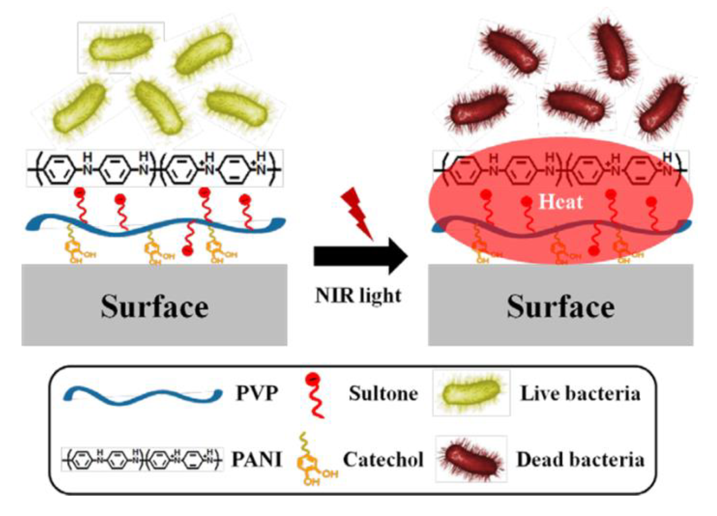
- Kim, S.H.; Kang, E.B.; Jeong, C.J.; Sharker, S.M.; In, I.; Park, S.Y. Light Controllable Surface Coating for Effective Photothermal Killing of Bacteria. ACS Appl. Mater. Interfaces 2015, 7, 15600–15606. [Google Scholar] [CrossRef]
- Korupalli, C.; Huang, C.-C.; Lin, W.-C.; Pan, W.-Y.; Lin, P.-Y.; Wan, W.-L.; Li, M.-J.; Chang, Y.; Sung, H.-W. Acidity-triggered charge-convertible nanoparticles that can cause bacterium-specific aggregation in situ to enhance photothermal ablation of focal infection. Biomaterials 2017, 116, 1–9. [Google Scholar] [CrossRef]
- Abel, S.B.; Yslas, E.I.; Rivarola, C.R.; Barbero, C.A. Synthesis of polyaniline (PANI) and functionalized polyaniline (F-PANI) nanoparticles with controlled size by solvent displacement method. Application in fluorescence detection and bacteria killing by photothermal effect. Nanotechnology 2018, 29, 125604. [Google Scholar] [CrossRef] [PubMed]
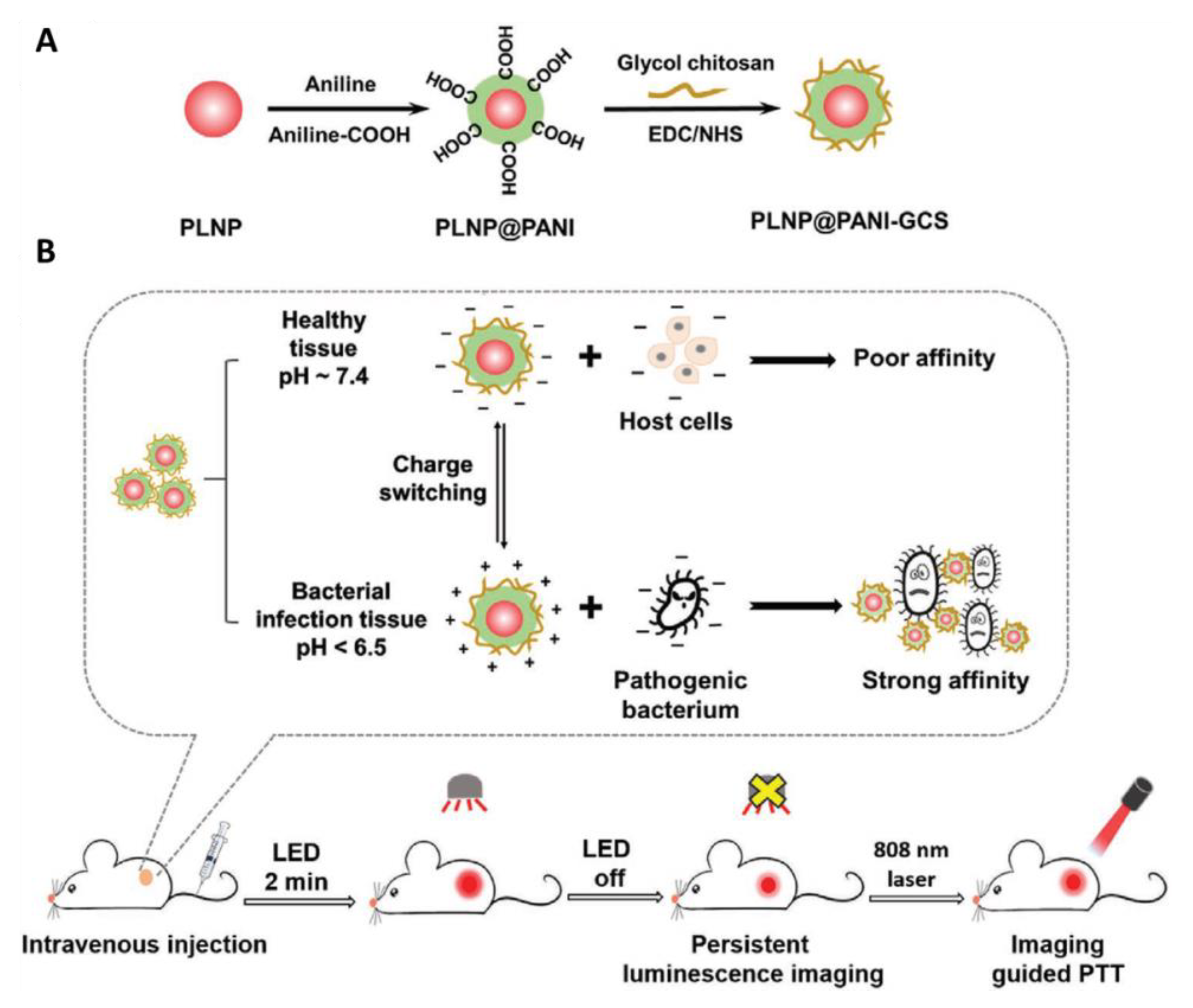
- Yan, L.X.; Chen, L.J.; Zhao, X.; Yan, X.P. pH Switchable Nanoplatform for In Vivo Persistent Luminescence Imaging and Precise Photothermal Therapy of Bacterial Infection. Adv. Funct. Mater. 2020, 30, 1909042. [Google Scholar] [CrossRef]
- Lardner, A. The effects of extracellular pH on immune function. J. Leukoc. Biol. 2001, 69, 522–530. [Google Scholar] [PubMed]


| Composition of PTA | PTA Structure | Irradiation Conditions | In Vitro | In Vivo | Function | Reference |
|---|---|---|---|---|---|---|
| PANI/PEG | NPs | 808, 2.45 W/cm2 | A431 | Yes | PTT | [14] |
| PANI/F-127 | NPs | 808, 0.5 W/cm2 | HCT116 | Yes | PTT | [30] |
| PANI/PVP | NPs | 785, 0.5 W/cm2 | LM2 | No | PTT | [39,40] |
| UiO66@PANI | NPs | 808, 0.7 W/cm2 | CT26 | Yes | PTT | [41] |
| PANI/PSS | NPs | 808, 2.08 W/cm2 | HeLa | No | PTT | [42] |
| PANI/GdIII/PVA/CET | NPs | 808, 1.5 W/cm2 | A431 | Yes | Targeting, MRI, PTT | [43] |
| PANI/Cu(II) | NPs | 808, 0.3 W/cm2 | HeLa | Yes | MRI, PTT | [44] |
| PANI/MnFe2O4/ pyrene/CET | NPs | 808, 5 W/cm2 | A431 | No | Targeting MRI, PTT | [45] |
| PANI/Lipid/FA | NPs | 808, 2.0 W/cm2 | Hela | Yes | PAI, PTT | [46] |
| PANI/Ag/ICG/ PEG | NPs | 808, 1.0 W/cm2 | Hela | Yes | PAI, FI, PTT, PDT | [47] |
| PANI/Si/HA-DA | NPs | 808, 2.0 W/cm2 | MDAMB-231 KB MDCK | Yes | FI, PTT | [48] |
| PANI/γ-PGA | NPs | 808, 1.5 W/cm2 | 4T1 | Yes | PAI, PTT | [23] |
| PANI/HA | NPs | 808, 0.64 W/cm2 | HFF HCT-116 HeLa | Yes | Targeting, PTT | [49] |
| PANI/LA/Tween80 | NPs | 808, 10 W/cm2 | MDA-MB-231 | No | Self-doping, PTT | [50] |
| NMPA/CS | Hydrogel | 808, 0.5 W/cm2 | Hep3B | Yes | Self-doping, PTT | [51] |
| PANI/Au/PEG | NPs | 808, 2 W/cm2 | HEK293T HepG2 HeLa | Yes | Charge transfer, PTT | [52] |
| PANI/BSA | NPs | 808, 1.0 W/cm2 | 4T1 | Yes | Self-doping, PAI, PTT | [31] |
| PANI/LT/MTX/PVP/SDS/PLGA/DSPE-PEG2000-mal | NPs | 808, 2 W/cm2 | MCF-7 MDA-MB-231 | Yes | Targeting, Chemotherapy PTT | [53] |
| PANI/5-FU/ZIF-8 | NPs | 980, 0.8 W/cm2 | MCF7 | Yes | Chemotherapy, PTT | [54] |
| PANI/cisplatin/lecithin/cRGD or FA-PEG-DSPE | NPs | 808, 1.54 W/cm2 | MGC-803 MDA-MB-231 | No | Targeting, Chemotherapy, PTT | [55] |
| PANI/DOX/Si | NPs | 808, 1.4 W/cm2 | 4T1 | Yes | Chemotherapy, PTT | [56] |
| PANI/cisplatin/lecithin/PEG-PCL/ Tmab | NPs | 808, 1.54 W/cm2 | SK-BR-3 | No | Targeting, Chemotherapy, PTT | [57] |
| PANI/Ce6/lecithin/cRGD-PEG-PCL | NPs | 808, 1.54 W/cm2 | MGC-803 MCF-7 | No | Targeting, Chemotherapy, PTT, PDT | [58] |
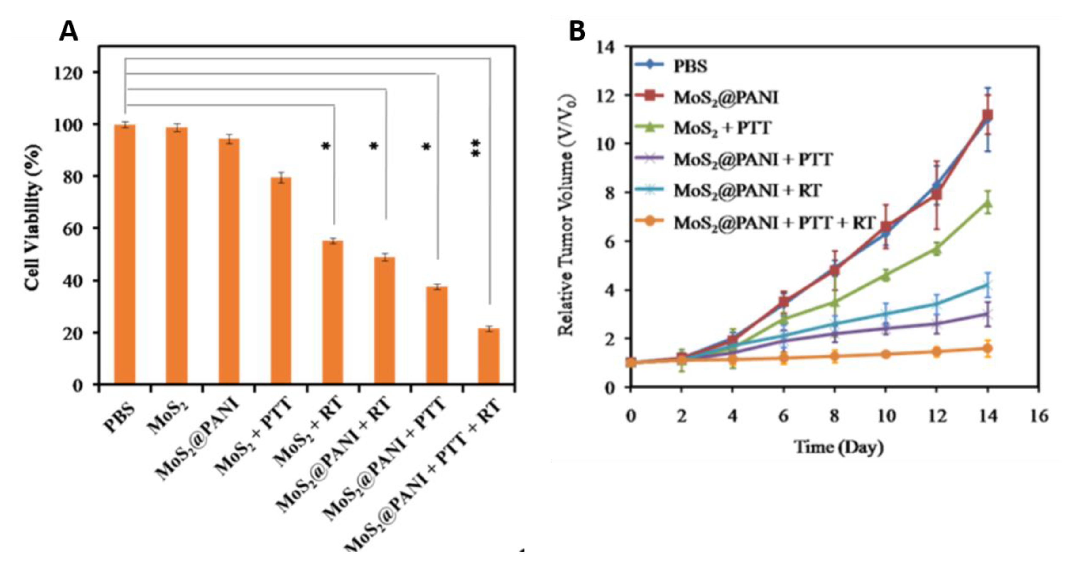
| PANI/MoS2/PEG | NPs | 808, 1.5 W/cm2 | 4T1 | Yes | PAI, CTI RT, PTT | [59] |
| PANI/WS2/Ce6/HA | NPs | 808, 1.5 W/cm2 | 4T1 | Yes | FI, PAI, CTI, PTT, PDT, RT | [60] |
| PANI/R848/GCS | NPs | 808, 0.9 W/cm2 | CT26 | Yes | Immunotherapy, PTT | [61] |
| Composition of PTA | PTA Structure | Irradiation Conditions | Bacteria | In Vivo | Functions | Reference |
|---|---|---|---|---|---|---|
| PANI/PVPS | NPs | 808, 2 W/cm2 | S. aureus,
E. coli | No | PTT | [94] |
| NMPA/CS | Hydrogel | 808, 0.5 W/cm2 | MRSA | Yes | PTT | [29] |
| PANI/GCS | NPs | 808, 0.6 W/cm2 | MRSA E. coli | Yes | Targeting, Imaging, PTT | [95] |
| PANI/polymer | NPs | 785, 0.5 W/cm2 | P. aeruginosa | No | Imaging | [96] |
| PANI/PLNP/GCS | NPs | 808, 1.5 W/cm2 | S. aureus, E. coli MRSA | Yes | Targeting, Imaging | [97] |
© 2020 by the authors. Licensee MDPI, Basel, Switzerland. This article is an open access article distributed under the terms and conditions of the Creative Commons Attribution (CC BY) license (http://creativecommons.org/licenses/by/4.0/).
Share and Cite
Korupalli, C.; Kalluru, P.; Nuthalapati, K.; Kuthala, N.; Thangudu, S.; Vankayala, R. Recent Advances of Polyaniline-Based Biomaterials for Phototherapeutic Treatments of Tumors and Bacterial Infections. Bioengineering 2020, 7, 94. https://0-doi-org.brum.beds.ac.uk/10.3390/bioengineering7030094
Korupalli C, Kalluru P, Nuthalapati K, Kuthala N, Thangudu S, Vankayala R. Recent Advances of Polyaniline-Based Biomaterials for Phototherapeutic Treatments of Tumors and Bacterial Infections. Bioengineering. 2020; 7(3):94. https://0-doi-org.brum.beds.ac.uk/10.3390/bioengineering7030094
Chicago/Turabian StyleKorupalli, Chiranjeevi, Poliraju Kalluru, Karthik Nuthalapati, Naresh Kuthala, Suresh Thangudu, and Raviraj Vankayala. 2020. "Recent Advances of Polyaniline-Based Biomaterials for Phototherapeutic Treatments of Tumors and Bacterial Infections" Bioengineering 7, no. 3: 94. https://0-doi-org.brum.beds.ac.uk/10.3390/bioengineering7030094