Left Pulmonary Artery from the Ascending Aorta: A Case Report and Review of Published Cases

 , ,
, ,
Abstract
:1. Introduction
2. Methods
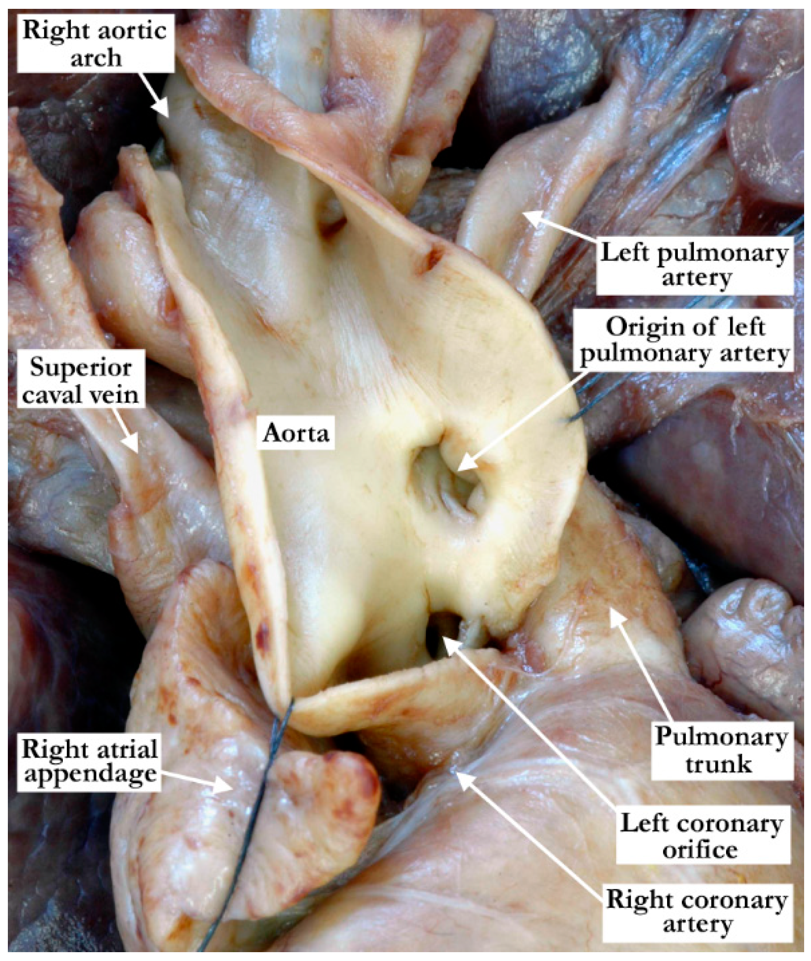
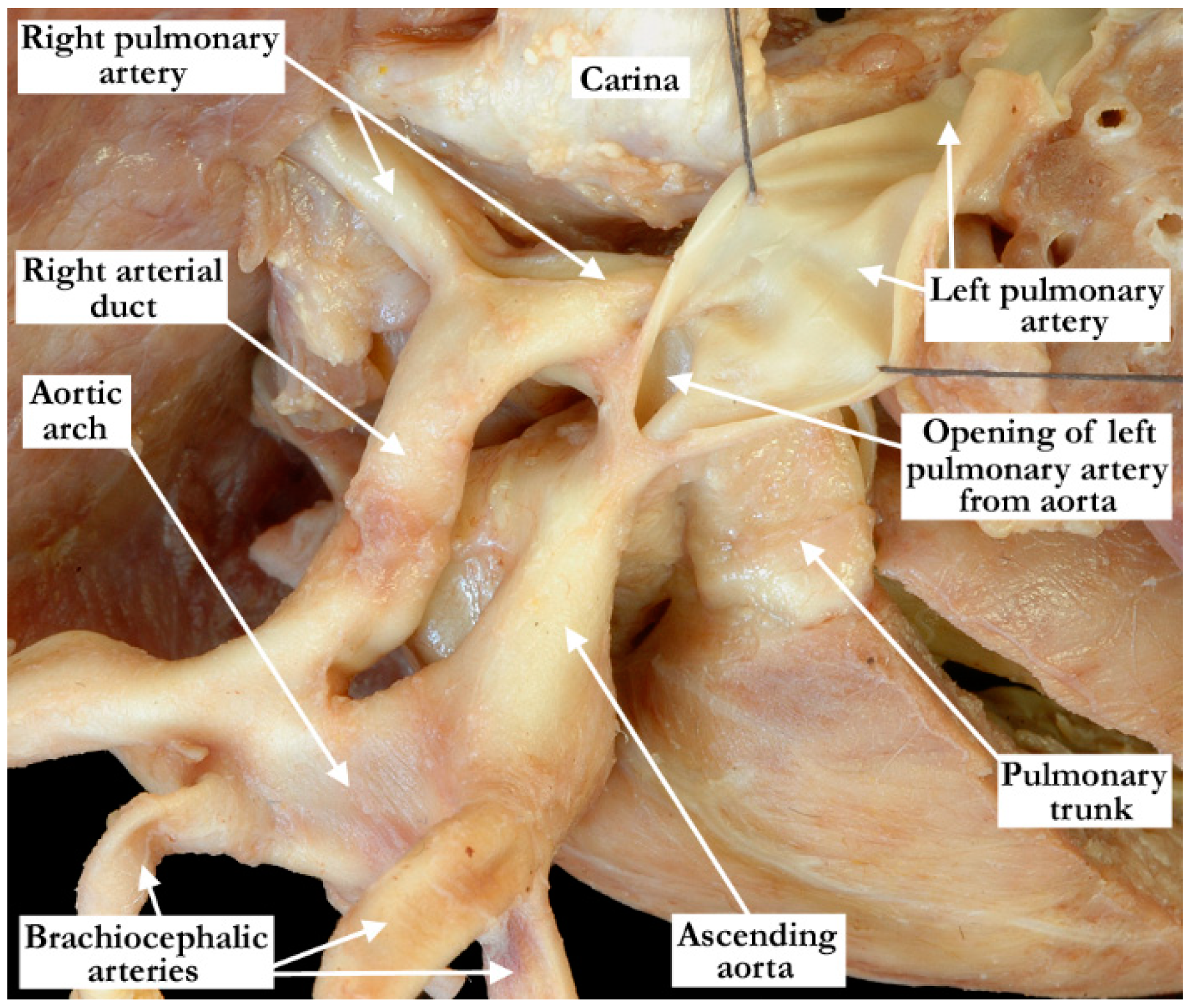
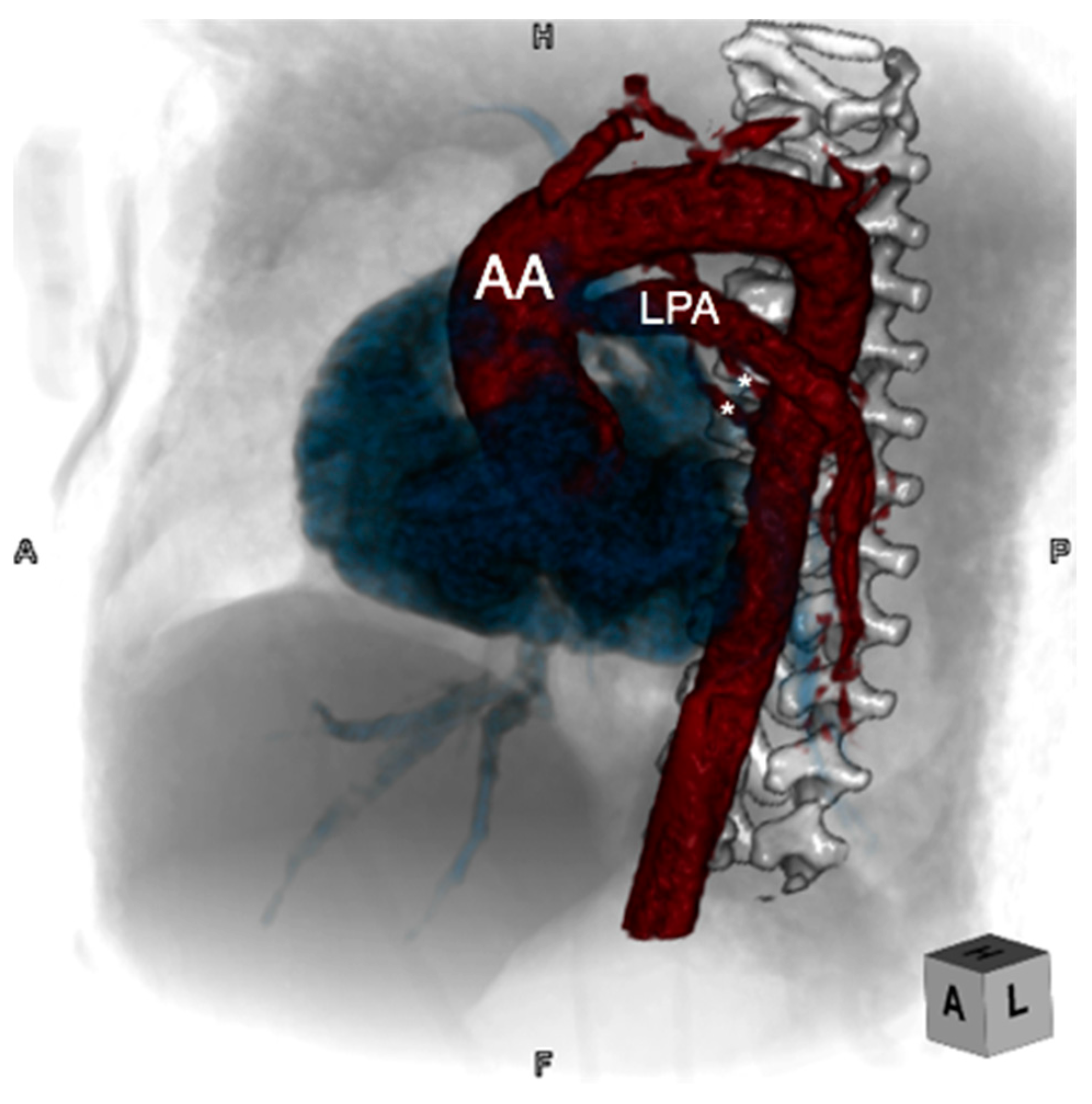
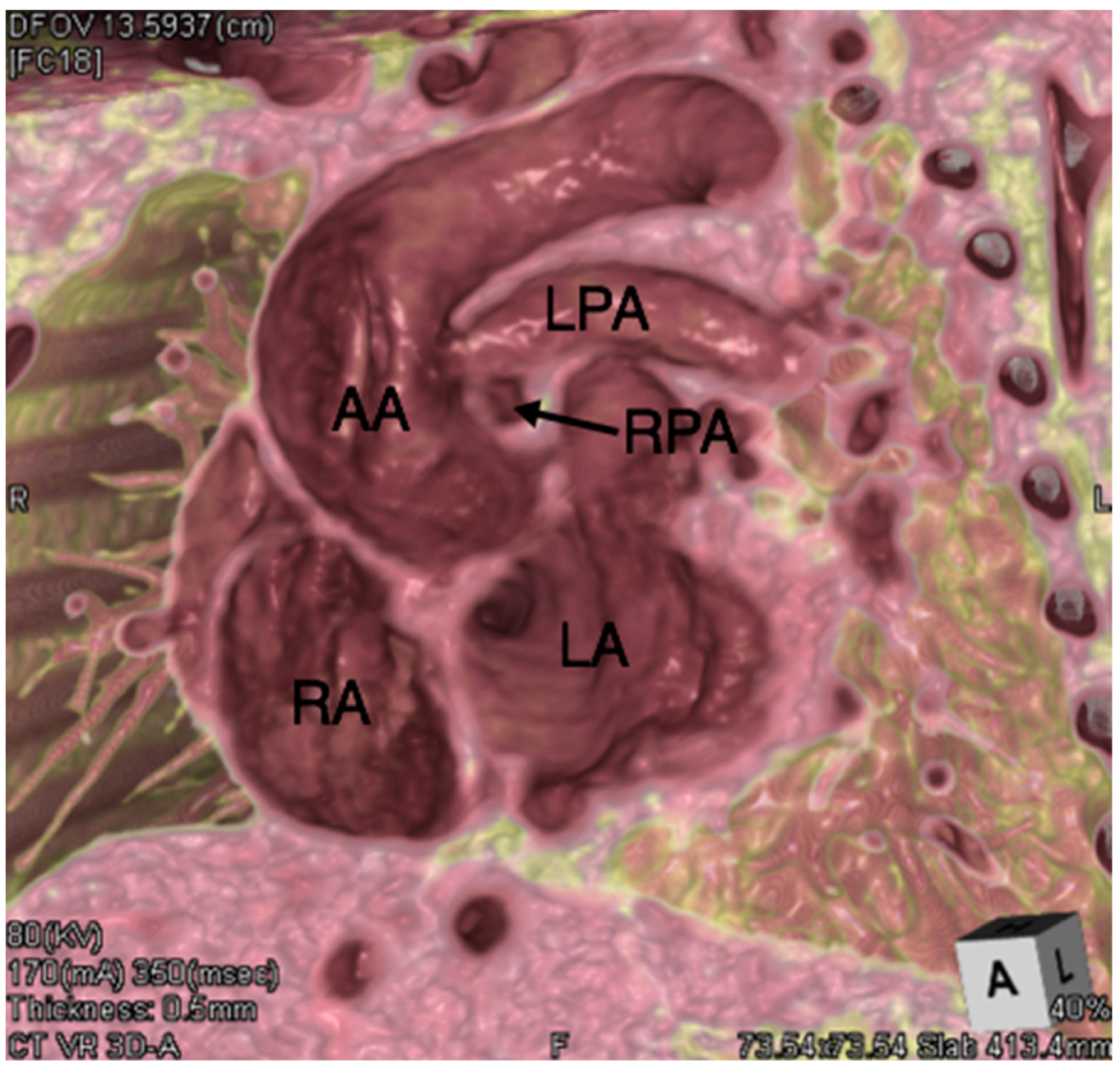
3. Case Report
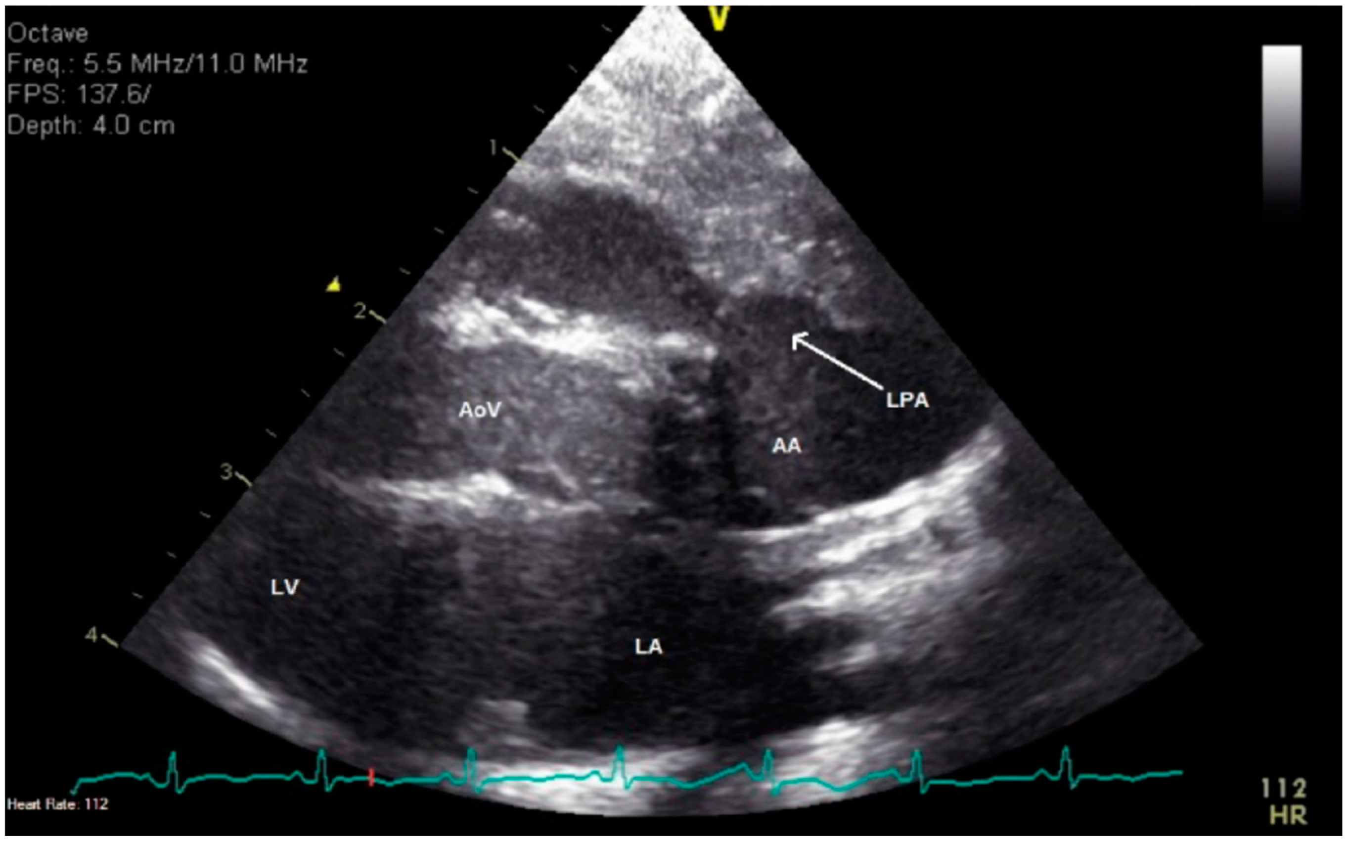
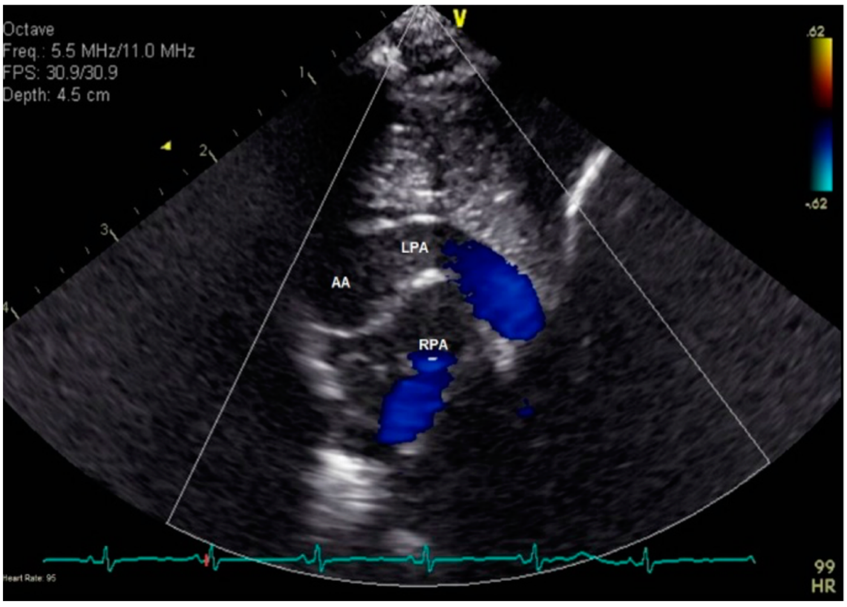
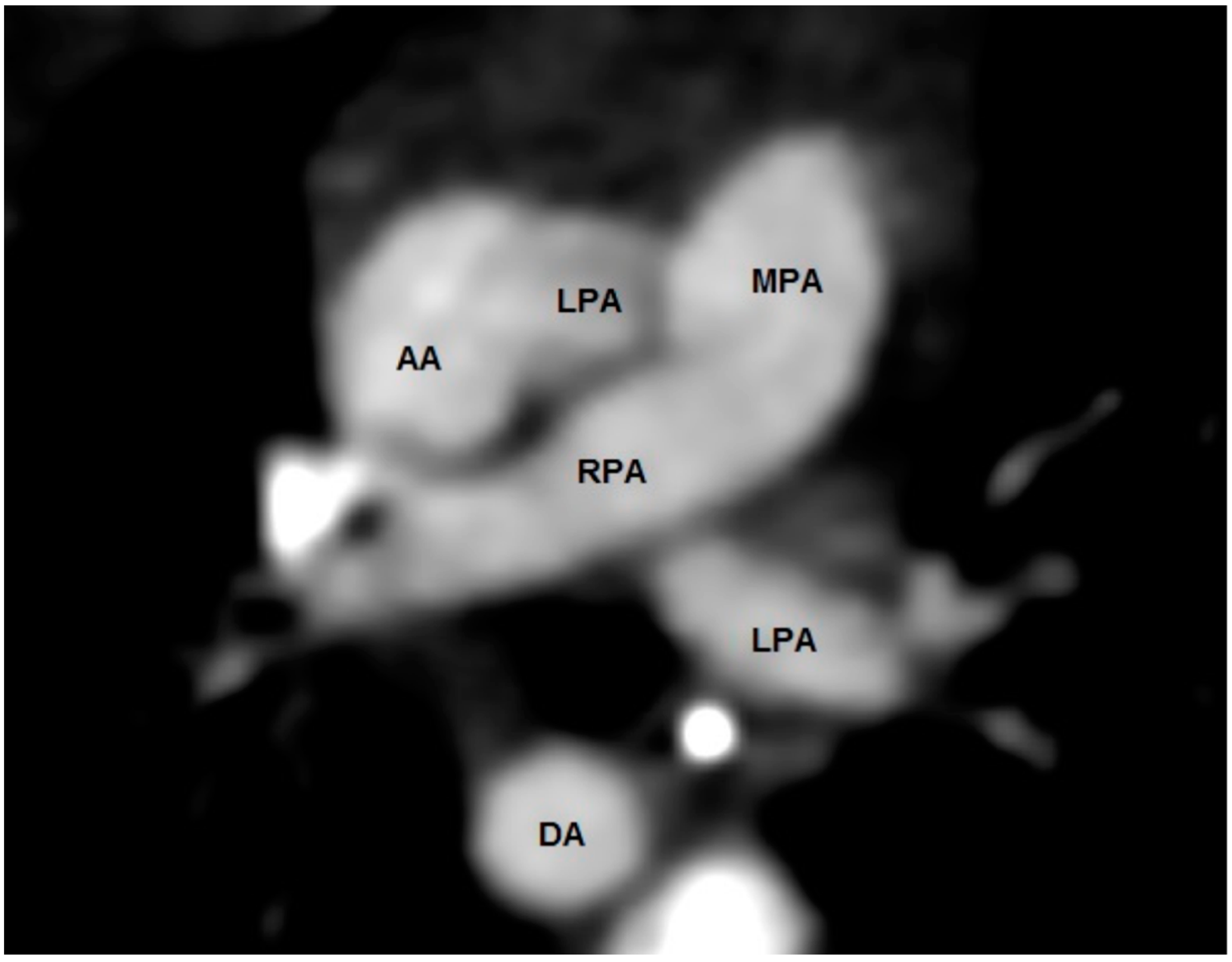
3.1. Clinical History
3.2. Review of Published Cases
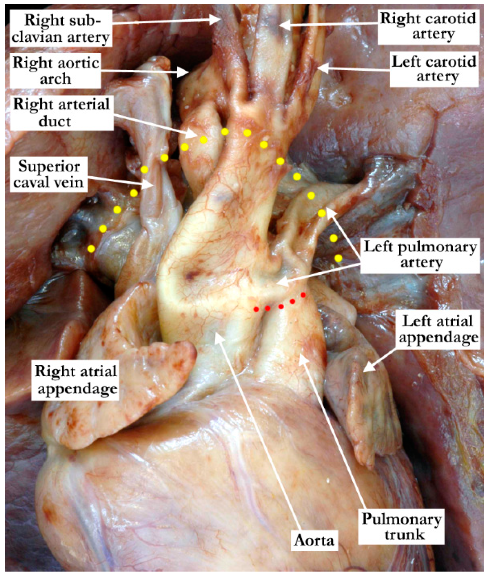
4. Discussion
5. Conclusions
Author Contributions
Funding
Institutional Review Board Statement
Data Availability Statement
Conflicts of Interest
References
- Fraentzel, O. Ein Fall von abnormer Communication der Aorta mit der Arteria pulmonalis. Virchows Archiv. 2005, 43, 420–426. [Google Scholar] [CrossRef] [Green Version]
- Carretero, J.; Rissech, M.; Mortera, C.; Mayol, J.; Caffarena, J.; Prada, F. Aortic Origin of the Left Pulmonary Artery in an Infant With Fallot’s Tetralogy. Rev. Española Cardiol. 2005, 58, 1124–1126. [Google Scholar] [CrossRef] [Green Version]
- Kutsche, L.M.; Van Mierop, L. Anomalous origin of a pulmonary artery from the ascending aorta: Associated anomalies and pathogenesis. Am. J. Cardiol. 1988, 61, 850–856. [Google Scholar] [CrossRef]
- Gerlis, L.M.; Ho, S.Y.; Smith, A.; Anderson, R.H. The site of origin of nonconfluent pulmonary arteries from a common arterial trunk or from the ascending aorta: Its morphological significance. Am. J. Cardiovasc. Pathol. 1990, 3, 115–120. [Google Scholar] [PubMed]
- Bastos, P.; De Sousa, A.R.; Casanova, J.; Moreira, J.; Alvares, S.; Mota, C.R.; Gomes, M.R. Vascular rings: Surgical treatment. Rev. Port. Cardiol. 1994, 13, 707–715. [Google Scholar]
- Libsona, R.; Derbekyan, V.; Novales-Diaz, J. Hemitruncus Anomaly as a Cause of Unilateral Absent Lung Perfusion with nor-mal ventilation on radionuclide lung scan. Clin. Nucl. Med. 1996, 21, 655–6556. [Google Scholar]
- Zhang, H.; Xu, Z.; Su, Z.; Ding, W. Single stage repair of anomalous origin of one pulmonary artery branch from ascending aorta with associated lesions. J. Shanghai Jiaotong Univ. 2007, 27, 1370–1372. [Google Scholar]
- Erdem, A.; Aydemir, N.A.; Demir, H.; Zeybek, C.; Sarıtaş, T.; Akdeniz, C.; Karaci, A.R.; Çelebi, A. Anomalous origin of one pulmonary artery branch from the ascending aorta: Ex-perience of our center. Turk Kardiyol. Dern. Arş. Arch Turk Soc. Cardiol. 2010, 38, 411–415. [Google Scholar]
- Goldstein, B.H.; Bergersen, L.; Powell, A.J.; Graham, D.A.; Bacha, E.A.; Lang, P. Long-Term Outcome of Surgically Repaired Unilateral Anomalous Pulmonary Artery Origin. Pediatr. Cardiol. 2010, 31, 944–951. [Google Scholar] [CrossRef]
- Weintraub, R.A.; Fabian, C.E.; Adams, D.F. Ectopic Origin of One Pulmonary Artery from the Ascending Aorta. Radiology 1966, 86, 666–676. [Google Scholar] [CrossRef]
- Caudill, D.R.; Helmsworth, J.A.; Daoud, G.; Kaplan, S. Anomalous origin of left pulmonary artery from ascending aorta. J. Thorac. Cardiovasc. Surg. 1969, 57, 493–506. [Google Scholar] [CrossRef]
- Haddadin, R.B. Left hemitruncus: A rare congenital heart condition. East. Mediterr. Heal. J. 2014, 19 (Suppl. 3), S224–S226. [Google Scholar]
- Nigam, A.; Trehan, V. Left hemitruncus with normal right-sided pressures in an adult. Cardiol. Young 2013, 24, 926–928. [Google Scholar] [CrossRef] [PubMed]
- Akyüz, M.; Işık, O.; Ayık, M.F.; Atay, Y. Total Correction without Cardiopulmonary Bypass of Isolated Left Hemitruncus Arteriosus. J. Pediatr. Res. 2015, 2, 233–235. [Google Scholar] [CrossRef]
- Vázquez, R.M.; Chávez, I.O.M.; Soto, M.E.; Bahena, E.J.P.; Zárate, R.C.; Flores, A.C.C.; Colmenero, J.C.; García, A.M.; Hernández, A.B.; Lans, V.G.; et al. Anomalous origin of pulmonary branches from the ascending aorta. A report of five cases and review of the literature. J. Cardiol. Cases 2015, 11, 1–6. [Google Scholar] [CrossRef] [PubMed] [Green Version]
- Schiller, M.; Williams, T.E.; Craenen, J.; Hosier, D.M.; Sirak, H.D. Anomalous Origin of Left Pulmonary Artery From the Ascending Aorta. Vasc. Surg. 1971, 5, 126–132. [Google Scholar] [CrossRef] [PubMed]
- Smallhorn, J.F.; Anderson, R.H.; Macartney, F.J. Two dimensional echocardiographic assessment of communications between ascending aorta and pulmonary trunk or individual pulmonary arteries. Heart 1982, 47, 563–572. [Google Scholar] [CrossRef] [Green Version]
- Benatar, A.; Kinsley, R.H.; Milner, S.; Dansky, R.; Hummel, D.A.; Levin, S.E. Surgical correction for one pulmonary artery arising from ascending aorta—Report of five cases. Int. J. Cardiol. 1987, 16, 249–255. [Google Scholar] [CrossRef]
- Mittal, P.K.; Agarwal, S.K.; Ghosh, P.K. Isolated anomalous origin of left pulmonary artery from the ascending aorta in an adult. J. Thorac. Cardiovasc. Surg. 1993, 106, 1220–1222. [Google Scholar] [CrossRef]
- Prasad, K.; Radhakrishnan, S.; Mittal, P. Anomalous origin of the left pulmonary artery from the aorta in an adult as an isolated anomaly: A case report. Int. J. Cardiol. 1993, 38, 326–329. [Google Scholar] [CrossRef]
- Tagliente, M.R.; Troise, D.; Milella, L.; Vairo, U. Isolated anomalous origin of left pulmonary artery from the ascending aorta. Am. Hear. J. 1996, 132, 1289–1292. [Google Scholar] [CrossRef]
- Aru, G.M.; English, W.P.; Gaymes, C.H.; Heath, B.J. Origin of the left pulmonary artery from the aorta: Embryologic considerations. Ann. Thorac. Surg. 2001, 71, 1008–1010. [Google Scholar] [CrossRef]
- Prifti, E.; Bonacchi, M.; Murzi, B.; Crucean, A.; Bernabei, M.; Luisi, V.S.; Bartolozzi, F.; Vanini, V. Anomalous origin of the left pulmonary artery from the aorta. Our experience and literature review. Hear. Vessel. 2003, 18, 79–84. [Google Scholar] [CrossRef] [PubMed]
- Sotomora, R.F.; Edwards, J.E. Anatomic identification of so-called absent pulmonary artery. Circulation 1978, 57, 624–633. [Google Scholar] [CrossRef] [PubMed] [Green Version]
- Purcaro, A.; Capestro, F.; Ciampani, N.; Brugnami, R. Tetralogy of Fallot with anomalous origin of the left pulmonary artery from the ascending aorta. Review of literature and description of a case. Minerva Cardioangiol. 1978, 26, 461–470. [Google Scholar] [PubMed]
- Calder, A.; Brandt, P.W.; Barratt-Boyes, B.G.; Neutze, J.M. Variant of tetralogy of fallot with absent pulmonary valve leaflets and origin of one pulmonary artery from the ascending aorta. Am. J. Cardiol. 1980, 46, 106–116. [Google Scholar] [CrossRef]
- Laborde, F.; de Riberolles, C.; Leca, F.; Rose-Pitet, L.; Hazan, E.; Neveux, J.Y. Abnormal pulmonary artery arising from the as-cending aorta. Seven case-reports (author’s transl). Ann. Pediatr. 1981, 28, 515–519. [Google Scholar]
- Duncan, W.J.; Freedom, R.M.; Olley, P.M.; Rowe, R.D. Two-dimensional echocardiographic identification of hemitruncus: Anoma-lous origin of one pulmonary artery from ascending aorta with the other pulmonary artery arising normally from right ventricle. Am. Heart J. 1981, 102, 892–896. [Google Scholar] [CrossRef]
- Zinkovskiĭ, M.F.; Buchnev, A.I. Case of successful correction of tetralogy of Fallot with abnormal branching-out of the left the pulmonary artery from the ascending aorta. Grud. Khirurgiia 1984, 3, 75–77. [Google Scholar]
- Nouri, S.; Wolverson, M.K. Anomalous Origin of a pulmonary artery from ascending aorta. In Surgery of Congenital Heart Dis-Ease: Pediatric Cardiac Care Consortium, 1984–1995; John Wiley & Sons: Hoboken, NJ, USA, 1998; pp. 99–110. [Google Scholar]
- Robida, A.; Fettich, D. Tetralogy of fallot with origin of left pulmonary artery from the ascending aorta. Pediatr. Radiol. 1985, 15, 422–423. [Google Scholar] [CrossRef]
- Makhmudov, M.M.; Abdumadzhidov, K.A.; Abdullaev, E.E.; Mirzakhmedov, B.M. A case of tetralogy of Fallot with atresia of the pulmonary trunk and origin of the left pulmonary artery from the ascending aorta. Grud. Khirurgiia 1987, 32, 82–83. [Google Scholar]
- Fong, L.V.; Anderson, R.H.; Siewers, R.D.; Trento, A.; Park, S.C. Anomalous origin of one pulmonary artery from the ascending aorta: A review of echocardiographic, catheter, and morphological features. Br. Heart J. 1989, 62, 389–395. [Google Scholar] [CrossRef] [PubMed] [Green Version]
- Thomas, H.W. Cardiovascular anomalies, congenital cardiac malformations. J. Tech. Methods 1941, 58, 21–29. [Google Scholar]
- Sasaki, T.; Shiina, Y.; Abe, K.; Kimura, H.; Hamada, Y.; Niitu, K. Surgical correction of tetralogy of Fallot with anomalous origin of left pulmonary artery from the ascending aorta. Nihon Kyobu Geka Gakkai Zasshi 1990, 38, 488–492. [Google Scholar] [PubMed]
- Saxena, A.; Shrivastava, S.; Sharma, S.K. Anomalous origin of the left pulmonary artery from the ascending aorta in a patient with tetralogy of Fallot and “absent pulmonary valve”. Int. J. Cardiol. 1991, 33, 315–317. [Google Scholar] [CrossRef]
- Endo, M.; Haneda, K.; Suzuki, Y.; Mohri, H.; Yamaki, S. Tetralogy of Fallot with anomalous origin of unilateral pulmonary ar-tery. Kyobu Geka. 1991, 44, 109–115. [Google Scholar]
- Sreeram, N.; Smith, A.; Peart, I. Fallot’s tetralogy with absent pulmonary valve and anomalous origin of the left pulmonary artery. Int. J. Cardiol. 1993, 42, 175–177. [Google Scholar] [CrossRef]
- Py, A.; Lazarus, A.; Spaulding, C.; Toussaint, M.; Planché, C.; Duboc, D.; Fouchard, J.; Guérin, F. Left pulmonary artery originating from the ascending aorta in tetralogy of Fallot. Therapeutic strategy. Arch. Mal. Coeur Vaiss. 1993, 86, 1069–1072. [Google Scholar]
- Abu-Sulaiman, R.M.; Hashmi, A.; McCrindle, B.W.; Williams, W.G.; Freedom, R.M. Anomalous origin of one pulmonary artery from the ascending aorta: 36 years’ experience from one centre. Cardiol. Young 1998, 8, 449–454. [Google Scholar] [CrossRef]
- Saliba, Z.; Le Bidois, J.; Sidi, D.; Kachaner, J.; Bonnet, D. Prenatal detection of a tetralogy of Fallot with origin of the left pulmo-nary artery from the ascending aorta in a familial 22 q11 microdeletion. Prenat Diagn. 1999, 19, 260–262. [Google Scholar] [CrossRef]
- Soylu, M.; Demir, A.D.; Tikiz, H.; Kisacik, H.; Korkmaz, U.; Korkmaz, Ş. Left hemitruncus associated with tetralogy of fallot: A case report. Catheter Cardiovasc Interv. 2000, 51, 58–60. [Google Scholar] [CrossRef]
- Amaral, F.; Teixeira, M.A.C.; Granzotti, J.A.; Manso, P.H.; Vicente, W.V.D.A. Anomalous origin of the left pulmonary artery from the ascending aorta. Successful surgical correction in an infant with Fallot’s tetralogy. Arq. Bras. Cardiol. 2002, 79, 541–543. [Google Scholar] [CrossRef] [PubMed] [Green Version]
- Krishnamoorthy, K.; Tharakan, J.; Padmakumar, R. Absent pulmonary valve and origin of left pulmonary artery from ascending aorta. Int. J. Cardiol. 2004, 96, 475–476. [Google Scholar] [CrossRef] [PubMed]
- Sikl, H. Casopis lekaru ceskych: Unusual malformation of the ductus arteriosus; departure of a principal branch of the pulmonary artery from the aorta. Cas. Lek. Ces. 1952, 14, 1366. [Google Scholar]
- Vida, V.L.; Sanders, S.P.; Bottio, T.; Maschietto, N.; Rubino, M.; Milanesi, O.; Stellin, G. Anomalous origin of one pulmonary artery from the ascending aorta. Cardiol. Young 2005, 15, 176–181. [Google Scholar] [CrossRef]
- Cheng, W.; Xiao, Y.; Zhong, Q.; Wen, R. Anomalous Origin of Left Pulmonary Artery Branch from the Aorta with Fallot’s Tetralogy. Thorac. Cardiovasc. Surg. 2008, 56, 432–434. [Google Scholar] [CrossRef]
- Diab, K.; Richardson, R.; Pophal, S.; Alboliras, E. Left Hemitruncus Associated with Tetralogy of Fallot: Fetal Diagnosis and Postnatal Echocardiographic and Cardiac Computed Tomographic Confirmation. Pediatr. Cardiol. 2009, 31, 534–537. [Google Scholar] [CrossRef]
- Sun, H.-F.; Qin, C.; Liang, J.-L. One-Stage Correction of Tetralogy of Fallot Associated with Anomalous Origin of the Left Pulmonary Artery from the Aorta and Aortopulmonary Window. J. Card. Surg. 2011, 27, 112–114. [Google Scholar] [CrossRef]
- Dwivedi, S.K.; Vijay, S.K.; Chandra, S.; Saran, R.K. Left Hemitruncus with Tetralogy of Fallot and Right Aortic Arch: Rare Survival beyond the First Decade. Pediatr. Cardiol. 2012, 33, 863–865. [Google Scholar] [CrossRef]
- Reid, D.A.; Jiwani, A.K.; Desai, P.H.; Aly, A.M. Absent Pulmonary Valve Syndrome with Left Hemitruncus. Am. J. Perinatol. Rep. 2012, 2, 51–54. [Google Scholar] [CrossRef] [Green Version]
- Garg, P.; Talwar, S.; Kothari, S.S.; Saxena, A.; Juneja, R.; Choudhary, S.K.; Airan, B. The anomalous origin of the branch pulmonary artery from the ascending aorta. Interact. Cardiovasc. Thorac. Surg. 2012, 15, 86–92. [Google Scholar] [CrossRef] [PubMed] [Green Version]
- Tantiwongkosri, K.; Chanthong, P.; Sriyodcharti, S. Hemitruncus Arteriosus. J. Am. Coll. Cardiol. 2013, 61, 1653. [Google Scholar] [CrossRef] [PubMed]
- Tsukimori, K.; Morihana, E.; Fusazaki, N.; Fujita, Y. Prenatal Images of Left Hemitruncus in Tetralogy of Fallot With an Absent Pulmonary Valve. Pediatr. Cardiol. 2012, 34, 1285–1287. [Google Scholar] [CrossRef] [PubMed]
- Talwar, S.; Meena, A.; Choudhary, S.K.; Kothari, S.S.; Gupta, S.K.; Saxena, A.; Juneja, R.; Airan, B. Anomalous branch of pulmonary artery from the aorta and tetralogy of Fallot: Morphology, surgical techniques and results. Eur. J. Cardiothoracic Surg. 2014, 46, 291–296. [Google Scholar] [CrossRef] [Green Version]
- Czarnecki, S.W.; Hopeman, A.R.; Child, P.L. Tetralogy of Fallot with Aortic Origin of the Left Pulmonary Artery: Radiographic and Angiocardiographic Considerations. Dis. Chest 1964, 46, 97–101. [Google Scholar] [CrossRef]
- Mathur, A.; Mathur, A.; Garg, P.; Sharma, N.; Mittal, P. Anomalous origin of left pulmonary artery from aorta with tetralogy of Fallot in an adult: What is protecting the lung? Indian J. Thorac. Cardiovasc. Surg. 2014, 31, 187–189. [Google Scholar] [CrossRef]
- Liu, H.; Juan, Y.-H.; Chen, J.; Xie, Z.; Wang, Q.; Zhang, X.; Liang, C.; Huang, H.; Kwong, R.Y.; Saboo, S.S. Anomalous Origin of One Pulmonary Artery Branch From the Aorta: Role of MDCT Angiography. Am. J. Roentgenol. 2015, 204, 979–987. [Google Scholar] [CrossRef]
- Paredes, A.M.; Caspi, J.; Lilje, C. Tetralogy of Fallot and Anomalous Origin of the Left Pulmonary Artery From the Ascending Aorta Associated With Absent Right Internal Carotid Artery. World J. Pediatr. Congenit. Hear. Surg. 2015, 6, 324–327. [Google Scholar] [CrossRef]
- Cho, S.; Kim, W.-H.; Choi, E.S.; Lee, J.R.; Kim, Y.J. Surgical Results of Anomalous Origin of One Pulmonary Artery Branch from the Ascending Aorta. Pediatr. Cardiol. 2015, 36, 1532–1538. [Google Scholar] [CrossRef]
- Wyler, F.; Rutishauser, M.; Olafsson, A.; Kaufmann, H.J. Congenital absence of the pulmonary valve in tetralogy of Fallot and origin of the left pulmonary artery from the aortic arch. Am. J. Roentgenol. 1970, 110, 505–508. [Google Scholar] [CrossRef] [Green Version]
- Morgan, J.R. Left Pulmonary Artery from Ascending Aorta in Tetralogy of Fallot. Circulation 1972, 45, 653–657. [Google Scholar] [CrossRef] [PubMed] [Green Version]
- Keane, J.F.; Maltz, D.; Bernhard, W.F.; Corwin, R.D.; Nadas, A.S. Anomalous Origin of One Pulmonary Artery from the Ascending Aorta. Circulation 1974, 50, 588–594. [Google Scholar] [CrossRef] [PubMed] [Green Version]
- Robin, E.; Silberberg, B.; Ganguly, S.N.; Magnisalis, K. Aortic origin of the left pulmonary artery. Am. J. Cardiol. 1975, 35, 324–329. [Google Scholar] [CrossRef]
- Calazel, P.; Martinez, J. Abnormal origin of 1 of 2 pulmonary arteries from the ascending aorta. Arch. Mal. Coeur Vaiss. 1975, 68, 397–403. [Google Scholar]
- Razavi, R.; Miquel, M.E.; Baker, E. Diagnosis of Hemi-Truncus Arteriosis by Three-Dimensional Magnetic Resonance Angiography. Circulation 2004, 109, e15–e16. [Google Scholar] [CrossRef] [Green Version]
- Khositseth, A.; Siripornpitak, S.; Pornkul, R. Pulmonary atresia and ventricular septal defect with collaterals to right lung associated with anomalous left pulmonary artery from the ascending aorta. Pediatr. Radiol. 2010, 40, 72–76. [Google Scholar] [CrossRef]
- Pepeta, L.; Takawira, F.F.; Cilliers, A.M.; Adams, P.; Ntsinjana, N.; Mitchell, B.J. Anomalous origin of the left pulmonary artery from the ascending aorta in two children with pulmonary atresia, subaortic ventricular septal defect and right-sided major aorto-pulmonary collateral arteries. Cardiovasc. J. Afr. 2011, 22, 268–271. [Google Scholar] [CrossRef] [Green Version]
- Wang, J.-N.; Wu, J.-M.; Yang, Y.-J. Double-lumen aortic arch with anomalous left pulmonary artery origin from the main pul-monary artery-bilateral persistent fifth aortic arch—A case report. Int. J. Cardiol. 1999, 69, 105–108. [Google Scholar] [CrossRef]
- Belenkov, Y.N.; Makhachev, O.A.; Sobolev, A.V.; Plakhova, V.V.; Gorbachevsky, S.V.; Zaets, S.B. Double Outlet Right Ventricle with Anomalous Left Pulmonary Artery. Asian Cardiovasc. Thorac. Ann. 2008, 16, e18–e20. [Google Scholar] [CrossRef]
- Nicholson, G.T.; Petit, C.J.; Pasquarella, T.; Ferguson, M.E. Persistent 5th Aortic Arch with Coexisting Discontinuous Pulmonary Arteries, Double-Outlet Right Ventricle, and Complete Atrioventricular Septal Defect. Tex. Hear. Inst. J. 2015, 42, 92–93. [Google Scholar] [CrossRef] [Green Version]
- Mudd, J.G.; Willman, V.L.; Riberi, A. Origin of One Pulmonary Artery From The Aorta. Am. Rev. Respir. Dis. 1964, 89, 255–263. [Google Scholar] [PubMed]
- Verel, D.; Grainger, R.G. Cardiac Catheterization and Angiocardiography. Cardionasc. Clin. 1971, 3, 81–96. [Google Scholar]
- Santos, M.A.; Manuel, V.; Azevedo, P. Anomalous Origin of One Pulmonary Artery from the Ascending Aorta: Surgical Repair Resolving Pulmonary Arterial Hypertension. Arq. Bras. Cardiol. 2004, 83, 503–507. [Google Scholar]
- Nathan, M.; Rimmer, D.; Piercey, G.; Del Nido, P.J.; Mayer, J.E.; Bacha, E.A.; Pigula, F.A. Early repair of hemitruncus: Excellent early and late outcomes. J. Thorac. Cardiovasc. Surg. 2007, 133, 1329–1335. [Google Scholar] [CrossRef] [PubMed] [Green Version]
- Li, L.; Su, J.; Wu, Y.; Luo, Y. A Simple and Feasible Technique for Repair of Anomalous Origin of the Left Pulmonary Artery from the Ascending Aorta. Pediatr. Cardiol. 2007, 29, 420–421. [Google Scholar] [CrossRef]
- Sanz, L.M.; Guillén, A.G.; Maresma, J.L.; Reyez, L.M.S. Repair of an Anomalous Origin of the Left Pulmonary Artery From the Ascending Aorta. Ann. Thorac. Surg. 2013, 96, e121–e123. [Google Scholar] [CrossRef]
- Matsubayashi, K.; Ueda, Y.; Ogino, H.; Matsumura, M. A Case of Anomalous Origin of the Pulmonary Arteries: Right Pulmonary Artery from the Descending Aorta and the Left Pulmonary Artery from the Ascending Aorta. Pediatr. Cardiol. 1999, 20, 298–300. [Google Scholar] [CrossRef]
- Salaymeh, K.J.; Kimball, T.R.; Manning, P.B. Anomalous pulmonary artery from the aorta via a patent ductus arteriosus: Repair in a premature infant. Ann. Thorac. Surg. 2000, 69, 1259–1261. [Google Scholar] [CrossRef]
- Amir, G.; Frenkel, G.; Bruckheimer, E.; Dagan, T.; Katz, J.; Berant, M.; Vidne, B.; Birk, E. Anomalous origin of the pulmonary artery from the aorta: Early diagnosis and repair leading to immediate physiological correction. Cardiol. Young 2010, 20, 654–659. [Google Scholar] [CrossRef]
- Sechtem, U.; Jungehulsingt, M.; Viviet, R.D.E.; Mennicken, U.; Höpp, H.W. Left hemitruncus in adulthood: Diagnostic role of magnetic resonance Imaging. Eur. Heart J. 1991, 12, 1040–1044. [Google Scholar]
- Hussain, A.S.; Shakir, M.; Ariff, S.; Ali, R.; Hassan, B. Anomalous Origin of the Left Pulmonary Artery: Hemi-Truncus Arteriosus. APSP J. Case Rep. 2017, 8, 9. [Google Scholar] [CrossRef] [PubMed] [Green Version]
- Herbert, W.H.; Rohman, M.; Farnsworth, P.; Swamy, S. Anomalous Origin of Left Pulmonary Artery from Ascending Aorta, Right Aortic Arch and Right Patent Ductus Arteriosus. Chest 1973, 63, 459–461. [Google Scholar] [CrossRef] [PubMed]
- Brill, R.; Velez, E.G.; Colagiuri, P.M. Anomalous left pulmonary artery arising from the ascending aorta. J. Med. Soc. N. J. 1973, 70, 568–570. [Google Scholar]
- Dodo, H.; Alejos, J.C.; Perloff, J.K.; Laks, H.; Drinkwater, D.C.; Williams, R.G. Anomalous origin of the left main pulmonary artery from the ascending aorta associated with Digeorge syndrome. Am. J. Cardiol. 1995, 75, 1294–1295. [Google Scholar] [CrossRef]
- Sett, S.S.; Sandor, G.G.; Mawson, J.B. Interrupted right aortic arch and origin of the left pulmonary artery from the aorta in DiGeorge syndrome. Cardiol. Young 2001, 11, 676–679. [Google Scholar] [CrossRef] [PubMed]
- Jacobs, M.L. Congenital Heart Surgery Nomenclature and Database Project: Truncus arteriosus. Ann. Thorac. Surg. 2000, 69, 50–55. [Google Scholar] [CrossRef]
- Penkoske, P.A.; Castañeda, A.R.; Fyler, D.C.; Van Praagh, R. Origin of pulmonary artery branch from ascending aorta. J. Thorac. Cardiovasc. Surg. 1983, 85, 537–545. [Google Scholar] [CrossRef]
- Selcuk, T.; Bilgili, C.; Otcu, H.; Savas, Y.; Cakmak, G.; Cengel, F. Anomalous Orıgın of the Left Pulmonary Artery from the Ascendıng Aorta wıth Pulmonary Atresıa ın a 13 Year-old Girl. Br. J. Med. Med. Res. 2015, 5, 719–723. [Google Scholar] [CrossRef]
- Gilboa, S.M.; Salemi, J.L.; Nembhard, W.N.; Fixler, D.E.; Correa, A. Mortality Resulting From Congenital Heart Disease among Children and Adults in the United States, 1999 to 2006. Circulation 2010, 122, 2254–2263. [Google Scholar] [CrossRef] [Green Version]
- Geyer, S.; Mohun, T.J.; Weninger, W.J. Visualizing Vertebrate Embryos with Episcopic 3D Imaging Techniques. Sci. World J. 2009, 9, 1423–1437. [Google Scholar] [CrossRef]
- Anderson, R.H.; Webb, S.; Brown, N.A.; Lamers, W.; Moorman, A. Development of the heart: (3) formation of the ventricular out-flow tracts, arterial valves, and intrapericardial arterial trunks. Heart 2003, 89, 1110–1118. [Google Scholar] [CrossRef] [PubMed]
- Chiu, C.C.; Wu, J.R.; Chen, H.M.; Lin, Y.T. Persistent fifth aortic arch: An ignored and underestimated disease. Jpn. Heart J. 2000, 41, 665–671. [Google Scholar] [CrossRef] [PubMed] [Green Version]
- Gupta, S.K.; Gulati, G.S.; Anderson, R.H. Clarifying the anatomy of the fifth arch artery. Ann. Pediatr. Cardiol. 2016, 9, 62–67. [Google Scholar] [CrossRef]
- Calder, A. Variants of tetralogy of fallot. Am. J. Cardiol. 1982, 53, 106–116. [Google Scholar]
- Cho, M.-J.; Park, J.A.; Lee, H.D. A Variant of Tetralogy of Fallot with Absent Pulmonary Valve: Left Pulmonary Artery Originate from Left Patent Ductus Arteriosus. Pediatr. Cardiol. 2009, 31, 153–154. [Google Scholar] [CrossRef]
- Changela, V.P.; John, C.; Maheshwari, S. Unusual Cardiac Associations with Tetralogy of Fallot—A Descriptive Study. Pediatr. Cardiol. 2010, 31, 785–791. [Google Scholar] [CrossRef]
- Dipchand, A.I.; Giuffre, M.; Freedom, R.M. Tetralogy of Fallot with non-confluent pulmonary arteries and aortopulmonary septal defect. Cardiol. Young 1999, 9, 75–77. [Google Scholar] [CrossRef]
- Kobayashi, D.; Cook, A.L.; Williams, D.A. Anomalous Origin of Left Pulmonary Artery from the Aorta with Partial Anomalous Pulmonary Venous Return. Pediatr. Cardiol. 2009, 31, 560–561. [Google Scholar] [CrossRef]
- Jedeikin, R.; Rheuban, K.S.; Carpenter, M.A.; Kron, I.L. Ductal origin of the left pulmonary artery in severe tetralogy of Fallot: Problems in management. Pediatr. Cardiol. 1984, 5, 323–326. [Google Scholar] [CrossRef]
- Peng, E.W.; Pollock, J.; Shanmugam, G.; MacArthur, K.J. Ascending aortic origin of a branch pulmonary artery—Surgical management and long-term outcome. Eur. J. Cardiothoracic Surg. 2004, 26, 762–766. [Google Scholar] [CrossRef] [Green Version]
- Godown, J.; Baker, C.; Kuhn, N.; Brown, K.; Buck, S.; Mill, M.; Price, W. Delayed Repair of Hemitruncus in an Extremely Low Birth Weight Infant. Congenit. Hear. Dis. 2011, 8, E13–E16. [Google Scholar] [CrossRef] [PubMed]
- Prifti, E.; Crucean, A.; Bonacchi, M.; Bernabei, M.; Leacche, M.; Murzi, B.; Bartolozzi, F.; Vanini, V. Postoperative outcome in patients with anomalous origin of one pulmonary artery branch from the aorta. Eur. J. Cardiothorac. Surg. 2003, 24, 21–27. [Google Scholar] [CrossRef] [Green Version]


| Year | Author | Age/Sex | Arch | Additional Defect | Surgical Procedure | Outcome | Follow Up |
|---|---|---|---|---|---|---|---|
| 1941 | Thomas | 3 mo/F | -- | TOF | None | Died | Died |
| 1952 | Sikl | Stillborn/M | Right | TOF | None | Died | Died |
| 1964 | Mudd | 1 yr/M | Left | VSD | None | Alive | Alive at 4 yr |
| 1964 | Czarneck | 4 yr/F | Left | TOF | TR | Died | Died intraop |
| 1966 | Weintraub | 7 yr/M | Right | None-Isolated | DR no CPB | Survived | Alive |
| 1969 | Caudill | 4.5 yr/F | Right | None-Isolated | DR no CPB | Survived | Alive at 3 yr |
| 1970 | Wyler | 2 day/M | Right | TOF/APV | None | Survived | Alive at 2 mo |
| 1971 | Schiller | 18 mo/F | Right | None-Isolated | DR | Survived | Alive at14 mo |
| 1972 | Morgan | 20 mo/F | Left | TOF | DR, TR | Survived | Alive at 9 mo |
| 1973 | Herbert | 15 mo/M | Right | R-PDA | DR no CPB, PDA ligation | Survived | -- |
| 1973 | Verel | 2 yr/F | Right | VSD, PS | -- | -- | -- |
| Verel | 16 yr/F | Right | VSD | -- | -- | -- | |
| 1973 | Brill | 3 mo/F | Right | R-PDA | None | Died | Infection |
| 1974 | Keane | 4 mo/F | Right | TOF, bilobed RL | None | Died | Unknown |
| 1975 | Robin | 5 mo/F | Left | TOF | Conservatively Managed | Died—9 mo | Pneum. meningitis |
| 1975 | Calazel | 14 mo/-- | -- | TOF | TR | Died | -- |
| Calazel | --/-- | -- | TOF | None | N/A | -- | |
| 1978 | Sotomora | Newborn/F | Right | TOF, R-PDA, ALSA | None | Died | Postmortem |
| 1978 | Purcaro | 43 yr/M | Left | TOF | -- | -- | -- |
| 1980 | Calder | 1 mo/M | Right | TOF/APV | None | Died | Pneumonia |
| Calder | 2 mo/F | Left | TOF/APV | None | Died | “Chest cold” | |
| 1981 | Laborde | 4 yr/ | -- | TOF | TR | Survived | -- |
| 1981 | Duncan | 22 mo/M | -- | TOF | TR, DR | Survived | Alive |
| 1982 | Smallhorn | -- | -- | PAVSD, MAPCA-RL | -- | -- | -- |
| Smallhorn | -- | -- | PAVSD | -- | -- | -- | |
| Smallhorn | -- | -- | None-Isolated | -- | -- | -- | |
| Smallhorn | -- | -- | R-PDA | -- | -- | -- | |
| 1984 | Zinkovskii | -- | -- | TOF | TR, DR | Survived | -- |
| 1984 | Nouri | 1–5 yr/F | TOF | TR, DR | Survived | -- | |
| 1–5 yr/F | TOF | TR, DR | Survived | -- | |||
| 6–12 mo/M | R-PDA | DR, PDA ligation | Survive | -- | |||
| 1985 | Robida | 4 yr/M | NR | TOF | TR | Died | PVOD |
| 1987 | Benatar | 2.5 mo/F | Right | None-Isolated | DR | Survived | Alive |
| 1987 | Makhmudov | -- | -- | TOF | -- | -- | -- |
| 1988 | Kutsche | 8 Dy/F | Right | R-PDA, ASD, ALSA | -- | -- | -- |
| Kutsche | 5 yr/M | Right | TOF, ALSA | -- | -- | -- | |
| Kutsche | 5 yr/F | Left | TOF, ARSA, PLSVC | -- | -- | -- | |
| 1989 | Fong | 26 mo/NR | Right | TOF | DR, TR | Died | -- |
| Fong | 3 mo/NR | Right | R-PDA, ALSA | DR, PDA ligation | Survived | -- | |
| 1990 | Gerlis | -- | Left | -- | -- | -- | Postmortem |
| 1990 | Sasaki | 3 yr/F | -- | TOF | DR, TOF repair | Survived | Alive |
| 1990 | Cherian* | 5 yr/M | Right | TOF | TR | Survived | -- |
| 1991 | Saxena | 2 mo/M | Left | TOF | NR | Survived | -- |
| 1991 | Endo | 13 yr/F | Right | TOF | DR, TR | Died | PVOD |
| Endo | 26 yr/F | Right | TOF | DR, TR | Survived | Alive | |
| 1991 | Sechtem | 32 yr/F | Right | R-PDA | DR, PDA ligation | Survived | Alive at 6 mo |
| 1993 | Sreeram | 1 Dy/-- | Right | TOF/APV | Deferred Repair | N/A | Alive |
| 1993 | Mittal | 20 yr/M | Right | None-Isolated | DR no CPB | Survived | Alive at 3 mo |
| 1993 | Py | 12/F | Right | TOF | DR, TR | Survived | Alive 9 yr |
| 1993 | Prasad | 23 yr/M | Left | None-Isolated | DR | Survived | Alive |
| 1994 | Bastos | -- | -- | -- | Vascular Ring Repair | Survived | -- |
| 1995 | Dodo | 7 wk/M | Right | R-PDA, 22 q11.2 microdeletion | DR, PDA ligation | Survived | -- |
| Dodo | 3 day/F | Right | R-PDA, 22 q11.2 microdeletion | DR, PDA ligation, interatrial closure | Survived | Constricted LPA-reoperation | |
| 1995 | Lisbona | 50 yr/F | -- | -- | -- | -- | -- |
| 1996 | Tagliente | 24 Dy/M | Right | None-Isolated | DR | Died | MOF |
| 1998 | Sulaimain | -- | -- | TOF, MAPCA-RL | Inoperable | -- | -- |
| Sulaimain | -- | -- | -- | -- | -- | -- | |
| Sulaimain | -- | -- | -- | -- | -- | -- | |
| Sulaimain | -- | -- | -- | -- | -- | -- | |
| 1999 | Wang | 44 Dy/F | Right | DORV, VSD, R-PDA, facial dysmorphism | Banding of MPA, DR, PDA ligation | Survived | Alive, 33 mo DORV repair |
| 1999 | Saliba | 1 mo/F | TOF, 22 q11 microdeletion | TR, DR | Survived | Alive at 6 mo | |
| 1999 | Matsubayashi | 3 mo/F | Left | VSD, RPA origin from descending Ao | VSD Repair, RVOT reconstruction, LPA and RPA anastomoses | Survived | Died post op Day 4 |
| 2000 | Salaymeh | 10 Dy/M | Right | R-PDA | DR without CPB | Survived | Alive at10 mo |
| 2000 | Soylu | 14 yr/M | -- | TOF | TR, DR | Survived | Alive at 6 mo |
| 2001 | Serr | 34 wk-gestation/F | Right | VSD, ASD, APV, ALSA, 22 q11 microdeletion, interrupted aortic arch | VSD repair, ASD repair, DR, Interrupted aortic arch repair | Survived | Stenotic LPA at 6 mo |
| 2001 | Aru | 3 wk/M | Right | None-Isolated | DR, without CPB | Survived | Alive at13 mo |
| 2002 | Amaral | 40 Dy/F | -- | TOF, absent thymus | DR, VSD closure with Dacron patch | Survived | Post-op Infection |
| 2003 | Prifti | 37 Dy/-- | -- | VSD, CHF | DR, VSD repair | Survived | Alive at61 mo |
| Prifti | 34 Dy/-- | -- | None-Isolated | DR | Survived | Alive at27 mo | |
| 2004 | Krishnamoorthy | 10 yr/M | -- | TOF/APV | -- | -- | -- |
| 2004 | Santos | 6 mo/M | Left | VSD | DR, VSD repair | Survived | Alive at 2 yr |
| 2004 | Razavi | 40 yr/M | -- | PAVSD, double aortic arch | Conservatively managed | N/A | Assessed for transplant |
| 2005 | Vida | 13 Dy/-- | Right | TOF, 22-q11 microdeletion | DR, VSD Repair, RVOT Repair | Survived | LPA stenosis at 48 mo |
| Vida | 9 wk/-- | Right | TOF, 22-q11 microdeletion | DR | Survived | Alive at 104 mo | |
| 2005 | Carretero | 23 Dy/M | Right | TOF, 22 q11 microdeletion | DR, VSD repair | Survived | Alive |
| 2006 | Nathan | 5 mo/-- | -- | VSD | DR, VSD Repair | Survived | Alive |
| Nathan | 25 day/-- | -- | PFO, VSD | DR, VSD Repair | Survived | Alive | |
| 2007 | Zhang | -- | -- | -- | -- | -- | -- |
| Zhang | -- | -- | -- | -- | -- | -- | |
| Zhang | -- | -- | -- | -- | -- | -- | |
| 2008 | Bockeria | 2 yr/M | -- | DORV, AORSA, | DR, DORV repair | Survived | Alive at 4 yr |
| 2008 | Cheng | 10 yr/M | -- | TOF | DR, TR | Survived | Alive at 3 mo |
| 2008 | Li | 33 m/F | -- | VSD | DR, VSD repair | Survived | Alive at20 mo |
| 2010 | Amir | 11 days | Right | PFO, R-PDA | DR | Survived | Alive |
| Amir | 6 mo | Right | R-PDA | DR | Survived | Alive | |
| 2010 | Khositseth | 10 mo/M | Right | PAVSD, MAPCA-RL | Staged Repair shunt LPA-Ao | -- | -- |
| 2010 | Diab | 2 mo/-- | Right | TOF | DR, TR | Survived | Alive at 1 yr |
| 2010 | Erdem | -- | -- | -- | DR | Survived | -- |
| 2010 | Goldstein | -- | -- | -- | -- | -- | -- |
| Goldstein | -- | -- | -- | -- | -- | -- | |
| 2011 | Pepeta | 10 mo/M | Left | PAVSD, ARSA, MAPCA-RL | Conservatively managed | N/A | -- |
| Pepeta | 6 yr/F | Left | PAVSD, MAPCA-RL | Conservatively managed | N/A | -- | |
| 2011 | Sun | 2 yr/M | -- | TOF | DR, TR | Survived | Alive at 1 yr |
| 2011 | Dwivedi | 12 yr/M | Right | TOF | DR, TR | Survived | Alive at 6 mo |
| 2012 | Aly | 13 day/F | Right | TOF, APV | DR, TR | Survived | Alive at10 mo |
| 2012 | Garg | 1 yr/M | Left | VSD, ASD | DR, VSD, ASD closure | Survived | Alive at 2 mo |
| Garg | 7 mo/M | Left | TOF | DR, RPA shunt—without CPB | Died | Died | |
| Garg | 6 yr/F | Left | TOF | DR, TR | Survived | Alive at 2 mo | |
| Garg | 13 yr/F | Right | TOF | DR, TR | Survived | Alive at 2 mo | |
| 2013 | Haddadin | 2 mo/M | Right | None-Isolated | DR | Survived | Alive at 4 wk |
| 2013 | Tantiwongkorsi | 9 yr/M | Right | TOF | DR, VSD closure | Survived | -- |
| 2013 | Tsukimori | 36 day/F | Left | TOF, APV | TR, RPA plication LPA reconstruction | Survived | Alive |
| 2013 | Sanz | 6 mo/F | Right | VSD | DR | Survived | Alive at 6 mo |
| 2014 | Nigam | 19 yr/M | Right | None-Isolated | -- | -- | -- |
| 2014 | Talwar | 7 mo/M | Left | TOF | DR, TR | Died | Severe Low CO Postop |
| Talwar | 72 mo/F | Left | TOF | DR, TR | Survived | Alive at18 mo | |
| Talwar | 156 mo/F | Right | TOF | DR, TR | Survived | Alive at56 mo | |
| Talwar | 42 mo/M | Right | TOF | DR, TR | Survived | Alive at 3 mo | |
| Talwar | 33 mo/F | Left | TOF | DR, TR | Survived | Alive at 6 mo | |
| Talwar | 7 mo/M | Left | TOF | DR without CPB | Died | Post op cardiac arrest | |
| 2014 | Mathur | 16 yrF | Left | TOF | TR, DR | Survived | Alive |
| 2015 | Liu | -- | Right | TOF | -- | -- | -- |
| Liu | -- | Right | VSD | -- | -- | -- | |
| 2015 | Paredes | 6 wk/M | Right | TOF, 22 q11 microdeletion | DR | Survived | TR at 6 mo |
| 2015 | Akyuz | 21 Dy/f | Left | None-Isolated | DR, without CPB | Survived | Alive, Post op pneumonia |
| 2015 | Cho | 2 yo | -- | TOF, APV, PFO | DR, VSD repair monocusp implantation | Survived | PVR, LPA Angioplasty-14 yr postop |
| Cho | 3 mo | -- | PDA, PFO | PFO closure, PDA Ligation, DR | Survived | Alive | |
| Cho | 10 mo | -- | VSD, PDA, PFO | PFO/VSD closure PDA Ligation, DR | Survived | Alive | |
| Cho | 28 Dy | -- | TOF, APV, ASD | DR, TR, ASD rep | Survived | Alive | |
| 2015 | Nicholson | 34 wk gestation | Left | DORV, VSD | DR | -- | -- |
| 2015 | Vasquez | 1 yr/M | -- | None-Isolated | No CPB, DR | Survived | Alive at 6 mo |
| Vasquez | 7 yr/F | -- | None-Isolated | No CPB, DR | Survived | Alive at 2 mo | |
| 2015 | Selcuk | 13 yr/F | Right | Right pulmonary artery atresia | Conservatively managed | N/A | Treat pulmonary infections |
| 2017 | Hussain | 1 mo/F | Right | PFO | DR | Survived | Alive at publication |
| 2017 | Loomba (Current Study) | 34 wk Gestation | Right | ALSA | None | Died | E. Coli Sepsis with necrotizing enterocolitis, support withdrawn |
| Associated Anomalies | |
|---|---|
| Total Reported | 113 |
| TOF (All Instances) | 52.2% (59) |
| TOF, APV | 7.9% (9) |
| Isolated | 13.3% (15) |
| R-PDA | 14.6% (16) |
| 22q11.1 | 7.1% (8) |
| VSD (non-TOF) | 17.7% (20) |
| ALSA | 4.5% (5) |
| ARSA | 2.7% (3) |
| MAPCA-RL | 4.4% (5) |
| DORV | 2.7% (3) |
| Adult | 4.4% (5) |
| Arch Reported | 74 |
| Right | 68.9% (51) |
| Left | 31.1% (23) |
| Year | Author | Age at Death | Defect | Surgical Procedure | Notes |
|---|---|---|---|---|---|
| 1941 | Thomas | 3 mo | TOF | None | Bronchopneumonia |
| 1952 | Sikl | Stillborn | TOF | None | None |
| 1964 | Czarneck | 4 yr | TOF | VSD repair, TR | Unable to wean from CPB |
| 1975 | Calazel | 14 mo | TOF | TR | None |
| 1978 | Brill | 3 mo | PDA | None | Died 30 min after admission, upper respiratory tract infection |
| 1974 | Keane | 4 mo | TOF, bi-lobed RL | None | Postmortem Finding |
| 1975 | Robin | 5 mo | TOF | Conservatively Managed | Died 1 h after admission, fever and cyanosis, |
| 1978 | Sotomora | Newborn | TOF, R-PDA, ALSA | None | Postmortem finding |
| 1980 | Calder | 1 mo | TOF/APV | None | Died 4 h after admission, inhalation pneumonia |
| Calder | 10.5 mo | TOF/APV | Conservatively Managed | Discharged, Died 8.5 mo later from “a chest cold” | |
| 1985 | Robida | 4 yr | TOF | DR, TR | Died immediate postop, advanced pulmonary vascular disease |
| 1989 | Fong | 26 mo | TOF | DR, TR | High RV pressure, poor CO, severe vascular changes on left lung |
| 1990 | Cherian | 5 yr | TOF | TR | None |
| 1992 | Endo | 13 yr | TOF | DR, TR | Died 38 th day post op, Low cardiac output syndrome, PVOD |
| 1996 | Tagliente | 56 Dy | None | DR | Died post op day 32, MOF |
| 1999 | Matsubayashi | 3 mo | VSD, RPA-dAO | VSD repair, Reconstruction | Died post op Day 4 |
| 2012 | Garg | 7 mo | TOF | DR without CPB | Died post op |
| 2014 | Talwar | 7 mo | TOF | DR without CPB | Died 1 day post op, Severe low cardiac output syndrome |
| 2014 | Talwar | 7 mo | TOF | DR without CPB | Died 6 h post op, cardiac arrest |
| 2017 | Loomba | 34 wk Gest. | ALSA | Support Withdrawn | E. Coli Sepsis with necrotizing enterocolitis |
Publisher’s Note: MDPI stays neutral with regard to jurisdictional claims in published maps and institutional affiliations. |
© 2020 by the authors. Licensee MDPI, Basel, Switzerland. This article is an open access article distributed under the terms and conditions of the Creative Commons Attribution (CC BY) license (http://creativecommons.org/licenses/by/4.0/).
Share and Cite
Loomba, R.S.; Aiello, S.; Tretter, J.T.; Gaffar, M.; Reppucci, J.; Brock, M.A.; Spicer, D.; Anderson, R.H. Left Pulmonary Artery from the Ascending Aorta: A Case Report and Review of Published Cases. J. Cardiovasc. Dev. Dis. 2021, 8, 1. https://0-doi-org.brum.beds.ac.uk/10.3390/jcdd8010001
Loomba RS, Aiello S, Tretter JT, Gaffar M, Reppucci J, Brock MA, Spicer D, Anderson RH. Left Pulmonary Artery from the Ascending Aorta: A Case Report and Review of Published Cases. Journal of Cardiovascular Development and Disease. 2021; 8(1):1. https://0-doi-org.brum.beds.ac.uk/10.3390/jcdd8010001
Chicago/Turabian StyleLoomba, Rohit S., Salvatore Aiello, Justin T. Tretter, Maira Gaffar, Jennifer Reppucci, Michael A. Brock, Diane Spicer, and Robert H. Anderson. 2021. "Left Pulmonary Artery from the Ascending Aorta: A Case Report and Review of Published Cases" Journal of Cardiovascular Development and Disease 8, no. 1: 1. https://0-doi-org.brum.beds.ac.uk/10.3390/jcdd8010001