
Prevalence and Intensity of Sarcocystis spp. Infections in Alpine Chamois (Rupicapra r. rupicapra) in Germany
Kathrinenhof Research Center, Boehringer Ingelheim Vetmedica GmbH, Walchenseestr. 8-12, 83101 Rohrdorf, Germany
*
Author to whom correspondence should be addressed.
Parasitologia 2024, 4(1), 61-70; https://0-doi-org.brum.beds.ac.uk/10.3390/parasitologia4010005
Submission received: 5 October 2023
/
Revised: 7 February 2024
/
Accepted: 7 February 2024
/
Published: 18 February 2024
(This article belongs to the Special Issue Sarcocystis in Domestic and Wildlife Animals)
Abstract
:Chamois are mountain ungulates (Artiodactyla: Caprinae) which inhabit several medium- and high-altitude mountain ranges from southern Europe to the Near East. The first findings of Sarcocystis cysts in the musculature of chamois were reported in the 1970s. However, only limited work on the epidemiology of sarcocystosis and the identification of the species of Sarcocystis in chamois has been carried out in the past. The present study aimed to provide, for the first time, data on the prevalence and intensity of Sarcocystis spp. Infection in native Alpine chamois using a histology examination of heart and/or diaphragm tissue samples collected from 216 chamois (40 kids [<1 year] and 176 chamois ranging up to 18 years of age). Sarcocysts were detected in either the heart or diaphragm of 167/216 chamois (77.3%), with 131 of 183 heart samples and 127 of 215 diaphragm samples testing sarcocyst-positive. Of the 181 chamois with both heart and diaphragm samples available (34 kids and 147 older animals), sarcocysts were detected in the heart and/or diaphragm of 142 animals, translating to an overall 78.5% prevalence of Sarcocystis spp. infection (95%CI 72.5–84.4%). Sarcocysts were more frequently recorded in the heart vs. diaphragm (72.4% vs. 56.4%; p = 0.0021), and diaphragm positivity was associated with heart positivity (p = 0.0001). The sarcocyst prevalence (heart and/or diaphragm) was significantly (p < 0.001) lower in the kids than in the older chamois (27.1% vs. 88.6%, respectively); however, it did not differ between the sexes, regardless of the chamois’ age (p > 0.3). The intensity of infection was generally low (<10 sarcocysts per cm2 muscle cut) in both heart-positive and diaphragm-positive animals (94.7% and 93.7%, respectively). The heart tissue yielded higher sarcocyst counts than the diaphragm tissue (p < 0.001). Both the heart and diaphragm sarcocyst counts were significantly (p < 0.001) lower in the kids than in the older chamois. Sarcocystis spp. infection was demonstrated to be prevalent in chamois in Germany, but its intensity is apparently low. Further studies are desired to identify the species of Sarcocystis parasitizing the chamois using both phenotypic and molecular characteristics.
1. Introduction
Chamois are mountain ungulates (Genus Rupicapra, Subfamily Caprinae, Family Bovidae) which inhabit numerous medium- and high-altitude mountain ranges from southern Europe to the Near East. The taxonomy of the chamois has been revised several times since the end of the 19th century. Currently, there is consensus to recognize two species of chamois: the northern chamois, Rupicapra rupicapra (with seven subspecies, occurring from central-eastern Europe to the Near East) and the southern chamois, Rupicapra pyrenaica (with three subspecies, occurring in south-western Europe). The Alpine chamois, R. r. rupicapra, is the most abundant subspecies of the two species of chamois. It is native to the whole Alpine mountain range and has also been successfully introduced into former Czechoslovakia (parts of both today’s Czech Republic and Slovakia) and into New Zealand, where it forms a significant population on the South Island. However, several subspecies of R. rupicapra and R. pyrenaica require conservation measures [1].
With respect to the intracellular-cyst-forming protozoan parasites of the genus Sarcocystis, a review of the literature has shown that findings of sarcocysts (Sarcocystis intramuscular stages) in chamois were apparently for the first time reported at almost the same time in Alpine chamois (R. r. rupicapra) from Switzerland [2,3], former Czechoslovakia [4,5] and Italy [6]. Subsequent work, mainly based on conventional histology, reported further findings of sarcocysts in Alpine chamois, Tatra chamois (R. r. tatrica), Carpathian chamois (R. r. carpatica), Cantabrian chamois (R. p. parva) and Pyrenean chamois (R. p. pyrenaica) (Table 1).
However, only limited work regarding the adequate identification of the species of Sarcocystis parasitizing chamois using ultrastructural and molecular methods has been conducted in the past. Two species were identified and named: S. cornagliai, described in fine structural studies among Alpine chamois from Austria, Germany and Italy [12,19], and S. tenella, from Tatra chamois from Poland, using both microscopical and molecular characteristics [22]. Sarcocysts, which are morphologically similar to S. tenella/S. capracanis-type cysts in sheep and goats, were previously described in Alpine chamois from Italy [12] and from Austria and Germany [19]. The sarcocysts of S. cornagliai are ultrastructurally very similar to one sarcocyst type reported from the Rocky Mountain goat (Oreamnos americanus) [19,29] and were recorded too in Alpine ibex (Capra ibex) [30]. Interestingly, a brief conference abstract from 1980 stated that sheep, goat and chamois are each the hosts of three sarcocyst forms which are microscopically indistinguishable [21]. It is further worth mentioning that there are short descriptions of two transmission experiments where sarcocyst-containing muscle tissues of chamois were fed to dogs, and the sporocysts shed by the dogs developed into sarcocysts of the S. tenella/S. capracanis type in both sheep and goats [21,31].
In addition to the scientific interest in identifying and characterizing the species of Sarcocystis parasitizing the (sub)species of chamois, which is also important for the understanding of the phylogenetic relationship of the species of intermediate hosts belonging to the Caprinae subfamily [32], several aspects of the epidemiology of these muscle parasites are still not well known in chamois and other wild ungulates, which constitute, at least regionally, an important source of meat for human consumption [16,33]. Therefore, this paper presents investigations on the prevalence, distribution and intensity of Sarcocystis spp. infection in Alpine chamois from Germany, where the chamois occur as native species in the Bavarian Alps, and, after the successful transfer in the 1930s of chamois from the Alps, in the Black Forest mountains [34].
2. Results
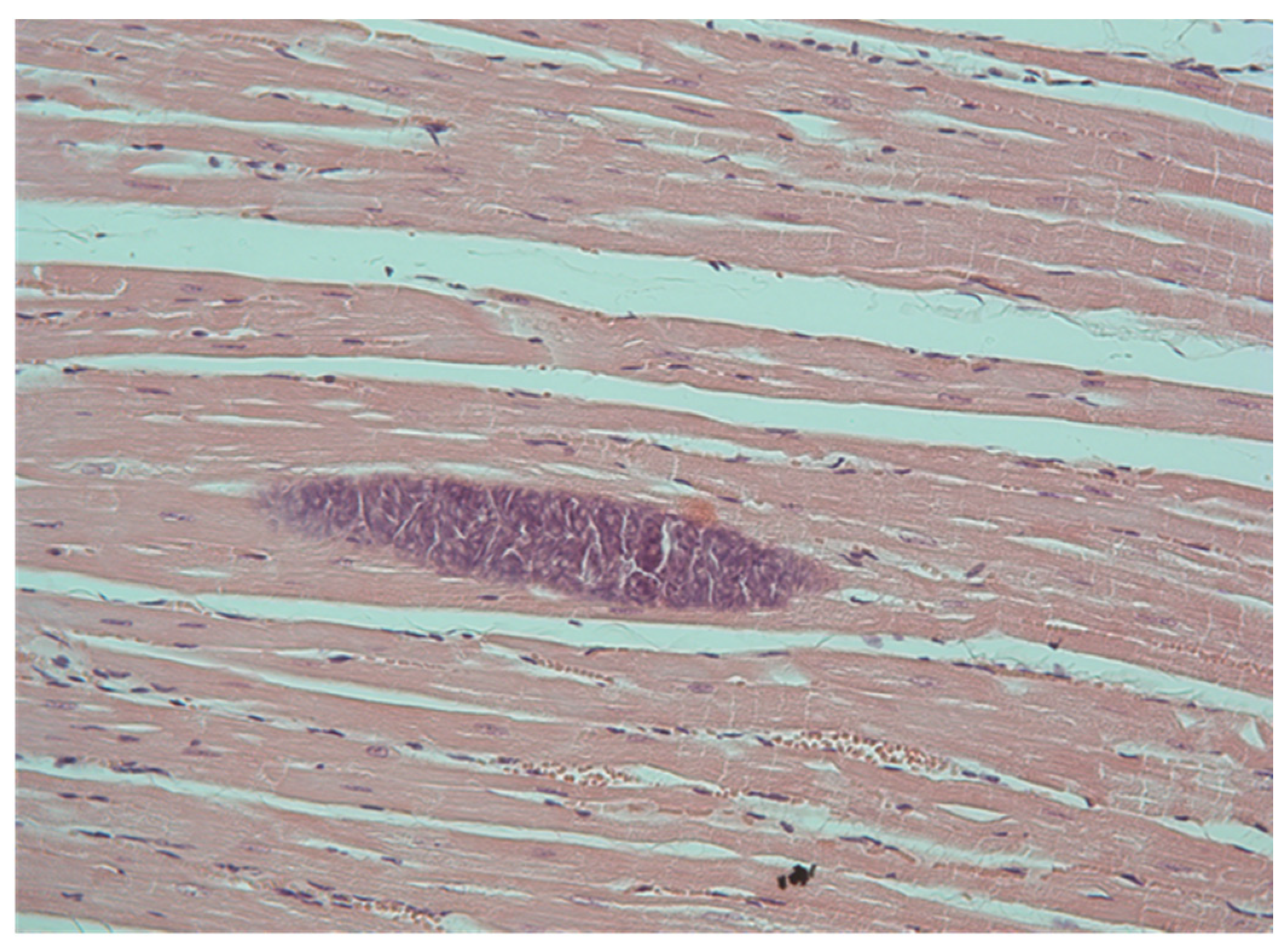
Overall, of the 216 chamois examined, 167 (77.3%) yielded evidence of infection with sarcocysts, as demonstrated by the histological detection of intramuscular sarcocysts in either the heart or diaphragm muscle specimens (Figure 1). There was no difference in sarcocyst positivity between the chamois from the Bavarian Alps and those from the Black Forest mountains (140/179, 78.2% and 27/37, 72.9%; p = 0.5196).
The examination for sarcocysts in the 181 chamois with both heart and diaphragm tissues available revealed a collective prevalence of Sarcocystis infection of 78.5% (95%CI 72.5–84.4%). Sarcocysts were more frequently recorded in the heart vs. diaphragm samples (131/181, 72.4% vs. 102/181, 56.4%; p = 0.0021). Diaphragm sarcocyst positivity was associated with heart sarcocyst positivity (diaphragm positive [heart positive]/heart positive: 91/131, 69.5% vs. diaphragm positive [heart negative]/heart negative: 11/50, 22.0%; p = 0.0001).
For the prevalence of infection in the heart and/or diaphragm, the chi-square test revealed a significant (p < 0.001) difference between the kids (27.5%) and the junior-, middle- and senior-class chamois (87.5%, 89.4% and 89.5%, respectively). Sarcocysts were significantly (p < 0.05) more often recorded in the samples from the junior-, middle- and senior-class chamois than in those from kids, but there was no difference in the prevalence of Sarcocystis spp. infection between the three classes of sexually mature chamois (p = 0.941). No significant difference in the prevalence of Sarcocystis spp. infection was found between the male and female chamois in the four age classes combined (80.7% vs. 74.7%; p = 0.3585) or in the three classes of sexually mature chamois combined (89.2% vs. 94.3%; p = 0.3762).
Similar results were obtained when considering sarcocyst positivity in the heart and diaphragm samples separately: there was a significant (p < 0.001) difference between the kids (17.1% and 25.0%, respectively) and the junior-, middle- and senior-class chamois (82.5% and 70.8%, 86.7% and 63.1%, and 81.3% and 68.4%, respectively), and sarcocysts were significantly (p < 0.05) more often recorded in the samples from the junior-, middle- and senior-class chamois than in those from the kids, but there was no significant difference in the prevalence of Sarcocystis spp. infection between the junior-, middle- and senior-class chamois (p > 0.5). No significant difference in the prevalence of Sarcocystis spp. infection was found between the male and female chamois in the four age classes combined and in the three classes of sexually mature chamois combined (p > 0.5).
The results of the examination of the samples of the 181 chamois with both heart and diaphragm tissues available for the intensity of infection are summarized in Table 2. As there was no statistical difference for the sarcocyst counts between the three classes of sexually mature chamois, overall and for the two sexes (p < 0.05, Kruskal–Wallis test), the data for the three classes were combined.
Of the 131 heart-sarcocyst-positive chamois, 124 and 7 had <10 and ≥10 sarcocysts per cm2 of muscle cut, respectively. Of the 127 diaphragm-sarcocyst-positive chamois, 119 had <10 and 8 had ≥10 sarcocysts per cm2 of muscle cut. The maximum intensity recorded was 18.7 sarcocysts per cm2 for the heart tissue and 14.6 sarcocysts per cm2 for the diaphragm tissue in a one-and-a-half-year-old male and a two-and-a-half-year-old female, respectively.
The diaphragm tissue had significantly lower counts of sarcocysts than the heart tissue in the four age classes combined and in the three classes of sexually mature chamois combined (p < 0.001), while the counts in the two tissues from the kids did not differ (p = 0.662).
There was a significantly positive correlation between the sarcocyst counts in both the heart and diaphragm tissues and the age of the chamois (rs = 0.4038, p < 0.001, and rs = 0.2093, p = 0.005, respectively). The kids had significantly lower counts of sarcocysts in both the heart and diaphragm tissues than the older chamois (p < 0.001).
There was no statistical difference between the male and female animals for the heart sarcocyst counts in the four age classes combined, the three classes of sexually mature chamois combined and the kids, or between the diaphragm sarcocyst counts in the four age classes combined and the kids (p > 0.1); however, among the three classes of sexually mature chamois combined, female animals had significantly higher diaphragm sarcocyst counts than male animals (p < 0.01).
There was a significant medium-size relationship between the sarcocyst counts of the two tissues for the four age classes combined (Spearman’s r = 0.4038; p < 0.001).
3. Discussion
The present work was part of a comprehensive study on the parasites of chamois in Germany [35,36], aiming to provide, for the first time, epidemiological data on the prevalence and intensity of Sarcocystis spp. infection in chamois from Germany using a histology examination of heart and/or diaphragm muscle samples collected from 216 chamois.
For the study of the prevalence and intensity of infection with Sarcocystis species in intermediate hosts, standard histology was used, which has been shown to be suitable for epidemiological studies of Sarcocystis spp. infections, although enzymatic digestion was more sensitive for the detection of the infection, as discussed previously [17,37]. Therefore, the histologic examination used to determine the prevalence of infection represents a minimal estimate. Apart from the technique used for the detection of infection, the type of muscle tissue examined for sarcocysts may influence the outcome of the testing, so that the results of various studies are not easily comparable [37].
While the majority of publications do not necessarily allow for conclusions as to the prevalence of Sarcocystis spp. infection in chamois, mainly because of the small sample size examined, a lack of quantification of information or accidental findings [2,3,4,5,6,7,9,10,11,14,15,16,17,18,19,20,21,22,23,27,28], the overall prevalence of Sarcocystis infection established in this study with almost 80% of the animals testing sarcocyst-positive is in line with the highest prevalences reported previously in the literature [12,25]. Cornaglia et al. [12] examined the heart tissue of 198 Alpine chamois from the western Alps in Italy by conventional histology and recorded sarcocysts in 79.8% of the animals, and Díez-Baños et al. [25], using tissue compression, recorded sarcocysts in the muscle tissues of 78.1% of 32 Cantabrian chamois originating from the Cantabrian range in northern Spain. A slightly lower prevalence of Sarcocystis spp. infection, 65%, was reported based on a histological examination of heart and diaphragm samples from 49 Alpine chamois from the central-eastern Alps in Italy [16]. Overall, however, it seems reasonable to assume that Sarcocystis spp. infections are common in chamois of both species and have been demonstrated in animals of several subspecies of chamois. In agreement with a study by Cornaglia et al. [12] on chamois and several studies on other wild ungulates in Europe, including mouflon and several species of cervids [18,37,38,39,40,41,42,43,44,45], the prevalence of sarcocyst positivity was positively correlated with the age of the animals, which reflects an age- and nutrition-associated risk of greater exposure to the infectious Sarcocystis stages for the older animals related to a lack of development of a robust protective immunity. Interestingly, the sarcocyst positivity of chamois kids found by Cornaglia et al. [12] was more than twice the frequency recorded in the present study—~57% vs. ~28%, respectively—while the sarcocyst positivity in the older animals was comparable.
Similarly, consistent with previous studies evaluating sex-related differences in the prevalence of Sarcocystis spp. infection in wild ungulates (see 37), no difference between male and female chamois was found.
To the best of the authors’ knowledge, only one previous study in chamois provided an estimation of the intensity of Sarcocystis spp. infection, based on counts of sarcocysts in pieces of muscle tissue examined by tissue compression [25]. The present investigation generated sarcocyst counts following standard histology, and it is not known to what extent histology sarcocyst counts correlate with the counts established by Díez-Baños et al. [25]. According to a score proposed for the classification of sarcocyst counts in histology muscle samples of red deer as ‘low intensity infection’ (<10 sarcocysts per cm2; [46]), the intensity of infection in the heart and diaphragm samples of almost all the Sarcocystis spp.-positive-tested chamois was low. This may also be concluded for the Cantabrian chamois examined by Díez-Baños et al. [25], given that the mean sarcocyst counts in 15 small pieces of muscle in chamois and roe deer were 1.9, with a range of 1 to 9, vs. 15.1, with a range of 1 to 155, respectively. In agreement with studies conducted on mouflon and cervids [38,45,47], the heart tissue of the Alpine chamois from Germany yielded higher sarcocyst counts than the diaphragm samples.
There are many studies which have found that the prevalence and intensity of parasite infections, especially infections with helminths, are higher among male than female hosts of different taxa of animals; however, male-biased parasitism is not necessarily the general rule, as there are several factors, ecological and physiological, which may influence the susceptibility and exposure to infection [47]. Regarding sarcocystosis, there are a couple of studies which indicate, at least partly, that male-biased parasitism may play a role in this infection. However, this did not apply to the prevalence of infection but only to the intensity of infection measured in terms of sarcocyst counts (but not for all tissues examined in a study), while no difference in the prevalence of infection between the male and female animals was found [37,44,45]. This study did not find male-biased sarcocyst counts. In contrast, the diaphragm sarcocyst counts of the sexually mature female chamois were significantly higher than the counts in the male animals.
The results of the present study demonstrated that sarcocystosis is highly prevalent in Alpine chamois in Germany, but the intensity of infections is apparently low. Further studies are desired to characterize the species of Sarcocystis parasitizing chamois using the current adequate methodology in Germany, but also in chamois in general, including experimental transmission studies to understand the role of carnivores or other animals in the life cycle of Sarcocystis species involving chamois, as well as to better understand the circulation of the same Sarcocystis species in various caprine hosts (intermediate host specificity).
4. Material and Methods
Over the course of the studies on the endoparasite fauna of Alpine chamois in Germany, [35,36] heart and/or diaphragm muscle samples of 216 chamois (40 kids [<1 year] and 176 chamois ranging up to 18 years of age) were obtained for examination for sarcocysts (Sarcocystis spp. infection). The samples were collected from chamois harvested according to the hunting regulations (Bundesjagdgesetz—‘Federal Hunting Act’) during the years 2004 to 2006 (179 chamois originated from the Bavarian Alps, ranging from the National Park Berchtesgaden in the east to the Allgäu Alps in the west, and 37 chamois from the Black Forest mountains); information on the age and sex of the animals was provided by the hunters. Both heart and diaphragm tissues were available for examination from 181 chamois; only heart or diaphragm muscle were available from 2 or 34 chamois, respectively.
For analysis, animals were stratified into four classes considering sexual maturity and contribution to the reproduction of the population in addition to age, which was determined by counting the horn rings [48]: kids (<1 year old); Class 3, junior class (1-to-2-year-old males, 1-to-3-year-old females); Class 2, middle class (3-to-7-year-old males, 4-to-9-year-old females); Class 1, senior class (≥8-year-old males, ≥10-year-old females). Reproduction among the chamois population is based on the sexually mature adult middle class (primarily) and senior class chamois, while kids (sexually immature) and junior-class chamois, although sexually mature, do not contribute to the reproduction of the population [37]. The total sample set of 216 chamois included 40 kids (17 male, 23 female), 72 junior class chamois (37 male, 33 female, 2 unknown sex), 85 middle class chamois (63 male, 22 female) and 19 senior class chamois (9 male, 10 female). The subsample of 181 chamois from which both heart and diaphragm tissues were available (160 and 21 originating from the Bavarian Alps and Black Forest mountains, respectively) included 34 kids (16 male, 18 female), 57 junior class chamois (31 male, 25 female, 1 unknown sex), 74 middle class chamois (55 male, 19 female) and 16 senior class chamois (7 male, 9 female).
A standard histological examination (light microscopy) was carried out on formalin-fixed tissue samples (right ventricular wall, diaphragmatic pillars) using 5 µm sections of paraffin-embedded tissue stained with hematoxylin–eosin. One section per tissue type per animal was screened for intramuscular sarcocysts (Sarcocystis cysts). The area of each tissue sample examined was measured using the dhs-Bilddatenbank® V5.0 (Dietermann & Heuser, Greifenstein, Germany) software to estimate the intensity of infection as the number of sarcocysts per cm2 of muscle cut.
To assess the relationship between sarcocyst presence (sarcocyst positivity, presented as prevalence of Sarcocystis spp. infection) and the chamois’ demographic factors and for the analysis of the intensity of infection (the count of sarcocysts per cm2 of tissue cut, sarcocyst count), the subsample of 181 chamois with both heart and diaphragm tissues examined was considered.
Associations between sarcocyst positivity and variables representing host demographic factors (age class, sex) were assessed using contingency tables, Fisher’s exact test, or chi-square statistics. The association of sarcocyst positivity in the two tissues was analyzed using McNemar’s test for matched-pair samples. The sarcocyst counts were analyzed using the Kruskal–Wallis test or the Mann–Whitney test, as appropriate. All testing was two-sided at a significance level of p < 0.05.
To test for the association of the sarcocyst counts of the two tissues and for the association of the sarcocyst counts with the age of the chamois, Spearman’s rank correlation coefficient (rs) was used.
Author Contributions
Conceptualization, S.R.; methodology, M.V.; formal analysis, S.R. and M.V.; investigation, M.V.; resources, S.R.; writing—original draft preparation, S.R.; writing—review and editing, S.R. and M.V. All authors have read and agreed to the published version of the manuscript.
Funding
This research received no external funding.
Institutional Review Board Statement
Not applicable.
Informed Consent Statement
Not applicable.
Data Availability Statement
All relevant data are reported in the paper.
Acknowledgments
The authors extend their gratitude to Leslie C. Schlegel for her assistance in examining the tissue samples and conducting the initial data analysis. While we made earnest attempts to reach L. C. Schlegel during the manuscript preparation, unfortunately, we were unable to establish contact. If you have any inquiries regarding this article, please do not hesitate to get in touch with the authors directly.
Conflicts of Interest
Both authors were employed by Boehringer Ingelheim Vetmedica GmbH, this research was conducted in the absence of any relationships which could be construed as a potential conflict of interest as the work does not have any commercial or financial implication.
References
- Corlatti, L.; Herrero, J.; Ferretti, F.; Anderwald, P.; Garcia-González, R.; Hammer, S.E.; Nores, C.; Rossi, L.; Lovari, S. Northern chamois Rupicapra rupicapra (Linnaeus, 1758) and Southern chamois Rupicapra pyrenaica Bonaparte, 1845. In Terrestrial Cetartiodactyla; Corlatti, L., Zachos, F.E., Eds.; Springer Nature: Cham, Switzerland, 2022; pp. 325–366. [Google Scholar]
- Dollinger, P. Beitrag zur Kenntnis des Endoparasitenspektrums des Gemswildes in der Schweiz. Zschr. Jagdwiss. 1974, 20, 115–118. [Google Scholar] [CrossRef]
- Burgisser, H. Compte-rendu sur les maladies des animaux sauvages (1973–1974). Schweiz. Arch. Tierheilkd. 1975, 117, 397–400. [Google Scholar] [PubMed]
- Ippen, R.; Blažek, K.; Henne, D.; Kotrly, A. Ein Beitrag zur Sarkosporidiose der Zoo- und Wildtiere. Verhandlungsber. Erkrank. Zootiere 1974, 16, 315–321. [Google Scholar]
- Blažek, K.; Kotrlý, A.; Ippen, R. Sarkosporidióza myokardu spárkaté zvěře. Vet. Med. 1976, 21, 75–80. [Google Scholar]
- Biocca, E.; Balbo, T.; Guarda, E.; Costantini, R. L’importanza della volpe (Vulpes vulpes) nella trasmissione della sarcosporidiosi dello stambecco (Capra ibex) nel Parco Nazionale del Gran Paradiso. Parassitologia 1975, 18, 17–23. [Google Scholar]
- Burgisser, H. Compte-rendu sur les maladies des animaux sauvages de 1975 à 1982. Schweiz. Arch. Tierheilkd. 1983, 125, 519–527. [Google Scholar]
- Chroust, K. Parazitofauna kamzíka obecného v oblasti Jeseníků. Fol. Venatoria 1991, 21, 77–88. [Google Scholar]
- Euzeby, J.; Hugonnet, L. Le parasitisme les chamois et mouflons de la reserve naturelle des Bauges et le parasitisme chez les chamois de Vanoise. Rapp. Maladies Parasitaires. Ecole Nat. Vét. Lyon France 1980. [Google Scholar]
- Hugonnet, L.; Euzeby, J.; Hars, J.; Gibert, P.; Chauve, C. Cinetique de l’infesttaion parasitaire des jaunes chamois dans la reserve nationale des Bauges. Bull. Mens. Off. Nat. Chass. 1983, 67, 35–43. [Google Scholar]
- Nevejans, Y. Contribution a L’etude des Causes de Mortalité du Chamois (Rupicapra rupicapra) Dans les Alpes du Nord. Ph.D. Thesis, French Veterinary School, Lyon, France, 2002. [Google Scholar]
- Cornaglia, E.; Guarda, F.; Guarda, F.; Misciattelli, M.E. Ricerca, frequenza, diagnosi, morfologia istopatologica ed ultrastrutturale della sarcosporidiosi cardiaca nei camosci e stambecchi. Ann. Fac. Med. Vet. Torino 1980, 27, 279–296. [Google Scholar]
- Guarda, F.; Guarda, F.; Cornaglia, E. Contributo allo studio della patologia cardiaca dei camosci e stambecchi. Ann. Fac. Med. Vet. Torino 1980, 27, 253–274. [Google Scholar]
- Guarda, F.; Peracino, V. Problemi di patologia nei camosci e stambecchi delle Alpi. Schweiz. Arch. Tierheilkd. 1987, 129, 327–331. [Google Scholar]
- Guarda, F.; Bollo, E.; Bassano, B.; Peracino, V. Cardiac pathology in alpine ibex (Capra ibex) and chamois (Rupicapra rupicapra) of the Gran Paradiso National Park. In Proceedings of the Abstracts 2nd World Conference on Mountain Ungulates, Aosta, Italy, 5–7 May 1997; p. 76. [Google Scholar]
- Bregoli, M.E.; Lucchini, R.; Debora, D.; Enrico, F.; Citterio, C.V.; Capelli, G.; Vascellari, M.; Farina, G. Survey of Sarcocystis spp. in game ungulates of central-eastern Italian Alps and report of a systemic sarcosporidiosis in a roe deer (Capreolus capreolus). In Trends in Game Meat Hygiene; Paulsen, P., Bauer, A., Smulders, F.J.M., Eds.; Wageningen Acad. Publ.: Wageningen, The Netherlands, 2014; pp. 183–187. [Google Scholar]
- Giacomelli, S.; Bianchi, A.; Rota Npdari, S.; Gibelli, L.R.; Vicari, N.; Archetti, I.; Bertoletti, I. Messa a punto di metodiche per la determinazione di presenza ed intensità di infestazione da Sarcocystis spp. in ungulati selvatici. In Proceedings of the Atti XVII Congresso Nazionale della Societa Italiana di Diagnostica di Laboratoria Veterinaria (S.I.Di.L.V.), Perugia, Italy, 7–9 November 2018; pp. 290–294. [Google Scholar]
- Goldová, M.; Tóth, Š.; Letková, V.; Mojžišová, J.; Ciberej, J.; Konjevič, D.; Kočišová, A.; Slavica, A. Sarcocystosis in cloven-hoofed game in Slovak Republic. Nat. Croat. 2008, 17, 303–309. [Google Scholar]
- Odening, K.; Stolte, M.; Bockhardt, I. On the diagnostics of Sarcocystis in chamois (Rupicapra rupicapra). Appl. Parasitol. 1996, 37, 153–160. [Google Scholar]
- Hoby, S.; Walzer, C.; Slotta-Bachmayr, L.; Segner, H.; Robert, N. Untersuchungen zur Pathologie von Wildungulaten im Nationalpark Hohe Tauern, Österreich. Wien. Tierärztl. Mschr. 2006, 93, 104–112. [Google Scholar]
- Erber, M. Specificity of Sarcocystis spp. in sheep, chamois and goat. In Proceedings of the Abstracts 3rd European Multicolloquium on Parasitology, Cambridge, UK, 7–13 September 1980; p. 142. [Google Scholar]
- Kolenda, R.; Schierack, P.; Zieba, F.; Zawijacz-Kozina, T.; Bednarski, M. First molecular characterization of Sarcocystis tenella in Tatra chamois (Rupicapra rupiccapra tatrica) in Poland. Parasitol. Res. 2015, 114, 3885–3892. [Google Scholar] [CrossRef] [PubMed]
- Iacob, O.; Cotofan, O.; Raileanu, G.; Pop, I. The multiple parasitic aggression on black goat species (Rupicapra rupicapra) and local reactivity. Magyar Állat. Lapja 2008, 130, 288–289. [Google Scholar]
- Díez-Baños, N.; Hidalgo-Argüello, M.R.; Martínez-González, B.; Fernández-García, R.; Rubio-Nistal, P. Prevalencia de la sarcosporidiosis en corzo (Capreolus capreolus) y rebeco (Rupicapra rupicapra parva) procedentes de la Cordillera Cantabrica de la provincia de León. In Proceedings of the Comunicaciones IV Congreso Ibérico de Parasitologia, Santiago de Compostela, Spain, 24–28 July 1995; p. 92. [Google Scholar]
- Díez-Baños, N.; Hidalgo-Argüello, M.R.; Morrondo-Pelayo, M.P.; Díez-Baños, P. Parasitofauna en pequeños ruminantes silvestres procedentes de la Cordillera Cantábrica de la provincia de León. In Proceedings of the Comunicaciones I Congreso Internacional Medio Ambiente y Veterinaria, Cáceres, Spain, 24–28 April 1997; pp. 547–548. [Google Scholar]
- Fernández Morán, J.; Gomez, S.; Ballesteros, F.; Quiros, P.; Benito, J.L.; Feliu, C.; Nieto, J.M. Epizootiology of sarcoptic mange in a population of Cantabrian chamois (Rupicapra pyrenaica parva) in north-western Spain. Vet. Parasitol. 1997, 73, 163–171. [Google Scholar] [CrossRef]
- Cruz Arnal, M.; Fernández de Luco, D. Estudio sanitario del sarrio en el Pireneo aragonés (1997–2002). In El Sarrio Pirenaico Rupicapra p. pyrenaica: Biología, Patología y Gestión; Herrero, J., Escudero, E., Fernández de Luco, D., García-González, D., Eds.; Publicaciones del Consejo Protección de la Naturaleza de Aragón: Zaragoza, Spain, 2004; pp. 125–132. [Google Scholar]
- Marco, I.; Lopez-Olvera, J.R.; Rosell, R.; Vidal, E.; Hurtado, A.; Juste, R.; Pumarola, M.; Lavin, S. Severe outbreak of disease in the southern chamois (Rupicapra pyrenaica) associated with border disease virus infection. Vet. Microbiol. 2007, 120, 33–41. [Google Scholar] [CrossRef]
- Calero-Bernal, R.; van Wilpe, E.; White, K.; Verma, S.K.; Cerqueira-Cézar, C.K.; Dubey, J.P. Sarcocystis oreamni, n. sp., (Apicomplexa: Sarcocystidae) from a mountain goat (Oreamnos americaus). Parasitol. Res. 2015, 114, 4135–4141. [Google Scholar] [CrossRef]
- Cornaglia, E.; Rossetto Giaccherino, A.; Peracino, V. Ultrastructural morphology of sarcosporidiosis in Alpine ibex (Capra ibex). Vet. Parasitol. 1998, 75, 21–32. [Google Scholar] [CrossRef] [PubMed]
- Rossi, L.; Lanfranchi, P.; Meneguz, P.G.; De Meneghi, D.; Guarda, F. Infezione sperimentale della capra e delle pecore con sarcosporidi del muflone e del camoscio. Parassitologia 1988, 30, 164–165. [Google Scholar]
- Odening, K.; Stolte, M.; Bockhardt, I. Phylogenetic relationship of chamois and mountain goat as demonstrated by parasites of the genus Sarcocystis. In Proceedings of the 2nd World Conference on Mountain Ungulates, Aosta, Italy, 5–7 May 1997; pp. 159–167. [Google Scholar]
- Guardone, L.; Armani, A.; Mancianti, F.; Ferroglio, E. A review on Alaria alata, Toxoplasma gondii and Sarcocystis spp. in mammalian game meat consumed in Europe: Epidemiology, risk management and future directions. Animals 2022, 12, 263. [Google Scholar] [CrossRef] [PubMed]
- Wotschikowsky, U. Ungulates and their management in Germany. In European Ungulates and Their Management in the 21st Century; Apollonio, M., Andersen, R., Putman, R., Eds.; Cambridge Univ. Press: Cambridge, UK, 2010; pp. 201–222. [Google Scholar]
- Hamel, D. Untersuchungen zum Parasitenbefall des Gamswildes in Deutschland—Helminthen des Gastrointestinaltraktes. Ph.D. Thesis, Ludwig-Maximilians-Univ., Munich, Germany, 2008. [Google Scholar]
- Schlegel, L.S. Untersuchungen zum Parasitenbefall des Gamswildes in Deutschland—Eimerien, Sarkosporidien, Lungenwürmer, Leberegel. Ph.D. Thesis, Ludwig-Maximilians-Univ., Munich, Germany, 2013. [Google Scholar]
- Rehbein, S.; Lindner, T.; Visser, M.; Lutz, W.; Reindl, H. Distribution, prevalence, and intensity of Sarcocystis infections in sika deer (Cervus nippon) of free-ranging populations in Germany and Austria. Parasitol. Res. 2022, 121, 2079–2086. [Google Scholar] [CrossRef]
- Ippen, R.; Hilgenfeld, M. Ergebnisse der reihenmäßigen Untersuchung des erlegten Rehwildes aus dem Wildforschungsgebiet Hakel bei Gatersleben. Beitr. Jagd- u. Wildforsch. 1961, 1, 49–52. [Google Scholar]
- Erber, M.; Boch, J.; Barth, D. Drei Sarkosporidienarten des Rehwildes. Berl. Münch. Tierärztl. Wschr. 1978, 91, 482–486. [Google Scholar]
- Entzeroth, R. Untersuchungen an Sarkosporidien (Mieschersche Schläuche) des einheimischen Rehwildes (Capreolus capreolus L.). Zschr. Jagdwiss. 1981, 27, 247–257. [Google Scholar] [CrossRef]
- Sugár, L. Sarcocystis-Infection in three Hungarian roe deer (Capreolus c. capreolus) populations. Symp. Biol. Hung. 1986, 33, 363–367. [Google Scholar]
- Haupt, W.; Eulenberger, K.H. Untersuchungen zum Befall des Damwildes mit Helminthen und Sarkosporidien. Beitr. Jagd- u. Wildforsch. 1988, 15, 8–54. [Google Scholar]
- Spickschen, C.; Pohlmeyer, K. Untersuchungen zum Vorkommen von Sarkosporidien bei Reh-, Rot- und Muffelwild in zwei unterschiedlichen Naturräumen des Bundeslandes Niedersachsen. Zschr. Jagdwiss. 2002, 48, 35–48. [Google Scholar]
- Prakas, P.; Rehbein, S.; Rudaiytė-Lukošienė, E.; Butkauskas, D. Molecular identification of Sarcocystis species in diaphragm muscle tissue of European mouflon (Ovis gmelini musimon) from Austria. Parasitol. Res. 2021, 120, 2695–2702. [Google Scholar] [CrossRef] [PubMed]
- Prakas, P.; Rehbein, S.; Rudaiytė-Lukošienė, E.; Butkauskas, D. Molecular identification of Sarcocystis species in sika deer (Cervus nippon) of free-ranging populations in Germany and Austria. Vet. Res. Comm. 2023, 47, 2165–2171. [Google Scholar] [CrossRef]
- Luzón, M.; Domíguez-González, J.; Soto-Carrión, A.M.; Alunda, J.M.; de la Fuente, C. Sarcocystosis in Cervus elaphus: Comparison of diagnostic methods. Int. J. Parasitol. Parasit. Wildl. 2015, 4, 396–400. [Google Scholar] [CrossRef] [PubMed]
- Wesołowska, A. Sex—the most underappreciated variable in research: Insights from helminth-infected hosts. BMC Vet. Res. 2022, 53, 94. [Google Scholar] [CrossRef] [PubMed]
- Knaus, W.; Schröder, W. Das Gamswild; Verl. P. Parey: Hamburg/Berlin, Germany, 1983. [Google Scholar]
Figure 1.
Thin-walled sarcocyst in hematoxylin–eosin-stained diaphragm muscle section.


Table 1.
Summary of publications reporting on the finding of sarcocysts or occurrence of sarcocystosis in chamois.
Table 1.
Summary of publications reporting on the finding of sarcocysts or occurrence of sarcocystosis in chamois.
| Chamois Subspecies | Country of Study | Period of Study | Type of Muscle Tissue Examined | Method of Examination | Number of Animals Examined | Number of Animals Testing Sarcocyst Positive; Percentage | Reference |
|---|---|---|---|---|---|---|---|
| Alpine chamois, Rupicapra r. rupicapra | Switzerland | 1972–1973 | Esophagus | Histology/LM1 | 4 | 3; 75% | [2] |
| 1973–1974 | No information | No information | 65 | ‘common’ | [3] | ||
| 1975–1982 | No information | No information | 375 | ‘sporadic’ | [7] | ||
| (former) Czechoslovakia | 1972–1973 | Heart | Histology/LM | 3 | 2; 66.7% | [4] | |
| 1972–1974 | Heart | Histology/LM | 4 | 3; 75% | [5] | ||
| 1980–1988 | Heart, esophagus, laryngeal and skeletal muscles | Histology/LM | 58 | 6; 10.3% | [8] | ||
| France | 1979 | Heart | No information | No information | No informationA | [9] | |
| 1978–1980 | Heart | No information | No information | No informationA | [10] | ||
| 1977–2001 | No information | No information | 466 | 1 case of ‘sarcosporidiosis’ | [11] | ||
| Italy | No information | Heart | Histology/LM | No infor- mation | ‘common’ | [6] | |
| No information | Heart | Histology/LM + TEM2 | 198 | 158; 79.8% | [12] | ||
| 1974–1977 | Heart | Histology/LM | 138 | 103; 74.6% | [13] | ||
| No information | Heart | Histology/LM | No information | No informationA | [14] | ||
| No information | Heart | Histology/LM | No information | No informationA | [15] | ||
| No information | Diaphragm | Histology/LM | 49 | 32; 65.3% | [16] | ||
| No information | Diaphragm, esophagus, heart | Histology/LM, tissue compression, tissue homogenization | 6 | 4; 66.7% | [17] | ||
| Slovakia | 2005–2007 | Heart, skeletal muscle | Histology/LM, trypsine digestion | 6 | 6; 100% | [18] | |
| Austria + Germany | No information | Diaphragm, esophagus, heart, skeletal muscle | Histology/LM + TEM | No information | No informationA | [19] | |
| Austria | 2002–2004 | Heart | Histology/LM | 90 | ‘common’ | [20] | |
| Germany | No information | No information | Histology/LM + TEM | No information | No informationA | [21] | |
| Tatra chamois, Rupicapra r. tatrica | Poland | 2012–2013 | Diaphragm, esophagus, heart, skeletal muscle, tongue | Histology/LM, PCR (cox1, ssu rRNA) | 3 | 3; 100% | [22] |
| Carpatian chamois, Rupicapra r. carpatica | Romania | No information | Heart | Histology/LM | No information | No informationA | [23] |
| Cantabrian chamois, Rupicapra pyrenaica parva | Spain | 1994 | Diaphragm, esophagus, heart | Tissue compression | 31 | 22; 74.2% | [24] |
| 1995 | Diaphragm, esophagus, heart | Tissue compression | 32 | 25; 78.1% | [25] | ||
| 1993–1995 | Heart | Histology/LM | 52 | 10; 19.2% | [26] | ||
| Pyrenean chamois, Rupicapra p. pyrenaica | Spain | 1997–2002 | Heart, skeletal muscle | Histology/LM | 74 | ‘in most of the animals’ | [27] |
| 2001–2002 | Heart, skeletal muscle | Histology/LM | 20 | ‘frequently’ | [28] |
1—LM = light microscopy. 2—TEM = transmission electron microscopy. A—only presence/detection of sarcocysts quoted.
Table 2.
Intensity of infection by histology (number of sarcocysts per cm2 of muscle cut) in the heart and diaphragm muscle tissues of 181 chamois from the Bavarian Alps and Black Forest mountains in Germany (animals with both heart and diaphragm muscle samples examined).
Table 2.
Intensity of infection by histology (number of sarcocysts per cm2 of muscle cut) in the heart and diaphragm muscle tissues of 181 chamois from the Bavarian Alps and Black Forest mountains in Germany (animals with both heart and diaphragm muscle samples examined).
| Class of Chamois | Sex | Number of Sarcocysts per cm2 Muscle Cut, Mean ± Standard Deviation | |
|---|---|---|---|
| Heart | Diaphragm | ||
| Kids (<1 year, n = 34) | Male | 0.26 ± 0.43 | 1.30 ± 1.95 |
| Female | 0.91 ± 1.51 | 0.88 ± 1.47 | |
| Male + Female | 0.60 ± 1.00 | 1.08 ± 1.72 | |
| Combined Classes 1, 2 and 3 (actually ~1 to 18 years, n = 147) | Male | 3.72 ± 2.72 | 1.72 ± 1.63 |
| Female | 3.44 ± 2.18 | 3.16 ± 2.64 | |
| Male + Female | 3.61 ± 2.51 | 2.26 ± 2.06 | |
| Combined kids and Classes 1, 2 and 3 (actually <1 to 18 years, n = 181) | Male | 3.22 ± 2.68 | 2.88 ± 2.36 |
| Female | 1.66 ± 1.69 | 2.58 ± 2.56 | |
| Male + Female | 3.05 ± 2.55 | 2.04 ± 2.07 | |
Age classes: Class 3, junior class (1-to-2-year-old males, 1-to-3-year-old females); Class 2, middle class (3-to-7-year-old males, 4-to-9-year-old females); Class 1, senior class (≥8-year-old males, ≥10-year-old females).
Disclaimer/Publisher’s Note: The statements, opinions and data contained in all publications are solely those of the individual author(s) and contributor(s) and not of MDPI and/or the editor(s). MDPI and/or the editor(s) disclaim responsibility for any injury to people or property resulting from any ideas, methods, instructions or products referred to in the content. |
© 2024 by the authors. Licensee MDPI, Basel, Switzerland. This article is an open access article distributed under the terms and conditions of the Creative Commons Attribution (CC BY) license (https://creativecommons.org/licenses/by/4.0/).
Share and Cite
MDPI and ACS Style
Rehbein, S.; Visser, M. Prevalence and Intensity of Sarcocystis spp. Infections in Alpine Chamois (Rupicapra r. rupicapra) in Germany. Parasitologia 2024, 4, 61-70. https://0-doi-org.brum.beds.ac.uk/10.3390/parasitologia4010005
AMA Style
Rehbein S, Visser M. Prevalence and Intensity of Sarcocystis spp. Infections in Alpine Chamois (Rupicapra r. rupicapra) in Germany. Parasitologia. 2024; 4(1):61-70. https://0-doi-org.brum.beds.ac.uk/10.3390/parasitologia4010005
Chicago/Turabian StyleRehbein, Steffen, and Martin Visser. 2024. "Prevalence and Intensity of Sarcocystis spp. Infections in Alpine Chamois (Rupicapra r. rupicapra) in Germany" Parasitologia 4, no. 1: 61-70. https://0-doi-org.brum.beds.ac.uk/10.3390/parasitologia4010005