materials-logo

Journal Browser

Journal Browser

Functional Materials and Digital Technology in Biomedical Applications for Oral and Maxillofacial Surgery

A special issue of Materials (ISSN 1996-1944). This special issue belongs to the section "Biomaterials".

Deadline for manuscript submissions: closed (20 April 2023) | Viewed by 8709

Special Issue Editor


E-Mail Website
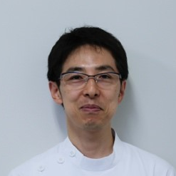
Guest Editor
Department of Oral and Maxillofacial Surgery/Orthodontics, Yokohama City University Medical Center, 4-57 Urafune-cho, Minami-ku, Yokohama, Kanagawa 232-0024, Japan
Interests: oral and maxillofacial reconstruction; dental implants; titanium materials; osteoblast behavior; bone healing

Special Issue Information

Dear Colleagues,

Various types of biomaterials for oral and maxillofacial surgery have been developed for functional and morphological reconstruction to ensure patient quality of life. Recent advancements of CAD/CAM technology enable the development of highly functional biomedical materials and devices. However, the clinical application of new technologies has a challenging aspect. Therefore, translational research of those materials and devices is quite important to go to further clinical or basic research. This Special Issue covers biomaterial research of “bench to bedside” or “laboratory to clinic” for patients with facial trauma, head and neck cancer, jaw deformity, and osteomyelitis/osteonecrosis of the jaw. To address this, interaction between osteogenic cells and dental-implant-related materials and maxillofacial reconstruction, between mechanical strength and bone reconstructive materials, between CAD/CAM technology and computational simulation, and between minimally invasive devices and patient QOL are topics of focus. Surface technology to enhance osteoblast attachment and behavior on titanium, in vivo imaging or quantification of physiological activities in the oral and maxillofacial region also provide useful information.

In conclusion, it is a great pleasure to invite all researchers dedicated to research and development of material sciences for returning or preserving QOL in the oral and maxillofacial field to submit your contributions to this exciting new Special Issue.

Dr. Makoto Hirota
Guest Editor

Manuscript Submission Information

Manuscripts should be submitted online at www.mdpi.com by registering and logging in to this website. Once you are registered, click here to go to the submission form. Manuscripts can be submitted until the deadline. All submissions that pass pre-check are peer-reviewed. Accepted papers will be published continuously in the journal (as soon as accepted) and will be listed together on the special issue website. Research articles, review articles as well as short communications are invited. For planned papers, a title and short abstract (about 100 words) can be sent to the Editorial Office for announcement on this website.

Submitted manuscripts should not have been published previously, nor be under consideration for publication elsewhere (except conference proceedings papers). All manuscripts are thoroughly refereed through a single-blind peer-review process. A guide for authors and other relevant information for submission of manuscripts is available on the Instructions for Authors page. Materials is an international peer-reviewed open access semimonthly journal published by MDPI.

Please visit the Instructions for Authors page before submitting a manuscript. The Article Processing Charge (APC) for publication in this open access journal is 2600 CHF (Swiss Francs). Submitted papers should be well formatted and use good English. Authors may use MDPI's English editing service prior to publication or during author revisions.

Keywords

  • Dental implant
  • Maxillofacial reconstruction
  • Surface technology
  • CAD/CAM technology
  • Titanium materials
  • Biodegradable metal composites
  • Computational simulation
  • Physiological activities in head and neck area
  • Osteoblast attachment and behavior
  • Mechanical strength of reconstructive material
  • Quality of life for oral cancer and facial trauma survivors

Published Papers (4 papers)

Order results
Result details
Select all
Export citation of selected articles as:

Research

13 pages, 4085 KiB  
Article
Osteoblast Attachment Compromised by High and Low Temperature of Titanium and Its Restoration by UV Photofunctionalization
by Takayuki Ikeda, Takahisa Okubo, Juri Saruta, Makoto Hirota, Hiroaki Kitajima, Naoki Yanagisawa and Takahiro Ogawa
Materials 2021, 14(19), 5493; https://0-doi-org.brum.beds.ac.uk/10.3390/ma14195493 - 23 Sep 2021
Cited by 13 | Viewed by 1497
Abstract
Titanium implants undergo temperature fluctuations during manufacturing, transport, and storage. However, it is unknown how this affects their bioactivity. Herein, we explored how storage (six months, dark conditions) and temperature fluctuations (5–50 °C) affected the bioactivity of titanium implants. Stored and fresh acid-etched [...] Read more.
Titanium implants undergo temperature fluctuations during manufacturing, transport, and storage. However, it is unknown how this affects their bioactivity. Herein, we explored how storage (six months, dark conditions) and temperature fluctuations (5–50 °C) affected the bioactivity of titanium implants. Stored and fresh acid-etched titanium disks were exposed to different temperatures for 30 min under wet or dry conditions, and their hydrophilicity/hydrophobicity and bioactivity (using osteoblasts derived from rat bone marrow) were evaluated. Ultraviolet (UV) treatment was evaluated as a method of restoring the bioactivity. The fresh samples were superhydrophilic after holding at 5 or 25 °C under wet or dry conditions, and hydrophilic after holding at 50 °C. In contrast, all the stored samples were hydrophobic. For both fresh and stored samples, exposure to 5 or 50 °C reduced osteoblast attachment compared to holding at 25 °C under both wet and dry conditions. Regression analysis indicated that holding at 31 °C would maximize cell attachment (p < 0.05). After UV treatment, cell attachment was the same or better than that before temperature fluctuations. Overall, titanium surfaces may have lower bioactivity when the temperature fluctuates by ≥20 °C (particularly toward lower temperatures), independent of the hydrophilicity/hydrophobicity. UV treatment was effective in restoring the temperature-compromised bioactivity. Full article
Show Figures

Figure 1

16 pages, 3394 KiB  
Article
Prolonged Post-Polymerization Biocompatibility of Polymethylmethacrylate-Tri-n-Butylborane (PMMA-TBB) Bone Cement
by Juri Saruta, Ryotaro Ozawa, Kosuke Hamajima, Makiko Saita, Nobuaki Sato, Manabu Ishijima, Hiroaki Kitajima and Takahiro Ogawa
Materials 2021, 14(5), 1289; https://0-doi-org.brum.beds.ac.uk/10.3390/ma14051289 - 08 Mar 2021
Cited by 17 | Viewed by 2099
Abstract
Polymethylmethacrylate (PMMA)-based acrylic bone cement is commonly used to fix bone and metallic implants in orthopedic procedures. The polymerization initiator tri-n-butylborane (TBB) has been reported to significantly reduce the cytotoxicity of PMMA-based bone cement compared to benzoyl peroxide (BPO). However, it is unknown [...] Read more.
Polymethylmethacrylate (PMMA)-based acrylic bone cement is commonly used to fix bone and metallic implants in orthopedic procedures. The polymerization initiator tri-n-butylborane (TBB) has been reported to significantly reduce the cytotoxicity of PMMA-based bone cement compared to benzoyl peroxide (BPO). However, it is unknown whether this benefit is temporary or long-lasting, which is important to establish given that bone cement is expected to remain in situ permanently. Here, we compared the biocompatibility of PMMA-TBB and PMMA-BPO bone cements over several days. Rat femur-derived osteoblasts were seeded onto two commercially-available PMMA-BPO bone cements and experimental PMMA-TBB polymerized for one day, three days, or seven days. Significantly more cells attached to PMMA-TBB bone cement during the initial stages of culture than on both PMMA-BPO cements, regardless of the age of the materials. Proliferative activity and differentiation markers including alkaline phosphatase production, calcium deposition, and osteogenic gene expression were consistently and considerably higher in cells grown on PMMA-TBB than on PMMA-BPO, regardless of cement age. Although osteoblastic phenotypes were more favorable on older specimens for all three cement types, biocompatibility increased between three-day-old and seven-day-old PMMA-BPO specimens, and between one-day-old and three-day-old PMMA-TBB specimens. PMMA-BPO materials produced more free radicals than PMMA-TBB regardless of the age of the material. These data suggest that PMMA-TBB maintains superior biocompatibility over PMMA-BPO bone cements over prolonged periods of at least seven days post-polymerization. This superior biocompatibility can be ascribed to both low baseline cytotoxicity and a further rapid reduction in cytotoxicity, representing a new biological advantage of PMMA-TBB as a novel bone cement material. Full article
Show Figures

Figure 1

12 pages, 11626 KiB  
Article
Ultraviolet Light Treatment of Titanium Enhances Attachment, Adhesion, and Retention of Human Oral Epithelial Cells via Decarbonization
by Kourosh Nakhaei, Manabu Ishijima, Takayuki Ikeda, Amirreza Ghassemi, Juri Saruta and Takahiro Ogawa
Materials 2021, 14(1), 151; https://0-doi-org.brum.beds.ac.uk/10.3390/ma14010151 - 31 Dec 2020
Cited by 20 | Viewed by 2028
Abstract
Early establishment of soft-tissue adhesion and seal at the transmucosal and transcutaneous surface of implants is crucial to prevent infection and ensure the long-term stability and function of implants. Herein, we tested the hypothesis that treatment of titanium with ultraviolet (UV) light would [...] Read more.
Early establishment of soft-tissue adhesion and seal at the transmucosal and transcutaneous surface of implants is crucial to prevent infection and ensure the long-term stability and function of implants. Herein, we tested the hypothesis that treatment of titanium with ultraviolet (UV) light would enhance its interaction with epithelial cells. X-ray spectroscopy showed that UV treatment significantly reduced the atomic percentage of surface carbon on titanium from 46.1% to 28.6%. Peak fitting analysis revealed that, among the known adventitious carbon contaminants, C–C and C=O groups were significantly reduced after UV treatment, while other groups were increased or unchanged in percentage. UV-treated titanium attracted higher numbers of human epithelial cells than untreated titanium and allowed more rapid cell spread. Hemi-desmosome-related molecules, integrin β4 and laminin-5, were upregulated at the gene and protein levels in the cells on UV-treated surfaces. The result of the detachment test revealed twice as many cells remaining adherent on UV-treated than untreated titanium. The enhanced cellular affinity of UV-treated titanium was equivalent to laminin-5 coating of titanium. These data indicated that UV treatment of titanium enhanced the attachment, adhesion, and retention of human epithelial cells associated with disproportional removal of adventitious carbon contamination, providing a new strategy to improve soft-tissue integration with implant devices. Full article
Show Figures

Figure 1

12 pages, 2681 KiB  
Article
Compromised Epithelial Cell Attachment after Polishing Titanium Surface and Its Restoration by UV Treatment
by Takahisa Okubo, Takayuki Ikeda, Juri Saruta, Naoki Tsukimura, Makoto Hirota and Takahiro Ogawa
Materials 2020, 13(18), 3946; https://0-doi-org.brum.beds.ac.uk/10.3390/ma13183946 - 07 Sep 2020
Cited by 18 | Viewed by 2361
Abstract
Titanium-based implant abutments and tissue bars are polished during the finalization. We hypothesized that polishing degrades the bioactivity of titanium, and, if this is the case, photofunctionalization-grade UV treatment can alleviate the adverse effect. Three groups of titanium disks were prepared; machined surface, [...] Read more.
Titanium-based implant abutments and tissue bars are polished during the finalization. We hypothesized that polishing degrades the bioactivity of titanium, and, if this is the case, photofunctionalization-grade UV treatment can alleviate the adverse effect. Three groups of titanium disks were prepared; machined surface, polished surface and polished surface followed by UV treatment (polished/UV surface). Polishing was performed by the sequential use of greenstone and silicon rubber burs. UV treatment was performed using a UV device for 12 min. Hydrophobicity/hydrophilicity was examined by the contact angle of ddH2O. The surface morphology and chemistry of titanium were examined by scanning electron microscopy (SEM) and X-ray photoelectron spectroscopy (XPS), respectively. Human epithelium cells were seeded on titanium disks. The number of cells attached, the spreading behavior of cells and the retention on titanium surfaces were examined. The polished surfaces were smooth with only minor scratches, while the machined surfaces showed traces and metal flashes made by machine-turning. The polished surfaces showed a significantly increased percentage of surface carbon compared to machined surfaces. The carbon percentage on polished/UV surfaces was even lower than that on machined surfaces. A silicon element was detected on polished surfaces but not on polished/UV surfaces. Both machined and polished surfaces were hydrophobic, whereas polished/UV surfaces were hydrophilic. The number of attached cells after 24 h of incubation was 60% lower on polished surfaces than on machined surfaces. The number of attached cells on polished/UV surfaces was even higher than that on machined surfaces. The size and perimeter of cells, which was significantly reduced on polished surfaces, were fully restored on polished/UV surfaces. The number of cells remained adherent after mechanical detachment was reduced to half on polished surfaces compared to machined surfaces. The number of adherent cells on polished/UV surfaces was two times higher than on machined surfaces. In conclusion, polishing titanium causes chemical contamination, while smoothing its surface significantly compromised the attachment and retention of human epithelial cells. The UV treatment of polished titanium surfaces reversed these adverse effects and even outperformed the inherent bioactivity of the original titanium. Full article
Show Figures

Figure 1

Back to TopTop