Article

Enzyme Properties of a Laccase Obtained from the Transcriptome of the Marine-Derived Fungus Stemphylium lucomagnoense

1
Biodiversité et Biotechnologie Fongiques, Aix-Marseille Université, INRAE, UMR1163 Marseille, France
2
Laboratoire de Biochimie et de Génie Enzymatique des Lipases, Ecole Nationale d’Ingénieurs de Sfax, Université de Sfax, Sfax 3029, Tunisia
3
Michael Smith Laboratories, University of British Columbia, Vancouver, BC V6T 1Z4, Canada
*
Author to whom correspondence should be addressed.
Int. J. Mol. Sci. 2020, 21(21), 8402; https://0-doi-org.brum.beds.ac.uk/10.3390/ijms21218402
Submission received: 13 October 2020 / Revised: 4 November 2020 / Accepted: 6 November 2020 / Published: 9 November 2020

Abstract

:
Only a few studies have examined how marine-derived fungi and their enzymes adapt to salinity and plant biomass degradation. This work concerns the production and characterisation of an oxidative enzyme identified from the transcriptome of marine-derived fungus Stemphylium lucomagnoense. The laccase-encoding gene SlLac2 from S. lucomagnoense was cloned for heterologous expression in Aspergillus niger D15#26 for protein production in the extracellular medium of around 30 mg L−1. The extracellular recombinant enzyme SlLac2 was successfully produced and purified in three steps protocol: ultrafiltration, anion-exchange chromatography, and size exclusion chromatography, with a final recovery yield of 24%. SlLac2 was characterised by physicochemical properties, kinetic parameters, and ability to oxidise diverse phenolic substrates. We also studied its activity in the presence and absence of sea salt. The molecular mass of SlLac2 was about 75 kDa, consistent with that of most ascomycete fungal laccases. With syringaldazine as substrate, SlLac2 showed an optimal activity at pH 6 and retained nearly 100% of its activity when incubated at 50°C for 180 min. SlLac2 exhibited more than 50% of its activity with 5% wt/vol of sea salt.

1. Introduction

The laccases (benzenediol:oxygen oxidoreductase, EC 1.10.3.2) belong to a small group of enzymes called the blue copper protein or copper oxidases [1] that catalyse the one-electron oxidation of four reducing-substrate molecules concomitant with the four-electron reduction of molecular oxygen to water [2]. Laccases are almost ubiquitous enzymes, widely distributed among plants, fungi, prokaryotes, and arthropods [3]. Before 2011, more than 100 laccases from Basidiomycota and Ascomycota were purified and characterised [4]. They are characterised by a molecular mass and an isoelectric point (pI) that range from about 50 to 100 kDa and 3 to 7, respectively [5,6]. Laccases have active sites containing four copper atoms (Cu) bound to three redox sites (Type 1, Type 2, and Type 3 Cu pair) involved in the catalytic mechanisms of these cuproproteins [7,8]. Christopher et al. [9] identified three types of copper using UV/visible and electronic paramagnetic resonance (EPR) spectroscopy. Type 1 Cu at its oxidised resting state is responsible for the blue colour of the protein and is EPR-detectable. Type 2 Cu is only EPR-detectable [9]. Type 3 Cu is composed of a pair of Cu atoms in a binuclear conformation, which is not detectable by EPR. The Type 2 and Type 3 copper sites form a trinuclear centre directly involved in the enzyme catalytic mechanism [9]. Laccases exhibit broad substrate ranges that vary from one laccase to another [10]. They are known as p-diphenol:oxygen oxidoreductases, preferentially oxidising monophenols such as 2,6-dimethoxyphenol or guaiacol. Laccase-catalysed reactions are strongly dependent on redox potential, temperature and reaction medium [11]. Laccases fall into two groups depending on their redox potential. Low-redox-potential enzymes occur in bacteria and plants, and high-redox-potential laccases are widely distributed in fungi [12,13]. The low-redox laccases (400–600 mV), unlike high-redox potential laccases (700–800 mV) and ligninolytic peroxidases (>1 V), enable only direct degradation of phenolic compounds of low redox potential and not oxidation of more recalcitrant aromatic compounds, such as some industrial dyes [14]. In the presence of a suitable redox mediator, for example 1-hydroxybenzotriazole (HBT), laccases are able to oxidise non-phenolic structures [15,16]. Many industrial applications for laccases have been proposed, including in pulp and paper [17], organic synthesis, environment, food, pharmaceuticals, and textile dye decolourisation [18].
Synthetic dyes are chemicals widely used in many industries including textiles, paper, printing, cosmetics, and pharmaceuticals [19]. The discharge of their effluents into water systems is a serious environmental concern [20,21]. Enzymatic decolourisation of dyes has shown several advantages over the physico-chemical methods, including low energy costs, ease of control, and eco-friendly impact on ecosystems [22]. However, biological treatments of dye wastewater usually involve an environment with high salinity and numerous organic solvents. Most laccases lose activity under these extreme conditions [23]. It is therefore important to identify new laccases with high tolerance to salt, organic solvents, and high temperature [24].
According to Bonugli-Santos et al. [25], studies of laccases from marine-derived fungi are still limited, and these enzymes may have different properties from those produced by terrestrial microorganisms, owing to different environmental conditions, such as salinity, temperature, pH and pressure. Stemphylium lucomagnoense was previously selected by screening fungi isolated from Tunisian coastal waters for its capacity to exert a laccase-like activity under saline conditions [26]. In addition, its secretome analysis confirmed the presence of laccase-like activities in the presence of sea salts [27]. In this study, we expressed a laccase encoding gene from S. lucomagnoense in Aspergillus niger and purified and characterised the corresponding protein to study the main properties of this marine-derived enzyme.

2. Results

2.1. Target Selection, Aspergillus Niger Transformation, and Screening

Among the seven putative full-length laccase-encoding genes annotated from the transcriptome of S. lucomagnoense [27] (NCBI BioSample accession ID:191 SAMN15897915 with corresponding NCBI BioProject accession ID: PRJNA659110, assembled contig sequences available at GitHub URL as 203 https://github.com/drabhishekkumar/Stemphylium-lucomagnoense-transcriptomics), one was selected for heterologous expression in A. niger, based on the divergence (i) of the predicted protein sequence and (ii) of the predicted pI compared with those of known laccases, and on (iii) the presence of the coordination site for Type 1, 2, and 3 coppers, as found in already-characterised laccases (Table 1 and Figure S1). The full-length gene encoding SlLac2 consisting of 1839 bp (613 amino acids including 21 amino acid for the signal peptide) was selected for the following reasons. SlLac2 shared only 44.1, 44.8, and 44.6% identity with the closest laccases Melanocarpus albomyces (MaLac), and Pestalotiopsis sp. PsLac1 and PsLac2, respectively. The calculated molecular weight was 64.6 kDa with a theoretical pI of 8.07, 3.4 to 2 points higher than the lowest and highest calculated pI related to APMZ2_prot2393 (pI 4.66) and APMZ2_7477 (pI 6.03) proteins. In comparison with the closest characterised laccases, PsLac1, PsLac 2, and MaLac have a calculated pI of 6.17, 4.13, and 5.13, respectively (accession numbers KY55480, KY554801, and Q70KY3, respectively). The theoretical extinction coefficients of SlLac2 at 280 nm was 111,645 M−1 cm−1.
To produce the recombinant SlLac2, protoplasts from A. niger D15#26 were co-transformed with a mixture of plasmid pAB4-1 and an expression vector containing the corresponding gene. Transformants were selected for their ability to grow without uridine supplementation, and co-transformants containing the laccase-encoding cDNA were screened for laccase expression by growth on minimum medium plates supplemented with 2,2′-azino-bis (3-ethylbenzothiazoline-6-sulphonic acid) (ABTS). Laccase-producing transformants were identified by the appearance of a green zone around the colonies after 3 to 6 days of culture. Coloured zones on plates were not observed for control transformants lacking the laccase-encoding cDNA (transformed only with pAB4-1). A total of 27 positive clones were cultured in standard liquid media and checked daily for protein production by sodium dodecyl sulfate-polyacrylamide gel electrophoresis (SDS-PAGE) and for enzymatic activity by spectrophotometric assays. Approximately 90% of the tested transformants exhibited laccase-like activity in the culture medium and the transformant SlLac2 reached a peak of laccase activity (85.9 nkat mL−1) on Day 9.

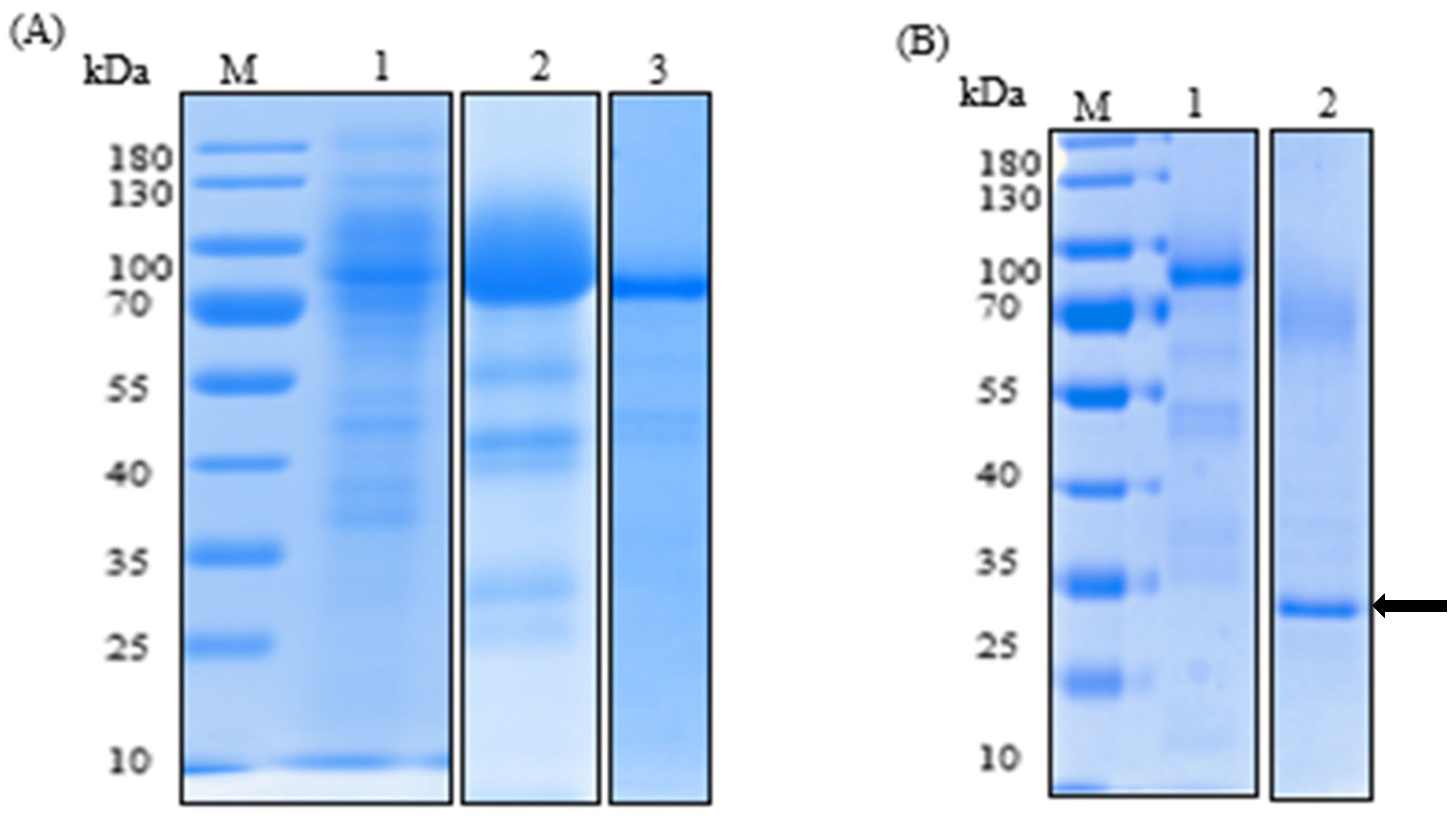
2.2. Purification of the Recombinant Laccase and Study of Physico-Chemical Properties

The recombinant laccase was purified from the culture medium of A. niger in a three-step procedure (Table 2): ultrafiltration, anion-exchange chromatography, and size exclusion chromatography. 930 mL of the filtered culture media (containing 176 mg of protein) was concentrated (1.9-fold) and separated from most impurities, which included a brown pigment absorbing strongly at 280 nm [28], by ultrafiltration through a polyethersulphone membrane (10 kDa molecular mass cut-off), with a resulting purification factor of 1.4-fold. The resulting concentrate was then loaded onto a carboxymethyl (CM)-Sepharose column to be further purified with a purification factor of 3-fold. In the last step, the resulting sample was concentrated through a 10 kDa molecular mass cut-off Amicon membrane and loaded onto a Sephacryl S-200HR size exclusion chromatography column. The resulting purification factor was 5.9-fold, yielding 7.2 mg of protein. The final recovery yield was 24.1%. The molecular mass of the purified laccase by SDS/PAGE was about 75 kDa (Figure 1A).
To check the N-terminal protein processing, the first five amino acids of the recombinant laccase were sequenced and aligned with the deduced mature protein. They showed 100% identity. This result demonstrated that the 24-amino-acid glucoamylase (GLA) prepropeptide from A. niger was correctly processed. For N-glycosylation, five sites (71, 115, 232, 404, and 424) were predicted. In addition, SlLac2 was deglycosylated using N-glycosidase PNGase (Figure 1B). On SDS-PAGE, the deglycosylated protein showed an apparent difference in its molecular mass from the untreated protein. The molecular masses of the purified and digested laccases were 75 kDa and <70 kDa, respectively. The molecular mass of the deglycosylated SlLac2 and the corresponding calculated molecular mass (64.6 kDa) confirmed the presence of N-glycosylations of approximately 10% of the total protein.

2.3. Kinetic Parameters

The kinetic constants (KM and Vmax) of SlLac2 were determined using various substrates such as 2,2′-azino-bis (3-ethylbenzothiazoline-6-sulphonic acid) (ABTS), 2,6-dimethoxyphenol (DMP) and syringaldazine under the optimal reaction conditions at 30 °C and pH (4.0, 5.0 and 6.0 respectively) (Table 3). Of the six substrates, SlLac2 exhibited the highest affinity for syringaldazine (KM = 0.0035 mM) followed by ABTS (KM = 0.0206 mμM) and DMP (KM = 0.024 mμM). The highest catalytic efficiency was found for ABTS (7.42 s−1 mM−1), followed by syringaldazine (2.11 s−1 mM−1) and DMP (0.24 s−1 mM−1).

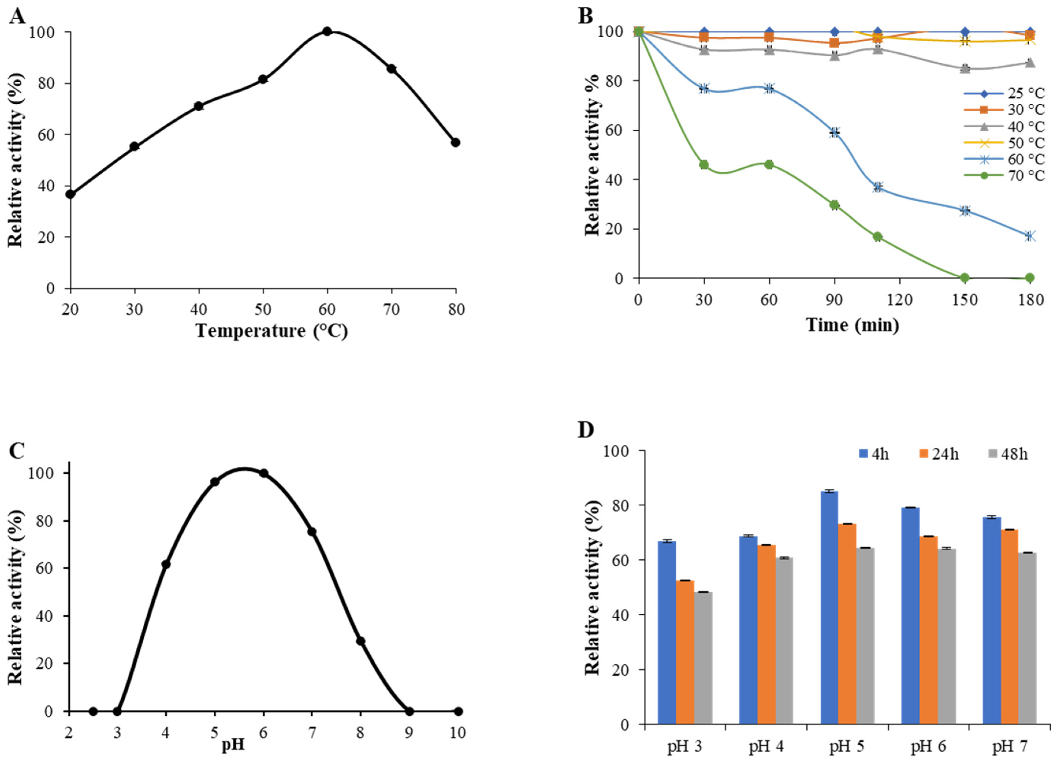
2.4. Enzyme Activity and Stability at Different pH and Temperature Values

SlLac2 activity increased gradually with increasing temperature up to 60 °C, the optimal temperature value (Figure 2A). Regarding thermal stability, SlLac2 was stable at temperatures ranging from 25 to 50 °C, but the activity was significantly lost above 60 °C (Figure 2B). The half-life value for SlLac2 was 95 min and 30 min, at 60 °C and 70 °C, respectively. The highest laccase activity level was recorded at pH 6.0 (Figure 2C). The enzyme possessed significant activity at pH 4.0 and 7.0 (60% and 75%, respectively). A lower activity could be measured at alkaline pH, with 30% of its initial activity at pH 8.0, and the activity almost disappeared above pH 8.0. When incubated at pH 5.0, 6.0 and 7.0 (Figure 2D), laccase activity of the purified enzyme SlLac2 retained around 80% initial activity after 4 h of incubation, about 70% after 24 h and 60% after 48 h. When incubated at pH 3.0 or 4, the laccase activity retained over 65% residual activity after 4 h, about 50–65% after 24 h and about 50–60% after 48 h. These results demonstrate that the enzyme endures at neutral to weak-acidic pH but has lower resistance to strong-acidic pH values.

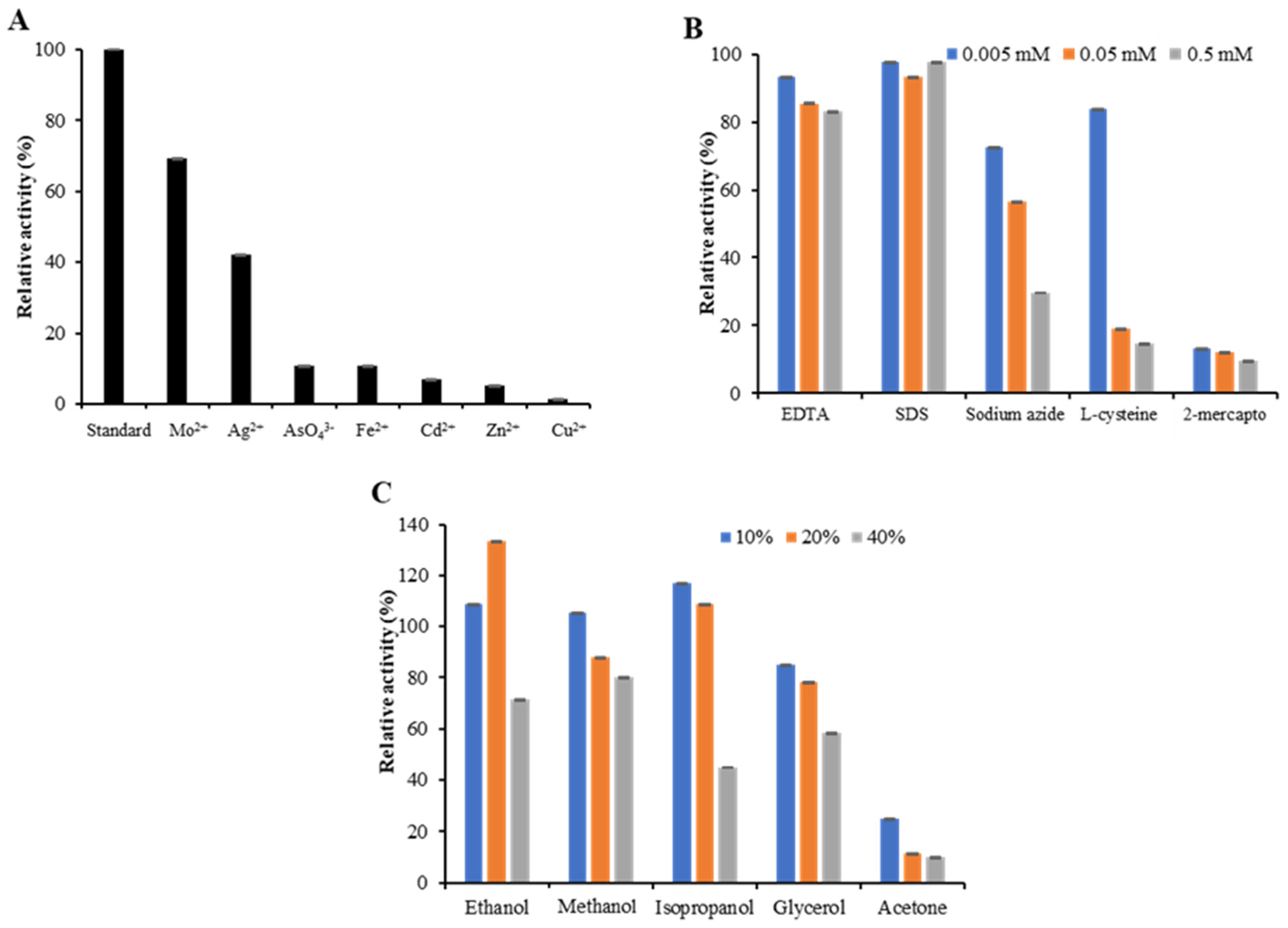
2.5. Effect of Metal Ions, Inhibitors and Solvents on Laccase Activity

Various metal ions (Mo2+, Ag2+, AsO43−, Fe2+, Cd2+, Zn2+, and Cu2+) at 10 mM concentration were tested on the activity of SlLac2 (Figure 3A). Mo2+ and Ag2+ caused the disappearance of 30% and 60% of SlLac2 activity, respectively. AsO43, Fe2+, Cd2+, and Zn2+ led to an even greater decrease in laccase activity (about 90% inhibition), and Cu2+ completely inhibited SlLac2 activity (98%).
Three concentrations (0.05 to 0.5 mM) of potential laccase inhibitors, ethylenediamine tetra-acetic acid (EDTA), sodium dodecyl sulfate (SDS), 2-mercaptoethanol, cysteine, and sodium azide (NaN3) were evaluated to check their effects on the SlLac2 activity (Figure 3B). In the tested concentration range, more than 85% of enzyme activity remained in the presence of EDTA and SDS. Sodium azide inhibited SlLac2 with a loss of 25–70% of its initial activity, while cysteine acted as a stronger inhibitor (15–85% of inhibition). The most efficient inhibitor was found to be 2-mercaptoethanol, which reduced activity by 88–91%.
The effect of different concentrations of solvents ethanol, methanol, isopropanol, glycerol and acetone on SlLac2 activity in the range 10–40% (vol/vol) was studied (Figure 3C). SlLac2 was relatively stable, with 10–20% (vol/vol) of ethanol, methanol and isopropanol, but its activity was increased to more than 130% of its initial activity with 20% (vol/vol) ethanol. However, the recombinant laccase was less stable towards 40% (vol/vol) of these solvents, with a decrease of about 20% of its activity (methanol) to 60% (isopropanol). Of all the organic solvents tested, acetone showed a markedly negative effect on SlLac2 activity, causing more than 80% activity inhibition at a concentration of 10% (vol/vol) and near-inactivation with 20% and 40% (vol/vol) of acetone.

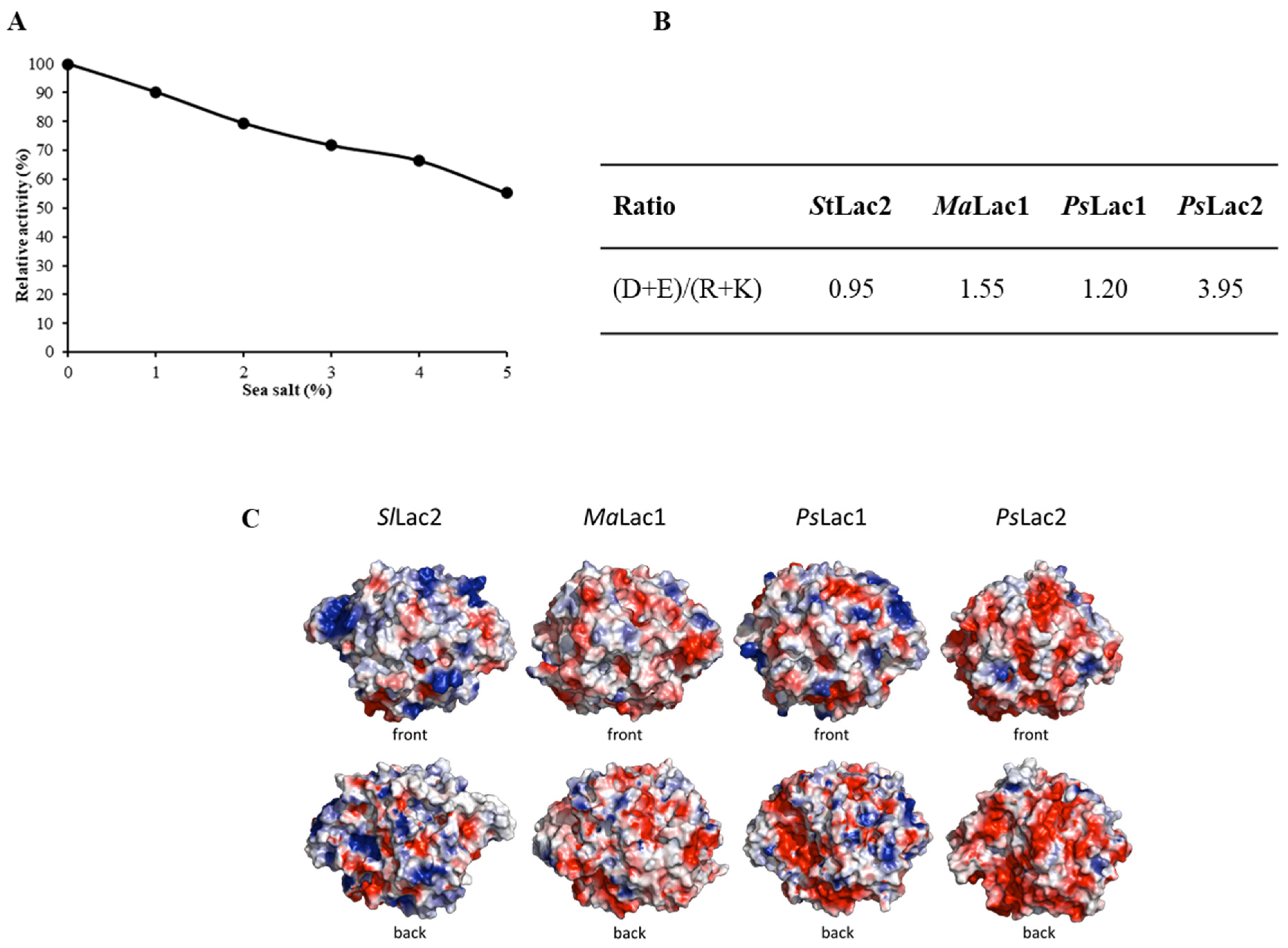
2.6. Effect of Sea Salt on Laccase Activity and Surface Charge of SlLac2

The effect of different concentrations of sea salt (1–5% wt/vol) on the purified laccase was analysed (Figure 4). The results showed that the activity gradually decreased as the salt concentration increased from 10% to almost half of its initial activity (Figure 3A). The enzyme could thus tolerate the presence of sea salt, retaining more than 50% of its activity with 5% wt/vol of sea salt. In parallel, analysis of the primary sequence and the overall surface charges of StLac2 were carried out and compared with those of the terrestrial-derived laccase, MaLac1, and of the marine-derived laccases, PsLac1 and PsLac2. The primary sequence of StLac2 exhibited a similar recurrence of negatively charged (D + E) over positively charged (R + K) amino acids, with the exception of PsLac2, which had 4.15 times higher (D + E)/(R + K) ratio (Figure 4B). MaLac1 and PsLac1 presented intermediate values, i.e., 1.55 and 1.20, respectively. The three-dimensional models of MaLac1 and PsLac1 showed an even charge distribution in their surface plots, whereas for PsLac2, the surface charge was strongly negative (Figure 4C). In contrast, StLac2 exhibited a slightly higher positive charge distribution at the surface.

3. Discussion

The objective of our research project was to seek new insights into the physiology of the marine-derived fungi, more specifically their lignocellulose enzyme machinery, and how they adapt in their marine environment. In previous work, we screened five fungal strains isolated from Tunisian coastal waters [26] and characterised for their laccase-like activities. Based on a microbial approach and on enzyme activity screening, one of these marine-derived strains, S. lucomagnoense, was selected for its adapted growth on xylan in saline conditions, and its improved laccase (seagrass-containing cultures) and cellulase (wheat straw-containing cultures) activities in the presence of sea salt [27]. To further study the selected marine-derived fungus, its transcriptome was sequenced and its proteome analysed when grown on wheat-straw and sea grass in the absence or presence of sea salt. From this study, we were unable to identify the laccases involved in the improved laccase activity in the presence of salts, possibly owing to the sensitivity limit of our combined transcriptomic-proteomic approach, because of undetected transcript domains. However, seven genes were identified in S. lucomagnoense transcriptome that encoded putative laccases, and one was selected for heterologous expression in A. niger and characterisation of the corresponding laccase, SlLac2.
Only a few biochemical characterisations of laccases from marine organisms are available [28,29,30,31]. Our aim was accordingly to obtain additional data on these enzymes and their potential for biotechnological applications. Marine-derived fungi are considered as a source of original enzymes active in saline conditions and pH ranging from 3.0 to 11.0 [24], which are properties of interest for many industrial applications. SlLac2 was produced in A. niger with a yield of about 30 mg L−1 in the culture medium, which is in the range of protein production obtained for the laccase of the terrestrial fungus Pycnoporus cinnabarinus, heterogously produced in A. niger (70 mg L−1) [32]. Recently, two laccase-encoding genes isolated from the marine-derived fungus Pestalotiopsis sp., were expressed in A. niger and lower yields were obtained: 2.6 and 6.2 mg L−1 for PsLac 1 and PsLac2, respectively [28]. The main physico-chemical properties of SlLac2 were determined to compare them with literature reports on laccases from marine and terrestrial fungi. The purified laccase was active on all typical laccase substrates including ABTS, DMP, and syringaldazine, and showed the highest affinity (lowest KM value) for syringaldazine (3.5 μM). However, the highest catalytic efficiency (Kcat/KM) was found for ABTS (7.42 s−1 mM−1) as its turnover value was much higher (Kcat 0.153 s−1) compared with that determined for syringaldazine (0.0074 s−1). Laccases are widespread in the natural environment, and very well-characterised for their kinetic parameters. Many previous studies found that syringaldazine and ABTS were their preferred substrates [33]. Laccase affinity for syringaldazine (tens of µM) is generally higher than that measured for ABTS (hundreds of µM), whereas KM constants for other phenolic compounds are considerably higher [24,34]. In our case, SlLac2 exhibited a preference (affinity 6-fold) for syringaldazine. By comparison, PsLac1 from the marine-derived fungus Pestalotiopsis sp., showed KM values of 4 µM and 24 µM for syringaldazine and ABTS, respectively, while PsLac2, did not exhibit a specific preference for either substrate, with higher KM values (hundreds of µM) [28]. Two other marine-derived fungal laccases from Trematosphaeria mangrovei [29] and Cerrena unicolor [31] showed different behaviours, with KM values of 1.42 mM and 54 μM against ABTS. Even though the affinity was stronger for ABTS, the catalytic efficiency (Kcat/KM) of SlLac2 was slightly higher for syringaldazine (7.42 s−1 mM−1). This value is very close to the Kcat/KM measured for Pestalotiopsis sp. Lac2 (5.67 s−1 mM−1) [28]. PsLac2 was described as a relatively versatile laccase, exhibiting a similar catalytic efficiency for syringaldazine, ABTS and DMP (5.67, 4.33 and 9.1 s−1 mM−1, respectively). By contrast, the second laccase of Pestalotiopsis sp., PsLac1 showed the highest catalytic efficiency for syringaldazine at 82.93 s−1 mM−1, and a 3-times lower Kcat/KM for the other two substrates ABTS and DMP (29.25 and 24.53 s−1 mM1, respectively). Higher catalytic efficiency for syringaldazine can be found in the literature, such as for the laccase-like enzyme cloned form a marine library with 29 s−1 mM−1 [23]. In conclusion for the kinetic parameters, SlLac2 was shown to possess the general characteristics of fungal laccases, with the highest efficiency for syringaldazine.
The physical and biochemical properties of the purified recombinant SlLac2 were tested for further comparisons with fungal laccases. As reported by Baldrian, fungal laccases typically exhibit an optimal pH in the acidic pH range (3.0–5.0), using ABTS as substrate [33], whereas the optimal pH for the oxidation of syringaldazine is higher (3.5–6.0) [33]. The purified recombinant SlLac2 was tested to determine its optimal pH in the range 3.0–9.0 using syringaldazine as a substrate, and it showed that the optimal pH value driving the maximum activity of the recombinant enzyme was pH 6.0, with a measurable activity at pH 8.0. For the laccases obtained from Pestalotiopsis sp. and T. mangrovei, the optimal pH was determined with ABTS as substrate: 5.0 and 4.0, respectively [28,29]. Usually, the stability of most fungal laccases is higher at acidic pH and decreases when close to neutral pH conditions, which is the case for the terrestrial P. cinnabarinus laccase [34]. In our work, we demonstrated that the purified recombinant SlLac2 was relatively resistance in several pH conditions tested (3.0 to 7.0). In addition, SlLac2 was more stable in the tested range of pH than Pestalotiopis sp. laccases, i.e., PsLac1 was less stable at alkaline pH. It showed 30% residual activity after 100 h of incubation at pH 6.0, whereas PsLac2, was instead more sensitive to low pH; its stability decreased with time between pH 3.0 and 6.0, and abruptly at pH 2.0 (no activity left after 40 min of incubation) [28].
Concerning the behaviour of fungal laccases with temperature, previous studies found that the optimal temperature was between 50 and 60 °C for most fungal laccases [32,33]. In our study, we also showed that the optimum temperature of the purified laccase SlLac2 was in this range (60 °C). Compared with marine laccases, T. mangrovei and PsLac 1 had slightly higher optimal temperatures: 65 °C (syringaldazine. 80 °C with ABTS) and 70 °C (ABTS), respectively [28,29]. Regarding thermal stability, SlLac2 was stable for 180 min at temperatures ranging from 25 to 50 °C, whereas the activity was significantly lost above 60 °C, as demonstrated for PsLac2 [28]. In addition, T. mangrovei laccase and PsLac1 showed curves with more pronounced thermal deactivation at temperatures above 60 °C [28,29,30]. We conclude that SlLac2 presents novel, useful properties due to its activity relative to temperature and pH, and to its stability compared with its marine laccase homologs.
Enzyme activity is often affected positively or negatively by metal ions present in the mixtures [35,36,37,38]. Because laccases are used in several processes containing metal ions, it is useful to characterise the effect of metal ions on their activities. It has been shown in many studies that laccase activities vary with the type of metal ion present and the enzyme source [38,39,40,41]. SlLac2 showed that adding a 10 mM concentration of Mo2+ and Ag2+ to the purified enzyme left 70% and 40% of the activity, respectively, but the presence of AsO43−, Fe2+, Cd2+, Zn2+, and especially Cu2+, caused the loss of all or most activity. For instance, Cu2+ has already been shown to inhibit Scytalidium thermophilum laccase activity [42]. Among several metal ions tested, Xu et al. showed that Ag+, Ag2+, Li+, and Pb2+ could inhibit the laccase of Cerenia sp. reversibly. Hg+ was shown to reduce pH and thermal stability, and dynamic simulation showed the presence of binding sites for Hg close to copper binding sites on the laccase molecule [43]. For T. mangrovei, the effect of metal ions was analysed at 1 mM, and only Fe2+ showed a complete inactivation of its activity, whereas Cu2+ was not tested [29]. At the same concentration, Fe3+ showed a 30% inactivation of the marine-derived fungus, C. unicolor laccase, whereas Cu2+ addition produced a slight effect with a loss of less than 10% of activity [31]. The most common laccase inhibitors tested include cysteine, EDTA, sodium fluoride, sodium azide, dithiothreitol, thioglycolic acid, and diethyldithiocarbamic acid. These inhibitors are not laccase-specific and their applications on phenoloxidases was because they could inhibit metalloenzymes [44,45]. They can act on the active site: EDTA binds to copper atoms and stops the transfer of electrons. Cysteine, dithiothreitol, thioglycolic acid, and diethyldithiocarbamic acid are reducing substances described as potential laccase inhibitors sequestering dioxygen and stopping the oxidation of the phenolic substrates. However, Johannes and Majcherczyk (2002) have tested several potential laccase inhibitors including dithiothreitol, thioglycolic acid (TGA), diethyldithiocarbamic acid (DDC), cysteine, and sodium azide (NaN3) [46]. They found that only NaN3 stopped the substrate oxidation by no oxygen uptake, whereas the sulfydryl compounds did not affect the oxygen consumption and even increased it through an autoxidation reaction. SlLac2 was significantly inactivated by NaN3, l-cysteine and 2-mercaptoethanol, whereas C. unicolor laccase was inhibited by 1 mM of NaN3 (95% inhibition) and 2-mercaptoethanol (67% inhibition), but not by l-cysteine (0.1 mM) [28]. For T. mangrovei laccase, a 50% inhibition was demonstrated for 1 mM of NaN3 and the most efficient inhibitor was shown to be NaCN (80% imbibition) [29].
Biochemical reactions involving laccases in compatible organic solvents give access to some insoluble substrates, which may help in the detoxification of several sparingly water-soluble persistent organic pollutants. However, several studies have shown negative effects of organic solvents on laccase activity [47,48]. For instance, Robles et al. [49] studied a laccase of the hyphomycete Chalara (syn. Thielaviopsis) paradoxa CH32 and showed an activity decrease of 27% and 36% in the presence of 25% (vol/vol) ethanol and methanol, respectively. In our study, we showed that with 40% (vol/vol) of ethanol and methanol, there was a similar decrease in the activity from about 30% and 20%, respectively. However, of the solvents tested in our experiments, acetone was the most damaging, probably by precipitating the proteins. Both free and immobilised laccases decreased their activity with an increase in water-miscible organic solvent concentrations, and in most reported cases, immobilised laccase is less strongly affected than the free enzyme by the organic solvent concentrations, which could offer an alternative, depending on the solvents needed to implement for the laccase application.
As SlLac2 was isolated from a marine-derived fungus, this enzyme behaviour was studied in relation to saline conditions. In the presence of sea salt, SlLac2 was affected, with a continuous decrease in its activity following the sea salt concentration increase. For the two laccases of the marine-derived fungus Pestalotiopsis sp., PsLac1 and 2, completely different behaviour was observed, as their activities were enhanced in the presence of increasing concentrations of sea salts [28]. PsLac2 activity was clearly stronger (1.6 times higher compared with PsLac2 at 5% sea salt). In addition, T. mangrovei laccase lost half of its activity in only 1 mM NaCl [29] while Pestalotiopsis sp. lytic polysaccharide monoxidases A (LMPOA) could act on cellulose for up to 6% sea salt [50]. A few studies have been carried out on enzymes originating from saline environments to identify the molecular determinants responsible for their salt tolerance. Recently, Li et al. [51] demonstrated that two amino acid sites were involved in the salt activation, as Cl ion could bind to specific local sites to interfere with substrate binding and/or electron transfer. In addition, salt-adapted enzymes were shown to be strongly negatively charged on their surface, a characteristic contributing to protein stability and to enzyme activities adapted to extreme osmotic conditions [52,53,54]. SlLac2 was shown to possess a slightly higher positive charge distribution at the protein surface. In contrast, MaLac1 and PsLac1 exhibited a relatively even balance of negative charges over positive charges (D + E/R + K ratio), whereas PsLac2 presented a very high ratio of 3.92. In a previous study on LMPOs identified from Pestalotiopsis sp., the calculated ratio also exhibited very high ratios greater than 4.0 [50] and an excess of negatively charged residues at their surface, typical of marine enzymes. Altogether, these results show that the resistance to salt is not yet fully understood, and that further research is still needed to elucidate the adaptation mechanisms.

4. Materials and Methods

4.1. Strains and Culture Conditions

Escherichia coli strain TOP 10 was used for vector storage and propagation. Aspergillus niger strain D15#26 (pyrG deficient) [55] was used for the heterologous expression of the SlLac2 encoding synthetic gene. After co-transformation with vectors respectively containing the pyrG gene and the laccase cDNA, transformants of A. niger were grown for selection on solid minimal medium without uridine and containing 70 mM of NaNO3, 7 mM of KCl, 11 mM of KH2PO4, 2 mM of MgSO4, glucose 1% (wt/vol), and trace elements (1000 × stock; 76 mM ZnSO4, 178 mM H3BO3, 25 mM MnCl2, 18 mM FeSO4, 7.1 mM CoCl2, 6.4 mM CuSO4, 6.2 mM Na2MoO4, and 174 mM EDTA). For the screening procedure of the positive transformants, 100 mL of culture medium containing 70 mM NaNO3, 7 mM KCl, 200 mM Na2HPO4, 2 mM MgSO4, glucose 5% (wt/vol), and trace elements was inoculated with 2 × 10⁶ spores mL−1 in a 250 mL baffled flask.

4.2. Cloning and Expression of SlLac2-Encoding Gene

The open reading frame sequence encoding SlLac2 was synthesised and codon bias optimised for A. niger (GeneArt, Regensburg, Germany), with some modifications. The amino acids of the signal peptide, which was predicted with the program SignalP hosted on the ExPASy Proteomics server (http://www.expasy.ch), were replaced by the 24-amino-acid glucoamylase (GLA) preprosequence from A. niger (MGFRSLLALSGLVCNGLANVISKR). Two restriction sites (BssHII and HindIII) were respectively added at the 5′ and 3′ ends of the sequence for cloning into the expression vector pAN52.4 (GenBank/EMBL accession number Z32699). In the final expression cassette, the Aspergillus nidulans glyceraldehyde-3-phosphate dehydrogenase-encoding gene (gpdA) promoter, the 5′ untranslated region of the gpdA mRNA, and the A. nidulans trpC terminator were used to drive the expression of the inserted coding sequences. The co-transformation was carried out as described by Punt and van den Hondel [25] using both the expression vector containing the expression cassette and pAB4-1 [56] containing the pyrG selection marker in a 10:1 ratio. Transformants were selected for uridine prototrophy by growth on selective solid minimal medium (without uridine).

4.3. Screening of Transformants and Laccase Activity Assay

To screen for the best clones for enzyme production in liquid medium, 100 mL of minimal medium (adjusted to pH 5.5 with 1 M of citric acid) was inoculated with 2 × 10⁶ spores mL−1 in a 250 mL flask. The cultures were incubated for 10 days at 30°C in a shaker incubator (110 rpm), and pH was adjusted daily to 5.5 with 1 M citric acid. From these liquid cultures, aliquots (2 mL) were withdrawn daily, and mycelia were pelleted (20 min at 15,000× g). Laccase activity of the resulting supernatant was assayed spectrophotometrically by monitoring the oxidation of syringaldazine (1 mM) as substrate at 436 nm (ɛ436 = 29,300 M−1 cm−1) in a sodium acetate buffer (50 mM, pH 6.0). The reaction was monitored for 1 min at 30 °C in an Uvikon XS spectrophotometer (BioTek Instruments, Colmar, France). Activity is expressed in nkat mL−1, 1 nkat corresponding to the oxidation of 1 nanomole of substrate per second. Measurements in all the experiments were performed in triplicate.

4.4. Production and Purification of Recombinant SlLac2

For protein production, the best positive clone corresponding to the clone with the highest laccase activity was selected and cultured. 930 mL of culture medium containing 70 mM NaNO3, 7 mM KCl, 200 mM K2HPO4, 2 mM MgSO4, glucose 10% (wt/vol), trace elements and adjusted to pH 5.0 with a 1 M citric acid solution, inoculated with 2 × 10⁶ spores mL−1, was prepared to initiate a large-scale protein production (1 L). The culture was harvested after 11 days. The culture medium was clarified by filtration through GF/D, GF/A and GF/F glass fibre filters (Whatman, Maidstone, UK), followed by filtrations through 0.45 μm and 0.22 μm polyethersulphone membranes (Express Plus, Merck Millipore). The collected filtrate was concentrated by ultrafiltration through a polyethersulphone membrane with a 10 kDa molecular mass cut-off (Vivaflow crossflow cassette, Sartorius, Les Ulis, France). After dialysing overnight at 4 °C against 100 mM Tris buffer pH 8.0, 150 mM NaCl and 1 mM EDTA, the sample was loaded onto on a CM-Sepharose fast flow column (GE Healthcare Life Science, Velizy-Villacoublay, France) using an AKTA purifier (GE Healthcare Life Science). The sample was loaded onto the column previously equilibrated with a binding buffer (0.2 mM sodium tartrate buffer pH 5.0, containing 25 mM NaCl). Protein elution was performed using a linear gradient of 0–100% of the elution buffer (0.2 mM of sodium tartrate buffer pH 5.0, containing 1 mM NaCl). Activity was determined in the 10 mL collected fractions, and protein production was evaluated by SDS-PAGE. The active fractions were concentrated through a 10 kDa molecular mass cut-off Amicon membrane (Millipore). The concentrated samples were loaded onto a Sephacryl S-200HR size exclusion chromatography column (GE Healthcare Life Science) equilibrated with a 50 mM sodium acetate buffer pH 5.0, containing 50 mM NaCl. 5 mL fractions were collected and assayed for laccase activity as described above, and protein homogeneity was tested by SDS-PAGE. For the different purification steps, the total protein concentration was determined with the Bradford assay using the BioRad Protein Assay Kit (BioRad, Marnes-la-Coquette, France) and bovine serum albumin (BSA) as a standard. Final protein concentration was determined spectrophotometrically at 280 nm using a NanoDrop 2000 (Thermo Fisher Scientific, Illkirch, France) and the theoretical molar extinction coefficients SlLac2 at 280 nm, 125,290 M−1 cm−1. The molecular mass of the purified protein was determined by 12% sodium dodecyl sulphate-polyacrylamide gel electrophoresis (SDS-PAGE).

4.5. Bioinformatic Analysis

The molecular mass, theoretical pI, and molar extinction coefficient of enzymes were predicted by the ProtParam tool (http://web.expasy.org/protparam/). Protein sequences were aligned using MUSCLE [57,58] and CLUSTAL W [59] at http://www.ebi.ac.uk/Tools/msa/. Signal peptides were predicted using Signal P [60] at http://www.cbs.dtu.dk/services/SignalP/. N-glycosylation sites [60] were predicted at http://www.cbs.dtu.dk/services/NetNGlyc/. The automated protein structure homology modelling online tool Phyre 2 [61] was used to predict the three-dimensional models of SlLac2 using the closest homologs of known structure available in the Protein Data Bank (PDB). Protein models c2q9oA (Melanocarpus albomyces laccase, 44% identity), c5lwxA (Aspergillus niger laccase McoG H253D variant, 37% identity), c3ppsD (Thielavia arenaria laccase, 45% identity) and c3sqrA (Botrytis aclada laccase, 44% identity) were used as templates for SlLac2. Surface charges were calculated, and all the figures were prepared with PyMOL.

4.6. Determination of N-Terminal Amino Acid Sequence and Glycosylation Level

The N-terminal sequence was determined according to Edman degradation. Analysis was carried out on an Applied Biosystem 476Asequencer by the proteomic platform of the Institut de Microbiologie de la Méditerrranée, CNRS, Aix-Marseille Université (France). Matrix-assisted laser desorption ionisation—time-of-flight mass spectrometry of samples was carried out on a Microflex II time-of-flight mass spectrometer (Bruker Daltonik, Germany).
To determine the level of glycosylation of the purified laccase SlLac2, a deglycosylation reaction was carried out using the PNGase F (New England Biolabs, France). 15 µg of purified laccase was mixed with 4 µL of the denaturation solution (5% SDS and 40 mM dithiothreitol (DTT)) denatured at 100 °C for 10 min. The denatured sample was deglycosylated in a final volume of 40 µL containing 1% Nonidet P-40, 50 mM sodium phosphate buffer (pH 7.5) and 2 µL of the deglycosylation enzyme PNgase F solution (500,000 units mL−1). The reactions were incubated at 37 °C for 2 h. After the deglycosylation, 10 µL of the digested enzyme was loaded onto a 12% homogeneous SDS-PAGE gel.

4.7. Substrate Specificity and Kinetics

Enzyme activities were measured using a UVIKONxs spectrophotometer (Bio-TEK Instruments) at 30 °C by following the oxidation of different aromatic substrates: 2,2′-azino-bis(3-ethylbenzothiazoline-6-sulphonic acid) (ABTS), 2,6-dimethoxyphenol (DMP) and syringaldazine. The absorbance increases at 436 nm (ɛ436 = 29,300 M−1 cm−1), 469 nm (ɛ469 = 27,500 M−1 cm−1) and 530 nm (ɛ530 = 65,000 M−1 cm−1) were followed for ABTS, DMP, and syringaldazine, respectively. Kinetic constants (enzyme concentration 0.18 mg mL−1) were determined for each of these compounds (substrate range of 0.05 to 1.1 mM for ABTS, and 0.025 to 0.1 mM for DMP and syringaldazine) with sodium acetate buffer (50 mM, pH 4.0, 5.0, and 6.0, respectively). All assays were performed in triplicate. Mean apparent affinity constant (Michaelis constant, Km) and enzyme turnover (catalytic constant, kcat) values and standard errors were obtained by nonlinear least-squares fitting to the Michaelis-Menten model. Fitting of these constants to the normalized Michaelis-Menten equation υ = (kcat/Km)[S]/(1 + [S]/Km) yielded enzyme efficiency values (kcat/Km) with their standard errors

4.8. Effect of pH and Temperature on the Activity and Stability of the Laccase

The effect of pH on the purified enzyme was determined by measuring the enzyme activity at 30 °C in various buffers at different pH (2.5–10.0) using the following buffers: 50 mM sodium acetate buffer (pH 2.5–7.0), phosphate buffer (pH 7.0–9.0), and sodium bicarbonate (pH 9.0–10.0), against 1 mM syringaldazine as substrate at 30°C. The pH stability of the purified enzyme was determined by incubating the enzyme in the different buffer solutions (50 mM) with different pH values (3.0–7.0) and allowing the mixture to stand for 4 h, 24 h and 48 h incubation at room temperature. The residual enzyme activity was determined under standard assay conditions after centrifugation to remove the denatured proteins.
To determine the effect of temperature on laccase, the activity of the purified enzyme was measured in a sodium acetate buffer (50 mM, pH 6.0) at different temperatures ranging from 20 to 80°C. The stability to thermal treatment of purified enzyme was determined by measuring residual activity after incubation at different temperatures (25–70°C) and pH 6.0 for 30 min to 3 h. All the reactions were run in triplicate.

4.9. Effect of Metal Ions, Inhibitors, Solvents, and Sea Salt on Laccase Activity.

To study the effect of metal ions, potential inhibitors, different solvents, and sea salt on the purified enzyme, the activity was determined in the presence of metal ions in standard laccase activity conditions. The following compounds were used as the source of metal ions: CuSO4.5H2O, Na2MoO4, FeSO4.7H2O, CdCl2, Na3AsO4.12H2O, and ZnSO4.7H2O. They were added at a final concentration of 10 mM. The potential inhibitors were l-cysteine, 2-mercaptoethanol, EDTA, SDS, and sodium azide (final concentrations of 0.5, 0.05 and 0.05 mM). The different organic solvents were ethanol, methanol, iso-propanol, glycerol, and acetone at different concentrations: 10, 20, and 40%. Different concentrations of sea salt (Sigma-Aldricht, Lyon, France) (1–5% wt/vol) were added to the reaction mixture and the activity was determined in standard conditions. All the reactions were run in triplicate.

4.10. Nucleotide Sequence Accession Number

The gene sequences encoding SlLac2 were deposited in the nucleotide sequence database (GenBank) under the accession code MT470191.

5. Conclusions

The present work carried out on the S. lucomagnoense gives new insights into the marine-derived laccases, which are catalysts of great interest for many biotechnological applications. In addition, SlLac2 produced in A. niger presented potential properties related to pH and some tolerance to sea salt, which could be exploited in biotechnological applications, such as the pulp and paper sector and enzymatic degradation of industrial dyes, for which alkaline-active enzymes and high tolerance to salt are needed, respectively

Supplementary Materials

The following are available online at https://0-www-mdpi-com.brum.beds.ac.uk/1422-0067/21/21/8402/s1, Supplementary Materials, Figure S1: Alignments of the closest characterized laccases from Melanocarpus albomyces (MaLac1, Q70KY3) and Pestalotiopsis sp. KF079 (PsLac1: KY554800 and PsLac 2: KY554801), using CLUSTAL W sequence alignment algorithm. Figure S2: Kinetics of SlLac2 on the three substrates.

Author Contributions

W.B.A., A.B.A., Y.M., and A.T.-D. provided experiment support; W.B.A., A.B.A., and A.T.-D. provided technical support and contributed to the discussion; W.B.A., E.R., T.M. designed the experiments; W.B.A., E.B., C.B.F., G.S., A.L., E.R., T.M. wrote the manuscript. All authors have read and agreed to the published version of the manuscript.

Funding

The authors thank PHC-Utique (39274UH 2018–2020, 18G0817) for financial support.

Conflicts of Interest

The authors declare they have no conflict of interest.

References

  1. Sivakumar, R.; Rajendran, R.; Balakumar, C.; Tamilvendan, M. Isolation, screening and optimization of production medium for thermostable laccase production from Ganoderma sp. Int. J. Eng. Sci. Technol. 2010, 2, 7133–7141. [Google Scholar]
  2. Kumar, R.; Kaur, J.; Jain, S.; Kumar, A. Optimization of laccase production from Aspergillus flavus by design of experiment technique: Partial purification and characterization. J. Genet. Eng. Biotechnol. 2016, 14, 125–131. [Google Scholar] [CrossRef] [PubMed] [Green Version]
  3. Giardina, P.; Faroca, V.; Pezzela, P.C.; Piscitelli, A.; Vanhilla, S.; Sannia, G. Laccase: A never-ending story. Cell. Mol. Life Sci. 2010, 62, 369–386. [Google Scholar] [CrossRef] [PubMed]
  4. Durán, N.; Rosa, M.A.; D’Annibale, A.; Gianfreda, L. Applications of laccases and tyrosinases (phenoloxidases) immobilized on different supports: A review. Enzym. Microb. Technol. 2002, 31, 907–931. [Google Scholar] [CrossRef]
  5. Arora, D.S.; Sharma, R.K. Ligninolytic fungal laccases and their biotechnological applications. Appl. Biochem. Biotechnol. 2010, 160, 1760–1788. [Google Scholar] [CrossRef]
  6. Xu, F. Oxidation of phenols, anilines, and benzenethiols by fungal laccases: Correlation between activity and redox potentials as well as halide inhibition. Biochemistry 1996, 35, 7608–7614. [Google Scholar] [CrossRef]
  7. Minussi, R.C.; Pastore, G.M.; Durán, N. Enzima de interés en enología: Lacasas. Aliment 1999, 304, 145–150. [Google Scholar]
  8. Christopher, F. The structure and function of fungal laccase. Microbiology 1994, 140, 19. [Google Scholar]
  9. Baldrian, P. Purification and characterization of laccase from the white-rot fungus Daedalea quercina and decolorization of synthetic dyes by the enzyme. Appl. Microbiol. Biotechnol. 2004, 63, 560–563. [Google Scholar] [CrossRef]
  10. Li, K.; Xu, F.; Eriksson, K.E. Comparison of fungal laccases and redox mediators in oxidation of a nonphenolic lignin model compound. Appl. Microbiol. Biotechnol. 1999, 65, 2654–2660. [Google Scholar] [CrossRef] [Green Version]
  11. Munk, L.; Sitarz, A.K.; Kalyani, D.C.; Mikkelsen, J.D.; Meyer, A.S. Can laccases catalyze bond cleavage in lignin? Biotechnol. Adv. 2015, 33, 13–24. [Google Scholar] [CrossRef] [PubMed]
  12. Janusz, G.; Pawlik, A.; Świderska-Burek, U.; Polak, J.; Sulej, J.; Jarosz-Wilkołazka, A.; Paszczyński, A. Laccase Properties, Physiological Functions, and Evolution. Int. J. Mol. Sci. 2020, 21, 966. [Google Scholar] [CrossRef] [Green Version]
  13. Xu, F.; Shin, W.; Brown, S.H.; Wahleithner, J.A.; Sundaram, U.M.; Solomon, E.I. A study of a series of recombinant fungal laccases and bilirubin oxidase that exhibit significant differences in redox potential, substrate specificity, and stability. Biochim. Biophys. Acta Protein Struct. Mol. Enzym. 1996, 1292, 303–311. [Google Scholar] [CrossRef]
  14. Bourbonnais, R.; Paice, M.G. Oxidation of non-phenolic substrates: An expanded role for laccase in lignin biodegradation. FEBS Lett. 1990, 267, 99–102. [Google Scholar] [CrossRef] [Green Version]
  15. Call, H.P.; Mücke, I. History, overview and applications of mediated lignolytic systems, especially laccase-mediator-systems (Lignozym®-process). J. Biotechnol. 1997, 53, 163–202. [Google Scholar] [CrossRef]
  16. Bajpai, P.; Kondo, R. Biotechnology for Environmental Protection in the Pulp and Paper Industry; Springer Science & Business Media: Berlin/Heidelberg, Germany, 2012; ISBN 3-642-60136-7. [Google Scholar]
  17. Wesenberg, D.; Kyriakides, I.; Agathos, S.N. White-rot fungi and their enzymes for the treatment of industrial dye effluents. Biotechnol. Adv. 2003, 22, 161–187. [Google Scholar] [CrossRef]
  18. Vinodhkumar, T.; Thiripurasundari, N.; Ramanathan, G.; Karthik, G. Screening of dye degrading bacteria from textile effluents. J. Chem. Pharm. Res. 2013, 3, 848–857. [Google Scholar]
  19. Michaels, G.B.; Lewis, D.L. Sorption and toxicity of azo and triphenylmethane dyes to aquatic microbial populations. Environ. Toxicol. Chem. 1985, 4, 45–50. [Google Scholar] [CrossRef]
  20. Chung, K.-T.; Stevens, S.E., Jr. Degradation azo dyes by environmental microorganisms and helminths. Environ. Toxicol. Chem. 1993, 12, 2121–2132. [Google Scholar]
  21. Ghoreishi, S.M.; Haghighi, R. Chemical catalytic reaction and biological oxidation for treatment of non-biodegradable textile effluent. Chem. Eng. J. 2003, 95, 163–169. [Google Scholar] [CrossRef]
  22. Santhanam, N.; Vivanco, J.M.; Decker, S.R.; Reardon, K.F. Expression of industrially relevant laccases: Prokaryotic style. Trends Biotechnol. 2011, 29, 480–489. [Google Scholar] [CrossRef]
  23. Yang, Q.; Zhang, M.; Zhang, M.; Wang, C.; Liu, Y.; Fan, X.; Li, H. Characterization of a novel, cold-adapted, and thermostable laccase-like enzyme with high tolerance for organic solvents and salt and potent dye decolorization ability, derived from a marine metagenomic library. Front. Microbiol. 2018, 9, 2998. [Google Scholar] [CrossRef] [Green Version]
  24. Bonugli-Santos, R.C.; Dos Santos Vasconcelos, M.R.; Passarini, M.R.Z.; Vieira, G.A.L.; Lopes, V.C.P.; Mainardi, P.H.; Dos Santos, J.A.; de Azevedo Duarte, L.; Otero, I.V.R.; da Silva Yoshida, A.M.; et al. Marine-derived fungi: Diversity of enzymes and biotechnological applications. Front. Microbiol. 2015, 6, 269. [Google Scholar] [CrossRef] [PubMed]
  25. Punt, P.J.; van den Hondel, C.A.M.J.J. Transformation of filamentous fungi based on hygromycin b and phleomycin resistance markers. Methods Enzymol. 1992, 216, 447–457. [Google Scholar]
  26. Ben Ali, W.; Chaduli, D.; Navarro, D.; Lechat, C.; Turbé-Doan, A.; Bertrand, E.; Faulds, C.B.; Sciara, G.; Lesage-Meessen, L.; Record, E.; et al. Screening of five marine-derived fungal strains for their potential to produce oxidases with laccase activities suitable for biotechnological applications. BMC Biotechnol. 2020, 20, 27. [Google Scholar] [CrossRef] [PubMed]
  27. Ben Ali, W.; Navarro, D.; Kumar, A.; Drula, E.; Turbé-Doan, A.; Correia, L.O.; Baumberger, S.; Bertrand, E.; Faulds, C.B.; Henrissat, B.; et al. Characterization of the CAZy Repertoire from the Marine-Derived Fungus Stemphylium lucomagnoense in Relation to Saline Conditions. Mar. Drugs 2020, 18, 461. [Google Scholar] [CrossRef]
  28. Wikee, S.; Hatton, J.; Turbé-Doan, A.; Mathieu, Y.; Daou, M.; Lomascolo, A.; Kumar, A.; Lumyong, S.; Sciara, G.; Faulds, C.B.; et al. Characterization and Dye Decolorization Potential of Two Laccases from the Marine-Derived Fungus Pestalotiopsis sp. Int. J. Mol. Sci. 2019, 20, 1864. [Google Scholar] [CrossRef] [Green Version]
  29. Atalla, M.M.; Zeinab, H.K.; Eman, R.H.; Amani, A.Y.; Abeer, A.A.E. Characterization and kinetic properties of the purified Trematosphaeria mangrovei laccase enzyme. Saudi J. Biol. Sci. 2013, 20, 373–381. [Google Scholar] [CrossRef] [Green Version]
  30. Mainardi, P.H.; Feitosa, V.A.; de Paiva, L.B.B.; Bonugli-Santos, R.C.; Squina, F.M.; Pessoa, A., Jr.; Sette, L.D. Laccase production in bioreactor scale under saline condition by the marine-derived basidiomycete Peniophora sp. CBMAI 1063. Fungal Biol. 2018, 122, 302–309. [Google Scholar] [CrossRef]
  31. D’Souza-Ticlo, D.; Sharma, D.; Raghukumar, C. A Thermostable Metal-Tolerant Laccase with Bioremediation Potential from a Marine-Derived Fungus. Mar. Biotechnol. 2009, 11, 725–737. [Google Scholar] [CrossRef]
  32. Record, E.; Punt, P.J.; Chamkha, M.; Labat, M.; van den Hondel, C.A.; Asther, M. Expression of the Pycnoporus cinnabarinus laccase gene in Aspergillus niger and characterization of the recombinant enzyme. Eur. J. Biochem. 2002, 269, 602–609. [Google Scholar] [CrossRef] [PubMed]
  33. Baldrian, P. Fungal laccases-occurrence and properties. FEMS Microbiol. Rev. 2006, 30, 215–242. [Google Scholar] [CrossRef] [Green Version]
  34. Eggert, C.; Temp, U.; Eriksson, K.-E. The ligninolytic system of the white rot fungus Pycnoporus cinnabarinus: Purification and characterization of the laccase. Appl. Environ. Microbiol. 1996, 62, 1151–1158. [Google Scholar] [CrossRef] [PubMed] [Green Version]
  35. Sun, K.; Kang, F.; Waigi, M.G.; Gao, Y.; Huang, Q. Laccase-mediated transformation of triclosan in aqueous solution with metal cations and humic acid. Environ. Pollut. 2017, 220, 105–111. [Google Scholar] [CrossRef]
  36. Murugesan, K.; Kim, Y.-M.; Jeon, J.-R.; Chang, Y.-S. Effect of metal ions on reactive dye decolorization by laccase from Ganoderma lucidum. J. Hazard. Mater. 2009, 168, 523–529. [Google Scholar] [CrossRef]
  37. Lu, L.; Zhao, M.; Wang, T.-N.; Zhao, L.-Y.; Du, M.-H.; Li, T.-L.; Li, D.-B. Characterization and dye decolorization ability of an alkaline resistant and organic solvents tolerant laccase from Bacillus licheniformis LS04. Bioresour. Technol. 2012, 115, 35–40. [Google Scholar] [CrossRef]
  38. Si, J.; Peng, F.; Cui, B. Purification, biochemical characterization and dye decolorization capacity of an alkali-resistant and metal-tolerant laccase from Trametes pubescens. Bioresour. Technol. 2013, 128, 49–57. [Google Scholar] [CrossRef] [PubMed]
  39. Siroosi, M.; Amoozegar, M.A.; Khajeh, K. Purification and characterization of an alkaline chloride-tolerant laccase from a halotolerant bacterium, Bacillus sp. strain WT. J. Mol. Catal. B Enzym. 2016, 134, 89–97. [Google Scholar] [CrossRef]
  40. Wang, S.-S.; Ning, Y.-J.; Wang, S.-N.; Zhang, J.; Zhang, G.-Q.; Chen, Q.-J. Purification, characterization, and cloning of an extracellular laccase with potent dye decolorizing ability from white rot fungus Cerrena unicolor GSM. Int. J. Biol. Macromol. 2017, 95, 920–927. [Google Scholar] [CrossRef]
  41. Yan, J.; Chen, D.; Yang, E.; Niu, J.; Chen, Y.; Chagan, I. Purification and characterization of a thermotolerant laccase isoform in Trametes trogii strain and its potential in dye decolorization. Int. Biodeter. Biodegr. 2014, 93, 186–194. [Google Scholar] [CrossRef]
  42. Younes, S.B.; Sayadi, S. Purification and characterization of a novel trimeric and thermotolerant laccase produced from the ascomycete Scytalidium thermophilum strain. J. Mol. Catal. B Enzym. 2011, 73, 35–42. [Google Scholar] [CrossRef]
  43. Xu, X.; Huang, X.; Liu, D.; Lin, J.; Ye, X.; Yang, J. Inhibition of metal ions on Cerrena sp. laccase: Kinetic, decolorization and fluorescence studies. J. Taiwan Inst. Chem. E 2018, 84, 1–10. [Google Scholar] [CrossRef]
  44. Bollag, J.-M.; Leonowicz, A. Comparative studies of extracellular fungal laccases. Appl. Environ. Microbiol. 1984, 48, 849–854. [Google Scholar] [CrossRef] [Green Version]
  45. Slomczynski, D.; Nakas, J.P.; Tanenbaum, S.W. Production and Characterization of Laccase from Botrytis cinerea 61. Appl. Environ. Microbiol. 1995, 61, 907–912. [Google Scholar] [CrossRef] [Green Version]
  46. Johannes, C.; Majcherczyk, A. Laccase activity tests and laccase inhibitors. J. Biotechnol. 2000, 78, 193–199. [Google Scholar] [CrossRef]
  47. Milstein, O.; Hüttermann, A.; Majcherczyk, A.; Schulze, K.; Fründ, R.; Lüdemann, H.-D. Transformation of lignin-related compounds with laccase in organic solvents. J. Biotechnol. 1993, 30, 37–48. [Google Scholar] [CrossRef]
  48. Luterek, J.; Gianfreda, L.; Wojtaś-Wasilewska, M.; Cho, N.S.; Rogalski, J.; Jaszek, M.; Malarczyk, E.; Staszczak, M.; Fink-Boots, M.; Leonowicz, A. Activity of free and immobilized extracellular Cerrena unicolor laccase in water miscible organic solvents. Holzforsch. Int. J. Biol. Chem. Phys. Technol. Wood 1998, 52, 589–595. [Google Scholar]
  49. Robles, A.; Lucas, R.; Martínez-Cañamero, M.; Ben Omar, N.; Pérez, R.; Gálvez, A. Characterization of laccase activity produced by the hyphomycete Chalara (syn. Thielaviopsis) paradoxa CH. Enzym. Microb. Technol. 2002, 31, 516–522. [Google Scholar] [CrossRef]
  50. Patel, I.; Kracher, D.; Ma, S.; Garajova, S.; Haon, M.; Faulds, C.B.; Berrin, J.-G.; Ludwig, R.; Record, E. Salt-responsive lytic polysaccharide monooxygenases from the mangrove fungus Pestalotiopsis sp. NCi. Biotechnol. Biofuels 2016, 9, 108. [Google Scholar] [CrossRef]
  51. Li, Z.; Jiang, S.; Xie, Y.; Fang, Z.; Xiao, Y.; Fang, W.; Zhang, X. Mechanism of the salt activation of laccase Lac. Biochem. Biophys. Res. Commun. 2020, 521, 997–1002. [Google Scholar] [CrossRef]
  52. Kern, M.; McGeehan, J.E.; Streeter, S.D.; Martin, R.N.A.; Besser, K.; Elias, L.; Eborall, W.; Malyon, G.P.; Payne, C.M.; Himmel, M.E.; et al. Structural characterization of a unique marine animal family 7 cellobiohydrolase suggests a mechanism of cellulase salt tolerance. Proc. Natl. Acad. Sci. USA 2013, 110, 10189–10194. [Google Scholar] [CrossRef] [Green Version]
  53. Paul, S.; Bag, S.K.; Das, S.; Harvill, E.T.; Dutta, C. Molecular signature of hypersaline adaptation: Insights from genome and proteome composition of halophilic prokaryotes. Genome Biol. 2008, 9, R70. [Google Scholar] [CrossRef] [Green Version]
  54. Lanyi, J.K. Salt-dependent properties of proteins from extremely halophilic bacteria. Bacteriol. Rev. 1974, 38, 272. [Google Scholar] [CrossRef] [Green Version]
  55. Gordon, C.L.; Khalaj, V.; Ram, A.F.; Archer, D.B.; Brookman, J.L.; Trinci, A.P.; Jeenes, D.J.; Doonan, J.H.; Wells, B.; Punt, P.J. Glucoamylase: Green fluorescent protein fusions to monitor protein secretion in Aspergillus niger. Microbiology 2000, 146, 415–426. [Google Scholar] [CrossRef] [Green Version]
  56. Van Hartingsveldt, W.; Mattern, I.E.; van Zeijl, C.M.; Pouwels, P.H.; van den Hondel, C.A. Development of a homologous transformation system for Aspergillus niger based on the pyrG gene. Mol. Gen. Genet. MGG 1987, 206, 71–75. [Google Scholar] [CrossRef]
  57. Edgar, R.C. MUSCLE: A multiple sequence alignment method with reduced time and space complexity. BMC Bioinform. 2004, 5, 113. [Google Scholar] [CrossRef] [Green Version]
  58. Chojnacki, S.; Cowley, A.; Lee, J.; Foix, A.; Lopez, R. Programmatic access to bioinformatics tools from EMBL-EBI update: 2017. Nucleic Acids Res. 2017, 45, W550–W553. [Google Scholar] [CrossRef] [Green Version]
  59. Armenteros, J.J.A.; Tsirigos, K.D.; Sønderby, C.K.; Petersen, T.N.; Winther, O.; Brunak, S.; von Heijne, G.; Nielsen, H. SignalP 5.0 improves signal peptide predictions using deep neural networks. Nat. Biotechnol. 2019, 37, 420–423. [Google Scholar] [CrossRef]
  60. Steentoft, C.; Vakhrushev, S.Y.; Joshi, H.J.; Kong, Y.; Vester-Christensen, M.B.; Schjoldager, K.T.-B.; Lavrsen, K.; Dabelsteen, S.; Pedersen, N.B.; Marcos-Silva, L. Precision mapping of the human O-GalNAc glycoproteome through SimpleCell technology. EMBO J. 2013, 32, 1478–1488. [Google Scholar] [CrossRef] [Green Version]
  61. Kelley, L.A.; Mezulis, S.; Yates, C.M.; Wass, M.N.; Sternberg, M.J. The Phyre2 web portal for protein modeling, prediction and analysis. Nat. Protoc. 2015, 10, 845–858. [Google Scholar] [CrossRef] [PubMed] [Green Version]
Figure 1. Sodium dodecyl sulfate-polyacrylamide gel electrophoresis (SDS-PAGE) (12% polyacrylamide gels) and N-glycosylation analysis of the purified SlLac2. Three-step purification procedure of SlLac2 (A). Lanes 1, culture supernatant concentrated by ultrafiltration; 2, purified SlLac2after carboxymethyl (CM)-Sepharose column; 3, purified SlLac2 obtained after Sephacryl S-200HR. N-glycosylation analysis by SDS-PAGE of the purified SlLac2 (B). Lanes 1, purified SlLac2; 2; purified SlLac2 deglycosylated by N-glycosidase PNGase F (marked with an arrow). M are molecular mass standards. Proteins were stained with Coomassie blue.
Ijms 21 08402 g001
Figure 2. Enzyme activity and stability of SlLac2 at different pH and temperature. (A) The oxidation of syringaldazine (1 mM) was determined for the temperature curve in a sodium acetate buffer (50 mM, pH 6.0), and (C) for the pH curve at 30 °C. Values were calculated as a percentage of maximum activity (set at 100%) at optimum temperature and pH. (B) Residual activities were estimated after 30, 60, 90, 120, and 180 min of incubation at six different temperatures ranging from 25 to 70 °C or (D) after 4, 24, and 48 h at five different pH values ranging from 3.0 to 7.0. Residual activities are expressed as a percentage of the initial activity (point at time 0, measured immediately after adding the enzyme), which was set at 100%. Assays were performed using syringaldazine as a substrate in standard conditions. Each data point (mean +/− standard deviation) is the result of triplicate experiments.
Ijms 21 08402 g002
Figure 3. Effect of metal ions, inhibitors, and solvents on SlLac2 activity. (A) Effects of various metal ions (Mo2+, Ag2+, AsO43−, Fe2+, Cd2+, Zn2+, or Cu2+) at 10 mM on the purified recombinant enzyme. (B) Effect of various inhibitors (EDTA, SDS, 2-mercaptoethanol, l-cysteine, and sodium azide) on SlLac2 activity at different concentrations (0.5, 0.05 and 0.005 mM). (C) Effect of solvents (ethanol, methanol, isopropanol, glycerol, and acetone) at different concentrations (10, 20, and 40% vol/vol) on SlLac2 activity.
Ijms 21 08402 g003
Figure 4. Effect of sea salt and surface charge of SlLac2. (A) Effect of different concentrations (1, 2, 3, 4 and 5% wt/vol) of sea salt on SlLac2 activity. (B) (D + E)/(R + K) amino acids ratio of SlLac2 compared with those from Melanocarpus albomyces laccase 1, MaLac1 (Q70KY3) and Pestalotiopsis sp. laccases 1 and 2, PsLac1 (KY554800) and PsLac2 (KY5548001). (C) Surface charge plots (negative and positive charges are in red and blue, respectively) of SlLac2 compared with those from M. albomyces laccase 1, MaLac1 and Pestalotiopsis sp. laccases 1 and 2, PsLac1 and PsLac2. The surface potentials were calculated using the vacuum electrostatics function of the PyMOL molecular graphics system (Schrödinger, New York, NY, USA).
Ijms 21 08402 g004
Table 1. Properties of the putative laccases of Stemphylium lucomagnoense and the closest characterised laccases from the terrestrial fungus, Melanocarpus albomyces (MaLac1: Q70KY3), and the marine-derived fungus Pestalotiopsis sp. KF079 (PsLac1: KY554800 and PsLac 2: KY554801), including the number of amino acids of the signal peptide and the mature protein, the calculated molecular mass and pI, and the amino acids involved in the enzyme coordination sites for the Type 1, 2 and 3 coppers. APMZ2_prot14771 * encoding SlLac2 was deposited in the nucleotide sequence database (GenBank) under the accession code MT470191.
Accession NumberSignal Peptide
(Amino Acids)
Mature Protein
(Amino Acids)
Molecular Mass (Da)PICoordination Sites
Q70KY322601689585.18IHWHG WYHSH HPMHLH  HCHIAWH
KY55480021555609046.09IHWHG WYHSH HPMHLH HCHIAWH
KY55480118543598134.10IHWHG WYHSH HPMHLH HCHIAWH
APMZ2_prot14771 *21592645818.07IHWHG WYHSH HPIHLH HCHIAFH
APMZ2_prot834523578634565.64IHWHG WYHSH HPIHLH HCHIAWH
APMZ2_prot717721615682216.03IHFHG WYHSH HPIHKH HCHINNH
APMZ2_prot10330No754834535.40IHFHG WYHAH HPFHLH HCHNMWH
APMZ2_prot15523No729801565.37LHAHG WYHSH HPMHLH HCHLAWH
APMZ2_prot2393No554604384.66LHFHG WYHSH HPFHLH HCHIEWH
APMZ2_prot919819576645325.08MHWHG FYHSH HPFHLH HCHVLQH
Table 2. Purification for the recombinant SlLac2 produced in Aspergillus niger D15#26. CM-Sepharose: carboxymethyl-Sepharose.
Purification StepsVolume (mL)Total Activity (nKat)Protein (mg)Specific Activity (nkat mg−1)Activity Yield (%)Purification Factor (fold)
Culture medium93078,0271764431001
Ultrafiltration50099,000156635126.91.4
CM-Sepharose32520,77515.4134926.63.0
Sephacryl S-200HR4018,7827.2260924.15.9
Table 3. Kinetic parameters of SlLac2.
Substrate KM (mM)Kcat (s−1)Kcat/KM (s−1 mM1)
ABTS Ijms 21 08402 i0010.0206 +/− 0.00390.153 +/− 0.00267.42
DMP Ijms 21 08402 i0020.0240 +/− 0.00390.0059 +/− 0.00030.24
Syringaldazine Ijms 21 08402 i0030.0035 +/− 0.000570.0074 +/− 0.00022.11
Publisher’s Note: MDPI stays neutral with regard to jurisdictional claims in published maps and institutional affiliations.

