Article

Vaginal Lactobacilli and Vaginal Dysbiosis-Associated Bacteria Differently Affect Cervical Epithelial and Immune Homeostasis and Anti-Viral Defenses

1
Department of Experimental and Clinical Medicine, University of Firenze, 50134 Florence, Italy
2
Clinical Microbiology and Virology Unit, Careggi University Hospital, 50139 Florence, Italy
3
Department of Neurosciences, Psychology, Drug Research and Child Health, Section of Pharmaceutical and Nutraceutical Sciences, University of Firenze, 50134 Firenze, Italy
*
Authors to whom correspondence should be addressed.
Int. J. Mol. Sci. 2021, 22(12), 6487; https://0-doi-org.brum.beds.ac.uk/10.3390/ijms22126487
Submission received: 16 April 2021 / Revised: 7 June 2021 / Accepted: 14 June 2021 / Published: 17 June 2021

Abstract

:
Persistent infection with High Risk-Human Papilloma Viruses (HR-HPVs) is a primary cause of cervical cancer worldwide. Vaginal-dysbiosis-associated bacteria were correlated with the persistence of HR-HPVs infection and with increased cancer risk. We obtained strains of the most represented bacterial species in vaginal microbiota and evaluated their effects on the survival of cervical epithelial cells and immune homeostasis. The contribution of each species to supporting the antiviral response was also studied. Epithelial cell viability was affected by culture supernatants of most vaginal-dysbiosis bacteria, whereas Lactobacillus gasseri or Lactobacillus jensenii resulted in the best stimulus to induce interferon-γ (IFN-γ) production by human mononuclear cells from peripheral blood (PBMCs). Although vaginal-dysbiosis-associated bacteria induced the IFN-γ production, they were also optimal stimuli to interleukin-17 (IL-17) production. A positive correlation between IL-17 and IFN-γ secretion was observed in cultures of PBMCs with all vaginal-dysbiosis-associated bacteria suggesting that the adaptive immune response induced by these strains is not dominated by TH1 differentiation with reduced availability of IFN-γ, cytokine most effective in supporting virus clearance. Based on these results, we suggest that a vaginal microbiota dominated by lactobacilli, especially by L. gasseri or L. jensenii, may be able to assist immune cells with clearing HPV infection, bypasses the viral escape and restores immune homeostasis.

1. Introduction

Microorganisms of the “cervico-vaginal microbiota” establish mutual relationships with the host and strongly contribute to defending the mucosal barrier against the invasion of sexually transmitted pathogens, including the Human Papilloma Virus (HPV). Lactobacillus species, evolutionally selected in the vaginal environment, are mainly represented by Lactobacillus crispatus, Lactobacillus gasseri, Lactobacillus iners and Lactobacillus jensenii and contribute to the host’s defenses against invading pathogens by lowering the pH (through the production of lactic acid) and secreting huge amounts of antimicrobial peptides (AMPs) [1]. Lactobacillus species dominate the vaginal microbiota in a large majority of women and define specific vaginal microbial communities [2,3]. In detail, L. crispatus is representative of community state type (CST)-I, L. gasseri of CST-II, L. iners of CST-III and L. jensenii of CST-V, respectively [2,3,4]. Loss of the Lactobacillus dominance and the colonization by anaerobic and aerobic species define the CST-IV vaginal microbiota. Strains of the genera Gardnerella, Atopobium, Prevotella, Megasphaera, Mobiluncus, Streptococcus and Ureaplasma are highly represented in CST-IV, with Gardnerella vaginalis usually representing the dominant species. This bacterial community produces lower amounts of AMPs and lactic acid and represents a condition of vaginal dysbiosis highly associated with bacterial vaginosis (BV), the most common bacterial infection of the lower female genital tract [5,6,7].
Vaginal-dysbiosis-associated bacteria often produce mucin-degrading enzymes [3,8] and induce a pro-inflammatory response [8] with impairment of mucosal barrier that facilitates invasion by sexually transmitted pathogens, including high-risk (HR) Human Papilloma Virus (HPV) [3,4]. Persistent infection with HR-HPVs occurs in 10% of infected women and is a primary cause of cervical cancer worldwide [9].
The antiviral-specific immune response is crucial to the eradication of HPV infection and requires the cooperation of CD4+ T-helper (TH) and cytotoxic CD8+ T cells (CTLs) [10]. In fact, the high levels of IFN-γ secreted by TH1 cells potentiate the cytotoxic activity of CTLs that specifically recognize and kill cells expressing viral antigens linked to MHC I molecules [11]. Clinical studies confirmed the strong association between the TH1 pattern and the clearance of HR-HPV infection [12]. In contrast, IL-17 has been shown to suppress the effectors of the immune response in HPV-associated diseases [13], and the role of TH17 and IL-17 seems more crucial in immune enhancement and disease progression but not in the eradication of HR-HPV infection [14].
HR-HPVs evolved different mechanisms of escape from host adaptive response, secreting lower amounts of proteins and manipulating the antigen processing machinery [15]. Despite this, infection clearance is not a rare event and is often associated with the composition of vaginal microbiota [3,16].
Vaginal microbiota composition is essential in preserving the integrity of the cervical epithelium and the functions of the cervical barrier against the invasion of sexually transmitted pathogens [3]. The production of lactate by Lactobacillus species maintains the vaginal pH at 3.5–4.5, preventing the overgrowth of opportunistic pathogens and maintaining low concentrations of Short Chain Fatty Acids (SCFAs). Differently than the gastrointestinal tract, an increase in vaginal SCFAs and a concomitant decrease in lactate is always a marker of dysbiosis, a condition that allows pathogens to propagate ascending intrauterine infection with adverse reproductive outcomes, including preterm birth [17]. Due to the high concentration of lactate, the concentration of SCFAs is usually low in Lactobacillus-dominated microbiota. An increase in vaginal SCFAs is always a marker of dysbiosis, a condition that allows infections by pathogenic microorganisms [1].
Vaginal bacteria also regulate the functions of antigen-presenting cells (APCs) and the activation of memory TH1, TH17 and regulatory T (Treg) lymphocytes in the submucosal compartment [18]. Activation of these cells may result in high concentrations of IFN-γ, IL-17 and IL-10 that can support or repress the host’s specific response against invading pathogens, including HR-HPV [12,13]. Vaginal-dysbiosis-associated bacteria were repeatedly linked with persistent HR-HPV infection and with cervical cancer [19,20]. However, the contribution of each species in epithelial cell damage and the antiviral response has not been fully defined.
Here, we selected strains of bacterial species representative of each vaginal CST and evaluated their effects on the production of inflammatory cytokines by HPV-transformed cervical epithelial cells and peripheral blood mononuclear cells (PBMCs) from healthy donors.

2. Results

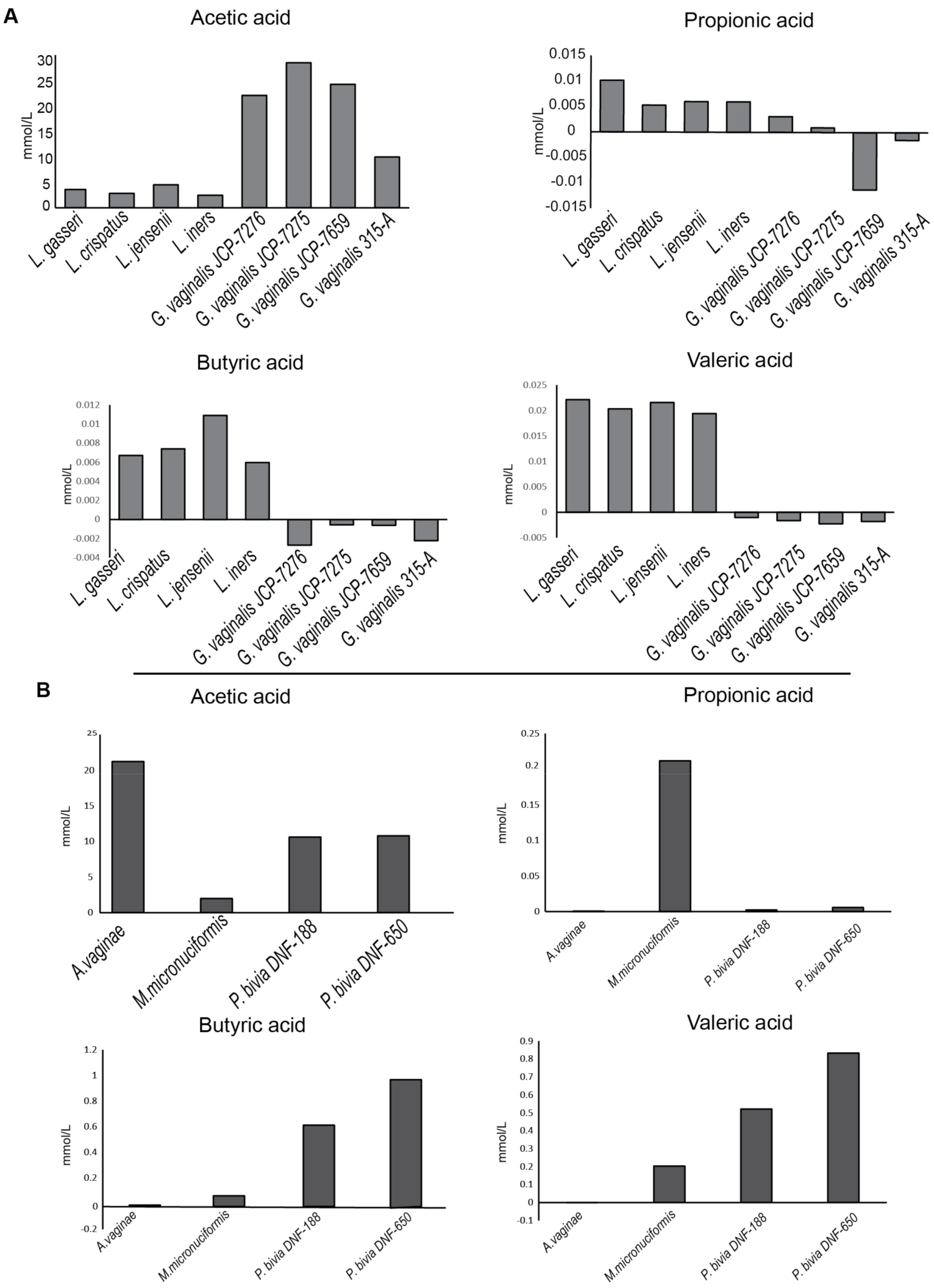
2.1. Bacterial Strains and Analysis of Short Chain Fatty Acids (SCFAs) Production

Strains of L. crispatus, L. gasseri, L. iners and L. jensenii were used as representatives of CST-I, II, III and V, respectively. G. vaginalis, Atopobium vaginae, Megasphaera micronuciformis and Prevotella bivia were used as representatives of CST-IV. Table 1 shows the related features of the used strains.
SCFAs have been reported to affect epithelial cell metabolism and innate inflammatory response [20,21]. Acetate, propionate, butyrate and isovalerate are usually found in the vaginal environment [22]. As the first step of our study, we assessed the production of SCFAs in culture supernatants of all bacterial strains by gas-chromatography coupled with a mass spectrometry (GC-MS) system.
Acetic acid turned out to be the dominant SCFA produced by all vaginal bacteria. Figure 1A shows that all vaginal lactobacilli produced a lower acetic acid amount than G. vaginalis that usually represents the dominant species of vaginal dysbiosis.
On the other hand, vaginal lactobacilli produced butyric and valeric acid amounts higher than G. vaginalis strains. Figure 1B shows that the metabolic profile of A. vaginae and P. bivia is largely superimposable to that of G. vaginalis. In contrast, M. micronuciformis produced low amounts of either acetic and butyric acid, and its metabolic profile was intermediate between lactobacilli and G. vaginalis strains.

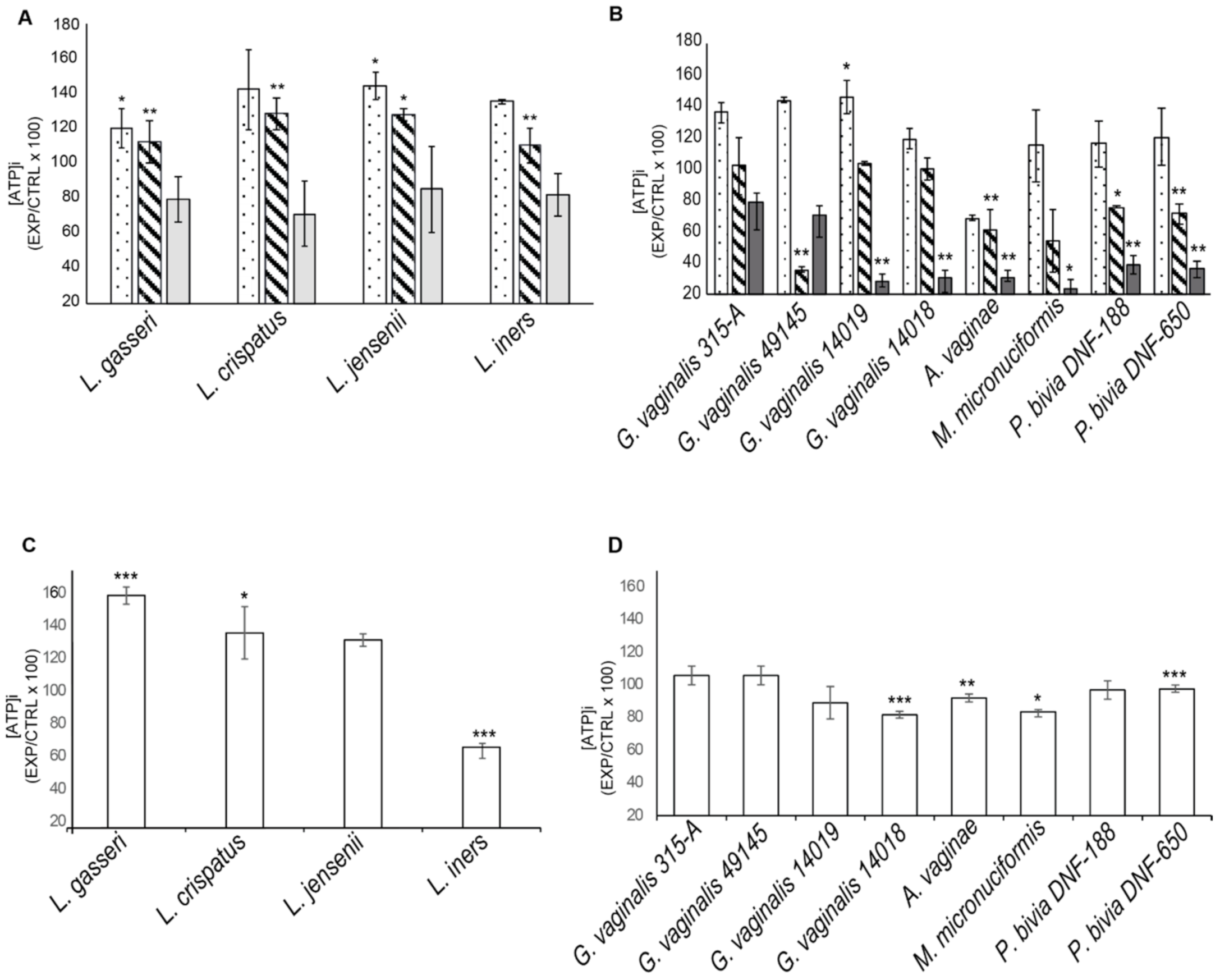
2.2. Effects of Supernatants and Bacterial Lysates on Viability of Cervical Epithelial Cell Lines

The cervical epithelial cell lines SiHa and CaSki were cultured in the presence or absence of different amounts of bacterial culture supernatants or bacterial lysates for 24 h. Intracellular ATP was measured as an index of metabolically active cells.
Figure 2A shows that supernatants from all lactobacilli cultures induced a significant increase in ATP production by SiHa cells. In contrast, supernatants from most vaginal dysbiosis bacteria affected cell viability in a dose-dependent manner. The greatest decrease in cell viability was observed in cultures with supernatants from A. vaginae culture. Lysates from vaginal dysbiosis bacteria and from L. iners induced a decrease in the viability of SiHa cells (20–50%).
The effects of bacterial supernatants on CaSki cell viability were comparable to those induced in SiHa cells (data not shown). Bacterial lysates from G. vaginalis, P. bivia and from L. jensenii significantly affected CaSki cell viability (Supplementary Figure S1).
The whole data suggest that most vaginal lactobacilli best support the viability and metabolic activity of cervical epithelial cells. In contrast, vaginal dysbiosis bacteria produce factors that affect the viability of epithelial cells and potentially compromise the cervical barrier.

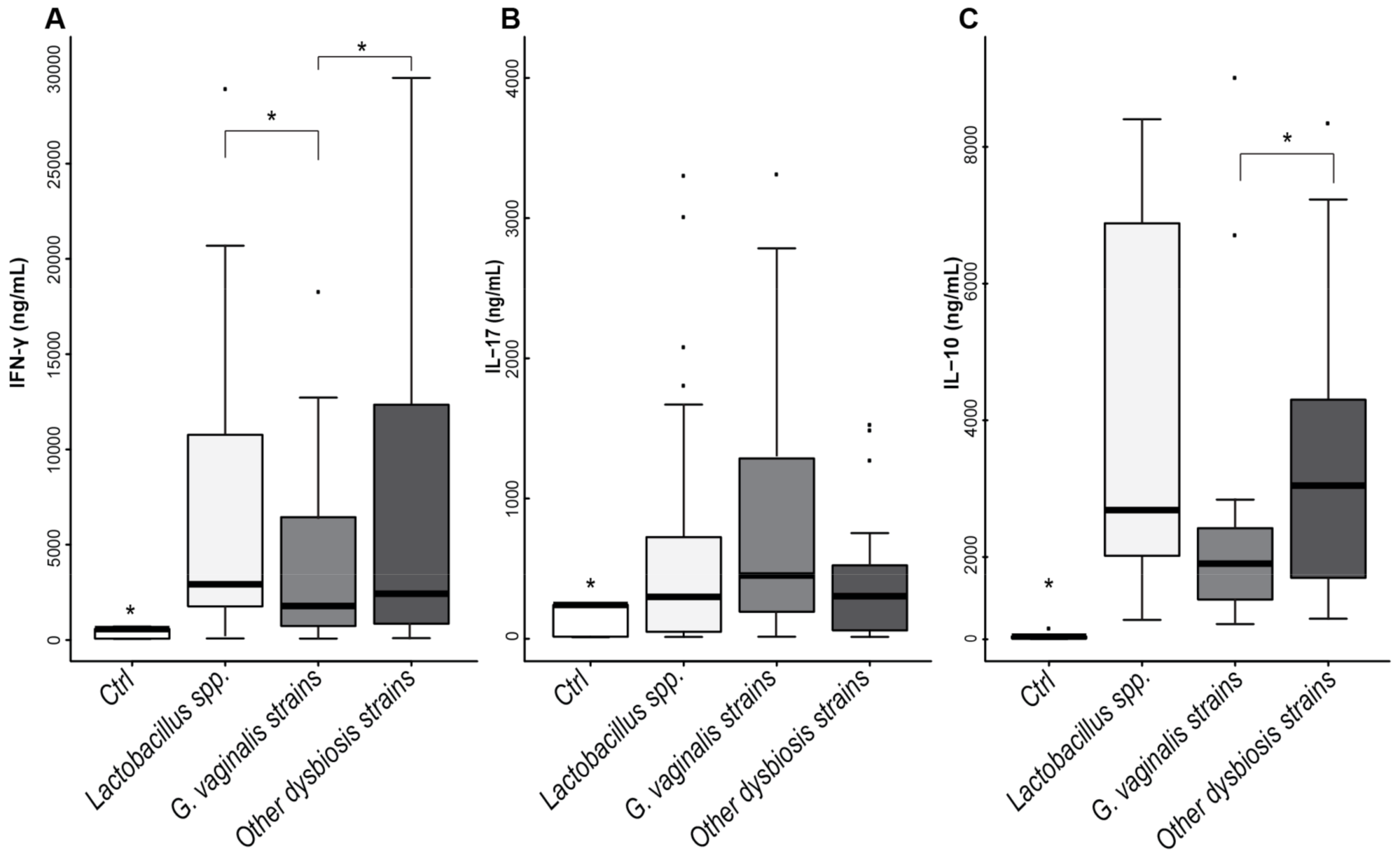
2.3. Cytokine’s Production by CaSki and SiHa Cells Cultured with Vaginal Bacteria

SiHa and CaSki cells were cultured with heat-inactivated bacteria (50 MOI/cell) from each species representative of the vaginal microbiota, and the concentration of TNF-α, IL-1β and IL-8 was assessed after 5 days of stimulation. The data obtained have shown that no significant increase in these cytokines compared to unstimulated control was evident in bacterial stimulated cultures (Supplementary Table S1). We noted, however, that, despite the huge amounts of IL-8 that were spontaneously produced by CaSki cells, vaginal dysbiosis bacteria slightly increase the production of this cytokine, while lactobacilli always led to a decrease (12–30%) (Supplementary Table S1).

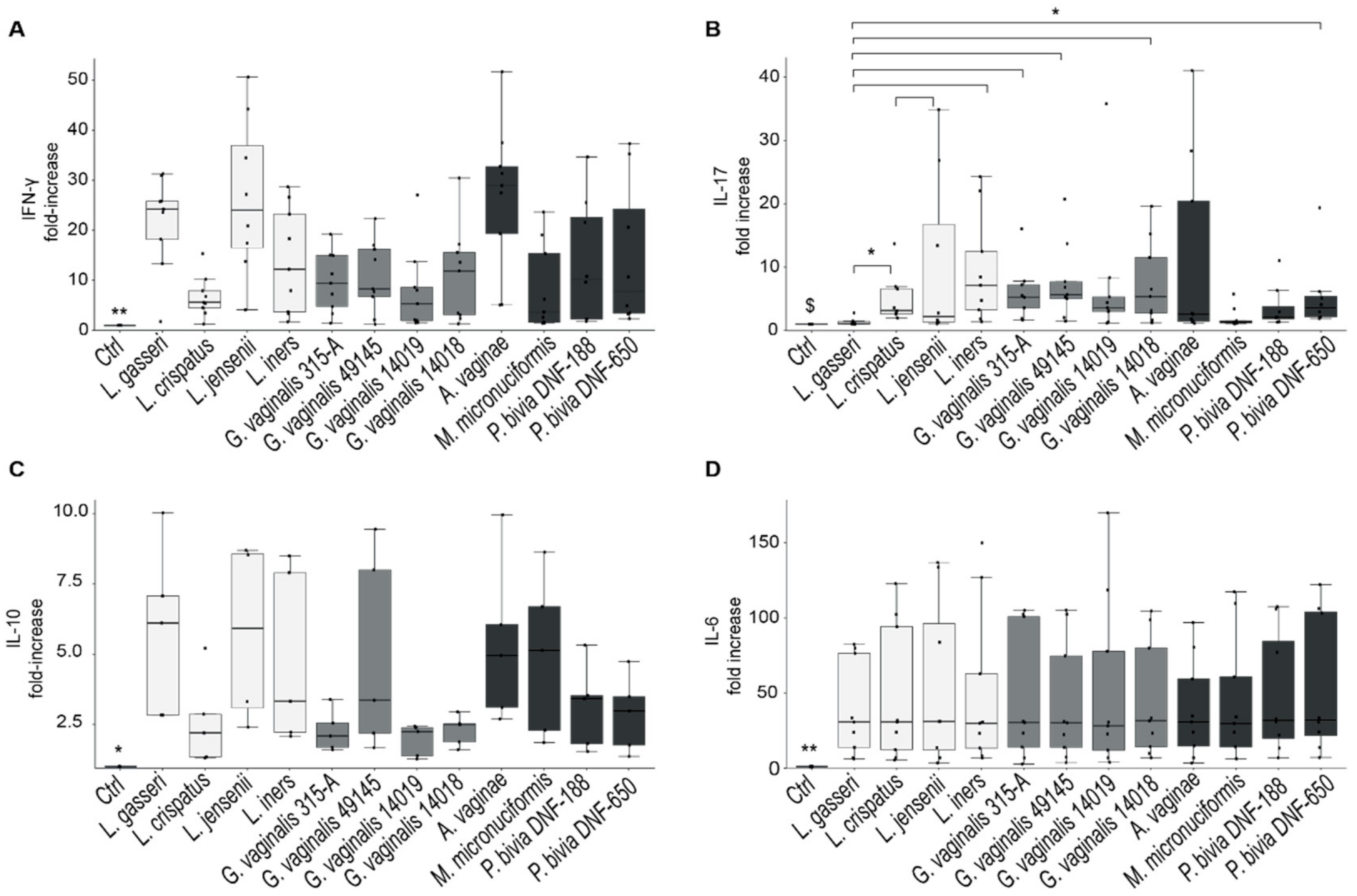
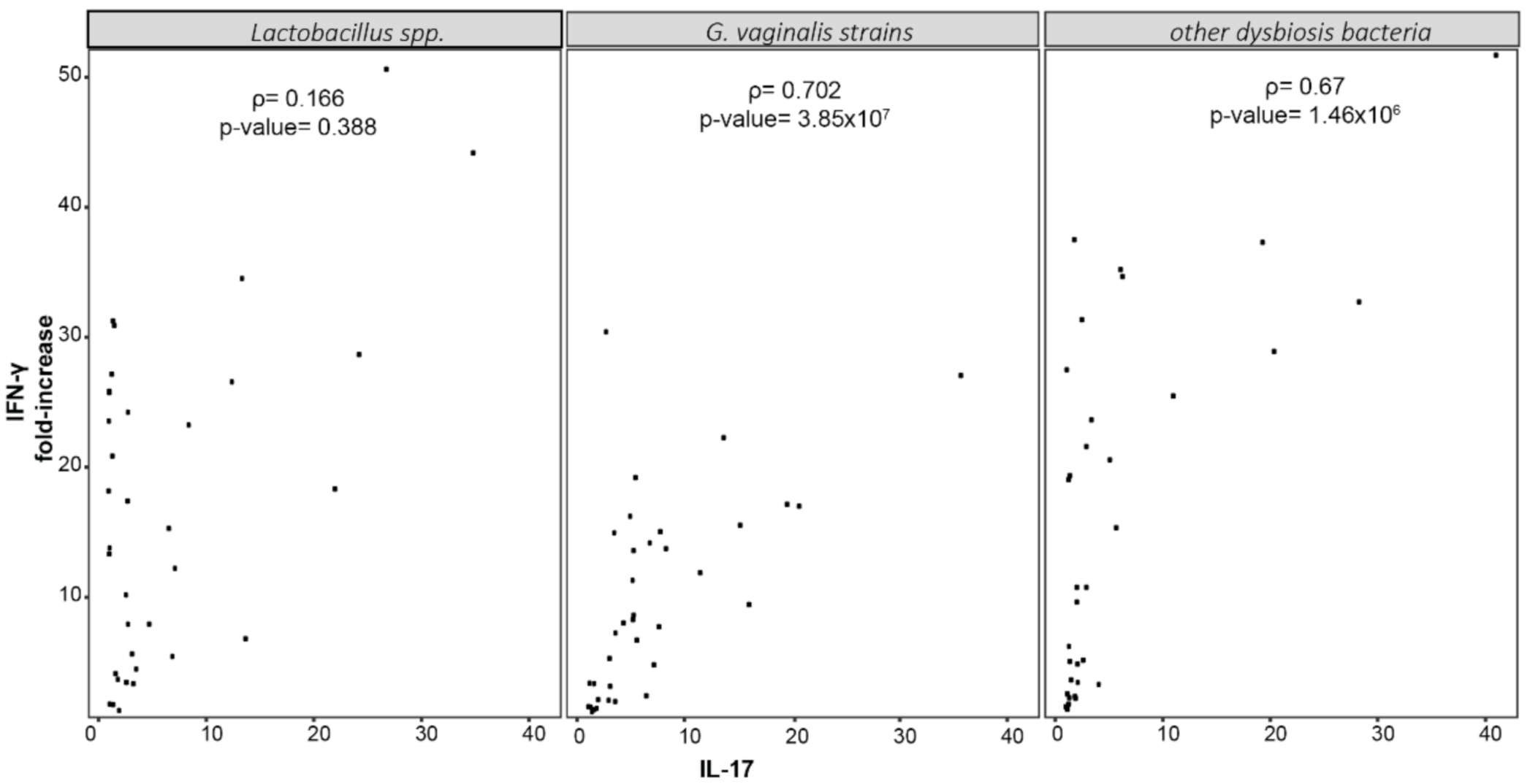
2.4. Cytokine’s Production by PBMCs Stimulated with Dominant Species of Vaginal Microbiota

As TH1 differentiation and IFN-γ production are essential to eradicate viral infections [12], we assessed whether vaginal Lactobacillus species induce the production of IFN-γ differently than vaginal dysbiosis bacteria. We cultured PBMCs from healthy donors (n = 9) with heat-inactivated bacteria (50 MOI/cell) from all strains reported in Table 1, and cytokine concentration was assessed in supernatants after 5 days of culture. In addition to IFN-γ, the production of IL-4, IL-17, IL-10, IL-6, IFN-γ and IP-10 was analyzed. Figure 3 shows that almost all species of vaginal microbiota were able to induce the production of a significant amount of IFN-γ, IL-17, IL-6 and IL-10 compared to unstimulated controls. In contrast, they did not induce significant production of IFN-α, IP-10 and IL-4 (Supplementary Figure S2).
Among lactobacilli, L. gasseri was the species that induced the highest amounts of IFN-γ and the lowest amounts of IL-17; L. iners, in contrast, was an optimal stimulus for either IFN-γ and IL-17 production.
G. vaginalis strains induced more IL-17 production compared to lactobacilli, and significant differences emerged, especially from the amounts induced by G. vaginalis strains and those induced by L. gasseri.
The anti-inflammatory cytokine IL-10 was significantly induced by all bacterial strains compared to unstimulated control, and in particular, Lactobacillus species induced amounts of IL-10 highest than G. vaginalis or P. bivia. However, statistical analysis did not reveal significant differences among the bacterial-stimulated cultures.
To further confirm the differences in cytokine’s production between lactobacilli and vaginal dysbiosis bacteria, we obtained pools of bacterial lysates from (1) Lactobacillus species (L. crispatus, L. jensenii, L. gasseri and L. iners); (2) G. vaginalis strains (315-A, 49145, 14019 and 14018); (3) other vaginal dysbiosis bacteria (A. vaginae, P. bivia (DNF-188, DNF-650) and M. micronuciformis) and used them to stimulate PBMCs as reported above. Culture supernatants were collected after 5 days of stimulation, and the production of IFN-γ, IL-17 and IL-10 was assessed.
Figure 4 shows that IFN-γ production induced by vaginal lactobacilli was significantly higher than those induced by G. vaginalis. In contrast, G. vaginalis promoted higher production of IL-17 and lower amounts of IL-10 compared to lactobacilli as well as to other species of vaginal dysbiosis.
Finally, Spearman’s correlation analysis among the cytokine levels in cultures with vaginal dysbiosis bacteria revealed a significant correlation between the production of IFN-γ and that of IL-17 (Figure 5 and Supplementary Figure S3). Notably, these data suggest that vaginal dysbiosis bacteria may compromise the anti-viral TH1 response by increasing the differentiation of an increased number of TH17 cells and IL-17 concentration in the vaginal environment.

3. Discussion

Epithelial and immune homeostasis in the vaginal microenvironment is crucial for host defenses against sexually transmitted pathogens [21,22,23,24,25]. Microbial and the host metabolites in the host microenvironment may affect the course of sexually transmitted infections [26].
We observed that products from most vaginal-dysbiosis-bacteria affected the cell viability of cervical epithelial cells in a dose-dependent manner while products from all lactobacilli (either secreted or not secreted) are able to maintain or increase cell viability. A single exception was observed with lysates from L. iners that induced a decrease in the viability of SiHa cells. Although we are aware that in vitro culture of cervico-vaginal epithelial cells is not representative of the complexity of the vaginal microenvironment, our data support the hypothesis that vaginal dysbiosis bacteria and, to a lesser extent, L. iners compromise the cervical epithelial barrier. Similar data were reported by Anton L. et al. [27], Randis T. et al. [28] and Lopez-Moreno et al. [29].
Following bacterial stimulation, the cervical epithelial cells produce IL-8, a potent chemoattractant and activator of polymorphonuclear leukocytes [30,31]. Huge amounts of this chemokine are spontaneously produced by HPV-transformed cervical epithelial cell lines [32]. We found that L. crispatus and L. gasseri were able to negatively interfere with the molecular pathways leading to the high constitutive production of IL-8. In contrast, the G. vaginalis increased chemokine production.
The high production of lactate and low production of SCFAs by lactobacilli could be responsible for the modulation of pro-inflammatory properties of cervicovaginal epithelial cells [17].
In addition, the results from the SCFAs profile, performed in supernatants from bacterial cultures, show that Lactobacillus species produced a much lower amount of the pro-inflammatory acetic acid compared to all vaginal-dysbiosis-associated bacteria. In contrast, and according to other reports, lactobacilli produced higher amounts of butyric and valeric acid, metabolites with anti-inflammatory activity [33].
A metabolomic analysis performed on vaginal samples from HPV+ and HPV- women ascertained that the metabolome of vaginal-dysbiosis-bacteria clustered differently from Lactobacillus-dominated microbiota [26].
To summarize, our data are in accordance with previous reports showing that most of the vaginal dysbiosis bacteria affect the viability and the inflammatory properties of cervical epithelial cells [34,35] and may therefore contribute to increasing the risk of sexually transmitted viral infections, including HR-HPV infection.
HR-HPVs are spontaneously cleared by the immune response in most infections, but viral persistence occurs in 10% of infected women and may induce carcinogenesis [9,36]. Impairment of the vaginal epithelial barrier, chronic inflammation, alterations of the metabolic signaling and of the immune response are all involved in carcinogenesis [37].
The clearance of HPV infections is associated with an optimal level of IFN-γ produced by the TH cells [38]. The importance of immune homeostasis is highlighted by immunosuppressed women who display the increase in the incidence and persistence of HR-HPVs infections [39]. Moreover, prolonged inflammatory response and high secretion of exosomes in the vaginal environment may promote the progress of intraepithelial lesions [37].
The vaginal microbiota compositions affect the rate of infection as well as its outcome, and the CST-IV microbiota profile, dominated by common vaginal dysbiosis bacteria, has emerged as a risk factor for persistent HPV infection [3,16]. However, women who recovered from HPV infection show a significant reduction of CST-IV and an increase in CST-I compared to the time of recruitment [40]. On the other hand, L. gasseri (CST-II) has been associated with the complete clearance of the virus [41]. Although the number of enrolled patients was rather limited in both reports, these data suggest that vaginal Lactobacillus species do not have comparable protective effects against HR-HPV infections.
In addition, it is not known whether and how vaginal bacteria affect the adaptive immune response to HR-HPV infection and so the viral clearance.
In fact, in literature, most of the studies are focused on the pro-inflammatory or immunomodulating activity induced by vaginal bacteria interacting with cervical epithelial cells or with cells of the innate immunity [34,35]. These important studies have established a significant correlation between vaginal dysbiosis bacteria (CST-IV) and the progression of HPV infections to preneoplastic (CIN1/2) or neoplastic (CIN 3) stages [19,33].
The connection between the persistence of infection and the neoplastic progression was represented by the inflammatory potential of vaginal bacteria [42,43]. Bacterial species of genera Ureaplasma, Atopobium, Prevotella, Gardnerella, Sneathia, and especially, Fusobacteria have been associated with an increased oncogenic risk [41,44]. Among lactobacilli, L. iners can promote the progression of infection [45,46].
A more in-depth investigation of the relationship between vaginal bacteria, immune response and persistent infection and, in detail, how some vaginal bacteria can affect the adaptive immune response was performed by van Teijlingen et al. [47]. The authors studied the effect of two species often reported in vaginal dysbiosis CST-IV, Megasphaera eldsenii and Prevotella timonensis, on the activation of dendritic cells (DCs) and compared their effects with those induced by L. crispatus. They found that P. timonensis induces a strong TH1 response while L. crispatus and M. elsdenii did not affect TH polarization. A different study reports that L. crispatus confers an anti-inflammatory phenotype to DCs through up-regulation of anti-inflammatory/regulatory IL-10 cytokine production and induction of Tregs at optimal dosage [48]. Finally, Mitra et al. reported that the depletion of Lactobacillus species and the presence of anaerobic taxa of genus Gardnerella, Megasphaera and Prevotella are associated with persistence and slower regression of CIN2 lesions [49].
Previous studies observed that the production of IFN-γ following HPV 16 peptide stimulation is higher in recovered or HPV-negative women compared to those identified in cervical intraepithelial lesions (CIN) [50]. Ondondo and co-workers recently reported that men with HPV clearance had significantly higher IFN-γ levels than those with persistent HPV infection [51]. These data underline the relevance of TH1 cell-mediated cytokine response in HPV clearance, but they do not define the role of each bacterial species in supporting the antiviral response.
Our data show that all vaginal bacteria induce the production of IFN-γ, with L. gasseri being the best inducer of the cytokine. The differences among Lactobacillus and vaginal dysbiosis species mostly reside in the bacteria’s ability to stimulate TH17 differentiation and the production of IL-17 at the same time. In this scenario, G. vaginalis strains induce the production of greater amounts of IL-17 compared to lactobacilli suggesting that the adaptive immune response induced by these bacteria is not dominated by TH1 differentiation and that the combined effects of lower IFN- γ availability and higher IL-17 concentration does not appropriately support the specific antiviral response. In support of this concept, a significant correlation between the production of IFN-γ and IL-17 emerged for all CST-IV species but not for lactobacilli. This suggests that TH17 differentiation is induced as well as that of TH1 cells and potentially compromises the antiviral response, which does not benefit from TH17 effectors [12,13,14,52]. Moreover, IL-17 may be crucial in immune enhancement and disease progression.
In accordance with our results, Gosmann et al. observed an increase in the numbers of activated mucosal TH cells in the concentrations of IL-17 and IL-17-inducing cytokines (IL-23 and IL-1β) in the cervicovaginal lavage obtained from women with CST-IV dominated microbiota [13].

4. Materials and Methods

4.1. Bacterial Strains

A collection of 12 bacterial reference strains were included in the study, and related features are reported in Table 1. L. crispatus (JV-V01), L. gasseri (SV-16A), L. iners (UPII-60-B) and L. jensenii (JV-V16) were used as representative of CST-I, II, III and V, respectively. G. vaginalis was selected as representative of CST-IV and four strains isolated respectively from healthy women (315-A) or from women with bacterial vaginosis with Nugent Score 5 (49145/JCP-7276), 8 (14019/JCP-7659), 10 (14018/JCP-7275) were selected. A. vaginae (DSM-15829), M. micronuciformis (DNF00954) and P. bivia (DNF 00188 and DNF-00650) were also used as representative of CST-IV.

4.2. Bacterial Cultures

Anaerobic bacteria were grown in Tryptic Soy Agar (TSA), composed by Tryptic Soy Broth (Oxoid, Basingstoke, UK) and 15mg/L of Bacto-Agar (Sigma Aldrich, St. Louis, MO, USA), with the addition of 5% Horse lysed whole-blood (Oxoid, Basingstoke, UK). The plates were incubated at 37 °C for 72 h in anaerobic conditions inside a jar (AnaeroGen™, Thermo Fisher Scientific, Waltham, MA, USA) to create ideal growth conditions (CO2: 9–13.0%).
Bacterial strains were also grown in liquid cultures using TSB with 5% horse lysed blood (Oxoid, Basingstoke, UK). The turbidity of the culture broth of each individual strain was measured by using the DensiCHECK densitometer after centrifuging 1 mL of culture at 4000× g for 5 min and resuspending the pellet in 1 mL of physiological solution.
Bacterial concentration was calculated according to the following formula:
[bacterial] = [McFarland ∗ 1.5/0.5] ∗ 108;
Optical Density assessment (OD) was performed using DensiCHECK™ densitometer (bioMérieux, Marcy l’Étoile, France).
To obtain supernatants and heat-inactivated bacteria, cultures were centrifuged at 4000× g for 5 min and suspended in RPMI-1640 medium or DMEM with the addition of 10% FBS (Fetal bovine serum) and 1% of L-glutamine (Euroclone, Pero, Italy) and incubated for 1 h at 37 °C. After centrifugation at 6000× g for 10 min, supernatants were collected. Bacterial inactivation was performed by heating at 95 °C for 15 min.
The bacterial lysates were obtained after incubation of bacterial cells with PBS 0.1% TRITON X-100 at 37 °C for 15 min. Before this step, Gram-positive bacteria were incubated with Lysozyme (10 mg/mL) for 1 h at 37 °C. Each sample was heated at 95 °C and immediately frozen in liquid nitrogen for at least three times. Protein concentration has been quantified by the BCA (Bicinchoninic Acid) method, and each sample was used as a stimulus at the concentration of 1 µg/mL.

4.3. Epithelial Cell Culture

SiHa cell line, isolated from squamous cell carcinoma and containing HPV-16 genome (1–2 copies per cell), was obtained from ATCC® (ATCC® HTB35™). SiHa cells were cultured in DMEM medium (Euroclone, Pero, Italy) supplemented with 10% FBS (Fetal bovine serum), 1% L-glutamine, 1% penicillin and streptomycin (Euroclone, Pero, Italy).
CaSki cell line, originally isolated from a cervical carcinoma and containing 600 copies of integrated HPV-16, was obtained from BEI-Resource. CaSki cells were cultured in RPMI-1640 medium (Euroclone, Pero, Italy) supplemented with 10% FBS (Fetal bovine serum), 1% L-glutamine, 1% penicillin and streptomycin (Euroclone, Pero, Italy).

4.4. PBMCs Isolation and Culture

Buffy coats from healthy donors (n = 9) were supplied by the Transfusional Center of Azienda Ospedaliera Careggi (Firenze, Italy). PBMCs were isolated by Ficoll-Paque density gradient (Cedarlane Labs, Burlington, Ontario, Canada) according to Paccosi et al. [53] and cultured in 6-well plates at the concentration of 106 cells/mL in RPMI-1640 medium (Euroclone, Pero, Italy) supplemented with 10% FBS, 1% L-glutamine and 1% penicillin and streptomycin (Euroclone, Pero, Italy). After 1 h at 37 °C, cells were stimulated with heat-inactivated bacteria (50 MOI/cell) and cultured for additional 5 days at 37 °C and 5% di CO2. Culture supernatants from unstimulated or bacteria-stimulated cultures were collected, centrifuged at 4000 rpm for 10 min and stored at −80 °C.

4.5. Viability Test

SiHa and CaSki cells were cultured in 96-multiwells at the concentration of 2.5 × 10³ cells/mL with bacterial cell supernatants (10–20–30% v/v) or bacterial lysates (1 µg/mL). Intracellular ATP was measured as an index of metabolic activity using CellTiter-Glo® LuminescentCell Viability Assay (Promega Corporation, Medison, WI, USA) after 24 h of culture.

4.6. Cytokines’ Evaluation

IFN-γ, IL-1β, IL-4, IL-6, IL-10, IL-17A, IP-10 and TNF-α cytokine’s concentration was measured using Milliplex® Map kit Human Cytokine/Chemokine/Growth Factor Panel A Magnetic Bead Panel (Merck Kgaa, Darmstadt, Germany) and Luminex apparatus following manufacturer instruction (Luminex 200 MAGPIX).
IL-8 was measured by IL-8 Human ELISA (Thermo Fisher Scientific, Waltham, MA, USA) following the manufacturer’s instructions.

4.7. SCFAs (Short Chain Fait Acids) Profile of Bacterial Strains

Supernatants of bacterial strains were prepared from liquid cultures in broth containing TSB and 5% Horse lysed blood) (Oxoid, Basingstoke, UK) following OD determination and ultracentrifugation.
The SCFAs were assessed through an isotope dilution (ID) quantitative method [54] that uses gas chromatography coupled with a mass spectrometry (GC-MS) system.
Briefly, the SCFAs were recovered from the samples by liquid-liquid extraction and then analyzed, as free acid form, by GC-MS instrument equipped with a Supelco Nukol column 30 m length, 0.25 mm internal diameter and 0.25 µm of film thickness. The SCFAs separation was carried out by the temperatures program as follows: initial temperature of 40 °C was held for 1 min, then it was increased to 150 °C at 30 °C/min, finally grow up to 220 °C at 20 °C/min. A 1 µL aliquot of extracted sample was injected in spitless mode (spitless time 1 min) at 250 °C, while the transfer line temperature was 280 °C. The carrier flow rate was maintained at 1 mL/min.
The quantitative SCFAs’ evaluation was carried out by ratios between the area abundances of the analytes with the area abundances of respective labelled internal standards (isotopic dilution method). The ionic signals and the reference internal standard, used for the quantitation of each SCFA, were reported in Table 2. 3 mL of prefermented medium sample was added of 50μL of internal standards (ISTD) mixture, 1 mL of tert-butyl methyl ether and 50 µL of 1 M HCl solution in 15 mL centrifuge tube. Then, each tube was shaken in vortex apparatus for 2 min, centrifuged at 10,000 rpm for 5 min, and finally, the solvent layer was transferred in autosampler vial and analyzed by GC-MS method. Each sample has been prepared and processed three times by the previously described method.

4.8. Statistical Analysis

Numerical data were expressed as Mean ± standard deviation (SD) if they were in a normal distribution, or median and interquartile range (IQR) if they were not in Gaussian distribution. Mann–Whitney U test, Wilcoxon rank sum test, or Student t-test for two-group comparison was used, whereas ANOVA or Kruskal–Wallis with Bonferroni and Holm–Bonferroni p-value correction was used in case of multiple groups comparisons. Spearman rank correlation coefficient was used to examine the relationship between two continuous variables. Statistical significance was defined as a p-value < 0.05. Statistical analysis was performed using R software version 4.0.5. R Core Team (2021). R: A language and environment for statistical computing. R Foundation for Statistical Computing, Vienna, Austria. URL https://www.R-project.org/ (accessed on 31 March 2021).

5. Conclusions

Our results defined in more detail the contribution of each individual species of vaginal microbiota to the host’s defense against viral infection and revealed that not all vaginal lactobacilli have comparable properties of stimulating an adequate immune response. Based on the support of IFN-γ and lack of TH17 differentiation, L. gasseri appears to be the species that better assist the host’s defenses against HR-HPV infection.

Supplementary Materials

Author Contributions

Conceptualization, M.G.T.; methodology, S.N., G.M., S.B., G.B. and M.M.; software and statistical analysis, M.T.; writing—review and editing, S.N., M.G.T. and A.A. (Amedeo Amedei); resources, A.A. (Alberto Antonelli), I.B., C.B., G.N. and G.M.R.; supervision, G.M.R.; funding acquisition, M.G.T. All authors have read and agreed to the published version of the manuscript.

Funding

This work was supported by Regione Toscana, V.A.M.P. Project; MIUR; Fondazione Cassa di Risparmio Firenze, Italia (grant number 2016.0961).

Institutional Review Board Statement

The study was conducted according to the guidelines of the Declaration of Helsinki and approved by Ethics Committee of the Azienda ospedaliero-universitaria Careggi in agreement with the D.M. of Italian Ministry of Health (15A09709) G.U., n.300 12.28.2015.

Informed Consent Statement

Informed consent was obtained from all subjects involved in the study.

Acknowledgments

The authors would like to thank Eloisa Perissi for technical support.

Conflicts of Interest

The authors declare no conflict of interest.

References

  1. Aldunate, M.; Srbinovski, D.; Hearps, A.C.; Latham, C.F.; Ramsland, P.A.; Gugasyan, R.; Cone, R.A.; Tachedjian, G. Antimicrobial and immune modulatory effects of lactic acid and short chain fatty acids produced by vaginal microbiota associated with eubiosis and bacterial vaginosis. Front. Physiol. 2015, 6, 164. [Google Scholar] [CrossRef] [PubMed]
  2. Ravel, J.; Gajer, P.; Abdo, Z.; Schneider, G.M.; Koenig, S.S.K.; McCulle, S.L.; Karlebach, S.; Gorle, R.; Russell, J.; Tacket, C.O.; et al. Vaginal microbiome of reproductive-age women. Proc. Natl. Acad. Sci. USA 2011, 108, 4680–4687. [Google Scholar] [CrossRef] [PubMed] [Green Version]
  3. Torcia, M.G. Interplay among Vaginal Microbiome, Immune Response and Sexually Transmitted Viral Infections. Int. J. Mol. Sci. 2019, 20, 266. [Google Scholar] [CrossRef] [PubMed] [Green Version]
  4. Mastromarino, P.; Di Pietro, M.; Schiavoni, G.; Nardis, C.; Gentile, M.; Sessa, R. Effects of vaginal lactobacilli in Chlamydia trachomatis infection. Int. J. Med. Microbiol. 2014, 304, 654–661. [Google Scholar] [CrossRef]
  5. Turovskiy, Y.; Noll, K.S.; Chikindas, M.L. The aetiology of bacterial vaginosis. J. Appl. Microbiol. 2011, 110, 1105–1128. [Google Scholar] [CrossRef]
  6. Pybus, V.; Onderdonk, A.B. Microbial interactions in the vaginal ecosystem, with emphasis on the pathogenesis of bacterial vaginosis. Microbes Infect. 1999, 1, 285–292. [Google Scholar] [CrossRef]
  7. Peebles, K.; Velloza, J.; Balkus, J.E.; McClelland, R.S.; Barnabas, R.V. High Global Burden and Costs of Bacterial Vaginosis: A Systematic Review and Meta-Analysis. Sex. Transm. Dis. 2019, 46, 304–311. [Google Scholar] [CrossRef]
  8. Amabebe, E.; Anumba, D.O.C. The Vaginal Microenvironment: The Physiologic Role of Lactobacilli. Front. Med. 2018, 5, 181. [Google Scholar] [CrossRef] [Green Version]
  9. Ferlay, J.; Ervik, M.; Lam, F.; Colombet, M.; Mery, L.; Piñeros, M.; Znaor, A.; Soerjomataram, I.; Bray, F. Global Cancer Observatory: Cancer Today; International Agency for Research on Cancer: Lyon, France, 2018; Available online: https://gco.iarc.fr/today (accessed on 31 March 2021).
  10. Stanley, M. Immune responses to human papillomavirus. Vaccine 2006, 24, S16–S22. [Google Scholar] [CrossRef]
  11. Hickey, D.K.; Patel, M.V.; Fahey, J.V.; Wira, C.R. Innate and adaptive immunity at mucosal surfaces of the female reproductive tract: Stratification and integration of immune protection against the transmission of sexually transmitted infections. J. Reprod. Immunol. 2011, 88, 185–194. [Google Scholar] [CrossRef] [Green Version]
  12. Scott, M.; Stites, D.P.; Moscicki, A.-B. Th1 cytokine patterns in cervical human papillomavirus infection. Clin. Diagn. Lab. Immunol. 1999, 6, 751–755. [Google Scholar] [CrossRef] [PubMed] [Green Version]
  13. Gosmann, C.; Mattarollo, S.R.; Bridge, J.A.; Frazer, I.H.; Blumenthal, A. IL-17 Suppresses Immune Effector Functions in Human Papillomavirus-Associated Epithelial Hyperplasia. J. Immunol. 2014, 193, 2248–2257. [Google Scholar] [CrossRef]
  14. Xue, J.; Wang, Y.; Chen, C.; Zhu, X.; Zhu, H.; Hu, Y. Effects of Th17 cells and IL-17 in the progression of cervical carcinogenesis with high-risk human papillomavirus infection. Cancer Med. 2017, 7, 297–306. [Google Scholar] [CrossRef]
  15. Jee, B.; Yadav, R.; Pankaj, S.; Shahi, S.K. Immunology of HPV-mediated cervical cancer: Current understanding. Int. Rev. Immunol. 2020, 1–20. [Google Scholar] [CrossRef]
  16. Di Paola, M.; Sani, C.; Clemente, A.M.; Iossa, A.; Perissi, E.; Castronovo, G.; Tanturli, M.; Rivero, D.; Cozzolino, F.; Cavalieri, D.; et al. Characterization of cervico-vaginal microbiota in women developing persistent high-risk Human Papillomavirus infection. Sci. Rep. 2017, 7, 1–12. [Google Scholar] [CrossRef]
  17. Hearps, A.C.; Tyssen, D.; Srbinovski, D.; Bayigga, L.; Diaz, D.J.D.; Aldunate, M.; A Cone, R.; Gugasyan, R.; Anderson, D.J.; Tachedjian, G. Vaginal lactic acid elicits an anti-inflammatory response from human cervicovaginal epithelial cells and inhibits production of pro-inflammatory mediators associated with HIV acquisition. Mucosal Immunol. 2017, 10, 1480–1490. [Google Scholar] [CrossRef] [Green Version]
  18. Belkaid, Y.; Hand, T.W. Role of the Microbiota in Immunity and Inflammation. Cell 2014, 157, 121–141. [Google Scholar] [CrossRef] [Green Version]
  19. Mitra, A.; MacIntyre, D.A.; Lee, Y.S.; Smith, A.; Marchesi, J.R.; Lehne, B.; Bhatia, R.; Lyons, D.; Paraskevaidis, E.; Li, J.V.; et al. Cervical intraepithelial neoplasia disease progression is associated with increased vaginal microbiome diversity. Sci. Rep. 2015, 5, 1–11. [Google Scholar] [CrossRef] [Green Version]
  20. Usyk, M.; Zolnik, C.P.; Castle, P.E.; Porras, C.; Herrero, R.; Gradissimo, A.; Gonzalez, P.; Safaeian, M.; Schiffman, M.; Burk, R.D.; et al. Cervicovaginal microbiome and natural history of HPV in a longitudinal study. PLoS Pathog. 2020, 16, e1008376. [Google Scholar] [CrossRef] [PubMed] [Green Version]
  21. Doerflinger, S.Y.; Throop, A.L.; Herbst-Kralovetz, M.M. Bacteria in the Vaginal Microbiome Alter the Innate Immune Response and Barrier Properties of the Human Vaginal Epithelia in a Species-Specific Manner. J. Infect. Dis. 2014, 209, 1989–1999. [Google Scholar] [CrossRef] [Green Version]
  22. Brotman, R.M.; Klebanoff, M.A.; Nansel, T.R.; Yu, K.F.; Andrews, W.W.; Zhang, J.; Schwebke, J.R. Bacterial Vaginosis Assessed by Gram Stain and Diminished Colonization Resistance to Incident Gonococcal, Chlamydial, and Trichomonal Genital Infection. J. Infect. Dis. 2010, 202, 1907–1915. [Google Scholar] [CrossRef] [PubMed]
  23. Martin, J.H.L.; Richardson, B.A.; Nyange, P.M.; Lavreys, L.; Hillier, S.L.; Chohan, B.; Mandaliya, K.; Ndinya-Achola, J.O.; Bwayo, J.; Kreiss, J. Vaginal Lactobacilli, Microbial Flora, and Risk of Human Immunodeficiency Virus Type 1 and Sexually Transmitted Disease Acquisition. J. Infect. Dis. 1999, 180, 1863–1868. [Google Scholar] [CrossRef] [PubMed]
  24. Myer, L.; Denny, L.; Telerant, R.; De Souza, M.; Wright, J.T.C.; Kuhn, L. Bacterial Vaginosis and Susceptibility to HIV Infection in South African Women: A Nested Case-Control Study. J. Infect. Dis. 2005, 192, 1372–1380. [Google Scholar] [CrossRef] [PubMed]
  25. Petrova, M.I.; van den Broek, M.; Balzarini, J.; Vanderleyden, J.; Lebeer, S. Vaginal microbiota and its role in HIV transmission and infection. FEMS Microbiol. Rev. 2013, 37, 762–792. [Google Scholar] [CrossRef] [Green Version]
  26. Borgogna, J.-L.; Shardell, M.D.; Santori, E.; Nelson, T.; Rath, J.; Glover, E.; Ravel, J.; Gravitt, P.; Yeoman, C.; Brotman, R. The vaginal metabolome and microbiota of cervical HPV-positive and HPV-negative women: A cross-sectional analysis. BJOG Int. J. Obstet. Gynaecol. 2019, 127, 182–192. [Google Scholar] [CrossRef]
  27. Anton, L.; Sierra, L.-J.; Devine, A.; Barila, G.; Heiser, L.; Brown, A.G.; Elovitz, M. Common Cervicovaginal Microbial Supernatants Alter Cervical Epithelial Function: Mechanisms by Which Lactobacillus crispatus Contributes to Cervical Health. Front. Microbiol. 2018, 9, 2181. [Google Scholar] [CrossRef]
  28. Randis, T.M.; Zaklama, J.; LaRocca, T.J.; Los, F.C.O.; Lewis, E.L.; Desai, P.; Rampersaud, R.; Amaral, F.E.; Ratner, A.J. Vaginolysin Drives Epithelial Ultrastructural Responses to Gardnerella vaginalis. Infect. Immun. 2013, 81, 4544–4550. [Google Scholar] [CrossRef] [Green Version]
  29. López-Moreno, A.; Aguilera, M. Vaginal Probiotics for Reproductive Health and Related Dysbiosis: Systematic Review and Meta-Analysis. J. Clin. Med. 2021, 10, 1461. [Google Scholar] [CrossRef]
  30. Fichorova, R.N.; Desai, P.J.; Gibson, F.; Genco, C.A. Distinct Proinflammatory Host Responses to Neisseria gonorrhoeae Infection in Immortalized Human Cervical and Vaginal Epithelial Cells. Infect. Immun. 2001, 69, 5840–5848. [Google Scholar] [CrossRef] [Green Version]
  31. Spear, G.T.; St John, E.; Reza, M.R. Bacterial vaginosis and human immunodeficiency virus infection. AIDS Res. Ther. 2007, 4, 25. [Google Scholar] [CrossRef] [Green Version]
  32. Sánchez-Reyes, K.; Pedraza-Brindis, E.J.; Hernández-Flores, G.; Bravo-Cuellar, A.; López-López, B.A.; Rosas-González, V.C.; Ortiz-Lazareno, P.C. The supernatant of cervical carcinoma cells lines induces a decrease in phosphorylation of STAT-1 and NF-κB transcription factors associated with changes in profiles of cytokines and growth factors in macrophages derived from U937 cells. Innate Immun. 2019, 25, 344–355. [Google Scholar] [CrossRef]
  33. Tayyeb, J.Z.; Popeijus, H.E.; Mensink, R.P.; Konings, M.C.J.M.; Mokhtar, F.B.A.; Plat, J. Short-Chain Fatty Acids (Except Hexanoic Acid) Lower NF-kB Transactivation, Which Rescues Inflammation-Induced Decreased Apolipoprotein A-I Transcription in HepG2 Cells. Int. J. Mol. Sci. 2020, 21, 5088. [Google Scholar] [CrossRef]
  34. Delgado-Diaz, D.J.; Tyssen, D.; Hayward, J.; Gugasyan, R.; Hearps, A.C.; Tachedjian, G. Distinct Immune Responses Elicited From Cervicovaginal Epithelial Cells by Lactic Acid and Short Chain Fatty Acids Associated With Optimal and Non-optimal Vaginal Microbiota. Front. Cell. Infect. Microbiol. 2020, 9, 446. [Google Scholar] [CrossRef] [Green Version]
  35. Ilhan, Z.E.; Laniewski, P.; Thomas, N.; Roe, D.J.; Chase, D.M.; Herbst-Kralovetz, M.M. Deciphering the complex interplay between microbiota, HPV, inflammation and cancer through cervicovaginal metabolic profiling. EBioMedicine 2019, 44, 675–690. [Google Scholar] [CrossRef] [Green Version]
  36. De Martel, C.; Ferlay, J.; Franceschi, S.; Vignat, J.; Bray, F.; Forman, D.; Plummer, M. Global burden of cancers attributable to infections in 2008: A review and synthetic analysis. Lancet Oncol. 2012, 13, 607–615. [Google Scholar] [CrossRef]
  37. Kumari, S.; Bhor, V.M. Association of cervicovaginal dysbiosis mediated HPV infection with cervical intraepithelial neoplasia. Microb. Pathog. 2021, 152, 104780. [Google Scholar] [CrossRef]
  38. Seresini, S.; Origoni, M.; Caputo, L.; Lillo, F.; Longhi, R.; Vantini, S.; Paganoni, A.M.; Protti, M.P. CD4+ T cells against human papillomavirus-18 E7 in patients with high-grade cervical lesions associate with the absence of the virus in the cervix. Immunology 2010, 131, 89–98. [Google Scholar] [CrossRef] [PubMed]
  39. Serraino, D.; Carrieri, P.; Pradier, C.; Bidoli, E.; Dorrucci, M.; Ghetti, E.; Schiesari, A.; Zucconi, R.; Pezzotti, P.; Dellamonica, P.; et al. Risk of invasive cervical cancer among women with, or at risk for, HIV infection. Int. J. Cancer 1999, 82, 334–337. [Google Scholar] [CrossRef]
  40. Caselli, E.; D’Accolti, M.; Santi, E.; Soffritti, I.; Conzadori, S.; Mazzacane, S.; Greco, P.; Contini, C.; Bonaccorsi, G. Vaginal Microbiota and Cytokine Microenvironment in HPV Clearance/Persistence in Women Surgically Treated for Cervical Intraepithelial Neoplasia: An Observational Prospective Study. Front. Cell. Infect. Microbiol. 2020, 10. [Google Scholar] [CrossRef]
  41. Brotman, R.M.; Shardell, M.D.; Gajer, P.; Tracy, J.K.; Zenilman, J.M.; Ravel, J.; Gravitt, P.E. Interplay Between the Temporal Dynamics of the Vaginal Microbiota and Human Papillomavirus Detection. J. Infect. Dis. 2014, 210, 1723–1733. [Google Scholar] [CrossRef] [Green Version]
  42. Mitra, A.; Macintyre, D.A.; Marchesi, J.R.; Lee, Y.S.; Bennett, P.R.; Kyrgiou, M. The Vaginal Microbiota, Human Papillomavirus Infection and Cervical Intraepithelial Neoplasia: What Do We Know and Where Are We Going Next? Microbiome 2016, 4, 58. [Google Scholar] [CrossRef] [PubMed] [Green Version]
  43. Laniewski, P.; Barnes, D.; Goulder, A.; Cui, H.; Roe, D.J.; Chase, D.M.; Herbst-Kralovetz, M.M. Linking cervicovaginal immune signatures, HPV and microbiota composition in cervical carcinogenesis in non-Hispanic and Hispanic women. Sci. Rep. 2018, 8, 1–13. [Google Scholar] [CrossRef] [PubMed]
  44. Drago, F.; Herzum, A.; Ciccarese, G.; Dezzana, M.; Casazza, S.; Pastorino, A.; Bandelloni, R.; Parodi, A. Ureaplasma parvum as a possible enhancer agent of HPV-induced cervical intraepithelial neoplasia: Preliminary results. J. Med. Virol. 2016, 88, 2023–2024. [Google Scholar] [CrossRef] [PubMed]
  45. Curty, G.; De Carvalho, P.S.; Soares, M.A. The Role of the Cervicovaginal Microbiome on the Genesis and as a Biomarker of Premalignant Cervical Intraepithelial Neoplasia and Invasive Cervical Cancer. Int. J. Mol. Sci. 2019, 21, 222. [Google Scholar] [CrossRef] [Green Version]
  46. Cheng, L.; Norenhag, J.; Hu, Y.; Brusselaers, N.; Fransson, E.; Ährlund-Richter, A.; Guðnadóttir, U.; Angelidou, P.; Zha, Y.; Hamsten, M.; et al. Vaginal microbiota and human papillomavirus infection among young Swedish women. NPJ Biofilms Microbiomes 2020, 6, 1–10. [Google Scholar] [CrossRef]
  47. Van Teijlingen, N.H.; Helgers, L.C.; Willems, E.M.Z.; van Hamme, J.L.; Ribeiro, C.M.; Strijbis, K.; Geijtenbeek, T.B. Vaginal dysbiosis associated-bacteria Megasphaera elsdenii and Prevotella timonensis induce immune activation via dendritic cells. J. Reprod. Immunol. 2020, 138, 103085. [Google Scholar] [CrossRef]
  48. Eslami, S.; Hadjati, J.; Motevaseli, E.; Mirzaei, R.; Bonab, S.F.; Ansaripour, B.; Khoramizadeh, M.R. Lactobacillus crispatus strain SJ-3C-US induces human dendritic cells (DCs) maturation and confers an anti-inflammatory phenotype to DCs. APMIS 2016, 124, 697–710. [Google Scholar] [CrossRef]
  49. Mitra, A.; MacIntyre, D.A.; Ntritsos, G.; Smith, A.; Tsilidis, K.K.; Marchesi, J.R.; Bennett, P.R.; Moscicki, A.-B.; Kyrgiou, M. The vaginal microbiota associates with the regression of untreated cervical intraepithelial neoplasia 2 lesions. Nat. Commun. 2020, 11, 1–13. [Google Scholar] [CrossRef] [Green Version]
  50. Paaso, A.; Koskimaa, H.; Welters, M.J.P.; Kero, K.; Rautava, J.; Syrjänen, K.; Van Der Burg, S.H.; Syrjänen, S. Interferon-γ and IL-5 associated cell-mediated immune responses to HPV16 E2 and E6 distinguish between persistent oral HPV16 infections and noninfected mucosa. Clin. Exp. Dent. Res. 2021. [Google Scholar] [CrossRef]
  51. Ondondo, R.; Bukusi, E.; Ng’Ang’A, Z.; Kiptoo, M.; Mpoke, S. Cellular immune responses against natural human papillomavirus infections among men in Kisumu, Kenya. Clin. Immunol. 2020, 212, 108211. [Google Scholar] [CrossRef]
  52. Sasagawa, T.; Takagi, H.; Makinoda, S. Immune responses against human papillomavirus (HPV) infection and evasion of host defense in cervical cancer. J. Infect. Chemother. 2012, 18, 807–815. [Google Scholar] [CrossRef]
  53. Paccosi, S.; Musilli, C.; Caporale, R.; Gelli, A.M.G.; Guasti, D.; Clemente, A.M.; Torcia, M.G.; Filippelli, A.; Romagnoli, P.; Parenti, A. Stimulatory Interactions between Human Coronary Smooth Muscle Cells and Dendritic Cells. PLoS ONE 2014, 9, e99652. [Google Scholar] [CrossRef] [Green Version]
  54. De Almeida, C.; Lulli, M.; di Pilato, V.; Schiavone, N.; Russo, E.; Nannini, G.; Baldi, S.; Borrelli, R.; Bartolucci, G.; Menicatti, M.; et al. Differential Responses of Colorectal Cancer Cell Lines to Enterococcus faecalis’ Strains Isolated from Healthy Donors and Colorectal Cancer Patients. J. Clin. Med. 2019, 8, 388. [Google Scholar] [CrossRef] [Green Version]
Figure 1. Short Chain Fatty Acids (SCFAs) profile. Supernatants of (A) Lactobacillus spp., Gardnerella spp. and (B) other vaginal dysbiosis bacteria were analyzed to the qualitative and quantitative determination of acetic, propionic, butyric and valeric acids using gas-chromatography coupled with mass spectrometry (GC-MS) system. The quantitative SCFAs’ evaluation was carried out by ratios between the area abundances of the analytes with the area abundances of respective labelled internal standards (isotopic dilution method).
Ijms 22 06487 g001
Figure 2. Effects of bacterial products on SiHa cell viability. (A,B) ATP production by cells were cultured with 10% (dotted-bar), 20% (ruled-bar), 30% (grey-bar) (v/v) of bacterial supernatants for 24 h. (C,D) ATP production by cells cultured with bacterial lysates (1 µg/mL) for 24 h. The bar-graph shows results from one representative experiment out of three performed. Data are expressed as ATP produced by stimulated cultures/unstimulated control ± standard deviation (EXP/CTRL × 100). Statistical analysis was performed by ANOVA and Student t-test. Significant differences among stimulated/ unstimulated cultures were reported * p-value < 0.05; ** p-value < 0.01; *** p-value < 0.001.
Ijms 22 06487 g002
Figure 3. Cytokine’s production by PBMCs stimulated with dominant species of the vaginal microbiota. PBMCs were stimulated with 50 MOI/cell of heat-inactivated bacteria for 5 days. (A) IFN-γ, (B) IL-17, (C) IL-10 and (D) IL-6 concentration was measured in culture supernatants of bacterial-stimulated cultures. Results are expressed as a fold increase in cytokine concentration with respect to unstimulated cultures. The bar graph shows the median, and the whisker is calculated on the formula IQR × 1.5. Differences in cytokine concentrations among cultures were evaluated by Kruskal–Wallis test with Holm–Bonferroni p-value adjustment. $ p-value referred to unstimulated cultures; * p < 0.05; ** p < 0.02.
Ijms 22 06487 g003
Figure 4. Cytokine production induced by lactobacilli or vaginal dysbiosis bacteria. Pooled bacterial lysates obtained by (1) Lactobacillus strains (L. crispatus, L. jensenii, L. gasseri and L. iners), (2) G. vaginalis strains (315-A, 49145, 14019 and 14018); (3) others vaginal dysbiosis bacteria (A. vaginae, M. micronuciformis, P. bivia DNF-188 and P. bivia DNF-650) were used as a stimulus for PBMCs (n = 9). (A) IFN-γ, (B) IL-17 and (C) IL-10 concentration was measured in culture supernatants of bacterial-stimulated cultures. The bar graph shows the median, and the whisker is calculated on the formula IQR × 1.5. Statistical analysis was performed by Kruskal–Wallis test, * p ≤ 0.01. Bonferroni p-value adjustment was used for IFN-γ and IL-10. Steel p-value adjustment was used for IL-17. Student t-test paired was used for IFN-γ and IL-10, p < 0.02.
Ijms 22 06487 g004
Figure 5. Spearman’s correlation analysis among cytokines produced in cultures with Lactobacillus spp., G. vaginalis strains, other vaginal dysbiosis bacteria. Correlation among cytokines produced under different stimulation was evaluated by Spearman rank correlation analysis.
Ijms 22 06487 g005
Table 1. Reference bacterial strains.
CSTFamily and GenusSpecieStrain’s Name
ILactobacillaceae, LactobacillusL. crispatusJV-V01
IILactobacillaceae, LactobacillusL. gasseriSV-16A
IIILactobacillaceae, LactobacillusL. inersUPII-60-B
VLactobacillaceae, LactobacillusL. jenseniJV-V16
IVBifidobacteriaceae, GardnerellaG. vaginalis315-A
IVBifidobacteriaceae, GardnerellaG. vaginalis49145/JCP-7276
IVBifidobacteriaceae, GardnerellaG. vaginalis14019/JCP-7659
IVBifidobacteriaceae, GardnerellaG. vaginalis14018/JCP-7275
IVAtopobiaceae, AtopobiumA. vaginalisDSM-15829
IVPrevotellaceae, PrevotellaP. biviaDNF-00188
IVPrevotellaceae, PrevotellaP. biviaDNF-00650
IVVeillonellaceae, MegasphaeraM. micronuciformisDNF-00954
Table 2. The ionic signal used for quali/quantitation and relative ISTD of each Short Chain Fat Acids (SCFAs) acquired by the ID-GC-MS method.
SCFAsQuan. IonQual. IonISTD
Acetic acid60-[2H3] Acetic
Propionic acid7473[2H3] Propionic
Butyric acid6073[2H3] Propionic
Valeric acid6073[2H9] iso-Valeric
Publisher’s Note: MDPI stays neutral with regard to jurisdictional claims in published maps and institutional affiliations.

