Review

Single- and Two-Electron Reduction of Nitroaromatic Compounds by Flavoenzymes: Mechanisms and Implications for Cytotoxicity

by
Narimantas Čėnas
1,*,
Aušra Nemeikaitė-Čėnienė
2 and
Lidija Kosychova
1
1
Institute of Biochemistry of Vilnius University, Saulėtekio 7, LT-10257 Vilnius, Lithuania
2
State Research Institute Center for Innovative Medicine, Santariškių St. 5, LT-08406 Vilnius, Lithuania
*
Author to whom correspondence should be addressed.
Int. J. Mol. Sci. 2021, 22(16), 8534; https://0-doi-org.brum.beds.ac.uk/10.3390/ijms22168534
Submission received: 13 July 2021 / Revised: 30 July 2021 / Accepted: 4 August 2021 / Published: 8 August 2021
(This article belongs to the Collection Feature Papers in Molecular Toxicology)

Abstract

:
Nitroaromatic compounds (ArNO2) maintain their importance in relation to industrial processes, environmental pollution, and pharmaceutical application. The manifestation of toxicity/therapeutic action of nitroaromatics may involve their single- or two-electron reduction performed by various flavoenzymes and/or their physiological redox partners, metalloproteins. The pivotal and still incompletely resolved questions in this area are the identification and characterization of the specific enzymes that are involved in the bioreduction of ArNO2 and the establishment of their contribution to cytotoxic/therapeutic action of nitroaromatics. This review addresses the following topics: (i) the intrinsic redox properties of ArNO2, in particular, the energetics of their single- and two-electron reduction in aqueous medium; (ii) the mechanisms and structure-activity relationships of reduction in ArNO2 by flavoenzymes of different groups, dehydrogenases-electrontransferases (NADPH:cytochrome P-450 reductase, ferredoxin:NADP(H) oxidoreductase and their analogs), mammalian NAD(P)H:quinone oxidoreductase, bacterial nitroreductases, and disulfide reductases of different origin (glutathione, trypanothione, and thioredoxin reductases, lipoamide dehydrogenase), and (iii) the relationships between the enzymatic reactivity of compounds and their activity in mammalian cells, bacteria, and parasites.

1. Introduction

Over the decades, nitroaromatic compounds (ArNO2) maintain their importance in relation to industrial processes, environmental pollution, and pharmaceutical application. Current estimates have their production, that is, the synthesis of pigments, polymers, pesticides, explosives, or pharmaceuticals, up to 108 tons per year ([1,2,3,4,5,6], and references therein). Because of contamination of groundwater and soil at military and industrial sites by ArNO2 that exhibit toxic, mutagenic, and cancerogenic activities, there has been a significant increase in research to understand and apply biological processes for their degradation.
On the other hand, the electron-attracting ability and redox activity make the nitro group a versatile and unique group in medicinal chemistry. Nitroaromatic compounds have a long history of use as antibacterial and antiparasitic drugs and their application as radiosensitizers and hypoxia-selective anticancer agents ([6], and references therein) (Figure 1 and Figure 2). The resurgence of interest in their use is caused by the reevaluation of the problems with their mutagenicity and the new potential fields of their application, e.g., the treatment of oxic tumors, including the development of antibody- or gene-directed therapies employing bacterial nitroreductases [7,8].
Importantly, both the biodegradation of environmental pollutants such as explosives such as 2,4,6-trinitrotoluene (TNT) (4) or 2,4,6-trinitrophenyl-N-methylnitramine (tetryl) (2) (Figure 3) and the manifestation of toxicity/therapeutic action of nitroaromatic drugs (Figure 1 and Figure 2) may involve similar initial steps, single- or two-electron reduction in ArNO2 performed by various flavoenzymes and/or their physiological redox partners, metalloproteins. However, in spite of the rapidly increasing amount of information in this area, the pivotal and still incompletely resolved questions are the identification of the specific enzymes that are involved in the bioreduction of nitroaromatics, the characterization of their reaction mechanisms, and the establishment of their contribution to cytotoxic/therapeutic action of ArNO2.
This review, although it is not meant to be exhaustive, addresses the above problems with special emphasis on the characterization of flavoenzymes performing single- and two-electron reduction in nitroaromatics, the mechanisms and structure-activity relationships of reactions, and the relationships between the reactivity of compounds and their activity in biological systems.

2. Redox Properties of Nitroaromatic Compounds and Their Reduction Products

The quantitative characterization of intrinsic redox properties of nitroaromatic compounds is instrumental in the analysis of their enzymatic reduction mechanisms. In this part, we attempt to address the energetics of single- and two-electron reduction in ArNO2 in aqueous medium and some relevant properties of their reduction products. Another important mechanism of their reduction, the formation of Meisenheimer-type hydride adducts [9], is beyond the scope of this review because it is more relevant to the biodegradation of ArNO2 rather than their cytotoxicity.
ArNO2 can be reduced by multistep net six-electron transfer into corresponding amines (ArNH2) with the formation of anion-radical (ArNO2), nitroso (ArNO), and hydroxylamine (ArNHOH) intermediates. In this aspect, the best-characterized is the energetics of first electron transfer, described by a midpoint redox potential of the ArNO2/ArNO2· couple (E1, or E17 at pH 7.0). Due to the instability of free radicals in aqueous media, the E17 values of nitroaromatics (Table A1, Appendix A) are usually obtained from anaerobic pulse radiolysis experiments [10,11,12,13,14,15,16,17,18,19,20,21,22,23,24]. The range of E17 values of ArNO2 with biomedical interest is from −0.6 V to −0.2 V. Further in the text, all the potentials will be given with respect to NHE. For most important groups of compounds, E17 decreases in the order nitropyridines > nitrofurans ≥ nitrothiophenes > nitrobenzenes > nitroimidazoles (Table A1). For the series of homologous compounds, their E17 values may be roughly correlated with the σ values of their substituents. In addition, the value of E17 decreases if a nitro group loses a coplanarity with the aromatic system due to sterical hindrances. The pKa values of ArNO2·span from 2.0 to 3.0 (nitrobenzenes, nitrofurans) [25,26] to 5.7–6.1 (nitroimidazoles) [27]. In terms of an “outer-sphere” electron transfer mechanism ([28,29,30], and Appendix B), the electron self-exchange rate constants of ArNO2 are ~106 M−1s−1 [25,26].
Alternatively, the values of E1 of the number of nitroaromatics were determined by cyclic voltammetry. Typically, the electrochemical reduction in ArNO2 in aqueous media proceeds irreversibly with the formation of ArNHOH. However, this process takes place in two steps, with the pH-independent transfer of the first electron and pH-dependent transfer of three electrons:
ArNO2 + e → ArNO2,
ArNO2 + 3e + 4H+ → ArNHOH + H2O.
At pH 11–12, the redox potential of a second step may become more negative than the potential of ArNO2/ArNO2· couple. In this case, a separate reversible process of single-electron transfer at Em = E17 is observed in cyclic voltammetry ([31], and references therein).
There is some interest in the prediction of E17 of ArNO2 from quantum mechanical calculations or the use of substitute descriptors such as the electron affinities of ArNO2 or the heats of formation (∆Hf) of ArNO2. However, the calculations in vacuo frequently do not provide reliable predicted E17 values due to the large data scattering or may be confined only to a series of homologous compounds [32,33,34,35]. Some improvement may be expected upon the introduction of solvent matrix effects into calculations [36]. On the other hand, the E17 values of ArNO2 may be predicted from linear log (rate constant) vs. E17 relationships in single-electron reduction in nitroaromatics by flavoenzymes dehydrogenases-electrontransferases or their redox partners, FeS proteins [11,12,13]. The use of the geometric average of rate constants obtained in several enzymatic systems improves the prediction accuracy. The calculated reduction potentials (E17(calc.), Table A1, Appendix A) deviate from the experimental ones by no more than 40 mV (standard deviation, ± 18 mV) and thus should be considered as realistic. Importantly, this approach may be applied for groups of structurally diverse ArNO2.
The reoxidation of ArNO2·by O2 and their dismutation are among the most important factors influencing their cytotoxicity. The oxidation of anion-radicals is accompanied by the formation of superoxide (O2) and subsequently, H2O2:
ArNO2·+ O2 → ArNO2 + O2,
2O2 + 2H+ → H2O2 + O2
The latter further form cytotoxic hydroxyl radical (OH·) in transition metal-catalyzed Fenton reaction. The rate constants of ArNO2 oxidation by O2 decrease with an increase in their E17, for example, 7.7 × 10⁶ M−1s−1 (nitrobenzene), 1.4 × 10⁶ M−1s−1 (p-nitroacetophenone), 2.5 × 10⁵ M−1s−1 (nitrofurantoin), 1.5 × 10⁵ M−1s−1 (nifuroxime) [25,26]. During the single-electron reduction in ArNO2 by NAD(P)H-oxidizing flavoenzymes, the reactions (3,4) are responsible for typical redox cycling events, oxidation of significant excess NAD(P)H over ArNO2, the stoichiometric to NAD(P)H consumption of O2, and superoxide dismutase-sensitive reduction in added cytochrome c. The dismutation of nitro anion-radicals yields the nitroso compounds:
2ArNO2 + 2H+ → ArNO2 + ArNO + H2O.
The dismutation rate constants (2kd) are structure-sensitive. For the radicals of o-, m-, and p-dinitrobenzenes, they are equal to 2.4 × 10⁶ M−1s−1, 8.0 × 10⁶ M−1s−1, and 3.3 × 10⁸ M−1s−1, respectively, whereas the radicals of nitroimidazoles and nitrofurans are more stable (2kd = 104 ÷ 105 M−1s−1 [25,26,27]). The competition between the dismutation of ArNO2 and their reoxidation by O2 is responsible for the formation of a fraction of stable reduction products under partial aerobic conditions [37].
Finally, ArNO2 possessing substituents with potential leaving groups may undergo fragmentation, which competes with their reoxidation by O2 (Equation (6)). This approach is used in the development of hypoxia-selective antitumour agents such as TH-302 (36) [20].
O2N-ArCH2-N+(CH3)(CH2CH2Cl)2 + e → [-O2N-ArCH2-N+(CH3)(CH2CH2Cl)2] →
→ O2N-ArCH2· + CH3-N(CH2CH2Cl)2.
The redox properties of ArNO2 multielectron reduction products are insufficiently characterized in quantitative terms. In aqueous medium, ArNO2 are electrochemically reduced into ArNHOH directly, bypassing ArNO (Equations (1) and (2)). On the other hand, the use of mixed ethanol-aqueous solution with pH 1.0–4.0 enabled the detection of reduction intermediate dihydroxylamine (ArN(OH)2), which further undergoes dehydration [38]:
ArNO2 + 2e + 2H+ → ArN(OH)2 → ArNO + H2O.
However, the voltammetric characteristics of this reaction could not be extrapolated into aqueous medium with pH 7.0. Following this approach, it was assumed that the rate-limiting step of enzymatic two-electron reduction in ArNO2 is a net hydride transfer with the formation of ArN(OH)O [39]. The calculated heats of formation (∆Hf(ArN(OH)O) increase with the electron-accepting potency of substituents and roughly correlate with ∆Hf(ArNO2) and E17 of nitroaromatics.
In aqueous medium, nitrosobenzene is reversibly electrochemically reduced into phenylhydroxylamine at E07 = 0.184 V [40]. It is suggested that the intermediate free radical ArNOH· is unstable, and that the redox potentials of first and second electron transfer are separated by −0.5 V. Because ArNO are more powerful oxidants than ArNO2, they can be directly reduced by NAD(P)H, GSH, ascorbate, and other reductants. For example, nitrosobenzene is reduced by NADPH and ascorbate with k = 124 M−1s−1 and 2.8 × 10³ M−1s−1, respectively [41,42]. The reactivity of nitrosobenzenes increases with their electron-accepting properties. The reactions of ArNO with GSH proceed with the formation of semimercaptal (ArN(OH)-SG) intermediate, which may rearrange into sulfinamide (ArNH-(O)SG), whose acid hydrolysis will yield ArNH2 or further oxidize GSH [43].
ArNHOH are relatively unstable in aqueous medium. Under aerobic conditions, the products of decomposition of phenylhydroxylamine are nitrosobenzene, nitrobenzene, and nitrophenol [44]. ArNHOH can also disproportionate yielding ArNO and ArNH2 [45].
The mechanisms of alkylation of DNA by ArNHOH, especially by the reduction products of bifunctional dinitrobenzenes CB-1954 (14) and SN-23682 (19) and their homologues, were thoroughly analyzed and reported elsewhere [7,46], therefore, will not be addressed in this review. Another type of reaction is the fragmentation of ArNHOH with leaving groups containing a drug or chromophore molecule (Scheme 1). Because the –NHOH or –NH2 groups possess electron-donating properties, the reduction products hydrolyze or react with nucleophiles more easily than the parent nitroaromatics. This approach is used in the design of hypoxia-selective alkylating agents, gene-directed therapy involving nitroreductases, hypoxic tumour imaging, or imaging of nitroreductases in transfected tumors [47,48,49,50,51].
The energetics of formation of ArNH2 from ArNHOH in aqueous medium is also incompletely characterized. At metal or carbonaceous electrodes, nitrobenzene is reduced into phenylhydroxylamine with half-wave potentials (E1/2) of −0.30–−0.45 V, whereas the latter is reduced into aniline with E1/2 = −0.55–−0.70 V at pH 7.0 [52]. However, the overvoltage of this reaction depends on the electrode material [53]. On the other hand, the second electron transfer in this reaction, reduction in aniline radical into aniline, is characterized by E1 = 1.03 V at pH 6.9 [10]. Thus, in spite of the uncertain value of E07 of phenylhydroxylamine/aniline redox couple, it is clear that the reduction in phenylhydroxylamine into aniline radical should proceed at very negative potential. This may impose certain barriers toward the enzymatic formation of ArNH2 from ArNHOH, in particular, single-electron transfer steps.

3. Mechanisms of Reduction in Nitroaromatic Compounds by Flavoenzymes

An early study of nonenzymatic reduction in nitroaromatics by reduced FMN under anaerobic conditions demonstrated a linear dependence of log k on E17 of ArNO2 [54]. Its extrapolation to ∆E17 = 0 gives k ~ 107 M−1s−1, which agrees with an “outer-sphere” electron transfer model (Appendix B). The products of the reduction in nitroaromatics were hydroxylamines. Since that time, a substantial amount of information accumulated in this area, evidencing the diversity of reaction mechanisms, which will be analyzed in subsequent subsections.

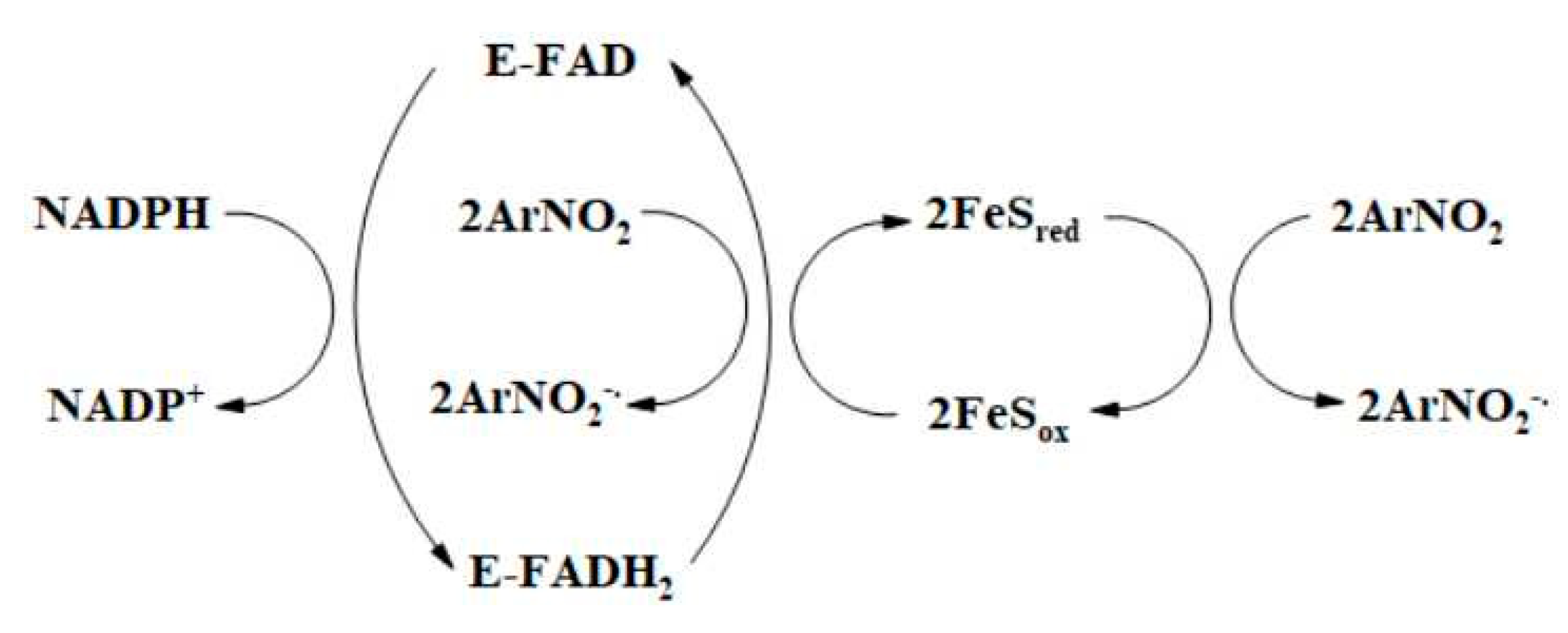
3.1. Single- and Mixed Single- and Two-Electron Reduction in Nitroaromatic Compounds by Flavoenzymes Dehydrogenases-Electrontransferases

Flavoenzymes dehydrogenases-electrontransferases transform two-electron (hydride) transfer into a single-electron one, and, most frequently, possess single-electron transferring redox partner, heme- or FeS-containing protein. Their action is characterized by the formation of neutral (blue) flavin semiquinone, FMNH or FADH·, as a reaction intermediate. In this section, the properties of flavohemoenzymes or heme-reducing flavoenzymes and flavoenzymes FeS reductases are discussed separately. This is related not to the different properties or action mechanisms of their flavin cofactors but to the different roles of the heme or FeS redox centers in the reduction in nitroaromatics.
NADPH: cytochrome P-450 reductase (P-450R) is a 78 kD enzyme associated with the endoplasmic reticulum of a variety of eukaryotic cells. It is responsible for electron transfer from NADPH to the cytochromes P-450 and to other microsomal enzyme systems ([55], and references therein). Rat liver P-450R has a hydrophobic 6 kD N-terminal membrane-binding domain, the FMN-binding domain next to it, the connecting domain, and the FAD- and NADPH-binding domains at the C-terminal side [56]. In catalysis, the transfer of redox equivalents follows the pathway NADPH → FAD → FMN → cytochrome P-450 (cytochrome c). The pyrimidine part of the isoalloxazine ring of FMN is accessible to solvent. The edge-to-edge distance between the isoalloxazine rings of FMN and FAD is 3.5–4.5 Å. The negatively charged FMN-binding domain and hydrophobic membrane-binding domain are involved in the complex formation between P-450R and cytochromes P-450 or cytochrome c. The potentiometric and kinetic characteristics of P-450R are presented in Table 1. The large differences between the redox potential values for the first and second electron transfer point to the high stability of flavin semiquinones. A catalytic cycle involving one-, two-, and three-electron-reduced states of reductase with FMNH2 acting as the principal donor of electrons to oxidants is proposed [57]. It is suggested that the rates of interflavin electron transfer are higher or similar to the rate of FAD reduction by NADPH [58]. Cytochromes P-450 reoxidize P-450R with low rates, 0.15–1.0 s−1 [59].
P-450R reduces nitroaromatic compounds in a single-electron way and is the enzyme used most frequently to demonstrate their redox cycling reactions. For this reaction, the linear dependence of logarithms of reaction rate or kcat/Km on E17 of ArNO2 was observed [68,69]. This is in line with an “outer-sphere” electron transfer model [28]. Moreover, ArNO2 are systematically less reactive than quinones with the same E17 values [69,70,71]. This additionally supports an “outer-sphere” reaction model because the electron self-exchange constants for ArNO2 are by two orders of magnitude lower than those for quinones [25,26,29]. Subsequently, the linear log (kcat/Km) vs. E17 relationships were used for the estimation of unknown E17 values of nitroaromatics [11,12,13].
The redox partners of P-450R, cytochromes P-450, catalyze the denitration of ArNO2 with the formation of corresponding hydroxyl derivatives [72,73] and reverse the accumulation of the amine products of polinitrobenzene reduction, catalyzing the formation of their hydroxylamines [74]. On the other hand, cytochrome P-450 101A1 is able to reduce m-nitroacetophenone into a corresponding amine [75]. However, the impact of these reactions on the cytotoxicity of ArNO2 has not been studied in detail.
Nitric oxide synthases (NOS) are dimeric flavohemoproteins that catalyze the conversion of L-arginine to citrulline and nitric oxide (NO·) at the expense of NADPH. Each monomer of NOS consists of a heme domain with tetrahydrobiopterin bound at its N-terminus and a FAD- and FMN-containing reductase domain at its C-terminus. The reductase and by domains are linked by a calmodulin (CAM)-binding sequence ([76], and references therein). The reductase domain is highly similar to P-450R. In catalysis, the redox equivalents are transferred in the pathway NADPH → FAD → FMN → heme (tetrahydrobiopterin), involving mainly one- and three-electron reduced states of the reductase domain in the turnover ([77,78], and references therein). The potentiometric and kinetic characteristics of neuronal NOS are presented in Table 1. Although these data may vary in various publications, it is accepted that the redox potential of cofactors decreases in the order FMN/FMNH·>> FMNH·/FMNH2 ≥ FAD/FADH > FADH·/FADH2 ≥ heme, and that the flavins are reduced much faster than heme. NOS reduces quinones and ArNO2 in a single-electron way via FMNH2 [79,80,81]. The reasons why the heme moiety is not involved in the reduction process are unclear. It may be partly explained by an increase in its redox potential after the binding of ligands. Under anaerobic conditions, nilutamide (15) and CB-1954 (14) are reduced into corresponding hydroxylamines [80,81]. TNT (4) and dinitrobenzenes inhibit the formation of NO· by NOS [82,83], most possibly by trapping of NO· with the product of their redox cycling, O2, resulting in the formation of peroxynitrite. An alternative mechanism is the diversion of electron flux from FMNH2 to heme. Like in P-450R-catalyzed reactions, the reactivity of ArNO2 was characterized by the linear log kcat/Km vs. E17 relationship with some possible discrimination against the negatively charged compounds [64]. ArNO2 were less reactive than quinones with the same E17 values.
Flavohemoglobins (FHbs) have been found in a wide variety of bacteria and fungi and play a key role in their resistance to nitrosative stress. They consist of an N-terminal heme-binding domain and of C-terminal FAD- and NAD(H)-binding modules. During turnover, NADH reduces FAD, which further reduces the Fe3+ form of hemoglobin (HbFe3+); oxyhemoglobin (HbFe2+O2) is finally formed under aerobic conditions. The reaction of the HbFe2+O2 with NO· leads to NO· detoxification, i.e., the formation of nitrate instead of the toxic peroxynitrite (ONOO). The reactions proceed with a high turnover rate, ca. 100 s−1 [84]. The crystal structures of FHb from various sources show that the pyrimidine ring of the FAD isoalloxazine is partly accessible to solvent, whereas the access to heme may be partly hampered by a bound phospholipid molecule 66 [85]. The potentiometric and kinetic characteristics of FHb are given in Table 1. The steady-state reduction in quinones or ArNO2 by S. aureus FHb follows a “ping-pong” mechanism with the oxidative half-reaction as a rate-limiting catalysis step [67]. During the turnover in the presence of an oxidant, the reduced FAD is reoxidized by >10 times more rapidly than HbFe2+O2 moiety, i.e., it acts as a preferred electron donor. The reoxidation of heme may be hampered by a bound phospholipid molecule; moreover, the binding of O2 may significantly increase the potential of Fe3+/Fe2+O2 couple. The log kcat/Km of nitrobenzenes and nitrofurans displays a well-expressed parabolic dependence on their E17. In contrast to reactions of other electrontransferases, TNT and p-nitrobenzaldehyde were reduced with a 35%–40% single-electron flux. The mixed character of reduction is possibly determined by the relatively low stability of the FAD semiquinone state of FHb, 15% at equilibrium [65].
Flavoenzymes FeS reductases either have the FeS proteins as redox partners or perform the intramolecular electron transfer to a FeS redox center. Some of their representatives may transfer electrons in both directions to and from FeS centers. Typically, both FeS reductases and their redox partners participate in the reduction in ArNO2 and other groups of redox-active xenobiotics (Scheme 2):
NADPH:adrenodoxin reductase (ADR) is a monomeric 51 kD FAD-containing enzyme, first isolated from bovine adrenal cortex mitochondria. It reduces 13.3 kD Fe2S2 protein adrenodoxin (ADX), which in turn reduces mitochondrial cytochromes P-450 that participate in the biosynthesis of steroid hormones. The ADR-ADX complex formation is determined by the electrostatic interaction between the positively charged amino acid residues of ADR and negatively charged residues of ADX ([86], and references therein). The potentiometric and kinetic characteristics of ADR and ADX are presented in Table 2.
NADPH reduces ADR with the final formation of the FADH2-NADP+ charge-transfer complex (Kd~10−8 M). NADP+ also binds to the oxidized and semiquinone form of ADR but with a lower affinity. NADPH binds to the FAD semiquinone more efficiently than NADP+ [99].
The steady-state reactions of quinone or ArNO2 reduction by ADR proceed with kcat = 20–25 s−1, which is close to the maximal rate of enzyme reduction by NADPH [100]. However, NADPH acts as a strong (Ki = 5.0–6.0 µM) inhibitor with respect to oxidants. Calculated by extrapolation to [NADPH] = 0, kcat/Km values for the reduction in nitrofurans by ADR are low, 1.0 ÷ 4.0 × 10³ M−1s−1 [101]. It was concluded that quinones and ArNO2 oxidize free enzyme form and its complexes with NADPH and NADP+, although with slower rates. Most probably, the rate-limiting step of this reaction is the oxidation of FAD semiquinone. On the other hand, ADX stimulates the reduction in ArNO2 by ADR, eliminating the inhibition by NADPH and providing a more efficient alternative reduction pathway via reduced ADX [100,101]. The kcat/Km values of nitroaromatics are much higher than in the oxidation of ADR. Their logarithms increase with their E17; moreover, ArNO2 are less reactive than quinones with the same E17 values [70,71].
Mycobacterium tuberculosis contains ADX-type 50 kD NADPH-feredoxin reductase FprA [102]. The E07 of FAD is equal to −0.235 V [103]. The rate of FAD reduction by NADPH exceeds 160 s−1; however, the reaction analysis is complicated by the enzyme reoxidation by NADP+. Blue FAD semiquinone is formed during the reduction in the enzyme by NADPH or during its photoreduction in the presence of NADP+. The enzyme reduces M. smegmatis Fe7S7 ferredoxin with kcat = 3.4 s−1 [102]. However, the data on the reactions of FprA and its redox partner(s) with ArNO2 are absent.
Most ferredoxin:NADP(H) oxidoreductases (FNRs) are 35–36 kD monomeric FAD-containing enzymes that transfer electrons from reduced FeS-protein ferredoxin (Fd) to NADP+, thus providing NADPH for CO2 assimilation in plants and cyanobacteria. However, some FNRs act in the opposite direction, reducing Fd at the expense of NADPH, thus supplying reduced Fd for nitrate assimilation (roots) or biosynthesis of isoprenoids (malaria parasite Plasmodium falciparum).
In this part, we discuss the mechanisms of ArNO2 reduction by enzymes that function in opposite directions, FNR from cyanobacterium Anabaena PCC7119 and from P. falciparum (PfFNR). Their potentiometric and kinetic characteristics are presented in Table 2. The isoalloxazine ring of FAD of both FNRs is partly exposed to solvent [104,105,106]. Electrostatic interaction between the negatively charged Fd and positively charged FNR and hydrophobic interactions are involved in the formation of the Anabaena FNR-Fd complex [89]. An analogous electrostatic complex is formed between PfFNR and PfFd [105]. Both enzymes reduce quinones and ArNO2 in a single-electron way [107,108,109]. Reactions follow the “ping-pong” mechanism with the oxidative half-reaction as a partly rate-limiting step, 100 s−1 for Anabaena FNR, and up to 63 s−1 for PfFNR. They are characterized by parabolic dependences of log kcat/Km of quinones and nitroaromatics on their E17 value. Again, ArNO2 comprised a separate series of compounds with lower reactivity. In both enzymes, the rate-limiting step in FADH reoxidation is the oxidation of FADH·. Like in reactions of ADR, Fd stimulated the reduction in nonphysiological oxidants by both FNRs, providing an alternative reduction pathway with lower kcat and higher kcat/Km of oxidants. In another study, several low-potential nitroimidazoles were found to oxidize reduced Anabaena Fd with k = 630 ÷ 3500 M−1s−1 [110], which was much faster than the reduction in their analogs by FNR alone [102]. PfFd also stimulated the reduction in quinones and nitroaromatics by PfFNR, providing the faster pathway of their reduction with kcat equal to the rate of PfFNR-PfFd electron transfer. Like spinach FNR, both Anabaena FNR and PfFNR catalyze the reductive denitration of tetryl (2) into N-methylpicramide (3) accompanied by redox cycling of free radical intermediate [111] (Scheme 3):
The above data indicate that although PfFNR possesses low homology, 20%–30%, with plant FNRs and functions in the opposite direction, its nitroreductase reaction mechanism and absence of substrate specificity are the same as of Anabaena FNR. However, because Fds possess higher nitroreduction rates, an important but insufficiently studied problem is their possible nitroaromatic substrate specificity.
NADH:ubiquinone reductase (CoQR, complex I) is localized in the inner mitochondrial membrane. It is a large (1 MD) enzyme of 45 subunits, catalyzing NADH oxidation by ubiquinone and performing transmembrane proton translocation. The data of numerous studies of bovine mitochondrial complex I [94,98,112,113,114] may be summarized as follows: (i) FMN and 8 FeS clusters N1-6, separated by 7.6–14 Å distances, are localized in the hydrophilic arm of L-shaped complex I that extends into the mitochondrial matrix. FMN is located in 51 kD subunit; (ii) the hydrophobic domain of the complex, localized in the inner mitochondrial membrane, pumps 4 protons from the matrix to intermembrane mitochondrial space per molecule of NADH oxidized; and (iii) the sequence of transfer of redox equivalents is NADH → FMN → N3(N1a) → N1b → N4 → N5 → N6a → N6b → N2 → bound ubiquinone. The reduction in ubiquinone is inhibited by tightly binding rotenone and piericidin. The potentiometric and kinetic characteristics of bovine complex I are presented in Table 3.
The mechanism of reduction in soluble quinones, nitroaromatics, and other artificial electron acceptors by CoQR is still a matter of debate. In this context, the reference reaction is its reduction in ferricyanide, where ferricyanide presumably directly oxidizes reduced FMN [96,97]. This reaction proceeds according to a “ping-pong” mechanism with double competitive substrate inhibition, which shows that both substrates compete for the same binding site in reduced and oxidized enzyme form. The use of 4-S2H- NADH decreases kcat of reaction and kcat/Km of NADH by two times, which shows that the rate-limiting step of the process is the reduction in FMN by NADH. The reduction in soluble quinones and nitroaromatics by complex I is insensitive to rotenone and is characterized by a common parabolic dependence of log kcat/Km on E17 of oxidants. For the most active oxidants, kcat of reaction reaches 100 s−1. Importantly, ArNO2 are reduced in a mixed single- and two-electron way with a single-electron flux of 45%–70%. The studies of the complex I inhibition by NADH, NAD+, redox inactive ADP-ribose, and slowly reacting quinones enabled us to conclude that quinones and nitroaromatics bind close to the ferricyanide binding site [115,116,117]. In contrast to ferricyanide, they may oxidize both free enzyme form and its complexes with NADH (Kd = 3.0 µM) and NAD+ (Kd = 30–60 µM), although with slower rates. Because the above Kd differ from NADH and NAD+ inhibition constants toward ferricyanide, it is possible that ferricyanide and quinones or ArNO2 oxidize different redox states of the enzyme. The possible involvement of FeS centers in nitroreduction warrants further studies.
Among the similar redox systems that may contribute to cytotoxic/therapeutic action of ArNO2, Trichomonas vaginalis contain a partly characterized Fd-dependent system. T. vaginalis ferredoxin (E17 = −0.347 V) plays a central role in hydrogenosomal electron transport, reversibly transferring electrons from pyruvate:ferredoxin oxidoreductase (PFOR) to hydrogenase or to the NADH dehydrogenase module that contains FMN in 51 kD subunit, and Fe2S2 cluster in 24 kD subunit (FOR) [118,119,120]. Hypothetically, FOR can reduce nitroaromatics; however, the data on its nitroreductase reactions are absent. On the other hand, using the hydrogenosomal extracts of T. vaginalis, PFOR catalyzed pyruvate-dependent reduction in a series of ArNO2 (E17 = −0.564 V–−0.243 V) under anaerobic conditions [121]. At fixed compound concentration, a linear log (reduction rate) vs. E17 relationship is observed. T. vaginalis Fd stimulated the reduction in ArNO2; however, the reaction rate almost did not depend on E17. Furthermore, it has been shown that T. vaginalis Fd reduces low-potential metronidazole (40) and other nitroimidazoles with an unexpectedly high rate, k = 4.2 × 10⁵ ÷ 1.0 × 10⁶ M−1s−1 [110]. On the other hand, metronidazole and another low-potential compound, chloramphenicol (23), are also rapidly reduced by another NADH oxidizing 26 kD FMN and FeS-containing protein, with kcat = 56 s−1 and kcat/Km = 2.0 × 10⁶ M−1s−1, and kcat = 130 s−1 and kcat/Km = 1.7 × 10⁶ M−1s−1, respectively [122]. The functions of this protein are unknown.
Microaerophilic bacterium Helicobacter pylori contains a similar partly characterized system, consisting of PFOR and flavodoxin:quinone oxidoreductase (FqrB) [123]. The electrons between these flavoenzymes are reversibly transferred by a low-potential electron carrier flavoprotein flavodoxin. Importantly, the reduction in NADP+ by FqrB was inhibited by nitrothiazole nitazoxanide (52) and a number of nitrochromanes, nitroben- zenes, and nitrobenzoxadiazoles, which were binding to flavodoxin [124]. The system consisting of PFOR, ferredoxin:NAD+ reductase, and ferredoxin, the latter participating in ArNO2 reduction, is also present in Giardia spp. [125]. However, the catalytic properties of its components are insufficiently characterized.
Mammalian xanthine oxidase (XOD) attracted some attention as a model system for the single-electron reduction in ArNO2. The reactions with nitroimidazoles [126] and nitroacridines [127] were characterized by the absence of structure specificity, i.e., an increase in log (reaction rate) with E17 of oxidants. However, one may note that XOD is a product of proteolysis of native NAD+-reducing xanthine dehydrogenase (XDH) under a variety of pathophysiological conditions ([128], and references therein). While XDH prevails intracellularly, XOD is prevalent in body fluids such as milk and plasma, where it may be secreted or released from dead cells. XDH is a 2 × 145 kD dimer, with each subunit containing molybdopterin cofactor, FAD, and two Fe2S2 clusters. During the catalysis, electrons are transferred from the purine substrate to molybdopterin, then to FAD via FeS clusters, and ultimately to the final electron acceptor, NAD+ (XDH) or O2 (XOD). The rate-limiting catalysis step is the reductive half-reaction [129]. Partly purified XDH under aerobic conditions reduces nitrofurazone into several products, including its amino metabolite [130]. The fractions of XDH and XOD in the cytosol under anaerobiosis reduced 1- and 2-nitropyrenes and 4-nitrobiphenyl into their amino metabolites [131]. However, the studies of nitroreductase reactions of XDH did not receive further attention.
Summing up, the single-electron reduction in ArNO2 by P-450, NOS, and FNR may be attributed to the high stability of their flavin semiquinone state. Evidently, the reaction follows an “outer-sphere” electron transfer mechanism. The distances of electron transfer (Rp) calculated according to this model (Appendix B, Equation (A3)), are equal to 4.2 (P-450R), 3.9 (nNOS), 4.4 (Anabaena FNR), 4.9–5.6 (PfFNR) [109], and 2.1–3.7 Å (bovine ADX) [71,101]. These orientational values are consistent with the partial exposure of their redox centers to solvent. In all these cases, however, the principal factor determining the reactivity of ArNO2 is their E17. This leaves relatively little space for the improvement of the enzymatic reactivity of compounds. The reasons for the mixed single- and two-electron way of ArNO2 reduction by CoQR and FHb are unclear. Because of the limited amount of data, the factors determining nitroaromatic oxidant specificity for the single-electron transferring flavoenzymes of M. tuberculosis, T. vaginalis, H. pylori, and Giardia spp. are unclear. On the other hand, the application of Equation (A3) in the analysis of reactions of T. vaginalis Fd [110] gives Rp ≤ 1.5 Å, which points to strong electronic coupling and deviation from an “outer-sphere” electron transfer model. This is in accordance with the possible binding of ArNO2 at the unique cavity near the FeS cluster of T. vaginalis Fd ([110], and references therein) and points to the possible substrate structure specificity.

3.2. Two-Electron Reduction in Nitroaromatic Compounds by NAD(P)H:Quinone Oxidoreductase (NQO1) and Bacterial Nitroreductases

Mammalian NAD(P)H:quinone oxidoreductase (NQO1, DT-diaphorase) is a dimeric 2 × 30 kD enzyme containing one molecule of FAD per subunit. It catalyzes two-electron reduction in quinones and ArNO2 at the expense of NADH or NADPH. The physiological functions of NQO1 are incompletely understood. It is supposed that it maintains vitamins K1, K2, and K3 in a reduced state and participates in the stabilization of transcription factor p53 ([132], and references therein). The activity of NQO1 is frequently elevated in various tumors ([133], and references therein). For this reason, there exists a continuing interest in the development of NQO1-directed bioreductive drugs. The studies of rat, mice, and human NQO1 revealed that the bound nicotinamide ring of NADP(H) displaces Phe178′, interacts with Tyr126′ and Tyr128′, and is stacked with FAD isoalloxazine ring at a distance of 3.4 Å. Duroquinone, other quinones, and dicoumarol, competitive to NAD(P)H, occupy the nicotinamide binding site [134,135,136,137]. In contrast, another inhibitor, cibacron blue, binds at the adenosine binding site and almost does not interact with the nicotinamide binding site. The potentiometric and kinetic characteristics of NQO1 are given in Table 3. Under redox equilibrium, NQO1 contains 8% red (anionic) semiquinone. During the reoxidation of reduced NQO1 by quinones and single-electron acceptor ferricyanide, the transient semiquinone formation was not observed. It means that the rate-limiting step of this process is the oxidation of two-electron reduced FAD [138].
The mechanism of reduction in quinones by NQO1 is understood better than the reduction in ArNO2. The reactions follow the “ping-pong” mechanism and are frequently characterized by quinone substrate inhibition due to the formation of dead-end complexes with the oxidized enzyme. The reactivity (log kcat/Km) of rat NQO1 with the number of quinones of different structures increased with their E17 and decreased with their van der Waals volume (VdWvol) [139]. Their optimal VdWvol was estimated to be ≤200 Å3. The entropies of activation (ΔS) calculated according to kcat/Km of reduction in rapidly reacting quinones were equal to −84 ÷ −76 J mol−1 K−1, whereas the reduction in “slow” quinones was characterized by ΔS = −36 ÷ −11 J mol−1 K−1. This demonstrates more efficient electronic coupling of “fast” oxidants in the transition state [140].
Table 3. Potentiometric and kinetic characteristics of mammalian NAD(P)H:quinone oxidoreductase and bacterial nitroreductases.
EnzymeRedox Potential
vs. NHE, pH 7.0
Rate Constants of Electron
(Hydride) Transfer, pH 7.0
Rat liver NAD(P)H:quinone oxidoreductase−0.159 V
(FAD/FADH),
−0.201 V
(FAD/FAD·)
[138]
>1000 s−1 (NADPH to FAD) [138];
kcat = 0.1–2300 s−1, kcat/Km = 3 × 10²–5.4 × 10⁸ M−1s−1 (quinones, steady-state) [139]; kcat = 0.04–75 s−1, kcat/Km = 5.0 × 101–5.7 × 10⁵ M−1s−1 (ArNO2, steady-state) [141]
E. coli NfsB >1000 s−1 (NADH to FMN) [142];
kcat = 225 s−1, kcat/Km = 1.5 × 10⁵ M−1s−1
(nitrofurazone), kcat/Km = 650 M−1s−1
(nitrobenzene, steady-state) [143];
kcat = 140 s−1, kcat/Km = 7.0 × 10³ M−1s−1 (CB-1954, steady-state) [144];
kcat = 60 s−1, kcat/Km = 1.3 × 10⁴ M−1s−1
(PR-104A, steady-state) [145]
E. cloacae NR-B−0.190 V (FMN/FMNH)
[146]
>1000 s−1 (NADH to ArNO2,
steady-state) [39];
700 s−1 (NADH to FMN), 1.9 s−1 (FMNH to p-nitrobenzoic acid, 4 °C) [147]
E. coli NfsA−0.215 V (FMN/FMNH) [148]>400 s−1 (NADPH to FMN),
kcat = 14–180 s−1, kcat/Km = 104–7.9 × 10⁶
M−1s−1 (ArNO2, steady-state) [149]
M. smegmatis
MsPnBA
−0.190 V (FMN/FMNH)
[150]
kcat = 3.4–19.2 s−1, kcat/Km = 1.2 × 10³–1.6 × 10⁵ M−1s−1 (ArNO2, steady-state) [150]
Peroxiredoxin-
nitroreductase
hybrid enzyme, Thermotoga maritima
−0.185 V (FMN/FMNH, pH 8.0) [151]146 s−1 (NADH to FMN), kcat ≤ 15 s−1, kcat/Km = 101–5.8 × 10⁵ M−1s−1 (ArNO2, steady-state) [152]
NQO1 reduces ArNO2 more slowly than quinones (Table 3) [141]. For example, CB-1954 (14) is reduced by rat NQO1 with kcat ≤ 0.1 s−1 and kcat/Km < 103 M−1s−1. The reactivity of human NQO1 is even lower because of a Tyr104Glu substitution [153]. Next, we will address the kinetic properties of rat NQO1. Its best substrates, tetryl (2) and tetranitrobenzimidazolone (42) are characterized by kcat ≥ 70 s−1 and kcat/Km > 105 M−1s−1. The ΔS of reduction in ArNO2 is even less negative than those of “slow” quinone substrates, which points to their less efficient electronic coupling. One should note that NQO1 performs the reductive N-denitration of tetryl (2) (Scheme 3) in a mixed single- and two-electron way [154]. This supports the possibility of a multistep (e,H+,e) hydride transfer in the reduction in other nitroaromatics with an initial single-electron transfer.
The reactivity of a series of nitrobenzimidazoles toward rat NQO1 increases with their E17(calc.) [155]. However, for a larger set of compounds, the dependence of log kcat/Km on E17 is almost absent. The reactivity of ArNO2 increases with the stability of their complexes with the oxidized form of NQO1 [141,155]. However, the criteria for efficient complex formation are poorly understood. Another factor enhancing the reactivity of ArNO2 is an increased torsion angle of the nitro group. This, in particular, explains why NQO1 reduces the 4-nitro group of CB-1954 (13). The combined studies of the effects of dicoumarol and cibacron blue and interdependent quinone and ArNO2 inhibition enabled us to conclude that nitroaromatics and bulky quinones bind partly outside the nicotinamide/dicoumarol binding pocket and may partly occupy adenosine/cibacron blue binding pocket [139,141].
Bacterial type I oxygen-insensitive nitroreductases (NRs) catalyze NAD(P)H-dependent multistep reduction in ArNO2 with the formation of nitroso, hydroxylamine and amino products. Among several groups of type I NRs, two major ones are distinguished according to their similarity with E. coli nitroreductases A or B [156]. The physiological functions and physiological electron acceptors for bacterial type I NRs are not clearly characterized. It is suggested that they participate in the antioxidant defense of microorganisms as part of the soxRS regulon, whose genes are upregulated in response to oxidative stress [157]. Oxygen-insensitive NRs play a considerable role in the bioreductive degradation of polynitroaromatic explosives such as 2,4,6-trinitrotoluene (TNT) (4) [3]. They also are potential candidates for gene-directed enzyme-produg cancer therapy (GDEPT) using nitroaromatic compounds. This approach is based on the much higher rates of two-electron reduction in ArNO2 by bacterial NRs than by human NQO1 [7,8,15]. In this case, genes encoding NRs are inserted into the tumor cells using either a virus or a plasmid. Further, we will address the properties of NRs with well-characterized catalysis mechanisms and/or well-established biomedical importance.
Most type I reductases are 2 × 24 ÷ 27 kD dimers, containing one FMN per subunit. They may reduce a wide spectrum of oxidants, including nitroaromatics, quinones, riboflavin derivatives, and inorganic complexes [142,158,159]. Typically, they are inhibited by micromolar or lower concentrations of dicoumarol, which binds at the dihydronicotinamide binding site of NAD(P)H, and acts as a competitive inhibitor toward NAD(P)H. Their reactions proceed according to the “ping-pong” mechanism. The potentiometric and kinetic characteristics of NRs are given in Table 3.
The best-characterized members of group B NRs are E. coli nitroreductase B (NfsB) and Enterobacter cloacae nitroreductase (E. cloacae NR). The X-ray studies of NfsB show that the FMN isoalloxazine ring is localized in the intersubunit domain, its re-plane is solvent-accessible [143,160,161]. The nicotinamide ring of bound NAD(P) is stacked between isoalloxazine and Phe124‘. Nitroaromatic compounds bind close to the nicotinamide binding domain of NfsB; however, there exist several potential binding sites of CB-1954 (14), with the participation of Lys14, Lys74, Ser12, or Phe124, Asn71, and Gly166 [150]. On the other hand, nitrofurazone interacts with Glu165 and Phe70 and binds in nonproductive conformation because its nitrofuran group does not stack over the isoalloxazine ring of FMN [143]. This may point to the flexibility of the active center of NfsB during catalysis and to its ability to accommodate the oxidants of different sizes and shapes. This is particularly indicated by the ability of NfsB to reduce either the 2- or 4-nitro group of CB-1954 and only the 2-nitro group of SN-23682 (19) [162]. The kinetic parameters of NfsB (Table 3) show that it reduces CB-1954 much more rapidly than does NQO1. As the maximal rates of reduction in nitroaromatics are lower than the rate of reduction in FMN (Table 3), the oxidative half-reaction is the rate-limiting step of catalysis of NfsB. The early studies [158,159] point to the possible absence of substrate specificity and the increase in their reactivity with reduction potential. According to the studies of a series of derivatives of CB-1954 and SN-23682, their kcat/Km varied in the range of 4.8 × 10²–6.3 × 10⁴ M−1s−1 [162]. This data scattering points to a definite effect of the size and position of substituents on the reactivity of compounds, which, however, is difficult to characterize. On the other hand, the reactivity of compounds starts to decrease at their VdWvol ≥ 400 Å3.
E. cloacae NR possesses 88% homology with NfsB [163]. The potentiometric and kinetic characteristics of the enzyme are given in Table 4. The semiquinone state of FMN of E. cloacae NR is extremely unstable, ca. 0.01% at equilibrium [146]. This possibly determines the two-electron character of reduction in ArNO2. The oxidative half-reaction is the rate-limiting step of the catalytical cycle. Using 4-R2H-NADH, kinetic isotope effects are observed in both reductive and oxidative half-reactions [147]. It shows that the H atom, transferred from dihydronicotinamide to N-5 position of isoalloxazine, is subsequently transferred to oxidant during two-electron reduction if the exchange of proton at N-5 position with the solvent is sufficiently slow. In contrast with single-electron transferring flavoenzymes and NQO1, E. cloacae NR reduced ArNO2 faster than quinones with the same E17 value [39,164]. The reactivity of nitroaromatics increased with their E17 or correlated with their ∆Hf(ArN(OH)O) or ∆Hf(ArNO2), thus showing little structure specificity. The kcat/Km of the most efficient oxidants, derivatives of tetryl (2), reached 107 M−1s−1. TNT (4) oxidized 4 NADH equivalents in two steps, apparently being reduced to dihydroxylamino derivative, whereas tetryl oxidized 6 NADH equivalents. Importantly, E. cloacae NR did not catalyze reductive N-denitration of tetryl (Scheme 3) and reduced it into currently unidentified products.
Salmonella typhimurium NR is a 2 × 24 kD dimer containing FMN and possessing 89% identity with E. cloacae NR [165]. It reduces nitrobenzene derivatives with kcat = 5.8–290 s−1 and kcat/Km = 1.7 × 10³–3.7 × 10⁵ M−1s−1 [150]. The reactivity of nitrobenzenes increases with the σ value of their substituents.
E. coli NfsA, an FMN-dependent NADPH-specific enzyme, is the best-characterized member of group A nitroreductases [166]. The kinetic and potentiometric characteristics of the enzyme are given in Table 3. NfsA follows the “ping-pong” mechanism with the rate-limiting oxidative half-reaction [149]. The reactivity of ArNO2 is systematically higher than the reactivity of quinones possessing the same E17. NfsA reduced tetryl (2) to the same unidentified products as did E. cloacae NR. The second step of a net four-electron reduction in ArNO2, the formation of ArNHOH from ArNO intermediate, is most likely the direct nonenzymatic reduction in ArNO by NADPH (Scheme 4, pathway (b)) [41]:
Like in NfsA, nitrofurantoin binds at the active center of NfsA in nonproductive orientation (Figure 4), its nitrofuran ring interacts with Arg15 and Lys167, and its imidazole group binding over the isoalloxazine ring interacts with Arg225 [142]. This shows that the catalysis of NfsA, NfsB, and E. cloacae NR may share some common features. However, there may exist certain differences in their action. The computer modeling study suggests that the binding CB-1954 to NfsA involves Ser40 (Ser41 in NfsB) and Ile129′ (Phe124′ in NfsB); however, it may also involve Phe42 that is absent in NfsB [167]. In contrast to E. cloacae NR and NfsB, dicoumarol does not quench the fluorescence of FMN, which points to its relatively weak interaction with isoalloxazine [149].
In NfsA-catalyzed reactions, log kcat/Km of a series of nitrobenzenes including CB-1954 and nitrofurans correlated well with their E17 [149]. The kcat/Km of the most efficient oxidant, tetryl, reaches 7.9 × 10⁶ M−1s−1. The reduction rate constants of several 2-nitroimidazoles (E17 ~ −0.390 V), dinitrobenzene PR-104 (13), and metronidazole (40) obtained in other studies [15,168,169] are also close to this correlation. Thus, the data available so far demonstrate that the reactivity is determined mainly by the reduction potential of ArNO2 and not by their structural peculiarities.
Mycobacterium smegmatis enzyme MsPnBA is classified as group A nitroreductase [150]. The kinetic and potentiometric characteristics of the enzyme are presented in Table 3. This nitroreductase reduces antitubercular benzothiazinones into their amines and confers M. smegmatis resistance to these drugs [170]. Like in NfsA-catalyzed reactions, the reactivity of a series of examined nitrobenzenes increases with the σ value of their substituents [150]. Other less-characterized NfsA-like nitroreductases from Neisseria meningitidis and Bartonella henselae reduce CB-1954 and metronidazole with similar rates to those of NfsA [169].
A relatively well-characterized oxygen-insensitive NR is the peroxiredoxin- nitroreductase (Prx-NR) hybrid enzyme of Thermotoga maritima, which consists of a Prx domain at the N-terminus, and an FMN-containing NR domain at the C-terminus. These domains function independently without the exchange of redox equivalents [151]. The NR domain of Prx-NR (residues 142–321) possesses a 20–24% homology with E. coli NfsB and E. cloacae NR, and 18% homology with NfsA [152], and does not contain the residues analogous to Phe124′, Phe70, Ser40, Lys14 and Lys74 of group B NRs. The rate-limiting step of Prx-NR catalysis is the oxidative half-reaction. Importantly, the substrate specificity of Prx-NR differs from that of E. cloacae NR and NfsA. Although the log kcat/Km of ArNO2 increased with their E17, nitroaromatics were less reactive than quinones with the same E17 values [152]. Because this phenomenon is characteristic of single-electron reduction (see Section 3.1), it is possible that ArNO2 are reduced in a multistep (e, H+, e) way with a rate-limiting first electron transfer.
Helicobacter pylori contains nitroreductase RdxA, an FMN-containing 2 × 26 kD dimer that shows no more than 29% sequence identity with other homologous structures of NRs [171,172]. RdxA exhibits high NADPH oxidase activity, 2.8 s−1. The maximal reduction rate of nitrofurazone and CB-1954 was similar to RdxA oxidase activity. They were reduced with kat/Km of 1.4 × 10⁶ M−1s−1 and 3.0 × 10⁵ M−1s−1, respectively. Metronidazole was reduced with a much lower rate, kcat = 0.22 ÷ 0.62 s−1 and kat/Km = 2.0 × 10³ M−1s−1. Nitrothiazole nitazoxanide (52) was reduced by RdxA with a rate similar to that of metronidazole [173]. Another H. pylori nitroreductase, FrxA, is even more scarcely characterized. In this case, the reactivity of ArNO2 increases in the order metronidazole < nitrofurans < nitazoxanide [173]. The factors determining substrate specificity of both NRs are unclear.
Nitroreductase-type enzymes (NTR) were found in parasites Trypanosoma cruzi and T. brucei [174,175,176]. They represent 2 × 30 kD dimers containing one FMN per monomer. T. cruzi NTR is NADPH-specific, whereas the T. brucei enzyme oxidizes both NADH and NADPH. They are inhibited by dicoumarol with Ki = 258 nM and Ki = 14 nM, respectively. These enzymes reduce nitrofurans, nitroimidazoles, and nitrobenzenes with kcat = 0.2 ÷ 1.2 s−1, and kcat/Km ranging from 7.3 × 10² to 2.5 × 10⁵ M−1s−1. The final product of reduction in nifurtimox under aerobic conditions is unsaturated open-chain nitrile, whose antitrypanosomal activity was close to that of nifurtimox. Benznidazole is metabolized into hydroxylamine products, which further undergoes secondary reactions. The factors determining enzyme-substrate specificity are unclear. It is evident that the activity of compounds does not correlate with their E17.
Leishmania spp. possess several types of NTR. An FMN-dependent mitochondrial 34.7 kD NADH and NADPH-oxidizing nitroreductase (NTR1) from Leishmania major reduces benznidazole, nifurtimox, CB-1954, and related compound without pronounced substrate specificity, with kcat = 0.01 ÷ 0.07 s−1 and kcat/Km = 2.5 × 10³ ÷ 1.0 × 10⁴ M−1s−1 [177]. This enzyme participates in the bioactivation of a novel antitrypanosomal and leishmanicidal agent fexinidazole (39); however, the kinetics data are not presented [178]. Because fexinidazole is a representative of 5-nitroimidazoles, one may expect its E17 in the range of −0.490 ÷ 0.430 V (Table 1). Another FMN-dependent cytosolic 39.6 kD enzyme (NTR2) was identified in L. donovani [179]. It is specific toward bicyclic low-potential nitroimidazooxazines such as R-PA–824 (57) and its analogs, whose turnovers are equal to 2.0 ÷ 10.1 s−1 at 100 µM compound concentration, whereas the more powerful monocyclic oxidants nifurtimox and fexinidazole are reduced much slower, with rates of 0.12 and <0.01 s−1, respectively [179].
There exist limited data on the formation of amines as the final product of reduction in ArNO2 by type I NRs, although this problem is relevant both to biomedicine and ecotoxicology. NfrA from Bacillus LMA exhibits 40% homology with NfsB and reduces 3,5-dinitro-trifluoromethylbenzene to diamine product with kcat = 18 s−1 [180]. Nitrofurazone is reduced to its amine derivative at a lower rate. S. typhimurium NR quantitatively reduces nitrobenzene into aniline [165]. It is unclear whether the tendency for amine formation is determined by the properties of ArNO2 or nitroreductase or by both factors. It has been suggested that the possibility of amine formation increases with the reduction potential of ArNO2 and the size of their aromatic system [150]. A recent study shows that Haemophilus influenza NR-B reduces chloroamphenicol (23) into a corresponding amine with kcat = 10.2 s−1 and kat/Km = 2.0 × 10⁴ M−1s−1 [181]. This NR possesses unusual and undisclosed substrate specificity because it reduces more powerful oxidant metronidazole (40) (Table 1) with a lower rate, kcat = 0.34 s−1 and kat/Km = 4.6 × 10³ M−1s−1 with the formation of its hydroxylamine metabolite.
There also exist several potentially important but insufficiently characterized flavin-independent enzymes with nitroreductase activity. In spite of the presence of nitroreductase MspnBA in M. smegmatis [170], this enzyme is absent in M. tuberculosis. In this case, the antitubercular drug S-PA-824 (57) is reduced by deazaflavin F-420 (7,8-didemethyl-8-hydroxy-5-deazariboflavin)-dependent nitroreductase [182]. This reaction with kcat = 0.1 s−1 leads to the formation of NO·. Under aerobic conditions, human aldo-keto reductase 1C3 catalyzes NADPH-dependent reduction in PR-104A (13) into its hydroxylamino metabolite with kcat = 0.013 s−1 [183].
Summing up, the two-electron reduction in ArNO2 by NQO1 and bacterial oxygen-insensitive NRs may be attributed to the low stability of their flavin semiquinone state. However, the relative stability of FAD of NQO1, 8% under equilibrium [138], may enable this enzyme to perform the reductive denitration of tetryl (2) (Scheme 3) in a mixed single- and two-electron way [143]. This reaction is not characteristic for E. cloacae NR-B and E. coli NfsA [39,149], evidently due to the much lower stability of their FMN semiquinone [146]. The crystallographic studies of NRs from E. coli [142,143,160,161] point to the flexibility of their active sites and to their ability to accommodate the substrates of various sizes. The kinetic studies of several A- and B-type NRs demonstrate that the reactivity of ArNO2 is strongly influenced by their reduction potential [39,149,150]. However, this leaves some space for the improvement of the activity of compounds. Another unresolved problem is the factors determining substrate specificity of nitroreductases from H. pylori, H. influenza, Leishmania, and Trypanosoma spp.

3.3. Single- and Two-Electron Reduction in Nitroaromatic Compounds by Flavoenzymes Disulfide Reductases

Flavoenzymes disulfide reductases contain FAD and redox-active disulfide group, which participate in the transfer of redox equivalents in a sequence NAD(P)H → FAD → catalytic disulfide → low-Mr or protein disulfide substrate. In most cases, they perform antioxidant functions. These reactions proceed via obligatory two-electron (hydride) transfer without the formation of free radical intermediates ([184,185], and references therein). Although being slow, the nitroreductase reactions of disulfide reductases received significant attention because of the combined action of ArNO2, redox cycling, and inhibition of physiological reactions of disulfide reductases. It is important to note that these compounds are reduced by flavin but not by reduced disulfide cofactor due to unfavorable energetics of single-electron oxidation of dithiols [186].
Glutathione reductase (GR) and trypanothione reductase (TR), the 2 × 55 kD homodimers, contain one FAD and catalytic disulfide per subunit. GR catalyzes the reduction in glutathione disulfide (GSSG), and TR catalyzes the reduction in trypanothione (TS2), a glutathione-spermidine conjugate. The structure and reaction mechanism of both enzymes are similar [187,188,189]. GR performs antioxidant functions in various organisms. TR is found exclusively in trypanosomes and leishmanias, the causative parasites of several tropical diseases, including African sleeping sickness and Chagas disease. These parasites do not contain GSSG/GSH, and their antioxidant defense relies mainly on TR-catalyzed regeneration of T(SH)2. The presence of amino acids with different charge in the disulfide substrate-binding domain of HGR and T. congolense TR [187,190] enable the discrimination between the negatively charged GSSG and positively charged TS2.
GR and TR are reduced by NADPH to two-electron reduced form (EH2), which is the FAD-thiolate charge-transfer complex with the main electron density being localized on thiolate. Next, EH2 is reoxidized by disulfide. These reactions follow a “ping-pong” mechanism with reductive half-reaction as a rate-limiting step. The kcat values of human erythrocyte GR (HGR), P. falciparum GR (PfGR), and TRs span between 120 and 240 s−1 [185,191,192,193,194,195]. More precisely, their mechanism should be classified as “hybrid ping-pong” because during turnover, GSSG reoxidizes not free EH2 form, but its tight complex with repeatedly bound NADPH (Kd = 2.1 µM, yeast GR [196]). In this case, GSSG oxidizes free EH2 and its complexes with NADPH and NADP+ with sufficiently close rates. The redox potentials of HGR, PfGR and T. congolense TR are equal to −0.227 (pH 8.2 [193]), −0.206 (pH 6.9 [197]), and −0.275 V (pH 7.5 [198]), respectively. Under artificial conditions, GR may be further reduced into the four-electron reduced state (EH4); however, this form is not formed during the enzyme turnover. This is attributed to the tight binding of NADPH, which stabilizes the EH2 form.
ArNO2 are reduced by GR and TR in a single-electron way. The most efficient oxidant of HGR and PfGR is tetryl (2) (kcat ≥ 5 s−1, kcat/Km = 2.0 ÷ 7.5 × 10³ M−1s−1 [195]). The low reaction rates complicate the substrate specificity studies. However, the introduction of basic substituents into nitrofuran molecule enhances their reactivity toward TR (kcat = 2.5 ÷ 3.0 s−1, kcat/Km = 3.3 ÷ 9.2 × 10⁴ M−1s−1 [199,200]). A specific feature of quinone and nitroreductase reactions of GR and TR is the activation by the reaction product NADP+ [192,201]. Although the main electron density in the FAD-thiolate charge-transfer complex is localized on thiolate, its minor part remains on FAD. The binding of NADP+ to EH2 with a concomitant displacement of NADPH increases the electron density on FAD, which may accelerate the reduction in xenobiotics. The order of reactivity of various redox forms of GR and TR with quinones and presumably with ArNO2 is EH2 < EH2-NADPH < EH2-NADP+ < EH4 [192,202]. However, the site(s) of their reduction are not characterized.
An important aspect of the interaction of ArNO2 with GR and TR is the inhibition of the reduction in their physiological disulfide oxidants [17,195,199,200,201,202,203]. In these cases, compounds act as non- or uncompetitive inhibitors with respect to NADPH and disulfide substrate and bind at the intersubunit domain of GR or TR close to the binding sites of GSSG or TS2. The amino acid residues of this domain of HGR, PfGR, and TR are strikingly different [189,204]. Currently, significant efforts are devoted to the studies of efficient and specific inhibitors targeting this domain, with an aim to develop new antimalarial or trypanocidal drugs [194,205]. The efficiency of ArNO2 or other redox-active compounds is evaluated by the ratio (kcat/Km)/Ki, which combines their ability to serve as redox cycling agents and inhibit GSSG/TS2 reduction [194]. For most active compounds, this ratio is above 1011 M−2s−1 (naphthoquinones) and above 1010 M−2s−1 (nitrofurans) [194,199,200].
Lipoamide dehydrogenase (LipDH, NADH:lipoamide oxidoreductase) is structurally similar to GR and TR, although it does not perform antioxidant functions. Mammalian LipDH catalyzes the NAD+-dependent oxidation of covalently bound dihydrolipoate of the pyruvate dehydrogenase or α-ketoglutarate dehydrogenase complexes [184]. Pig heart enzyme catalyzes the rapid transfer of two redox equivalents in both directions, from dihydrolipoamide to NAD+ and from NADH to lipoamide (LipS2NH2). In the latter case, the rate-limiting step of the process is the oxidative half-reaction [184,206]. Like GR or TR, LipDH shuttles between Eox and EH2 states.
Pig heart and T. cruzi LipDH reduce nitroaromatic compounds with kcat = 0.1 ÷ 2.0 s−1 [203,207] that makes 0.02%–0.5% of LipS2NH2 reduction rate. Some information on the reaction mechanism may be obtained from the studies of quinone reduction, which takes place with 20% (pig heart LipDH [206]), or even with 80% of maximal disulfide oxidant reduction rate (M. tuberculosis LipDH [208]). It was concluded that the EH4 fraction of LipDH, which is formed in the absence of NAD+, is mostly responsible for quinone reduction. Because M. tuberculosis LipDH efficiently reduces quinones, kcat =165–190 s−1 [208], one may also expect its significant nitroreductase activity, which, however, was not investigated.
NAD(P)H-thioredoxin reductases (TrxRs) are important antioxidant enzymes in both prokaryotic and eukaryotic organisms [209]. There exist two groups of TrxRs: high-Mr (2 × 54–58 kD) enzymes contain FAD, catalytic disulfide, and a third cofactor, selenylsulfide (mammalians), or additional disulfide (P. falciparum) in their active center. Low-Mr enzymes (2 × 35 kD) from plants, bacteria, and archaea contain FAD and catalytic disulfide. The physiological substrates of TrxRs are 10–12 kD disulfide proteins thioredoxins (Trxs), and sometimes disulfide or monothiol-containing glutaredoxins (Grxs). Apart from the antioxidant action, they perform other numerous physiological functions [210,211]. It is important to note that human TrxR is overexpressed in numerous cancers [212].
First, let us discuss the properties of mammalian (rat, human) TrxR. The catalytic cycle of the enzyme is similar to GR, except for the participation of selenylsulfide, which is located at the C-end of the protein [213,214]. The rate-limiting catalysis step, 30 ÷ 40 s−1, is the two-electron transfer between dithiolate and selenylsulfide, which is responsible for the reduction in Trx and other numerous substrates of mammalian TrxR. During catalysis, the enzyme cycles between EH2 and EH4 redox states (E07 = −0.294 V [215]). Mammalian TrxR may directly reduce 5,5-dithiobis(2-nitrobenzoic acid) (DTNB), with the maximal rate being equal to that of Trx reduction. Other less efficient oxidants are cystine, lipoate, alloxan, dehydroascorbate, vitamin E, coenzyme Q10, and vitamin K, lipid peroxides, and H2O2.
Rat TrxR reduces ArNO2 either in a two-electron way (p-dinitrobenzene) or in a single-electron way (tetryl) [216]. During the reaction, TrxR cycles between four- and two-electron reduced states. The reactivity of ArNO2, kcat = ≤ 0.1–2.8 s−1, and kcat/Km = < 102–1.4 × 10⁴ M−1s−1 [216,217], is comparable with that of quinones, which follow poorly expressed linear log kcat/Km vs. E17 relationship [215]. The reduction in nitroaromatics that may alkylate -SH/-Se groups (1-chloro−2,4-dinitrobenzene, tetryl (2), dinitrobenzo- furoxane (48), 2-phenylsulfonylnitropyridines) is accompanied by a rapid covalent modification of the enzyme with a loss of DTNB and Trx reductase activity and to an increase in NADPH oxidase activity [216,218,219]. The most important feature of TrxR is that in addition to reduced FAD, the reduced selenylsulfide may participate in the reduction in fully substituted quinones such as phenanthrene quinone or toxoflavin [215,220], and, presumably, nitroaromatics. This was demonstrated by the selective suppression of these reactions by Au-thioglucose, which specifically reacts with selenol. The participation of selenocysteine but not cysteine in these reactions may be explained by the relative ease of its single-electron oxidation since E7(Sec·/Sec) is equal to 0.43 V, whereas E7(Cys·/CysH) is equal to 0.92 V [186]. Dinitrobenzenes and nitrofurans acted as weak noncompetitive inhibitors with respect to DTNB (Ki = 17–400 µM), whereas tetryl and dinitrobenzofuroxane (48) acted as competitive inhibitors with Ki values of 12.5 and 5.0 µM, respectively [216]. This points to the existence of several binding sites of ArNO2.
Another representative of high-Mr enzymes, P. falciparum TrxR, contains FAD and two catalytic disulfides, Cys88, Cys93, and Cys535, Cys540 in the active center [221]. The enzyme reduces its physiological oxidant, PfTrx1, and quinones with relatively high and similar rates, 51.7 and 31–67 s−1, respectively [222]. This is >10 times faster than the reduction in quinones by PfGR or HGR. However, the nitroreductase reactions of PfTrxR have not been examined.
Low-Mr TrxRs from E.coli and other species contain one FAD and one redox-active disulfide per subunit. In contrast with GR and TR, their FAD and disulfide redox groups function separately. The E07 values of FAD/FADH2 and S2/(SH)2 redox couples are equal to −0.243 ÷ −0.260 and −0.254 ÷ −0.271 V, respectively [223]. In E. coli TrxR, NADP(H) binds at the vicinity of catalytic disulfide and at 17 Å distance from the isoalloxazine ring of FAD. In order to reduce FAD, the disulfide/NADP(H)-binding domain should undergo substantial rotation, which in turn exposes the catalytic disulfide into solution and makes it accessible to Trx [224]. This makes the formation of charge-transfer complex formation impossible [225,226]. The catalysis rate-limiting step, 25 ÷ 30 s−1, is either the conformational transition or Trx reduction. In catalysis, TrxR cycles between two- and four-electron reduced forms, FADH2/S2 and FADH2/(SH)2. Interestingly, in spite of an obligatory two-electron character of physiological reactions of E. coli TrxR, it forms neutral (blue) FAD semiquinone during irradiation under anaerobic conditions [227].
The reactions of ArNO2 and other prooxidant xenobiotics with E. coli TrxR were not studied. However, they were examined with structurally similar TrxRs from Arabidopsis thaliana (E07(FAD/FADH2) = −0.244 V) [228,229,230] and Thermotoga maritima (E07(FAD/FADH2) = −0.230 V) [151,231,232,233]. The latter enzyme preferentially oxidizes NADH, and its physiological oxidant is a 25.2 kD Grx-1. Although its crystal structure is unavailable, this enzyme possesses two catalytic cysteines Cys147 and Cys150, a vicinal NAD(P)H bonding motif, FAD bonding motif, and interdomain motif Gly251, Pro254, which possibly participates in domain rotation [231]. Both enzymes reduced ArNO2 with 70%–90% single-electron flux [230,232] and catalyzed N-denitration of tetryl with the formation of N-methylpicramide (Scheme 3). This reaction was characterized by kcat = 1.5 s−1 and kcat/Km = 2.7 × 10⁴ M−1s−1 (A. thaliana TrxR), and kcat = 6.3 s−1 and kcat/Km = 6.6 × 10⁴ M−1s−1 (T. maritima TrxR). In both cases, the log kcat/Km of reduction in ArNO2 increased with E17. The reactivity of nitroaromatics was by one order of magnitude lower than that of quinones with the same E17 values. On the analogy with quinone reduction by T. maritima TrxR, the rate-limiting reaction step in nitroreductase reactions of low-Mr TrxRs may be the oxidation of FAD semiquinone [233]. It is important to note that the single-electron character of ArNO2 reduction and reactivity of A.thaliana and T. maritima TrxRs are similar to the properties of low-Mr TrxRs from T. vaginalis and Giardia lamblia [234,235]. Thus, they may be considered as the model enzymes for the studies of this pathway of ArNO2 activation in parasites.
The structure and functions of Salmonella typhimurium alkyl hydroperoxide reductase AhpF share some similarities both with high-Mr and low-Mr TrxRs ([236], and references therein). Its 58 kD monomer contains FAD and two redox-active disulfide centers, Cys345-Cys348 and Cys129-Cys132. C-terminal FAD-binding domain of AhpF possesses 34% identity with the analogous domain of E. coli TrxR. Through putative large interdomain movements, electrons are transferred in a sequence NADH → FAD → Cys345-Cys348 → Cys129-Cys132. The latter center, located in N-terminus, transfers electrons to the catalytic disulfide of another hydroperoxide reductase, AhpC. In contrast to E. coli TrxR, reduced AhpF may form 540 nm absorbing charge-transfer complex under high ionic strength of the solution. AhpF from E. coli reduces nitrofurans in a single-electron way [237]. Although the kinetic parameters of these reactions are not reported, AhpH is partly responsible for the sensitivity of E. coli to nitrofurans.
Summing up, the low nitroreductase activity of disulfide reductases may be attributed to the low electron density on FAD in FAD-thiolate charge-transfer complexes. This diminishes their role in redox cycling or other modes of activation of ArNO2 in the cell. However, low-Mr TrxRs where FAD and catalytic disulfide function separately may more significantly contribute to the redox cycling of ArNO2. The frequently observed single-electron reduction in nitroaromatics is not inconsistent with the redox properties of FAD of disulfide reductases. Apart from the formation of FADH· of low-Mr TrxR of E. coli under the artificial conditions [227], the complex of reduced GR with NADP+ also gradually converts into a product, characterized as the complex of NADP+ with FAD·([184], and references therein). Another relevant problem is the exploitation of the bioreductive potential of a reduced selenylsulfide moiety of mammalian TrxR in nitroreductase reactions.

4. Role of Enzymatic Reduction in Nitroaromatic Compounds in Their Cytotoxicity/Therapeutic Action

4.1. Role of Bioreductive Processes in Mammalian Cell Cytotoxicity of Nitroaromatics

Redox cycling is an intrinsic property of ArNO2; therefore, the oxidative stress will obligatorily take place during their action under aerobic conditions. In our opinion, the deviation from the limits predicted by the redox cycling activity could be instrumental in the characterization of additional mechanisms of cytotoxicity or therapeutic action of nitroaromatics. It is commonly accepted that if the main cytotoxicity factor is the rate of enzymatic formation of ArNO2· and their redox cycling, their cytotoxicity may be described by a following quantitative structure-activity relationship (QSAR):
log cL50 = a + b E17 + c log P (log D),
where cL50 is compound concentration causing 50% cell death or analogous quantitative parameter, log P is octanol/water partition coefficient, and log D is octanol/water distribution coefficient at pH 7.0 [13,155,238,239,240,241] (Table 4). These dependences mirror the log (rate constant) vs. E17 relationships in single-electron reduction in ArNO2 by P-450R, NOS, or other single-electron transferring flavoenzymes (see Section 3.1). An additional diagnostic test is antioxidant protection against cytotoxicity. However, this approach may not be applied in the assessment of the mechanisms of action of ArNO2 that possess alkylating, bioreductively alkylating, or leaving groups (see below), or may directly interact with DNA, e.g., the positively charged nitracrine (49) derivatives [242]. Nevertheless, this approach, in particular, the use of E17(calc.) obtained from enzymatic single-electron reduction reactions (Table A1, Appendix A), enabled to demonstrate that the main cytotoxicity mechanism of explosives pentryl (1), tetryl (2), TNT (4), tetranitrobenzimidazolone (42), dinitrobenzofuroxane (48), ANTA (54), NTO (55), and nitroaromatic antiandrogens nilutamide (15) and flutamide (16) could be oxidative stress [13,71,155,241,243,244,245,246]. On the other hand, although nifurtimox (27) induces oxidative stress in neuroblastoma cells and is synergistic with the GSH synthesis inhibitor buthionine sulfoximine, it is unclear whether this is the main mechanism of its anticancer action [247,248].
Table 4. Structure-activity relationships in mammalian cell cytotoxicity of nitroaromatics with possible relevance to bioreductive activation.
Compounds and CellsStructure-Activity Relationships
Aerobic Conditions
Nitroimidazoles, nitrofurans, nitrobenzenes, Chinese hamster V79–379A cells [238,239]; nitrobenzenes, nitrofurans, nitrobenzimidazoles, bovine leukemia virus-transformed lamb kidney fibroblast FLK cells [144,240,243]; nitrobenzenes, nitrofurans, nitrothiophenes, murine hepatoma MH22a cells, human colon carcinoma HCT116 cells [13,71]∆log cL50/∆E17 = −8 ÷ −11 V−1,
∆log cL50/∆log P(D) = 0 ÷ −0.3;
CB-1954 (14), SN-23682 (19) and their derivatives follow the same correlation [240]
Nitrobenzenes, FLK cells [245], primary mice splenocytes [241]∆log cL50/∆E17 = −7.5 ÷ 8.5 V−1,
increased cytotoxicity of amino and hydroxylamino metabolites of TNT (4)
Derivatives of nitracrine (49), CHO-AA8 cells [242]The absence of a relationship between E17 and cytotoxicity
4-(Alkylamino)-5-nitroquinolines, Chinese hamster ovary CHO-AA8 cells [249]Cytotoxicity roughly increases with the rate of drug-stimulated O2 consumption
N,N-bis(2-chloroethyl)-N-methyl-N-(2-nitrobenzyl)
ammonium chloride and its derivatives, CHO-AA8 cells [250]
Alkylating bioreductively activated leaving group does not affect the cytotoxicity, the relationship between E17 and cytotoxicity is absent
Derivatives of NitroCBI (54), Skov3 and HT29 cells [23]The relationship between E17 and cytotoxicity is absent
Nitrofurans, nitrothiophenes, nitropyrroles and nitropyrazoles with alkyl-N-aziridine or alkyloxirane side chains, V79–379A cells [251,252,253]Alkylating side chains increase the cytotoxicity, the relationship between E17 and cytotoxicity is absent
Hypoxic Conditions
Nitroimidazoles, nitrofurans, V79–379A cells [254]∆log cL50/∆E17 = −10 V−1
4-(Alkylamino)-5-nitroquinolines, CHO-AA8 cells [249]Cytotoxicity and hypoxic selectivity roughly increase with the rate of drug-stimulated O2 consumption
Nitrofurans and nitrothiophenes with alkyl-N-aziridine or alkyloxirane side chains, V79–379A cells [251,252,253]Alkylating side chains do not increase hypoxic selectivity
N,N-bis(2-chloroethyl)-N-methyl-N-(2-nitrobenzyl)
ammonium chloride and its derivatives, CHO-AA8 cells [250]
Alkylating leaving groups increase cytotoxicity, no relationship between E17 and cytotoxicity or hypoxic selectivity
Nitrochloromethylbenzindolines (nitroCBIs, 56), Skov3 and HT29 cells [23]Hypoxic toxicity does not correlate with E17, hypoxic selectivity roughly increases with E17
In this context, one may note that because of the similarity of log (rate constant) vs. E17 relationships in the reduction in ArNO2 by flavoenzymes dehydrogenases-electron-transferases, it is difficult to assess their individual contribution. Some information may be obtained from the hypoxic cytotoxicity studies (see below), assuming that the extent of formation of reduced ArNO2 metabolites is proportional to the rate of their single-electron enzymatic reduction. In this case, the main enzyme responsible for redox cycling of ArNO2 should be NADPH:cytochrome P-450 reductase (P-450R) [255,256]. The roles of other P-450R-type enzymes such as inducible NO synthase, human novel oxidoreductase NR1 [257], and methionine synthase reductase [258] depend on the cell type. Although the nitroreductase reactions of the latter two enzymes are uncharacterized, one may expect the similarity of their log (rate constant) vs. E17 relationships with those of P-450R and NOS. On the other hand, the contribution of mitochondrial NADH:ubiquinone dehydrogenase and, possibly, other flavoenzymes of mitochondrial respiratory chain to the cytotoxicity of ArNO2 was recently demonstrated [259]. The comparison of reactivities of P-450R and NOS with those of disulfide reductases (Table 1 and Table 2 and Section 3.3) clearly rules out the latter enzymes as the important sources of free radicals of ArNO2. The inhibitors of cytochromes P-450 protect against the cytotoxicity of nitrobenzenes, nitrofurans, and nitrothiophenes [71,205] in several cell lines; however, the roles of cytochromes P-450 in ArNO2 cytotoxicity remain unclear so far.
The presence of directly alkylating alkyl-N-aziridine or alkyloxirane groups in ArNO2 molecules evidently increases their cytotoxicity and abrogates the log (cytotoxicity) vs. E17 relationships (Table 4). It is important to distinguish the contribution of redox cycling and alkylation in their action, in particular, the role of putative alkylation targets. It is suggested that 2-phenylsulfonylnitropyridines, which possess micromolar cL50 levels against the NCI cancer cell lines, exert their action through thioredoxin reductase inactivation [219]. However, because of the high E17 value of nitropyridine moiety (Table A1), the redox cycling reactions with flavoenzymes electrontransferases may significantly contribute to their cytotoxicity. The influence of bioreductively activated alkylating groups such as aziridine of nitrogen mustard or bioreductively activated leaving tetraalkylammonium group in the aerobic cytotoxicity of ArNO2 remains equivocal (Table 4). In these cases, an increase in cytotoxicity of ArNO2 may be attributed to the formation of a fraction of stable reduction products in cell compartments with reduced O2 tension such as nucleus and mitochondria. In cell lines with 100–200 U/mg NQO1 (determined according to quinone reduction rate), dicoumarol partly protects against the cytotoxicity of ArNO2. However, the cytotoxicity of CB-1954, SN-23682, and their derivatives is close to the limits predicted by their E17 [240], and dicoumarol similarly protects against the cytotoxicity of ArNO2 that do not contain aziridine or nitrogen mustard groups [71,243].
ArNO2 are considered to be the hypoxia-selective toxins or therapeutic agents because their hydroxylamine reduction products formed under hypoxia may alkylate DNA and other nucleophiles. Frequently, their -NHOH or -NH2 metabolites demonstrate even higher cytotoxicity than the parent ArNO2 ([23,260,261], and references therein). Additionally, the electron-donating character of these groups increases the reactivity of alkylating aziridine or nitrogen mustard groups and accelerates the dissociation of leaving groups. In certain cases, the hypoxic selectivity, i.e., the ratio of cL50 under oxic and hypoxic conditions, may exceed 1000. It was established that NADPH:cytochrome P-450 reductase (P-450R) plays a major role in the formation of reduced metabolites of ArNO2 under hypoxia [255,256]. One may expect the correlation of hypoxic toxicity with E17 analogously with the rate of ArNO2 reduction. However, this type of correlation is frequently absent (Table 4). Evidently, this reflects the superposition of two opposite factors, the ease of reduction increasing with E17 of the parent compound and the alkylating reactivity, decreasing with electron-accepting potency of substituents. The studies of CB-1954, SN-23682, and their reduction product have shown that the cytotoxicity is determined mainly by the alkylating reactivity of reduction products, their stability, and interconversion rate [260].
Nitroreductase-transfected cancer cells are the objects of gene-directed enzyme-prodrug therapy (GDEPT). Most typically, NRs, initially E. coli NfsB, activated the alkylating compounds such as CB-1954 and SN-23682 or released a drug molecule bound to ArNO2 after its reduction (Scheme 1) [7]. Further, we will address the reductive activation of alkylating ArNO2, because in the other case, the cytotoxic outcome mostly depends on the properties of the released drug. Early studies have shown that in the presence of exogenous NfsB and NADH in the cell growth medium, the cytotoxicity of CB-1954, SN-23682, and their derivatives roughly correlated with the rate of their enzymatic reduction [162]. The sensitivity to CB-1954 was shown to correlate closely with the level of nitroreductase expression in the cell ([7], and references therein). CB-1954 demonstrated a substantial bystander effect, i.e., killing of non-transfected cells, shown to be mediated via the cell-permeable hydroxylamine metabolite. Among ArNO2 that do not possess bioreductively activated alkylating groups, nitrofurazone, but not misonidazole (35) or nitracrine (50) showed significant activity in NfsB-transfected cell lines. This may be attributed to its better activity as an enzyme substrate.
However, the further use of NfsB in GDEPT was complicated by the relatively low tolerance of patients to large concentrations of CB-1954, required for its efficient NfsB-catalyzed conversion in the cell ([8,15], and references therein). This has spurred a search for alternative nitroreductase enzymes that are more effective at converting CB-1954 at its low concentrations and for alternative nitroaromatic compounds. The large-scale screening of various NRs pointed to the versatility of NfsA-group nitroreductases, which in general possess higher kcat/Km values of CB-1954 than NfsBs. It is suggested that not a higher kcat but a much higher affinity (i.e., much lower Km) for a given substrate is a defining characteristic of the efficiency of the process [262]. However, the nitroreductase gene expression was unpredictable in transfected HCT-116 cells, and there was little correlation between the CB-1954 sensitivity of those cell lines and the kcat/Km of the purified nitroreductases. Possibly, the long-term stable expression of some nitroreductases is not tolerated in human cell lines. Another potential drawback of the NfsA enzymes is their preference for NADPH, as NADPH is usually present at lower concentrations than NADH in mammalian cells. Currently, efforts are undertaken for the construction of selective mutants of NRs and the improvement of their expression [263,264]. Further studies are performed with new compounds such as PR-104A (13), TH-302 (36), and nitroCBI (56).
Nitroaromatic compounds induce both necrotic and apoptotic mammalian cell death. Thus, finally, one has to address the expression of cell signaling proteins during the apoptotic action of ArNO2. The current studies are confined to the changes of expression and phosphorylation of the ‘classical’ signaling proteins such as tumor suppressor p53, cell cycle inhibitor p21, proapoptotic protein Bax, antiapoptotic protein Bcl-2, protooncogene protein N-Myc, various caspases (cysteine-dependent aspartate-directed proteases, which are the executioners of the apoptotic cell death), mitogen-activated kinases JNK, p38 and ERK, and other kinases (Table 5). In certain cases, the upregulation of p21, p53, Bax/Bcl-2 ratio, and caspase expression was attenuated by antioxidants [265,266,267]. This shows that ArNO2 initiated oxidative stress contributes to these events.

4.2. Role of Bioreductive Processes in Antibacterial and Antiparasitic Action of Nitroaromatics

The current progress in the design and development of nitroaromatic antibacterial and antiparasitic agents was recently presented in several comprehensive reviews [6,268]. In this subsection, we will address the cases with evidence and, preferentially, quantitative demonstration of the contribution of reductive processes to the action of ArNO2.
Table 5. Expression of cell signaling proteins during the apoptotic action of nitroaromatic compounds.
Cell.
Signaling Protein
EffectCompound, Cells
p53UpregulationFurazolidone, human hepatocytes LO2 [265]; phenylnitrobenzene B16, human cervical carcinoma HeLa [269]
Upregulation, downregulation of mutant p53Nitrofuran NSC59984, colorectal cancer [270]
Activation of phosphorylation1-Nitropyrene, human hepatoma HepG2 [271]
p21UpregulationIndolin-2-one-subsituted nitrobenzene HO1-02, esophageal cancer [271]; 1,3-dinitrobenzene, rat Sertoli cells [272]
DownregulationFurazolidone, HepG2 [267]
Transcription factor STAT-3Inhibition of phosphorylationNifuroxazide, human myeloma [273]
Bax/Bcl-2 ratioUpregulationPhenylnitrobenzene B16, HeLa [269]; 1,3-dinitrobenzene, rat Sertoli cells [272]; furazolidone, HepG2 [267]; N-pentyl-furantoin, human promyelocytic leukemia HL-60 [274]; 2-(2-(5-nitrofuran-2-yl)-vinyl)-quinoline derivative, non-small cell lung cancer H1299 [275]; nifuroxazide, human breast cancer MCF-6 [276]
N-MycDownregulationNifurtimox, neuroblastoma cells [247]
CaspasesUpregulationTetryl, HeLa [216]; HO1-02, esophageal cancer [266]; furazolidone, HepG2 [267]; nifuroxazide, MCF-6 [276]; CB-1954, NR-transfected ovarian carcinoma SKOV3 [277]; TNT, HepG2 [278]
JNKActivation of phosphorylation1,3-dinitrobenzene, rat Sertoli cells [272]
p382-(2-(5-Nitrofuran-2-yl)-vinyl)-quinoline derivative H1299 [275]
ERKInhibition of phosphorylationNifurtimox, neuroblastoma cells [248]; 5-nitroimidazole DYT-40, human glyoma U251 and U87
Activation of phosphorylation1,3-dinitrobenzene, rat Sertoli cells [272]
AKT kinaseInhibition of phosphorylation2-(2-(5-Nitrofuran-2-yl)-vinyl)-quinoline derivative, H1299 [275]; 5-nitroimidazole DYT-40, U251 and U87 [279]; nifurtimox, human neuroblastoma [280]
Nitroreductase-deficient mutants of E. coli were resistant to all types of nitroaro- matic compounds [281,282]. This points to the principal role of NRs in their bioactivation. Reflecting the correlation between the reactivity of NfsA and NfsB and E17 of nitroaromatics, the rates of ArNO2 aerobic reduction by E. coli increased with their electron-accepting potency [283]. However, the activity of ArNO2 against E. coli and other aerobic bacteria poorly correlated with their electron affinity [284]. This may point to the role(s) of their other insufficiently studied targets such as AhpF [237] or to a parallel activity dependence on the alkylating potency of reduction products, which decreases with the electron-donating potency of substituents [285]. In particular, the activity of a large series of nitroaromatics against anaerobic Bacteroides fragilis decreased with their E17 [284]. On the other hand, there was a reasonable correlation between the kcat/Km of 20 purified NRs with CB-1954 as substrate and their ability to induce CB-1954-mediated DNA damage in E. coli host cells [15].
Metronidazole and other 5-nitroimidazoles exert a high antitrichomonal activity under both aerobic and anaerobic conditions; in most experiments, however, inhibitory concentrations are higher in aerobiosis than in anaerobiosis [286]. Trichomonad flagellates and anaerobic bacteria become resistant to metronidazole after a loss of PFOR and NADH:ferredoxin reductase (FOR) functions [120,286]. The unexpectedly high rate of reaction of nitroimidazoles by ferredoxin that initiates redox cycling [110] possibly explains the high sensitivity of T. vaginalis to metronidazole. Because the rate of reduction ArNO2 by ferredoxin is apparently insensitive to their E17 [121], it points to the perspectives of application of low-potential compounds that are nontoxic to mammalian cells. However, the further steps are complicated by the uncharacterized nitroreductase reactions of FOR and a parallel involvement of 26 kD FMN and FeS-containing protein with uncharacterized functions [122], which, under anaerobiosis, reduces metronidazole to amino metabolite and contributes to its detoxification. Because of low nitroreductase activity, the role of TrxR in the activation of ArNO2 in T. vaginalis remains a matter of debate [234,235].
The current studies of Helicobacter pylori most frequently address the mechanisms of its resistance to metronidazole [287]. The sensitivity to metronidazole correlated with NADH- and NADPH-dependent nitroreductase activity in the cytosolic fraction of clinical isolates [288]. In turn, resistance to metronidazole results from loss-of-function mutations in genes encoding nitroreductases RdxA and FrxA ([268], and references therein). However, the individual roles of the above NRs in the action of other nitroaromatic compounds remain poorly understood so far. Moreover, another potential candidate for the efficient reduction in ArNO2 is the complex of PFOR, FqrB, and flavodoxin [124]. Nitroaromatic compounds that bind to flavodoxin at micromolar concentrations exhibit activity against H. pylori at similar concentrations. Importantly, these compounds were active against metronidazole-resistant isolates, which may point to a different mechanism of their action [289]. Further studies in this direction warrant detailed examination of nitroreductase activity of FqrB and, especially, flavodoxin.
Giardiasis is treated primarily with metronidazole, but other ArNO2, including benznidazole, nitazoxanide, and nitrofurans, are also effective [125]. Giardia spp. possess numerous potential sources of their activation, such as PFOR, ferredoxin, thioredoxin reductase, and several nitroreductases ([125,290,291], and references therein). However, their individual contributions to the action of particular drugs remain a matter of debate.
The recent review [6] did not reveal the relationships between the antitubercular activity of nitroaromatics and their redox properties. The activity of a series of derivatives of PA-824 (57) against M. tuberculosis did not depend on their E17 value in the range of −0.570–−0.338 V [24] or their reduction potentials in acetonitrile [292]. On the other hand, 6,8-dinitro-1,3-benzothiazin-4-ones were more active than their 6-trifluoromethyl-8-nitro analogs [293]. Although the downregulation of F-420-dependent nitroreductase confers the resistance of M. tuberculosis to PA-824 and other nitroaromatics [294], the studies of several series of PA-824 derivatives did not demonstrate the relationship between their enzymatic reactivity and antibacterial activity [182,295]. In our opinion, the further development of antitubercular nitroaromatics could be extended by studies of their interaction with M. tuberculosis flavoenzymes, e.g., FprA [102] or LipDH [208].
Nifurtimox (27) and benznidazole (34) were the only nitroaromatic compounds to be recently considered for antitrypanosomal drug development ([296], and references the- rein). Because of its genotoxicity, megazol (38) was not further developed. The mechanisms of antitrypanosomal action of nifurtimox and other nitrofurans are a matter of debate. The early studies suggested that the main mechanism of action is their single-electron reduction and redox cycling of free radicals, the potential candidates being trypanothione reductase, lipoamide dehydrogenase, and, possibly, P-450R-type enzymes [199,203,297]. However, redox cycling was only observed at high nifurtimox concentrations, which were by two orders of magnitude higher than its micromolar concentrations required for antiproliferative activity [297]. On the other hand, the two-electron transferring trypanosomal NTRs may play a leading role in the bioactivation of nifurtimox and, possibly, other nitrofurans and nitroimidazoles [174,175,176]. Their upregulation or downregulation increased the sensitivity or resistance of parasites, respectively, toward nifurtimox, benznidazole, and fexinidazole. However, because of limited kinetic data on NTR reactions, including the new groups of ArNO2 such as 3-nitrotriazoles and nitroquinolin-2(1H)-ones [298,299,300,301,302], it is difficult to demonstrate the relationship between their trypanocidal activity and reactivity toward NTR. Moreover, in certain cases, the upregulation or downregulation of NTR causes limited effects on parasite sensitivity. In this context, parallel studies of other flavoenzyme targets of nitroaromatics may be suggested. For example, the nitroreductase activity of trypanothione reductase (TR) is similar to that of NTRs [199,200], although the reaction proceeds in a single-electron way. Moreover, nitrofurans act as TR inhibitors [199,200]. There did not exist a relationship between the trypanocidal activity and TR inhibition potency of a series of nitrofurans, which were weak enzyme inhibitors (Ki = 61–1180 µM) [303]. However, the trypanocidal activity of chinifur (24) and its derivatives which were more efficient TR inhibitors (Ki = 2.3–110 µM), increased with their TR inhibition potency [304].
NR1 of Leishmania spp. participates in the activation of the new antiparasitic agent fexinidazole (38) and nifurtimox (27), because its upregulation increases the parasite sensitivity to these drugs. On the other hand, the knockout of NR2 caused the parasite complete resistance R-PA-824 (57) and only marginal resistance to nifurtimox [179,296]. This is in line with their relative efficiency as NR2 substrates.
The action of ArNO2 against the malaria parasite P. falciparum received relatively little attention, although a number of nitrofurans, nitrobenzenes, nitroimidazoles, and 4-nitrobenzothiadiazole were shown to possess in vitro antiplasmodial activity at micromolar or lower concentrations [17,195,305,306]. The antiplasmodial activity of a series of nitrobenzenes and nitrofurans increases with their E17, log D, and their efficiency of inhibition of P. falciparum glutathione reductase (PfGR) [195]. The relationship between the activity of ArNO2 and the inhibition of erythrocyte GR is uncertain. Given the data available, P. falciparum ferredoxin:NADP+ oxidoreductase (PfFNR), and, possibly, ferredoxin may act as the most efficient reductants of ArNO2 in plasmodia. P. falciparum thioredoxin reductase may be next to it according to quinone reductase activity [221]. Because P. falciparum, during its intraerythrocyte stage, adopts microaerophilic metabolism and relies mainly on anaerobic processes [307], an increase in the activity of ArNO2 with E17 may equally well reflect the rates of formation of free radicals or DNA-damaging hydroxylamines under the action of PfFNR. On the other hand, the series of tetrazole-containing nitroimidazooxazines and quinoline-based nitroimidazoles exhibited antiplasmodial activity at micromolar concentrations [306], which is beyond the limits expected from their redox potentials (Table A1). This may be attributed to additional modes of their action arising from the hybrid structure of these compounds.

5. Conclusions

This review is not meant to be exhaustive in addressing the numerous and complex ways of interaction of nitroaromatic compounds with flavoenzymes and their cytotoxic/therapeutic consequences. However, one may conclude that with some minor exceptions, the single-electron reduction potential of nitroaromatics is the principal factor determining the rate of their single-electron reduction by flavoenzymes dehydrogenases-electrontransferases and their FeS redox partners. This leaves relatively little space for the improvement of the enzymatic reactivity of compounds. On the other hand, although the studies of several two-electron transferring bacterial nitroreductases point to an important role of redox potential in the reactivity of nitroaromatics, the factors determining substrate specificity of nitroreductases from H. pylori, H. influenza, Leishmania, and Trypanosoma spp. remain unclear. This leaves more space for the improvement of the activity of compounds.
Low nitroreductase activity of flavoenzymes disulfide reductases diminishes their role in redox cycling or other modes of activation of nitroaromatics in the cell. However, low-Mr TrxRs where FAD and catalytic disulfide function separately may more significantly contribute to the redox cycling of nitroaromatics. Another relevant problem is the exploitation of the bioreductive potential of a reduced selenylsulfide moiety of mammalian TrxR. In our opinion, this review may highlight the potential relevance of a number of flavoenzymes and/or their metalloprotein redox partners whose nitroreductase reactions were studied less comprehensively. Because of the definite perspectives of nitroaromatic compounds in the treatment of oxic tumors, the potential but insufficiently characterized targets of their action are the mitochondrial respiratory chain, in particular, its complex I and cytochromes P-450. Concerning the flavoenzymes of bacteria and parasites, more thorough studies of nitroreductase reactions of FprA and LipDH of M. tuberculosis, flavodoxin of H. pylori, TrxR, and Fd of P. falciparum, and PFOR/Fd/FOR systems of Trichomonas and Giardia spp. may foster the development of new groups of nitroaromatic agents or the repurposing of existing ones.

Author Contributions

L.K., N.Č. and A.N.-Č. wrote the drafts for Section 2, Section 3 and Section 4, respectively, and N.Č. wrote the final version of this manuscript. All authors have read and agreed to the published version of the manuscript.

Funding

This research was funded by the European Social Fund (Measure No. 09.33-LMT-K-712, grant No. DOTSUT-34/09.3.3.-LMT-K712-01-0058/LSS-600000-58).

Acknowledgments

We thank Benjaminas Valiauga for technical assistance and Mindaugas Lesanavičius for checking the stylistics of the manuscript.

Conflicts of Interest

The authors declare no conflict of interest.

Appendix A

Table A1. Single-electron reduction midpoint potentials of nitroaromatic compounds determined by pulse radiolysis (E17) or calculated according to the data of enzymatic single-electron reduction (E17(calc.)).
No.CompoundE17 (V) aE17(calc.) (V) b
Nitrobenzenes
12,4,6-Trinitrophenyl-N-nitramino-ethylnitrate (pentryl)-−0.175
22,4,6-Trinitrophenyl-N-methylnitramine (tetryl)-−0.191
3N-Methylpicramide-−0.247
42,4,6-Trinitrotoluene (TNT)−0.253 c−0.279
51,4-Dinitrobenzene−0.257−0.240
61,2-Dinitrobenzene−0.287−0.311
73,5-Dinitrobenzamide-−0.311
84-Nitrobenzaldehyde−0.325−0.342
93,5-Dinitrobenzoic acid−0.344−0.336
101,3-Dinitrobenzene−0.348−0.332
112-Hydroxylamino-4,6-dinitrotoluene-−0.351
124-Nitroacetophenone−0.355−0.356
132-((2-Bromoethyl)(2,4-dinitro-6-((2-(phosphonooxy)ethyl)carbamoyl)phenyl)amino)ethyl
methanesulfonate (PR-104A)
−0.366 d
145-(Aziridin-1-yl)-2,4-dinitrobenzamide (CB-1954)−0.385−0.352
155,5-Dimethyl-3-(4-nitro-3-(trifluoromethyl)phenyl)
imidazoline-2,4-dione (nilutamide)
-−0.399
162-Methyl-N-(4-nitro-3-(trifluoromethyl)phenyl)
propanamide (flutamide)
-−0.408
172-Amino-4,6-dinitrotoluene−0.417 c−0.423
184-Nitrobenzoic acid−0.425−0.423
195-(Bis(2,2′-chloroethyl)amino)-2,4-dinitrobenzamide
(SN-23682)
−0.425−0.398
204-Hydroxylamino-2,6-dinitrotoluene-−0.429
214-Amino-2,6-dinitrotoluene−0.449 c−0.453
22Nitrobenzene−0.485−0.489
23D-(-)-threo-2-dichloroacetamido-1-(4-nitrophenyl)-1,3-
propandiol (chloramphenicol)
−0.546 e
Nitrofurans and nitrothiophenes
242-(5′-Nitrofurylvinyl)-quinoline-4-carboxydiethylamino-
1-methyl-but-1-ylamide (chinifur, quinifuryl)
−0.225 f−0.221
255-Nitro-2-furaldoxime (nifuroxime)−0.255−0.279
26N-(5-nitro-2-furfurylindene)-1-aminohydantoin
(nitrofurantoin)
−0.265−0.263
274-(5-Nitrofurfurylidenamino)-3-methyltiomorpholine-
1,1-dioxide (nifurtimox)
−0.260 g−0.285
282-Nitrothiophene-5-aldehyde−0.260 h
292-Nitrothiophene-5-aldoxime−0.280 h
302-Nitrothiophene-5-carboxymorpholide−0.305 h
312-Nitrofuran−0.330
322-Nitrothiophene−0.390
Nitroimidazoles
331-Methyl-2-nitroimidazole-5-carboxamide−0.321
34N-Benzyl-2-(2-nitro-1H-imidazol-1-yl)acetamide
(benznidazole)
−0.380
351-Methoxy-3-(2-nitroimidazol-1-yl)propan-2-ol
(misonidazole)
−0.389
361-(Methyl-2-nitro-1H-imidazole-5-yl)-methyl-N,N’-bis(2-
bromoethyl)phosphorodiamidate (TH-302, evofosfamide)
−0.407 i
372-Nitroimidazole−0.418
381-Methyl-2-(5-amino-1,3,4-thiadazole)-
5-nitroimidazole (megazol)
−0.438 g
391-Methyl-2-((4-(methylthio)phenoxy)methyl)-5-nitro-1H-
imidazole (fexinidazole)
−0.458
402-Methyl-5-nitroimidazole-1-ethanol (metronidazole)−0.486
414-Nitroimidazole≤−0.527
Nitrobenzimidazoles
424,5,6,7-Tetranitrobenzimidazolone-−0.197
434,5,6-Trinitrobenzimidazolone-−0.224
445,6-Dinitrobenzimidazolone-−0.273
452-Nitrobenzimidazole−0.300-
465-Nitrobenzimidazolone-−0.355
Miscellaneous
474-Nitropyridine−0.190
484,6-Dinitrobenzofuroxane-−0.258
495-Nitro-4-(3′-dimethylaminopropylamino)-quinoline
(nitraquine)
−0.286 j
501-Nitro-9-(3′-dimethylaminopropylamino)-acridine
(nitracrine)
−0.303 j−0.285
511-N-alkyl-3-nitro-1,2,4-triazines−0.310–−0.330 k
522-((5-Nitrothiazol-2-yl)carbamoyl)phenyl acetate (nitazoxanide) −0.380
535-Nitrothiazole−0.400
545-Amino-3-nitro-1,2,4-triazole (ANTA)-−0.466
553-Nitro-1,2,4-triazolone (NTO)-−0.472
561-(Chloromethyl)-3-(5-(2-(dimethylamino)-ethoxy)indol-
2-carbonyl)-5-nitro-1,2-dihydro-3H-benzo[e]-indole
(representative of nitroCBIs)
−0.512 l
572-Nitro-6((4-(trifluoromethoxy)benzyl)oxy)-6,7-
dihydro-5H-imidazo [2,1-b][1,3]oxazine (PA-824)
−0.534 m
a Taken from Ref. [10] unless specified otherwise, b taken from Refs. [11,12,13], c taken from Ref. [14], d taken from Ref. [15], e taken from Ref. [16], f taken from Ref. [17], g taken from Ref. [18], h taken from Ref. [19], I taken from Ref. [20], j taken from Ref. [21], k taken from Ref. [22], l taken from Ref. [23], m taken from Ref. [24].

Appendix B

The single-electron reduction in nitroaromatics is frequently treated according to an “outer-sphere” electron transfer model, which describes an electron transfer with weak electronic coupling between the reactants ([28], and references therein). In the simplest form, the rate constants of single-electron transfer between reagents (k12) depend on the electron self-exchange rate constants of reagents (k11 and k22) and equilibrium constant of reaction (K) (log K = ∆E1 (V)/0.059, where ∆E1 is the difference in the standard single-electron transfer potentials of reactants):
k12 = (k11 × k22 × K × f)1/2
and
log f = (log K)2/4log (k11 × k22/Z2),
where Z is a frequency factor (1011 M−1s−1). According to Equations (A1) and (A2), in the reaction of electron donor with a series of homologous electron acceptors (k22 = constant), k12 will exhibit parabolic (square) dependence on ∆E1 with a slope ∆log k/∆∆E1 = 8.45 V−1 at ∆E1 = ±0.15 V. At ∆E1 = 0, k12 = (k11 × k22)1/2. For ArNO2, the self-exchange constants are around 106 M−1s−1 [25,26]. For quinones and free flavins, they are around 108 M−1s−1 ([29], and references therein). In the reactions of redox proteins, the protein k11 values may be used to evaluate the orientational distances of electron transfer (Rp) [30]:
Rp (Å) = 6.3 − 0.35 ln k11.

References

  1. Spain, J.C. Biodegradation of nitroaromatic compounds. Annu. Rev. Microbiol. 1995, 49, 523–555. [Google Scholar] [CrossRef]
  2. Purohit, V.; Basu, A.K. Mutagenicity of nitroaromatic compounds. Chem. Res. Toxicol. 2000, 13, 674–692. [Google Scholar] [CrossRef] [PubMed]
  3. Roldan, M.D.; Perez-Reinado, E.; Castillo, F.; Moreno-Vivian, E. Reduction of polynitroaromatic compounds: The bacterial nitroreductases. FEMS Microbiol. Rev. 2008, 32, 474–500. [Google Scholar] [CrossRef] [Green Version]
  4. Kovacic, P.; Somanathan, R. Nitroaromatic compounds: Environmental toxicity, carcinogenicity, mutagenicity, therapy and mechanism. J. Appl. Toxicol. 2014, 34, 810–824. [Google Scholar] [CrossRef] [PubMed]
  5. Singh, D.; Mishra, K.; Ramanathan, G. Bioremediation of nitroaromatic compounds. In Wastewater Treatment Engineering; Samer, M., Ed.; IntechOpen: London, UK, 2015; pp. 51–83. [Google Scholar]
  6. Nepali, K.; Lee, H.-J.; Liou, J.-P. Nitro-group-containing drugs. J. Med. Chem. 2019, 62, 2851–2893. [Google Scholar] [CrossRef] [PubMed]
  7. Denny, W.A. Nitroreductase-based GDEPT. Curr. Pharm. Des. 2002, 8, 1349–1361. [Google Scholar] [CrossRef] [PubMed]
  8. Williams, E.M.; Little, R.F.; Mowday, A.M.; Rich, M.H.; Chan-Hyans, J.V.E.; Copp, J.N.; Smaill, J.B.; Patterson, A.V.; Ackerley, D.F. Nitroreductase gene-directed enzyme prodrug therapy: Insights and advances toward clinical utility. Biochem. J. 2015, 471, 131–153. [Google Scholar] [CrossRef]
  9. Williams, R.E.; Rathbone, D.A.; Scrutton, N.S.; Bruce, N.C. Biotransformation of explosives by the old yellow enzyme family of flavoenzymes. Appl. Environ. Microbiol. 2004, 70, 3566–3574. [Google Scholar] [CrossRef] [Green Version]
  10. Wardman, P. Reduction potentials of one-electron couples involving free radicals in aqueous solutions. J. Phys. Chem. Ref. Data 1989, 18, 1637–1755. [Google Scholar] [CrossRef] [Green Version]
  11. Šarlauskas, J.; Nivinskas, H.; Anusevičius, Ž.; Misevičienė, L.; Marozienė, A.; Čėnas, N. Estimation of single-electron reduction potentials (E17) of nitroaromatic compounds according to the kinetics of their single-electron reduction by flavoenzymes. Chemija 2006, 17, 31–37. [Google Scholar]
  12. Šarlauskas, J.; Lesanavičius, M.; Aliverti, A.; Čėnas, N. Reduction of nitroaromatic explosives by Plasmodium falciparum ferredoxin: NADP+ reductase: Estimation of their single-electron reduction potentials. In Proceedings of the Abstracts of the 21st Seminar on New Trends in Research of Energetic Materials, Pardubice, Czech Republic, 18–20 April 2018; Pachman, J., Šelešovský, J., Eds.; University of Pardubice: Pardubice, Czech Republic, 2018; p. 135. [Google Scholar]
  13. Nemeikaitė-Čėnienė, A.; Marozienė, A.; Misevičienė, L.; Tamulienė, J.; Yantsevich, A.V.; Čėnas, N.K. Flavoenzyme-catalyzed single-electron reduction of nitroaromatic antiandrogens: Implications for their cytotoxicity. Free Radic. Res. 2021, 55, 246–254. [Google Scholar] [CrossRef]
  14. Riefler, R.G.; Smets, B.F. Enzymatic reduction of 2,4,6-trinitrotoluene and related nitroarenes: Kinetics linked to one-electron redox potentials. Environ. Sci. Technol. 2000, 34, 3900–3906. [Google Scholar] [CrossRef]
  15. Prosser, G.A.; Copp, J.N.; Mowday, A.M.; Guise, C.P.; Syddall, S.P.; Williams, E.M.; Horvat, C.N.; Swe, P.M.; Ashoorzadeh, A.; Denny, W.A.; et al. Creation and screening of a multi-family bacterial oxidoreductase library to discover novel nitroreductases that efficiently activate the bioreductive prodrugs CB1954 and PR-104A. Biochem. Pharmacol. 2013, 85, 1091–1103. [Google Scholar] [CrossRef]
  16. Kapoor, S.; Varshney, L. Redox reactions of chloramphenicol and some aryl peroxyl radicals in aqueous solution: A pulse radiolytic study. J. Phys. Chem. A 1997, 101, 7778–7782. [Google Scholar] [CrossRef]
  17. Grellier, P.; Šarlauskas, J.; Anusevičius, Ž.; Marozienė, A.; Houee-Levin, C.; Schrevel, J.; Čėnas, N. Antiplasmodial activity of nitroaromatic and quinoidal compounds: Redox potential vs. inhibition of erythrocyte glutathione reductase. Arch. Biochem. Biophys. 2001, 393, 199–206. [Google Scholar] [CrossRef]
  18. Viode, C.; de Albuquerque, C.; Chauviere, G.; Houee-Levin, C.; Perie, J. Comparative study by pulse radiolysis of the radical anion derived from compounds used in Chaga’s disease therapy. New J. Chem. 1997, 21, 1331–1338. [Google Scholar]
  19. Breccia, A.; Busi, F.; Gattavechia, E.; Tamba, M. Reactivity of nitro-thiophene derivatives with electron and oxygen radicals studied by pulse radiolysis and polarographic techniques. Radiat. Environ. Biophys. 1990, 29, 153–160. [Google Scholar] [CrossRef] [PubMed]
  20. Meng, F.; Evans, J.W.; Bhupatti, D.; Banica, M.; Lan, L.; Lorente, G.; Duan, J.-X.; Cai, X.; Mowday, A.M.; Guise, C.P.; et al. Molecular and cellular pharmacology of the hypoxia-activated prodrug TH-302. Mol. Cancer Ther. 2012, 11, 740–751. [Google Scholar] [CrossRef] [Green Version]
  21. Wilson, W.R.; Siim, B.G.; Denny, W.A.; van Zijl, P.L.; Taylor, M.L.; Chambers, D.M.; Roberts, P.B. 5-Nitro-4- (N,N-dimethyl-aminopropylamino)quinoline (5-nitraquine), a new DNA-affinic hypoxic cell radiosensitizer and bioreductive agent: Comparison with nitracrine. Radiat. Res. 1992, 131, 257–265. [Google Scholar] [CrossRef]
  22. Fielden, E.M.; Adams, G.E.; Cole, S.; Naylor, M.A.; O’Neill, P.; Stephens, M.A.; Stratford, I.J. Assessment of a range of novel nitro-aromatic radiosensitizers and bioreductive drugs. Int. J. Radiat. Oncol. Biol. Phys. 1992, 22, 707–711. [Google Scholar] [CrossRef]
  23. Tercel, M.; Atwell, G.J.; Yang, S.; Stevenson, R.J.; Botting, K.J.; Boyd, M.; Smith, E.; Anderson, R.F.; Denny, W.A.; Wilson, W.R.; et al. Hypoxia-activated prodrugs: Substituent effects on the properties of nitro seco-1,2,9,9a- tetrahydrocyclopropa[c]benz[e]indol-4-one (nitroCBI) prodrugs of DNA minor groove alkylating agents. J. Med. Chem. 2009, 52, 7258–7272. [Google Scholar] [CrossRef] [PubMed]
  24. Thompson, A.M.; Blaser, A.; Anderson, R.F.; Shinde, S.; Franzblau, S.G.; Ma, Z.; Denny, W.A.; Palmer, B.D. Synthesis, reduction potentials, and antitubercular activity of ring A/B analogues of the bioreductive drug (6S)-2-nitro-6-{[4-trifluoro methoxy)benzyl]oxy}-6,7-dihydro-5H-imidazo[2,1-b][1,3]oxazine (PA-824). J. Med. Chem. 2009, 52, 637–645. [Google Scholar] [CrossRef] [PubMed]
  25. Neta, P.; Simic, M.G.; Hoffman, M.Z. Pulse-radiolysis and electron spin resonance studies of nitroaromatic radical anions. Optical absorption spectra, kinetics, and one-electron reduction potentials. J. Phys. Chem. 1976, 80, 2018–2023. [Google Scholar] [CrossRef]
  26. Meotner, M.; Neta, P. Kinetics of electron transfer from nitroaromatic radical anions in aqueos solutions. Effects of temperature and steric configuration. J. Phys. Chem. 1986, 90, 4648–4650. [Google Scholar] [CrossRef]
  27. Guissany, A.; Henry, A.; Lougmani, N.; Hickel, B. Kinetic studies of four types of nitroheterocyclic radicals by pulse radiolysis: Correlation of pharmaceutical properties to decay rates. Free Radic. Biol. Med. 1990, 8, 173–189. [Google Scholar] [CrossRef]
  28. Marcus, R.; Sutin, N. Electron transfers in chemistry and biology. Biochim. Biophys. Acta 1985, 811, 265–322. [Google Scholar] [CrossRef]
  29. Grampp, G.; Jaenicke, W. ESR-spectroscopic investigation of the parallel electron and proton exchange between quinones and their radicals: Part I. Measurements at 298 K. J. Electroanal. Chem. 1987, 229, 297–303. [Google Scholar] [CrossRef]
  30. Mauk, A.G.; Scott, R.A.; Gray, H.B. Distances of electron transfer to and from metalloprotein redox sites in reactions with inorganic complexes. J. Am. Chem. Soc. 1980, 102, 4360–4363. [Google Scholar] [CrossRef]
  31. La-Scalea, M.A.; Menezes, C.M.S.; Juliao, M.S.S.; Chung, M.C.; Serrano, S.H.P.; Ferreira, E.I. Voltammetric behavior of nitrofurazone and its hydroxymethyl prodrug with potential anti-chagas activity. J. Braz. Chem. Soc. 2005, 16, 774–782. [Google Scholar] [CrossRef] [Green Version]
  32. Gal, M.; Kolivoška, W.; Ambrova, M.; Hiveš, J.; Sokolova, R. Correlation of the first reduction potential of selected radiosensitizers determined by cyclic voltammetry with theoretical calculations. Collect. Czechoslov. Chem. Commun. 2011, 76, 937–946. [Google Scholar] [CrossRef]
  33. Phillips, K.L.; Sandler, S.I.; Chiu, P.C. A method to calculate the one-electron reduction potentials for nitroaromatic compounds based on gas-phase quantum mechanics. J. Comput. Chem. 2011, 32, 226–239. [Google Scholar] [CrossRef]
  34. Fareghi-Alamdari, R.; Zandi, F.; Keshavarz, M.H. A new model for prediction of one electron reduction potential of nitroaryl compounds. Z. Anorg. Allg. Chem. 2015, 641, 2641–2648. [Google Scholar] [CrossRef]
  35. Salter-Blanc, A.J.; Bylaska, E.J.; Johnston, H.J.; Tratnyek, P.G. Predicting reduction rates of energetic nitroaromatic compounds using calculated one-electron reduction potentials. Environ. Sci. Technol. 2015, 49, 3778–3786. [Google Scholar] [CrossRef] [PubMed] [Green Version]
  36. Gooch, A.; Sizochenko, N.; Sviatenko, L.; Gorb, L.; Leszczynski, J. A quantum chemical based toxicity study of estimated reduction potential and hydrophobicity in series of nitroaromatic compounds. SAR QSAR Environ. Res. 2017, 28, 133–150. [Google Scholar] [CrossRef]
  37. Holtzman, J.; Crankshaw, D.; Peterson, F.; Polnaszek, C. The kinetics of the aerobic reduction of nitrofurantoin by NADPH-cytochrome P-450 c reductase. Mol. Pharmacol. 1981, 20, 669–673. [Google Scholar] [PubMed]
  38. Darchen, A.; Moinet, C. Mecanisme e.c.e. de reduction du para-dinitrobenzene en para-nitrophenylhydroxylamine. J. Electroanal. Chem. 1977, 78, 81–88. [Google Scholar] [CrossRef]
  39. Nivinskas, H.; Koder, R.L.; Anusevičius, Ž.; Šarlauskas, J.; Miller, A.F.; Čėnas, N. Quantitative structure-activity relationships in two-electron reduction of nitroaromatic compounds by Enterobacter cloacae NAD(P)H: Nitroreductase. Arch. Biochem. Biophys. 2001, 385, 170–178. [Google Scholar] [CrossRef] [Green Version]
  40. Laviron, E.; Vallat, A.; Meuner-Prest, R. The reduction mechanisms of aromatic nitrocompounds in aqueous medium. Part V. The reduction of nitrosobenzene between pH 0.4 and 13. J. Electroanal. Chem. 1994, 379, 427–435. [Google Scholar] [CrossRef]
  41. Valiauga, B.; Misevičienė, L.; Rich, M.H.; Ackerley, D.F.; Šarlauskas, J.; Čėnas, N. Mechanism of two-/four-electron reduction of nitroaromatics by oxygen-insensitive nitroreductases: The role of a non-enzymatic reduction step. Molecules 2018, 23, 1672. [Google Scholar] [CrossRef] [Green Version]
  42. Uršič, S.; Vrčik, V.; Ljubas, D.; Vinkovič, I. Interaction of L-ascorbate with substituted nitrosobenzenes. Role of the ascorbate 2-OH group in antioxidant reactions. New J. Chem. 1998, 22, 221–223. [Google Scholar] [CrossRef]
  43. Leung, K.H.; Yao, M.; Stearns, R.; Chiu, S.K. Mechanisms of bioactivation and covalent binding of 2,4,6-trinitrotoluene. Chem. Biol. Interact. 1995, 97, 37–51. [Google Scholar] [CrossRef]
  44. Becker, A.R.; Sternson, L.A. Oxidation of phenylhydroxylamine in aqueous solution: A model for study of the carcinogenic effect of primary aromatic amines. Proc. Natl. Acad. Sci. USA 1981, 78, 2003–2007. [Google Scholar] [CrossRef] [Green Version]
  45. Corbett, M.D.; Corbett, B.R. Bioorganic chemistry of the arylhydroxylamine and nitrosoarene functional groups. In Biodegradation of Nitroaromatic Compounds; Spain, J.C., Ed.; Plenum Press: New York, NY, USA, 1995; pp. 151–181. [Google Scholar]
  46. Helsby, N.A.; Atwell, G.J.; Yang, S.; Palmer, B.D.; Anderson, R.F.; Pullen, S.M.; Ferry, D.M.; Hogg, A.; Wilson, W.R.; Denny, W.A. Aziridinyldinitrobenzamides: Synthesis, and structure-activity relationships for activation by E. coli nitroreductase. J. Med. Chem. 2004, 47, 3295–3307. [Google Scholar] [CrossRef] [PubMed]
  47. Hay, M.P.; Sykes, B.M.; Denny, W.A.; Wilson, W.R. A 2-nitroimidazole carbomate prodrug of 5-amido-1- (chloromethyl)-3-[(5,6,7-trimethoxyindol-2-yl)carbonyl]-1,2-dihydro-3H-benz[e]indole (amino-seco-cbi-tmi) for use with ADEPT and GDEPT. Bioorg. Med. Chem. Lett. 1999, 9, 2237–2242. [Google Scholar] [CrossRef]
  48. Sykes, B.M.; Hay, M.P.; Bohinc-Herceg, D.; Helsby, N.A.; O’Connor, C.J.; Denny, W.A. Leaving group effects in reductively triggered fragmentation of 4-nitrobenzyl carbamates. J. Chem. Soc. Perkin Trans. 2000, 1, 1601–1608. [Google Scholar] [CrossRef]
  49. Borch, R.F.; Liu, J.; Schmidt, J.P.; Markovits, J.T.; Joswig, C.; Gipp, J.J.; Mulcahy, R.T. Synthesis and evaluation of nitroheterocyclic phosphoramidates as hypoxia-selective alkylating agents. J. Med. Chem. 2000, 43, 2258–2265. [Google Scholar] [CrossRef] [PubMed]
  50. Hu, J.; Van Valckenborg, E.; Xu, D.; Menu, E.; De Raeve, H.; De Bruyne, E.; Xu, S.; Van Camp, B.; Handisides, D.; Hart, C.P.; et al. Synergistic induction of apoptosis in multiple myeloma cells by bortezomib and hypoxia-activated prodrug TH-302, in vivo and in vitro. Mol. Cancer Ther. 2013, 12, 1763–1773. [Google Scholar] [CrossRef] [Green Version]
  51. Gruber, T.D.; Krishnamurthy, C.; Grimm, J.B.; Tadross, M.R.; Wysocki, L.M.; Gartner, Z.J.; Lavis, L.D. Cell-specific chemical delivery using a selective nitroreductase-nitroaryl pair. ACS Chem. Biol. 2018, 13, 2888–2896. [Google Scholar] [CrossRef] [Green Version]
  52. Cyr, A.; Laviron, E.; Lessard, J. Electrochemical behavior of nitrobenzene and phenylhydroxylamine on copper rotating disk electrodes. J. Electroanal. Chem. 1989, 263, 69–78. [Google Scholar] [CrossRef]
  53. Iwase, K.; Fujinani, N.; Hashimoto, K.; Kamiya, K.; Nakanishi, S. Cooperative electrocatalytic reduction of nitrobenzene to aniline in aqueous solution by copper modified covalent triazine network. Chem. Lett. 2018, 47, 304–307. [Google Scholar] [CrossRef]
  54. Clarke, E.D.; Wardman, P.; Goulding, K.H. Anaerobic reduction of nitroimidazoles by reduced flavin mononucleotide and by xanthine oxidase. Biochem. Pharmacol. 1980, 29, 2684–2687. [Google Scholar] [CrossRef]
  55. Porter, T.D.; Kasper, C.B. Coding nucleotide sequence of rat NADPH-cytochrome P-450 reductase cDNA and identification of flavin-binding domains. Proc. Natl. Acad. Sci. USA 1985, 83, 973–977. [Google Scholar] [CrossRef] [PubMed] [Green Version]
  56. Wang, M.; Roberts, D.L.; Paschke, R.; Shea, T.M.; Masters, B.S.S.; Kim, J.-J.P. Three-dimensional structure of NADPH- cytochrome P450 reductase: Prototype for FMN- and FAD-containing enzymes. Proc. Natl. Acad. Sci. USA 1997, 94, 8411–8416. [Google Scholar] [CrossRef] [Green Version]
  57. Oprian, D.D.; Coon, M.J. Oxidation-reduction states of FMN and FAD in NADPH-cytochrome P-450 reductase during the reduction by NADPH. J. Biol. Chem. 1982, 257, 8935–8944. [Google Scholar] [CrossRef]
  58. Gutierrez, A.; Paine, M.; Wolf, C.R.; Scrutton, N.S.; Roberts, G.C.K. Relaxation kinetics of cytochrome P450 reductase: Internal electron transfer is limited by conformational change and regulated by coenzyme binding. Biochemistry 2002, 41, 4626–4637. [Google Scholar] [CrossRef] [PubMed]
  59. Guengerich, F.P. Oxidation-reduction properties of rat liver cytochromes P-450 and NADPH-cytochrome P-450 reductase related to catalysis in reconstituted systems. Biochemistry 1983, 22, 2811–2820. [Google Scholar] [CrossRef] [PubMed]
  60. Das, A.; Sligar, S.G. Modulation of the cytochrome P-450 reductase redox potential by the phospholipid bilayer. Biochemistry 2009, 48, 12104–12112. [Google Scholar] [CrossRef] [PubMed] [Green Version]
  61. Hubbard, P.A.; Shen, A.L.; Paschke, R.; Kasper, C.B.; Kim, J.-J.P. NADPH-cytochrome P450 oxidoreductase. Structural basis for hydride and electron transfer. J. Biol. Chem. 2001, 276, 29163–29170. [Google Scholar] [CrossRef] [Green Version]
  62. Gao, Y.T.; Smith, S.M.E.; Weinberg, J.B.; Montgomery, H.J.; Newman, E.; Guillemette, J.G.; Ghosh, D.K.; Roman, L.J.; Martasek, P.; Salerno, J.C. Thermodynamics of oxidation-reduction reactions in mammalian nitric-oxide synthase isoforms. J. Biol. Chem. 2004, 279, 18759–18766. [Google Scholar] [CrossRef] [PubMed] [Green Version]
  63. Miller, T.; Martasek, P.; Omura, T.; Masters, B.S.S. Rapid kinetic studies of electron transfer in the three isoforms of nitric oxide synthase. Biochem. Biophys. Res. Commun. 1999, 265, 184–188. [Google Scholar] [CrossRef]
  64. Anusevičius, Ž.; Nivinskas, H.; Šarlauskas, J.; Sari, M.A.; Boucher, J.-L.; Čėnas, N. Single-electron reduction of quinone and nitroaromatic xenobiotics by recombinant rat neuronal nitric oxide synthase. Acta Biochim. Pol. 2013, 60, 207–222. [Google Scholar] [CrossRef]
  65. Cooper, C.E.; Ioannidis, N.; D’mello, R.; Poole, R.K. Haem, flavin and oxygen interactions in Hmp, a flavohemoglobin from Escherichia coli. Biochem. Soc. Trans. 1994, 22, 709–713. [Google Scholar] [CrossRef]
  66. Nobre, L.S.; Goncalves, V.L.; Saraiva, L.M. Flavohemoglobin of Staphylococcus aureus. Methods Enzymol. 2008, 436, 203–216. [Google Scholar]
  67. Moussaoui, M.; Misevičienė, L.; Anusevičius, Ž.; Marozienė, A.; Lederer, F.; Baciou, L.; Čėnas, N. Quinones and nitroaromatic compounds as substrates of Staphylococcus aureus flavohemoglobin. Free Radic. Biol. Med. 2018, 123, 107–115. [Google Scholar] [CrossRef] [PubMed]
  68. Orna, M.; Mason, R. Correlation of kinetic parameters of nitroreductase enzymes with redox properties of nitroaromatic compounds. J. Biol. Chem. 1989, 264, 12379–12384. [Google Scholar] [CrossRef]
  69. Čėnas, N.; Anusevičius, Ž.; Bironaitė, D.; Bachmanova, G.I.; Archakov, A.I.; Öllinger, K. The electron transfer reactions of NADPH-cytochrome P450 reductase with nonphysiological oxidants. Arch. Biochem. Biophys. 1994, 315, 400–406. [Google Scholar] [CrossRef]
  70. Nemeikaitė-Čėnienė, A.; Šarlauskas, J.; Jonušienė, V.; Marozienė, A.; Misevičienė, L.; Yantsevich, A.V.; Čėnas, N. Kinetics of flavoenzyme-catalyzed reduction of tirapazamine derivatives: Implications for their proxidant cytotoxicity. Int. J. Mol. Sci. 2019, 20, 4602. [Google Scholar] [CrossRef] [PubMed] [Green Version]
  71. Nemeikaitė-Čėnienė, A.; Šarlauskas, J.; Jonušienė, V.; Misevičienė, L.; Marozienė, A.; Yantsevich, A.V.; Čėnas, N. QSARs in proxidant mammalian cell cytotoxicity of nitroaromatic compounds: The roles of compound lipophilicity and cytochrome P-450 and DT-diaphorase-catalyzed reactions. Chemija 2020, 31, 170–177. [Google Scholar] [CrossRef]
  72. Li, H.; Lin, D.; Peng, Y.; Zheng, J. Oxidative bioactivation of nitrofurantoin in rat liver microsomes. Xenobiotica 2017, 47, 103–111. [Google Scholar] [CrossRef] [PubMed]
  73. Le Campion, L.; Delaforge, M.; Noel, J.P.; Ouazzani, J. Metabolism of 14C-labelled 5-nitro-1,2,4-triazol-3-one by rat liver microsomes. Evidence for the participation of cytochromes P450. Eur. J. Biochem. 1996, 248, 401–406. [Google Scholar] [CrossRef] [PubMed]
  74. Guengerich, F.P.; Chun, Y.-J.; Kim, D.; Gillam, E.M.J.; Shimada, T. Cytochrome P450 1B1: A target for inhibition in anticarcinogenesis. Mutat. Res. 2003, 523–524, 173–182. [Google Scholar] [CrossRef]
  75. Pochapski, T.C.; Wong, N.; Zhuang, Y.; Futcher, J.; Pandelia, M.-E.; Teitz, D.R.; Colthart, A.M. NADH reduction of nitroaromatics as a probe for residual ferric form high spin in a cytochrome P450. Biochim. Biophys. Acta 2018, 1866, 126–133. [Google Scholar] [CrossRef] [PubMed]
  76. Daff, S. NO synthase: Structures and mechanism. Nitric Oxide 2010, 23, 1–11. [Google Scholar] [CrossRef]
  77. Guan, Z.W.; Kamatani, D.; Kimura, S.; Iyanagi, T. Mechanistic studies on the intramolecular one-electron transfer between the two flavins in the human neuronal nitric oxide synthase and inducible nitric oxide synthase flavin domains. J. Biol. Chem. 2003, 278, 30859–30868. [Google Scholar] [CrossRef] [PubMed] [Green Version]
  78. Haque, M.M.; Tejero, J.; Bayachou, M.; Kenney, T.; Stuehr, D.J. A cross-domain charge interaction governs the activity of NO synthase. J. Biol. Chem. 2018, 293, 4545–4554. [Google Scholar] [CrossRef] [PubMed] [Green Version]
  79. Fu, Y.J.; Yamamoto, K.; Guan, Z.W.; Kimura, T.; Iyanagi, T. Human neuronal nitric oxide synthase can catalyze one-electron reduction of adriamycin: Role of flavin domain. Arch. Biochem. Biophys. 2004, 427, 180–187. [Google Scholar] [CrossRef] [PubMed]
  80. Ask, K.; Dijols, S.; Giroud, C.; Casse, L.; Frapart, Y.M.; Sari, M.A.; Kim, K.S.; Stuehr, D.J.; Mansuy, D.; Camus, P.; et al. Reduction of nilutamide by NO synthases: Implications for the adverse effects of this nitroaromatic antiandrogen drug. Chem. Res. Toxicol. 2003, 16, 1547–1554. [Google Scholar] [CrossRef]
  81. Chandor, A.; Dijols, S.; Ramassamy, B.; Frapart, Y.; Mansuy, D.; Stuehr, D.; Helsby, N.; Boucher, J.L. Metabolic activation of the antitumor drug 5-(Aziridin-1-yl)-2,4-dinitrobenzamide (CB1954) by NO synthases. Chem. Res. Toxicol. 2008, 21, 836–843. [Google Scholar] [CrossRef]
  82. Miller, R.T. Dinitrobenzene-mediated production of peroxynitrite by neuronal nitric oxide synthase. Chem. Res. Toxicol. 2002, 15, 927–934. [Google Scholar] [CrossRef]
  83. Kumagai, Y.; Kikushima, M.; Nakai, Y.; Shimojo, N.; Kanimoto, M. Neuronal nitric oxide synthase (NNOS) catalyzes one-electron reduction of 2,4,6-trinitrotoluene, resulting in decreased nitric oxide production and increased nNOS gene expression: Implications for oxidative stress. Free Radic. Biol. Med. 2004, 37, 350–357. [Google Scholar] [CrossRef]
  84. Gardner, A.M.; Martin, L.A.; Gardner, P.R. Steady-state and transient kinetics of Escherichia coli nitric oxide dioxygenase (flavohemoglobin). The B10 tyrosine hydroxyl is essential for dioxygen binding and catalysis. J. Biol. Chem. 2000, 275, 12581–12589. [Google Scholar] [CrossRef] [PubMed] [Green Version]
  85. Ermler, U.; Siddiqui, R.A.; Cramm, R.; Salzman, A.L. Crystal structure of the flavohemoglobin from Alcaligenes euthrophus at 1.75 Å resolution. EMBO J. 1995, 14, 6067–6077. [Google Scholar] [CrossRef]
  86. Hanukoglu, I. Interfaces in FAD and NADP binding adrenodoxin reductase—A ubiquitous enzyme. J. Mol. Evol. 2017, 85, 205–218. [Google Scholar] [CrossRef]
  87. Lambeth, J.D.; Kamin, H. Adrenodoxin reductase and adrenodoxin. Mechanism of reduction of ferricyanide and cytochrome c. J. Biol. Chem. 1977, 252, 2908–2917. [Google Scholar] [CrossRef]
  88. Lambeth, J.D.; Seybert, D.W.; Kamin, H. Ionic effects on adrenal steroidgenetic electron transport. The role of adrenodoxin as an electron shuttle. J. Biol. Chem. 1979, 254, 7255–7264. [Google Scholar] [CrossRef]
  89. Hurley, J.K.; Morales, R.; Martinez-Julvez, M.; Brodie, T.B.; Medina, M.; Gomez-Moreno, C.; Tollin, G. Structure-function relationships in Anabaena ferredoxin/ferredoxin:NADP+ reductase electron transfer: Insight from site-directed mutagenesis, transient absorption spectroscopy and X-ray crystallography. Biochim. Biophys. Acta 2002, 1554, 5–21. [Google Scholar] [CrossRef] [Green Version]
  90. Martinez-Julvez, M.; Nogues, I.; Faro, M.; Hurtley, J.K.; Brodie, T.B.; Sanz-Apariccio, J.A.; Stankovich, M.T.; Medina, M.; Tollin, G.; Gomez-Moreno, C. Role of a cluster of hydrophobic residues near the FAD cofactor in Anabaena PCC7119 ferredoxin-NADP+ reductase for optimal complex formation and electron transfer to ferredoxin. J. Biol. Chem. 2001, 276, 27498–27510. [Google Scholar] [CrossRef] [PubMed] [Green Version]
  91. Faro, M.; Gómez-Moreno, C.; Stankovich, M.; Medina, M. Role of critical charged residues in reduction potential modulation of ferredoxin-NADP+ reductase. Eur. J. Biochem. 2002, 269, 2656–2661. [Google Scholar] [CrossRef] [Green Version]
  92. Kimata-Ariga, Y.; Kurisu, G.; Kusunoki, M.; Aoki, S.; Sato, K.; Kobayashi, T.; Kita, K.; Horii, T.; Hase, T. Cloning and characterization of ferredoxin and ferredoxin-NADP+ reductase from human malaria parasite. J. Biochem. 2007, 141, 421–428. [Google Scholar] [CrossRef]
  93. Balconi, E.; Pennati, A.; Crobu, D.; Pandini, V.; Cerutti, R.; Zanetti, G.; Aliverti, A. The ferredoxin-NADP+ reductase/ ferredoxin electron transfer system of Plasmodium falciparum. FEBS J. 2009, 276, 3825–3836. [Google Scholar] [CrossRef]
  94. Sled, V.D.; Rudnitzky, N.I.; Hatefi, Y.; Ohnishi, T. Thermodynamic analysis of flavin in mitochondrial NADH:ubiquinone oxidoreductase (complex I). Biochemistry 1994, 33, 10069–10075. [Google Scholar] [CrossRef] [PubMed]
  95. Ingledew, W.J.; Ohnishi, T. An analysis of some thermodynamic properties of iron-sulfur centres in site I of mitochondria. Biochem. J. 1980, 186, 111–117. [Google Scholar] [CrossRef] [Green Version]
  96. Dooijewaard, G.; Slater, E.C. Steady-state kinetics of high molecular weight (type-I) NADH dehydrogenase. Biochim. Biophys. Acta 1976, 440, 1–15. [Google Scholar] [CrossRef]
  97. Čėnas, N.K. Reactions of complex I of mitochondrial electron-transport chain with artificial electron acceptors. Ukr. Biokhimicheskii Zhurnal 1989, 61, 23–29. (In Russian) [Google Scholar]
  98. Fedor, J.G.; Jones, A.J.Y.; Di Luca, A.; Kaila, V.R.I.; Hirst, J. Correlating kinetic and structural data on ubiquinone binding and reduction by respiratory complex I. Proc. Natl. Acad. Sci. USA 2017, 114, 12737–12742. [Google Scholar] [CrossRef] [PubMed] [Green Version]
  99. Kobayashi, K.; Miura, S.; Ichikawa, Y.; Tagawa, S. Interactions of NADPH-adrenodoxin reductase with NADP+ as studied by pulse-radiolysis. Biochemistry 1995, 34, 12932–12936. [Google Scholar] [CrossRef]
  100. Čėnas, N.K.; Marcinkevičienė, J.A.; Kulys, J.J.; Usanov, S.A. A negative cooperativity between NADPH and adrenodoxin on binding to NADPH:adrenodoxin reductase. FEBS Lett. 1990, 259, 338–340. [Google Scholar] [CrossRef] [Green Version]
  101. Marcinkevičienė, J.; Čėnas, N.; Kulys, J.; Usanov, S.A.; Sukhova, N.M.; Seleznieva, I.S.; Gryazev, V.F. Nitroreductase reactions of the NADPH–adrenodoxin reductase. Biomed. Biochim. Acta 1990, 49, 167–172. [Google Scholar]
  102. Fischer, F.; Raimondi, D.; Aliverti, A.; Zanetti, G. Mycobacterium tuberculosis FprA, a novel bacterial NADPH-ferredoxin reductase. Eur. J. Biochem. 2002, 269, 3005–3013. [Google Scholar] [CrossRef]
  103. McLean, K.; Scrutton, N.S.; Munro, A.W. Kinetic, spectroscopic and thermodynamic characterization of the Mycobacterium tuberculosis adrenodoxin reductase homologue FprA. Biochem. J. 2003, 372, 317–327. [Google Scholar] [CrossRef] [PubMed] [Green Version]
  104. Serre, L.; Vellieux, F.M.; Medina, M.; Gomez-Moreno, C.; Fontecilla-Camps, J.C.; Frey, M. X-ray structure of the ferredoxin:NADP+ reductase from the cyanobacterium Anabaena PCC7119 at 1.8 Å resolution, and crystallographic studies of NADP+ binding at 2.25 Å resolution. J. Mol. Biol. 1996, 263, 20–39. [Google Scholar] [CrossRef]
  105. Kimata-Ariga, Y.; Yuasa, S.; Saitoh, T.; Fukuyama, H.; Hase, T. Plasmodium-specific basic amino acid residues important for the interaction with ferredoxin on the surface of ferredoxin-NADP+ reductase. J. Biochem. 2018, 164, 231–237. [Google Scholar] [CrossRef] [PubMed]
  106. Milani, M.; Balconi, E.; Aliverti, A.; Mastrangelo, E.; Seeber, F.; Bolognesi, M.; Zanetti, G. Ferredoxin-NADP+ reductase from Plasmodium falciparum undergoes NADP+-dependent dimerization and inactivation: Functional and crystallographic analysis. J. Mol. Biol. 2007, 367, 501–513. [Google Scholar] [CrossRef]
  107. Anusevičius, Ž.; Martinez Julvez, M.; Genzor, C.G.; Nivinskas, H.; Gomez-Moreno, C.; Čėnas, N. Electron transfer reactions of Anabaena PCC 7119 ferredoxin:NADP+ reductase with nonphysiological oxidants. Biochim. Biophys. Acta 1997, 1320, 247–255. [Google Scholar] [CrossRef] [Green Version]
  108. Grellier, P.; Marozienė, A.; Nivinskas, H.; Šarlauskas, J.; Aliverti, A.; Čėnas, N. Antiplasmodial activity of quinones: Roles of aziridinyl substituents and the inhibition of Plasmodium falciparum glutathione reductase. Arch. Biochem. Biophys. 2010, 494, 32–39. [Google Scholar] [CrossRef]
  109. Lesanavičius, M.; Aliverti, A.; Šarlauskas, J.; Čėnas, N. Reactions of Plasmodium falciparum ferredoxin:NADP+ oxidoreductase with redox cycling xenobiotics: A mechanistic study. Int. J. Mol. Sci. 2020, 21, 3234. [Google Scholar] [CrossRef]
  110. Vidakovic, M.; Crossnoe, C.R.; Neidre, C.; Kim, K.; Krause, K.L.; Germanas, J.P. Reactivity of reduced [2Fe-2S] ferredoxin parallels host susceptibility to nitroimidazoles. Antimicrob. Agents Chemother. 2003, 47, 302–308. [Google Scholar] [CrossRef] [Green Version]
  111. Shah, M.M.; Spain, J.C. Elimination of nitrite from the explosive 2,4,6-trinitrophenylmethylnitramine (tetryl) catalyzed by ferredoxin NADP oxidoreductase from spinach. Biochem. Biophys. Res. Commun. 1996, 220, 563–568. [Google Scholar] [CrossRef] [PubMed]
  112. Walker, J.E. The NADH:ubiquinone oxidoreductase (complex I) of respiratory chain. Quart. Rev. Biophys. 1992, 25, 253–324. [Google Scholar] [CrossRef] [PubMed]
  113. Hirst, J. Mitochondrial complex I. Annu. Rev. Biochem. 2013, 82, 551–557. [Google Scholar] [CrossRef]
  114. Agip, A.-N.A.; Blaza, J.N.; Fedor, J.G.; Hirst, J. Mammalian respiratory complex I through the lens of cryo-EM. Annu. Rev. Biophys. 2019, 48, 165–184. [Google Scholar] [CrossRef]
  115. Bironaitė, D.A.; Čėnas, N.K.; Kulys, J.J. The rotenone-insensitive reduction of quinones and nitrocompounds by mitochondrial NADH:ubiquinone reductase. Biochim. Biophys. Acta 1991, 1060, 203–209. [Google Scholar] [CrossRef]
  116. Čėnas, N.K.; Bironaitė, D.A.; Kulys, J.J. On the mechanism of rotenone-insensitive reduction of quinones by mitochondrial NADH-ubiquinone reductase: The high-affinity binding of NAD+ and NADH to the reduced enzyme form. FEBS Lett. 1991, 284, 192–194. [Google Scholar] [CrossRef] [Green Version]
  117. Bironaitė, D.A.; Čėnas, N.K.; Anusevičius, Ž.J.; Medentsev, A.G.; Akimenko, V.K.; Usanov, A.A. Fungal quinone pigments as oxidizers and inhibitors of mitochondrial NADH-ubiquinone reductase. Arch. Biochem. Biophys. 1992, 297, 253–257. [Google Scholar] [CrossRef]
  118. Hrdy, I.; Hirt, R.P.; Dolezal, P.; Bardonová, L.; Foster, P.G.; Tachezy, J.; Embley, T.M. Trichomonas hydrogenosomes containthe NADH dehydrogenase module of mitochondrial complex I. Nature 2004, 432, 618–622. [Google Scholar] [CrossRef] [PubMed]
  119. Do, P.M.; Angerhofer, A.; Hrdy, I.; Bardonova, L.; Ingram, L.O.; Shanmugam, K.T. Engineering Escherichia coli for fermentative dihydrogen production: Potential role of NADH-ferredoxin oxidoreductase from the hydrogenosome of anaerobic protozoa. Appl. Biochem. Biotechnol. 2009, 153, 21–33. [Google Scholar] [CrossRef]
  120. Graves, K.J.; Novak, J.; Secor, W.E.; Kissinger, P.J.; Schwebke, J.R.; Muzny, C.A. A systematic review of the literature on mechanisms of 5-nitroimidazole resistance in Trichomonas vaginalis. Parasitology 2020, 147, 1383–1391. [Google Scholar] [CrossRef] [PubMed]
  121. Yarlett, N.; Gorrell, T.E.; Marczak, R.; Müller, G. Reduction of nitroimidazole derivatives by hydrogenosomal extracts of Trichomonas vaginalis. Mol. Biochem. Pharmacol. 1985, 14, 29–40. [Google Scholar] [CrossRef]
  122. Smutná, T.; Pilarová, K.; Tarábek, J.; Tachezy, J.; Hrdý, I. Novel functions of an iron-sulfur flavoprotein from Trichomonas vaginalis hydrogenosomes. Antimicrob. Agents Chemother. 2014, 58, 3224–3232. [Google Scholar] [CrossRef] [PubMed] [Green Version]
  123. St. Maurice, M.; Cremades, N.; Croxen, M.A.; Sisson, G.; Sancho, J.; Hoffman, P.S. Flavodoxin:quinone reductase (FqrB): A redox partner of Pyruvate: Ferredoxin oxidoreductase that reversibly couples pyruvate oxidation to NADPH production in Helicobacter pylori and Campylobacter jejuni. J. Bacteriol. 2007, 189, 4764–4773. [Google Scholar] [CrossRef] [Green Version]
  124. Galano, J.J.; Alias, M.; Perez, R.; Velazquez-Campoy, A.; Hoffman, P.S.; Sancho, J. Improved flavodoxin inhibitors with potential therapeutic effects against Helicobacter pylori infection. J. Med. Chem. 2013, 56, 6248–6258. [Google Scholar] [CrossRef] [PubMed]
  125. Leitsch, D.; Burgess, A.G.; Dunn, L.A.; Krauer, K.G.; Tan, K.; Duchene, M.; Upcroft, P.; Eckmann, L.; Upcroft, J.A. Pyruvate: Ferredoxin oxidoreductase and thioredoxin reductase are involved in 5-nitroimidazole activation while flavin metabolism is linked to 5-nitroimidazole resistance in Giardia lamblia. J. Antimicrob. Chemother. 2011, 66, 1756–1765. [Google Scholar] [CrossRef] [PubMed] [Green Version]
  126. Wardman, P.; Anderson, R.F.; Clarke, E.D.; Jones, N.R.; Milchinton, A.I.; Patel, K.B.; Stratford, I.J.; Watts, M.E. Reduction of nitroimidazoles in model chemical and biological systems. III. Effects of basic substituents in nitroimidazole sidechains. Int. J. Radiat. Oncol. Biol. Phys. 1982, 8, 777–780. [Google Scholar] [CrossRef]
  127. O’Connor, C.J.; McLennan, D.J.; Sutton, B.M.; Denny, W.A.; Wilson, W.R. Effect of reduction potential on the rate of reduction of nitroacridines by xanthine oxidase and by dihydro-flavin mononucleotide. J. Chem. Soc. Perkin Trans. 1991, 951–954. [Google Scholar] [CrossRef]
  128. Batelli, M.G.; Polito, L.; Bortolotti, M.; Bolognesi, A. Xanthine oxidoreductase in drug metabolism: Beyond a role as a detoxifying enzyme. Curr. Med. Chem. 2016, 23, 4027–4036. [Google Scholar] [CrossRef] [Green Version]
  129. Harris, C.M.; Massey, V. The oxidative half-reaction of xanthine dehydrogenase with NAD: Reaction kinetics and steady-state mechanism. J. Biol. Chem. 1997, 272, 28335–28341. [Google Scholar] [CrossRef] [Green Version]
  130. Kutcher, W.W.; McCalla, D.R. Aerobic reduction of 5-nitro-2-furaldehyde by rat liver xanthine dehydrogenase. Biochem. Pharmacol. 1984, 33, 799–805. [Google Scholar] [CrossRef]
  131. Ueda, O.; Sugihara, K.; Ohta, S.; Kitamura, K. Involvement of molybdenum hydroxylases in reductive metabolism of nitro polycyclic aromatic hydrocarbons in mammalian skin. Drug Metab. Dispos. 2005, 33, 1312–1318. [Google Scholar] [CrossRef]
  132. Anoz-Carbonell, E.; Timson, D.J.; Pey, A.L.; Medina, M. The catalytic cycle of the antioxidant and cancer associated human NQO1 enzyme: Hydride transfer, conformational dynamics and functional cooperativity. Antioxidants 2020, 9, 772. [Google Scholar] [CrossRef] [PubMed]
  133. Fitzsimmons, A.; Workman, P.; Grever, M.; Paull, K.; Camalier, R.; Lewis, A.D. Reductase enzyme expression across the National Cancer Institute tumor cell line panel—Correlation with sensitivity to mitomycin C and EO9. J. Natl. Cancer Inst. 1996, 88, 259–269. [Google Scholar] [CrossRef]
  134. Li, R.; Bianchet, M.A.; Talalay, P.; Amzel, L.M. The three-dimensional structure of NAD(P)H:quinone reductase, a flavoprotein involved in cancer chemoprotection and chemotherapy: Mechanism of the two-electron reduction. Proc. Natl. Acad. Sci. USA 1995, 92, 8846–8850. [Google Scholar] [CrossRef] [Green Version]
  135. Faig, M.; Bianchet, M.A.; Talalay, P.; Chen, S.; Winski, S.; Ross, D.; Amzel, L.M. Structures of recombinant human and mouse NAD(P)H: Quinone oxidoreductases: Species comparison and structural changes with substrate binding and release. Proc. Natl. Acad. Sci. USA 2000, 97, 3177–3182. [Google Scholar] [CrossRef]
  136. Bianchet, M.A.; Faig, A.; Amzel, L.M. Structure and mechanism of NAD[P}H:quinone acceptor oxidoreductases (NQO). Meth. Enzymol. 2004, 382, 144–174. [Google Scholar]
  137. Asher, G.; Dym, O.; Tsvetkov, P.; Adler, J.; Shaul, Y. The crystal structure of NAD(P)H quinone oxidoreductase 1 in complex with its potent inhibitor dicoumarol. Biochemistry 2006, 45, 6372–6378. [Google Scholar] [CrossRef]
  138. Tedeschi, G.; Chen, S.; Massey, V. DT-diaphorase. Redox potential, steady-state, and rapid reaction studies. J. Biol. Chem. 1995, 270, 1198–1204. [Google Scholar] [CrossRef] [PubMed] [Green Version]
  139. Anusevičius, Ž.; Šarlauskas, J.; Čėnas, N. Two-electron reduction of quinones by rat liver NAD(P)H: Quinone oxidoreductase: Quantitative structure-activity relationships. Arch. Biochem. Biophys. 2002, 404, 254–262. [Google Scholar] [CrossRef]
  140. Hubig, S.; Rathore, R.; Kochi, J. Steric control of electron transfer. Changeover from outer-sphere to inner-sphere mechanisms in arene/quinone redox pairs. J. Am. Chem. Soc. 1999, 121, 617–626. [Google Scholar] [CrossRef]
  141. Misevičienė, L.; Anusevičius, Ž.; Šarlauskas, J.; Čėnas, N. Reduction of nitroaromatic compounds by NAD(P)H:quinone oxidoreductase (NQO1): The role of electron-accepting potency and structural parameters in the substrate specificity. Acta Biochim. Pol. 2006, 53, 569–576. [Google Scholar] [CrossRef] [PubMed]
  142. Day, M.A.; Jarrom, D.; Christofferson, A.J.; Graziano, A.E.; Anderson, J.L.R.; Searle, P.F.; Hyde, E.I.; White, S.A. The structures of E. coli NfsA bound to the antibiotic nitrofurantoin: To 1,4-benzoquinone and FMN. Biochem. J. 2021, 478, 2601–2617. [Google Scholar] [CrossRef]
  143. Race, P.R.; Lovering, A.L.; Green, R.M.; Ossor, A.; White, S.A.; Searle, P.F.; Wrighton, C.J.; Hyde, E.I. Structural and mechanistic studies of Escherichia coli nitroreductase with the antibiotic nitrofurazone: Reversed binding orientations in different redox states of the enzyme. J. Biol. Chem. 2005, 280, 13256–13264. [Google Scholar] [CrossRef] [Green Version]
  144. Jarrom, D.; Jaberipour, M.; Guise, C.P.; Daff, S.N.; White, S.A.; Searle, P.F.; Hyde, E.I. Steady-state and stopped-flow kinetic studies of three Escherichia coli NfsB mutants with enhanced activity for the prodrug CB1954. Biochemistry 2009, 48, 7665–7672. [Google Scholar] [CrossRef]
  145. Prosser, G.A.; Copp, J.N.; Syddall, S.P.; Williams, E.M.; Smaill, J.B.; Wilson, W.R.; Patterson, A.V.; Ackerley, D.F. Discovery and evaluation of Escherichia coli nitroreductases that activate the anti-cancer prodrug CB1954. Biochem. Pharmacol. 2010, 79, 678–687. [Google Scholar] [CrossRef]
  146. Koder, R.L.; Haynes, C.A.; Rodgers, M.E.; Rodgers, D.W.; Miller, A.F. Flavin thermodynamics explain the oxygen insensitivity of enteric nitroreductases. Biochemistry 2002, 41, 14197–14205. [Google Scholar] [CrossRef]
  147. Pitsawong, W.; Hoben, J.P.; Miller, A.F. Understanding the broad substrate repertoire of nitroreductase based on its kinetic mechanism. J. Biol. Chem. 2014, 289, 15203–15214. [Google Scholar] [CrossRef] [Green Version]
  148. Valiauga, B. Studies of Reduction Mechanisms of Quinones and Nitroaromatic Compounds by Flavoenzymes Dehydrogenases-Transhydrogenases. Ph.D. Thesis, Vilnius University, Vilnius, Lithuania, 2020. [Google Scholar]
  149. Valiauga, B.; Williams, E.M.; Ackerley, D.F.; Čėnas, N. Reduction of quinones and nitroaromatic compounds by Escherichia coli nitroreductase A (NfsA): Characterization of kinetics and substrate specificity. Arch. Biochem. Biophys. 2017, 624, 14–22. [Google Scholar] [CrossRef]
  150. Miller, A.F.; Park, J.T.; Ferguson, K.L.; Pitsawong, W.; Bommarius, A.S. Informing efforts to develop nitroreductase for amine production. Molecules 2018, 23, 211. [Google Scholar] [CrossRef] [Green Version]
  151. Couturier, J.; Prosper, P.; Winger, A.M.; Hecker, A.; Hirasawa, M.; Knaff, D.B.; Gans, P.; Jacquot, J.-P.; Navazza, A.; Haouz, A.; et al. In the absence of thioredoxins, what are the reductants for peroxiredoxins in Thermotoga maritima? Antioxid. Redox Signal. 2013, 18, 1613–1622. [Google Scholar] [CrossRef] [PubMed]
  152. Anusevičius, Ž.; Misevičienė, L.; Šarlauskas, J.; Rouhier, N.; Jacquot, J.-P.; Čėnas, N. Quinone- and nitroreductase reactions of Thermotoga maritima peroxiredoxin-nitroreductase hybrid enzyme. Arch. Biochem. Biophys. 2012, 528, 50–56. [Google Scholar] [CrossRef] [PubMed] [Green Version]
  153. Chen, S.; Wu, K.; Zhang, D.; Sherman, M.; Knox, R.; Yang, C.S. Molecular characterization of binding of substrates and inhibitors to DT-diaphorase: Combined approach involving site-directed mutagenesis, inhibitor-binding analysis, and computer modeling. Mol. Pharmacol. 1999, 56, 272–278. [Google Scholar] [CrossRef]
  154. Anusevičius, Ž.; Šarlauskas, J.; Nivinskas, H.; Segura-Aguilar, J.; Čėnas, N. DT-diaphorase catalyzes N-denitration and redox cycling of tetryl. FEBS Lett. 1998, 436, 144–148. [Google Scholar] [CrossRef] [Green Version]
  155. Šarlauskas, J.; Dičkancaitė, E.; Nemeikaitė, A.; Anusevičius, Ž.; Nivinskas, H.; Segura-Aguilar, J.; Čėnas, N. Nitrobenzimidazoles as substrates for DT-diaphorase and redox cycling compounds: Their enzymatic reactions and cytotoxicity. Arch. Biochem. Biophys. 1997, 346, 219–229. [Google Scholar] [CrossRef]
  156. Akiva, E.; Copp, J.N.; Tokuriki, N.; Babbitt, P.C. Evolutionary and molecular foundations of multiple contemporary functions of the nitroreductase superfamily. Proc. Natl. Acad. Sci. USA 2017, 114, E9549–E9558. [Google Scholar] [CrossRef] [Green Version]
  157. Paterson, E.; Boucher, S.; Lambert, I. Regulation of the nfsA Gene in Escherichia coli by SoxS. J. Bacteriol. 2002, 184, 51–58. [Google Scholar] [CrossRef] [PubMed] [Green Version]
  158. Zenno, S.; Koike, H.; Kumar, A.N.; Jayaraman, R.; Tanokura, M.; Saigo, K. Biochemical characterization of NfsA, the Escherichia coli major nitroreductase exhibiting a high amino acid sequence homology to Frp, a Vibrio harveyi flavin oxidoreductase. J. Bacteriol. 1996, 178, 4508–4514. [Google Scholar] [CrossRef] [PubMed] [Green Version]
  159. Zenno, S.; Kobori, T.; Tanokura, M.; Saigo, K. Conversion of NfsA, the major Escherichia coli nitroreductase, to a flavin reductase with an activity similar to that of Frp, a flavin reductase in Vibrio harveyi, by a single amino acid substitution. J. Bacteriol. 1998, 180, 422–425. [Google Scholar] [CrossRef] [PubMed] [Green Version]
  160. Parkinson, G.; Skelly, J.; Neidle, S. Crystal structure of FMN-dependent nitroreductase from Escherichia coli B: A prodrug-activating enzyme. J. Med. Chem. 2000, 43, 3624–3631. [Google Scholar] [CrossRef]
  161. Johansson, E.; Parkinson, G.N.; Denny, W.A.; Neidle, S. Studies on the nitroreductase prodrug-activating system. Crystal structures of complexes with the inhibitor dicoumarol and dinitrobenzamide prodrugs and of the enzyme active form. J. Med. Chem. 2003, 46, 4009–4020. [Google Scholar] [CrossRef]
  162. Anlezark, G.M.; Melton, R.G.; Sherwood, R.F.; Wilson, W.R.; Denny, W.A.; Palmer, B.D.; Knox, R.J.; Friedlos, F.; Williams, A. Bioactivation of dinitrobenzamide mustards by an E. coli B nitroreductase. Biochem. Pharmacol. 1995, 50, 609–618. [Google Scholar] [CrossRef]
  163. Koder, R.L.; Miller, A.F. Steady state kinetic mechanism, stereospecificity, substrate and inhibitor specificity of Enterobacter cloacae nitroreductase. Biochim. Biophys. Acta 1998, 1387, 394–405. [Google Scholar] [CrossRef]
  164. Nivinskas, H.; Staškevičienė, S.; Šarlauskas, J.; Koder, R.L.; Miller, A.F.; Čėnas, N. Two-electron reduction of quinones by Enterobacter cloacae NAD(P)H:nitroreductase: Quantitative structure-activity relationships. Arch. Biochem. Biophys. 2002, 403, 249–258. [Google Scholar] [CrossRef]
  165. Yanto, Y.; Hall, M.; Bommarius, A.S. Nitroreductase from Salmonella typhimurium: Characterization and catalytic activity. Org. Biomol. Chem. 2010, 8, 1826–1832. [Google Scholar] [CrossRef]
  166. Kobori, T.; Sasaki, H.; Lee, W.C.; Zenno, S.; Saigo, K.; Murphy, M.; Tanokura, M. Structure and site-directed mutagenesis of a flavoprotein from Escherichia coli that reduces nitrocompounds: Alteration of pyridine nucleotide binding by a single amino acid substitution. J. Biol. Chem. 2001, 276, 2816–2823. [Google Scholar] [CrossRef] [Green Version]
  167. Yang, J.; Zhan, J.; Bai, J.; Liu, P.; Xue, Y.; Yang, Q. Residue Phe42 is critical for the catalytic activity of Escherichia coli major nitroreductase NfsA. Biotechnol. Lett. 2013, 35, 1693–1700. [Google Scholar] [CrossRef]
  168. Williams, E.M. Development of Bacterial Nitroreductase Enzymes for Noninvasive Imaging in Cancer Gene Therapy. Ph.D. Thesis, Victoria University, Wellington, Australia, 2013. [Google Scholar]
  169. Rich, M.H.; Sharrock, A.V.; Hall, K.R.; Ackerley, D.F.; MacKichan, J.K. Evaluation of NfsA-like nitroreductases from Neisseria meningitidis and Bartonella henselae for enzyme-prodrug therapy, targeted cellular ablation, and dinitrotoluene bioremediation. Biotechnol. Lett. 2018, 40, 359–367. [Google Scholar] [CrossRef] [PubMed]
  170. Manina, G.; Bellinzoni, M.; Pasca, M.R.; Neres, J.; Milano, A.; Ribeiro, A.L.J.L.; Buroni, S.; Skovierova, H.; Dianiskova, P.; Mikusova, K.; et al. Biological and structural characterization of the Mycobacterium smegmatis nitroreductase NfnB, and its role in benzothiazinone resistance. Mol. Microbiol. 2010, 77, 1172–1185. [Google Scholar] [CrossRef]
  171. Olekhnovich, I.N.; Goodwin, A.; Hoffman, P.S. Characterization of the NAD(P)H oxidase and metronidazole reductase activities of the RdxA nitroreductase of Helicobacter pylori. FEBS J. 2009, 276, 3354–3364. [Google Scholar] [CrossRef] [PubMed] [Green Version]
  172. Martinez-Júlvez, M.; Rojas, A.L.; Olekhnovich, I.; Angarica, V.E.; Hoffman, P.S.; Sancho, J. Structure of RdxA—An oxygen-intensitive nitroreductase essential for metronidazole activation in Helicobacter pylori. FEBS J. 2012, 279, 4306–4317. [Google Scholar] [CrossRef] [Green Version]
  173. Sisson, G.; Goodwin, A.; Raudonikiene, A.; Hughes, N.J.; Mukhopadhyay, A.K.; Berg, D.E.; Hoffman, P.S. Enzymes associated with reductive activation and action of nitazoxanide, nitrofurans, and metronidazole in Helicobacter pylori. Antimicrob. Agents Chemother. 2002, 46, 2116–2123. [Google Scholar] [CrossRef] [Green Version]
  174. Wilkinson, S.R.; Bot, C.; Kelly, J.M.; Hall, B.S. Trypanocidal activity of nitroaromatic prodrugs: Current treatments and future perspectives. Curr. Top. Med. Chem. 2011, 11, 2072–2084. [Google Scholar] [CrossRef]
  175. Hall, B.S.; Bot, C.; Wilkinson, S.R. Nifurtimox activation by trypanosomal type I nitroreductase generates cytotoxic nitrile metabolites. J. Biol. Chem. 2011, 286, 130088–130095. [Google Scholar] [CrossRef] [PubMed] [Green Version]
  176. Hall, B.S.; Meredith, E.L.; Wilkinson, S.R. Targeting the substrate preference of a tupe 1 nitroreductaseto develop antitrypanosomal quinone-based prodrugs. Antimicrob. Agents Chemother. 2011, 56, 5821–5830. [Google Scholar] [CrossRef] [PubMed] [Green Version]
  177. Voak, A.A.; Gobalakrishnapillai, V.; Seifert, K.; Balczo, E.; Hu, L.; Hall, B.S.; Wilkinson, S.R. An essential type I nitroreductase from Leishmania major can be used to activate leishmanicidal prodrugs. J. Biol. Chem. 2013, 288, 28466–28476. [Google Scholar] [CrossRef] [Green Version]
  178. Wyllie, S.; Patterson, S.; Stojanovski, L.; Simeons, F.R.C.; Norval, S.; Kime, R.; Read, K.D.; Fairlamb, A.H. The anti-trypanosome drug fexinidazole shows potential for treating visceral leishmaniasis. Sci. Transl. Med. 2012, 4, 119re1. [Google Scholar] [CrossRef] [Green Version]
  179. Wyllie, S.; Roberts, A.J.; Norval, S.; Patterson, S.; Foth, B.J.; Berriman, M.; Read, K.D.; Fairlamb, A.H. Activation of bicyclic nitro-drugs by a novel nitroreductase (NTR2) in Leishmania. PloS Pathog. 2016, 12, e1005971. [Google Scholar] [CrossRef] [PubMed] [Green Version]
  180. Chaignon, P.; Cortial, S.; Ventura, A.P.; Lopes, P.; Halgand, F.; Laprevote, O.; Ouazzani, J. Purification and identification of a Bacillus nitroreductase: Potential use in 3,5-DNBTF biosensoring system. Enzyme Microb. Technol. 2006, 39, 1499–1506. [Google Scholar] [CrossRef]
  181. Crofts, T.S.; Sontha, P.; King, A.O.; Wang, B.; Biddy, B.A.; Zanolli, N.; Gaumnitz, J.; Dantas, G. Discovery and characterization of a nitroreductase capable of conferring bacterial resistance to chloramphenicol. Cell Chem. Biol. 2019, 26, 559–570. [Google Scholar] [CrossRef]
  182. Gurumurthy, M.; Mukherjee, T.; Dowd, C.; Singh, R.; Niyomrattanakit, P.; Tay, J.A.; Nayar, A.; Lee, Y.S.; Cherian, S.; Boshoff, H.I.; et al. Substrate specificity of the deazaflavin-dependent nitroreductase from Mycobacterium tuberculosis responsible for the bioactivation of bicyclic nitroimidazoles. FEBS J. 2012, 279, 113–125. [Google Scholar] [CrossRef]
  183. Guise, C.P.; Abbattista, M.R.; Singleton, R.S.; Holford, S.D.; Connolly, J.; Dachs, G.U.; Fox, S.B.; Pollock, R.; Harvey, J.; Guilford, P.; et al. The bioreductive prodrug PR-104A is activated under aerobic conditions by human aldo-keto reductase 1C3. Cancer Res. 2010, 70, 1573–1584. [Google Scholar] [CrossRef] [Green Version]
  184. Williams, C.H., Jr. Lipoamide dehydrogenase, glutathione reductase, thioredoxin reductase, and mercuric ion reductase—A family of flavoenzyme transhydrogenases. In Chemistry and Biochemistry of Flavoenzymes, 2nd ed.; Miiller, F., Ed.; CRC Press: Boca Raton, FL, USA, 1992; Volume 3, pp. 121–211. [Google Scholar]
  185. Argyrou, A.; Blanchard, J.S. Flavoprotein disulfide reductases: Advances in chemistry and function. Prog. Nucleic Acid Res. Mol. Biol. 2004, 78, 89–142. [Google Scholar]
  186. Nauser, T.; Dockheer, S.; Kissner, R.; Koppenol, W.H. Catalysis of electron transfer by selenocysteine. Biochemistry 2006, 45, 6038–6043. [Google Scholar] [CrossRef]
  187. Karplus, P.; Pai, E.F.; Schulz, G.E. A crystallographic study of the glutathione binding site of glutathione reductase at 0.3-nm resolution. Eur. J. Biochem. 1989, 178, 693–703. [Google Scholar] [CrossRef]
  188. Kuriyan, J.; Kong, X.; Krishna, T.; Sweet, R.; Murgolo, N.; Field, H.; Cerami, A.; Henderson, G. X-ray structure of trypanothione reductase from Crithidia fasciculata at 2.4-Å resolution. Proc. Natl. Acad. Sci. USA 1991, 88, 8764–8768. [Google Scholar] [CrossRef] [PubMed] [Green Version]
  189. Sarma, G.N.; Savvides, S.N.; Becker, K.; Schirmer, M.; Schirmer, R.H.; Karplus, P.A. Glutathione reductase of the malarial parasite Plasmodium falciparum: Crystal structure and inhibitor development. J. Mol. Biol. 2003, 328, 893–907. [Google Scholar] [CrossRef]
  190. Sullivan, F.X.; Sobolov, S.B.; Bradley, M.; Walsh, C.T. Mutational analysis of parasite tripanothione reductase: Acquisition of glutathione reductase activity in triple mutant. Biochemistry 1991, 30, 2761–2767. [Google Scholar] [CrossRef] [PubMed]
  191. Krauth-Siegel, R.L.; Ender, S.B.; Henderson, G.B.; Fairamb, A.H.; Schirmer, R.H. Trypanothione reductase from Trypanosoma cruzi. Purification and characterization of the crystalline enzyme. Eur. J. Biochem. 1987, 164, 123–128. [Google Scholar] [CrossRef] [PubMed]
  192. Čėnas, N.K.; Arscott, D.; Williams, C.H., Jr.; Blanchard, J.S. Mechanism of reduction of quinones by Trypanosoma congolense trypanothione reductase. Biochemistry 1994, 33, 2509–2515. [Google Scholar] [CrossRef]
  193. Veine, D.; Arscott, L.; Williams, C.H., Jr. Redox potentials for yeast, Escherichia coli and human glutathione reductase relative to the NAD+/NADH redox couple: Enzyme forms active in catalysis. Biochemistry 1998, 37, 15575–15582. [Google Scholar] [CrossRef]
  194. Salmon-Chemin, L.; Buisine, E.; Yardley, V.; Kohler, S.; Debreu, M.A.; Ladry, V.; Sergheraert, C.; Croft, S.L.; Krauth-Siegel, R.L.; Davioud-Charvet, E. 2- And 3-substituted 1,4-naphthoquinone derivatives as subversive substrates of trypanothione reductase and lipoamide dehydrogenase fron Trypanosoma cruzi: Synthesis and correlation between redox cycling activities and in vitro cytotoxicity. J. Med. Chem. 2001, 44, 548–565. [Google Scholar] [CrossRef] [PubMed]
  195. Marozienė, A.; Lesanavičius, M.; Davioud-Charvet, E.; Aliverti, A.; Grellier, P.; Šarlauskas, J.; Čėnas, N. Antiplasmodial activity of nitroaromatic compounds: Correlation with their reduction potential and inhibitory action on Plasmodium falciparum glutathione reductase. Molecules 2019, 24, 4509. [Google Scholar] [CrossRef] [Green Version]
  196. Bulger, J.E.; Brandt, K.G. Yeast glutathione reductase. II. Interaction of dinucleotide phosphate. J. Biol. Chem. 1971, 246, 5578–5587. [Google Scholar] [CrossRef]
  197. Böhme, C.C.; Arscott, L.D.; Becker, K.; Schirmer, R.H.; Williams, C.H., Jr. Kinetic characterization of gliutathione reductase from malarial parasite Plasmodium falciparum. Comparison with the human enzyme. J. Biol. Chem. 2000, 275, 37317–37323. [Google Scholar] [CrossRef] [Green Version]
  198. Zheng, R.J.; Čėnas, N.; Blanchard, J.S. Catalytic and potentiometric characterization of E201 and E201Q mutants of Trypanosoma congolense trypanothione reductase. Biochemistry 1995, 34, 12697–12703. [Google Scholar] [CrossRef]
  199. Henderson, G.B.; Ulrich, P.; Fairlamb, A.H.; Rosenberg, I.; Pereira, M.; Sela, M.; Cerami, A. “Subversive” substrates of the enzyme trypanothione disulfide reductase: Alternative approach to chemotherapy of Chagas disease. Proc. Natl. Acad. Sci. USA 1988, 85, 5374–5378. [Google Scholar] [CrossRef] [PubMed] [Green Version]
  200. Čėnas, N.; Bironaitė, D.; Dičkancaitė, E.; Anusevičius, Ž.; Šarlauskas, J.; Blanchard, J.S. Chinifur, a selective inhibitor and subversive substrate for Trypanosoma congolense trypanothione reductase. Biochem. Biophys. Res. Commun. 1994, 204, 224–229. [Google Scholar] [CrossRef]
  201. Čėnas, N.K.; Bironaitė, D.A.; Kulys, J.J.; Sukhova, N.M. Interaction of nitrofurans with glutathione reductase. Biochim Biophys. Acta 1991, 1073, 195–199. [Google Scholar]
  202. Bauer, H.; Fritz-Wolf, K.; Winzer, A.; Kühner, S.; Little, S.; Yardley, V.; Vezin, H.; Palfey, B.; Schirmer, R.H.; Davioud-Charvet, E. A fluoro analogue of the menadione derivative 6-[2′-(3′-methyl)-1′,4′-naphthoquinolyl]hexanoic acid is a suicide substrate of glutathione reductase. Crystal structure of the alkylated human enzyme. J. Am. Chem. Soc. 2006, 23, 10784–10794. [Google Scholar] [CrossRef]
  203. Blumenstiel, K.; Schöneck, R.; Yardley, V.; Croft, S.L.; Krauth-Siegel, R.L. Nitrofuran drugs as common subversive substrates of Trypanosoma cruzi lipoamide dehydrogenase and lipoamide dehydrogenase. Biochem. Pharmacol. 1999, 58, 1791–1799. [Google Scholar] [CrossRef]
  204. Taylor, M.C.; Kelly, J.M.; Chapman, C.J.; Fairlamb, A.H.; Miles, M.A. The structure, organization, and expression of the Leishmania donovani gene encoding trypanothione reductase. Mol. Biochem. Parasitol. 1994, 64, 293–301. [Google Scholar] [CrossRef]
  205. Belorgey, D.; Lanfranchi, D.A.; Davioud-Charvet, E. 1,4-Naphthoquinones and other NADPH-dependent glutathione reductase-catalyzed redox cyclers as antimalarial agents. Curr. Pharm. Des. 2013, 19, 2512–2528. [Google Scholar] [CrossRef]
  206. Vienožinskis, J.; Butkus, A.; Čėnas, N.; Kulys, J. The mechanism of quinone reductase reaction of pig heart lipoamide dehydrogenase. Biochem. J. 1990, 269, 101–105. [Google Scholar] [CrossRef] [Green Version]
  207. Tsai, C.S. Nitroreductase activity of heart lipoamide dehydrogenase. Biochem. J. 1987, 242, 447–452. [Google Scholar] [CrossRef] [Green Version]
  208. Argyrou, A.; Sun, G.; Palfey, B.A.; Blanchard, J.S. Catalysis of diaphorase reactions by Mycobacterium tuberculosis lipoamide dehydrogenase occurs at the EH4 level. Biochemistry 2003, 42, 2218–2228. [Google Scholar] [CrossRef]
  209. Arner, E.S.; Holmgren, A. Physiological function of thioredoxin and thioredoxin reductase. Eur. J. Biochem. 2000, 267, 6102–6109. [Google Scholar] [CrossRef]
  210. Florencio, F.; Yee, B.; Johnson, T.; Buchanan, B.B. An NADP/thioredoxin system in leaves: Purification and characterization of NADP-thioredoxin reductase and thioredoxin h from spinach. Arch. Biochem. Biophys. 1988, 266, 496–507. [Google Scholar] [CrossRef]
  211. Meyer, Y.; Buchanan, B.B.; Vignols, F.; Reichheld, J.P. Thioredoxins and glutaredoxins: Unifying elements in redox biology. Annu. Rev. Genet. 2009, 43, 335–367. [Google Scholar] [CrossRef]
  212. Lincoln, D.T.; Ali Emadi, E.M.; Tonissen, K.F.; Clarke, F.M. The thioredoxin-thioredoxin reductase system: Over-expression in human cancer. Anticancer Res. 2003, 23, 2425–2433. [Google Scholar]
  213. Arscott, L.; Gromer, S.; Schirmer, R.; Becker, K.; Williams, C.H., Jr. The mechanism of thioredoxin reductase from human placenta is similar to the mechanisms of lipoamide dehydrogenase and glutathione reductase and is distinct from the mechanism of thioredoxin reductase from Escherichia coli. Proc. Natl. Acad. Sci. USA 1997, 94, 3621–3626. [Google Scholar] [CrossRef] [Green Version]
  214. Sandalova, T.; Zhong, L.; Lindqvist, Y.; Holmgren, A.; Schneider, G. Three-dimensional structure of a mammalian thioredoxin reductase: Implications of mechanism and evolution of a selenocysteine-dependent enzyme. Proc. Natl. Acad. Sci. USA 2001, 98, 9533–9538. [Google Scholar] [CrossRef] [Green Version]
  215. Čėnas, N.; Nivinskas, H.; Anusevičius, Ž.; Šarlauskas, J.; Lederer, F.; Arner, E.S.J. Interactions of quinones with thioredoxin reductase: A challenge to the antioxidant role of the mammalian selenoprotein. J. Biol. Chem. 2004, 279, 2583–2592. [Google Scholar] [CrossRef] [Green Version]
  216. Čėnas, N.; Prast, S.; Nivinskas, H.; Šarlauskas, J.; Arner, E.S.J. Interactions of nitroaromatic compounds with the mammalian selenoprotein thioredoxin reductase and the relation to induction of apoptosis in human cancer cells. J. Biol. Chem. 2006, 281, 5593–5603. [Google Scholar] [CrossRef] [Green Version]
  217. Millet, R.; Urig, S.; Jacob, J.; Amtmann, E.; Moulinoux, J.P.; Gromer, S.; Becker, K.; Davioud-Charvet, E. Synthesis of 5-nitro-2-furancarbohydrazides and their cis-diamminedichloroplatinum complexes as bitopic and irreversible human thioredoxin reductase inhibitors. J. Med. Chem. 2005, 48, 7024–7039. [Google Scholar] [CrossRef]
  218. Nordberg, J.; Arnér, E.S. Reactive oxygen species, antioxidants, and the mammalian thioredoxin system. Free. Radic. Biol. Med. 2001, 31, 1287–1312. [Google Scholar] [CrossRef]
  219. Stafford, W.C.; Peng, X.; Olofsson, M.H.; Zhang, X.; Luci, D.K.; Lu, L.; Cheng, Q.; Tresauges, L.; Dexheimer, T.S.; Coussens, N.P.; et al. Irreversible inhibition of cytosolic thioredoxin reductase 1 as a mechanistic basis for anticancer therapy. Sci. Transl. Med. 2018, 10, eaaf7444. [Google Scholar] [CrossRef] [Green Version]
  220. Gencheva, R.; Cheng, Q.; Arner, E.S.J. Efficient selenocysteine-dependent reduction of toxoflavin by mammalian thioredoxin reductase. Biochim. Biophys. Acta 2018, 1862, 2511–2517. [Google Scholar] [CrossRef]
  221. Wang, P.F.; Arscott, L.D.; Gilberger, T.W.; Müller, S.; Williams, C.H., Jr. Thioredoxin reductase from Plasmodium falciparum: Evidence for interaction between the C-terminal cysteine residues and the active site disulfide-dithiol. Biochemistry 1999, 38, 3187–3196. [Google Scholar] [CrossRef] [PubMed]
  222. Morin, C.; Besset, T.; Moutet, J.C.; Fayolle, M.; Brückner, M.; Limosin, D.; Becker, K.; Davioud-Charvet, E. The aza-analogues of 1,4-naphthoquinones are potent substrates and inhibitors of plasmodial thioredoxin and glutathione reductase and of human erythrocyte glutathione reductase. Org. Biomol. Chem. 2008, 6, 2731–2742. [Google Scholar] [CrossRef]
  223. O’Donnell, M.E.; Williams, C.H., Jr. Graphical analysis of interactions between oxidation-reduction sites in two site oxidation-reduction proteins. Anal. Biochem. 1984, 136, 235–246. [Google Scholar] [CrossRef] [Green Version]
  224. Williams, C.H., Jr. Mechanism and structure of thioredoxin reductase from Escherichia coli. FASEB J. 1995, 13, 1267–1276. [Google Scholar] [CrossRef] [Green Version]
  225. Lennon, B.W.; Williams, C.H., Jr. Reductive half-reaction of thioredoxin reductase from Escherichia coli. Biochemistry 1997, 36, 9464–9477. [Google Scholar] [CrossRef]
  226. Mulrooney, S.; Williams, C.H., Jr. Evidence for two conformational states of thioredoxin reductase from Escherichia coli: Use of intrinsic and extrinsic quenchers of flavin fluorescence as probes to observe domain rotation. Protein Sci. 1997, 6, 2188–2195. [Google Scholar] [CrossRef] [Green Version]
  227. Zanetti, G.; Williams, C.H., Jr.; Massey, V. Influence of photoirradiation on the oxidation-reduction state of thioredoxin reductase. J. Biol. Chem. 1968, 243, 4013–4019. [Google Scholar] [CrossRef]
  228. Dai, S.; Saarinen, M.; Ramaswami, S.; Meyer, Y.; Jacquot, J.-P.; Eklund, H. Crystal structure of Arabidopsis thaliana NADPH dependent thioredoxin reductase at 2.5 Å resolution. J. Mol. Biol. 1996, 264, 1044–1057. [Google Scholar] [CrossRef] [PubMed]
  229. Bironaitė, D.; Anusevičius, Ž.; Jacquot, J.-P.; Čėnas, N. Interaction of quinones with Arabidopsis thaliana thioredoxin reductase. Biochim. Biophys. Acta 1998, 1383, 82–92. [Google Scholar] [CrossRef]
  230. Miškinienė, V.; Šarlauskas, J.; Jacquot, J.-P.; Čėnas, N. Nitroreductase reactions of Arabidopsis thaliana thioredoxin reductase. Biochim. Biophys. Acta 1998, 1366, 275–283. [Google Scholar] [CrossRef] [Green Version]
  231. Yang, X.; Ma, K. Characterization of a thioredoxin-thioredoxin reductase system from the hyperthermophilic bacterium Thermotoga maritima. J. Bacteriol. 2010, 192, 1370–1376. [Google Scholar] [CrossRef] [Green Version]
  232. Valiauga, B.; Rouhier, N.; Jacquot, J.-P.; Čėnas, N. Quinone- and nitroreductase reactions of Thermotoga maritima thioredoxin reductase. Acta Biochim. Pol. 2015, 62, 303–309. [Google Scholar] [CrossRef]
  233. Valiauga, B.; Rouhier, N.; Jacquot, J.-P.; Čėnas, N. Characterization of redox properties of FAD cofactor of Thermotoga maritima thioredoxin reductase. Chemija 2020, 31, 191–195. [Google Scholar] [CrossRef]
  234. Leitsch, D.; Kolarich, D.; Binder, M.; Stadlmann, J.; Altmann, F.; Duchêne, M. Trichomonas vaginalis: Metronidazole and other nitroimidazole drugs are reduced by the flavin enzyme thioredoxin reductase and disrupt the cellular redox system. Implication for nitroimidazole toxicity and resistance. Mol. Microbiol. 2009, 72, 518–536. [Google Scholar] [CrossRef]
  235. Leitsch, D.; Müller, J.; Müller, N. Evaluation of Giardia lamblia thioredoxin reductase as drug activating enzyme and as drug target. Int. J. Parasitol. Drug Resist. 2016, 6, 148–153. [Google Scholar] [CrossRef] [Green Version]
  236. Jönsson, T.J.; Ellis, H.R.; Poole, L.B. Cysteine reactivity and thiol-disulfide interchange pathways in AhpF and AhpC of the bacterial alkyl hydroperoxide reductase system. Biochemistry 2007, 46, 5709–5721. [Google Scholar] [CrossRef] [Green Version]
  237. Le, V.V.H.; Davies, I.G.; Moon, C.D.; Wheeler, D.; Biggs, P.J.; Rakonjac, J. Novel 5-nitrofuran-activating reductase of Escherichia coli. Antimicrob. Agents Chemother. 2019, 63, e00868-19. [Google Scholar] [CrossRef] [PubMed] [Green Version]
  238. Adams, G.E.; Clarke, E.D.; Jacobs, R.S.; Stratford, I.J.; Wallace, R.G.; Wardman, P.; Watts, M.E. Mammalian cell toxicity of nitro compounds: Dependence upon reduction potential. Biochem. Biophys. Res. Commun. 1976, 72, 824–829. [Google Scholar] [CrossRef]
  239. Adams, G.E.; Clarke, E.D.; Gray, P.; Jacobs, R.S.; Stratford, I.J.; Wardman, P.; Watts, M.E.; Parrick, J.; Wallace, R.G.; Smithen, C.E. Structure-activity relationships in the development of hypoxic cell radiosensitizers. II. Cytotoxicity and therapeutic ratio. Int. J. Radiat. Biol. 1979, 35, 151–160. [Google Scholar]
  240. Miškinienė, V.; Sergedienė, E.; Nemeikaitė, A.; Segura-Aguilar, J.; Čėnas, N. Role of redox cycling and activation by DT-diaphorase in cytotoxicity of 5-(aziridin-1-yl)-2,4-dinitrobenzamide (CB-1954) and its analogs. Cancer Lett. 1999, 146, 217–222. [Google Scholar] [CrossRef]
  241. Miliukienė, V.; Čėnas, N. Cytotoxicity of nitroaromatic explosives and their biodegradation products in mice splenocytes: Implications for their immunotoxicity. Z. Naturforsch. C 2008, 63, 519–525. [Google Scholar] [CrossRef] [Green Version]
  242. Wilson, W.R.; Anderson, R.F.; Denny, W.A. Hypoxia-selective antitumor agents. 1. Relationships between structure, redox properties and hypoxia-selective cytotoxicity for 4-substituted derivatives of nitracrine. J. Med. Chem. 1989, 32, 23–30. [Google Scholar] [CrossRef] [PubMed]
  243. Čėnas, N.; Nemeikaitė-Čėnienė, A.; Sergedienė, E.; Nivinskas, H.; Anusevičius, Ž.; Šarlauskas, J. Quantitative structure-activity relationships in enzymatic single-electron reduction of nitroaromatic explosives: Implications for their cytotoxicity. Biochim. Biophys. Acta 2001, 1528, 31–38. [Google Scholar] [CrossRef]
  244. Šarlauskas, J.; Nemeikaitė-Čėnienė, A.; Anusevičius, Ž.; Misevičienė, L.; Marozienė, A.; Markevičius, A.; Čėnas, N. Enzymatic redox properties of novel nitrotriazole explosives: Implications for their toxicity. Z. Naturforsch. C 2004, 59, 399–404. [Google Scholar] [CrossRef]
  245. Šarlauskas, J.; Nemeikaitė-Čėnienė, A.; Anusevičius, Ž.; Misevičienė, L.; Martines-Julvez, M.M.; Medina, M.; Gomez-Moreno, C.; Čėnas, N. Flavoenzyme-catalyzed redox cycling of hydroxylamino and amino metabolites of 2,4,6-trinitrotoluene for their cytotoxicity. Arch. Biochem. Biophys. 2004, 425, 184–192. [Google Scholar] [CrossRef]
  246. Nemeikaitė-Čėnienė, A.; Šarlauskas, J.; Misevičienė, L.; Anusevičius, Ž.; Marozienė, A.; Čėnas, N. Enzymatic redox reactions of explosive 4,6-dinitrobenzofuran (DNBF): Implications for its toxic action. Acta Biochim. Pol. 2004, 51, 1081–1086. [Google Scholar]
  247. Cabanillas Stanchi, K.M.; Bruchelt, G.; Handgretinger, R.; Holzer, U. Nifurtimox reduces N-Myc expression and aerobic glycolysis in neuroblastoma. Cancer Biol. Ther. 2015, 16, 1353–1363. [Google Scholar] [CrossRef] [Green Version]
  248. Du, M.; Zhang, L.; Scorsone, K.A.; Woodfield, S.E.; Zage, P.E. Nifurtimox is effective against tumour cells and is synergistic with buthionine sulfoximine. Sci. Rep. 2016, 6, 27458. [Google Scholar] [CrossRef] [Green Version]
  249. Siim, B.G.; Atwell, G.J.; Anderson, R.F.; Wardman, P.; Pullen, S.M.; Wilson, W.R.; Denny, W.A. Hypoxia-selective antitumor agents. 15. Modification of rate of nitroreduction and extent of lysosomal uptake by polysubstitution of 4-(alkylamino)-5-nitroquinoline bioreductive drugs. J. Med. Chem. 1997, 40, 1381–1390. [Google Scholar] [CrossRef]
  250. Tercel, M.; Wilson, W.R.; Anderson, R.F.; Denny, W.A. Hypoxia-selective antitumor agents. 12. Nitrobenzyl quaternary salts as bioreductive prodrug of the alkylating agent mechlorethamine. J. Med. Chem. 1996, 39, 1084–1094. [Google Scholar] [CrossRef]
  251. Naylor, M.A.; Stephens, M.A.; Cole, S.; Threadgill, M.D.; Stratford, I.J.; O’Neill, P.; Fielden, E.M.; Adams, G.E. Synthesis and evaluation of novel electrophilic nitrofuran carboxamides and carboxylates as radiosensitizers and bioreductively activated cytotoxins. J. Med. Chem. 1990, 33, 2508–2513. [Google Scholar] [CrossRef]
  252. Naylor, M.A.; Stephens, M.A.; Stratford, I.J.; Keohane, A.; O’Neill, P.; Threadgill, M.D.; Webb, P.; Fielden, E.M.; Adams, G.E. Aziridinyl nitropyrroles and nitropyrazoles as hypoxia-selective cytotoxins and radiosensitizers. Anticancer Drug Des. 1991, 6, 151–167. [Google Scholar]
  253. Threadgill, M.D.; Webb, P.; O’Neill, P.; Naylor, M.A.; Stephens, M.A.; Stratford, I.J.; Cole, S.; Adams, G.E.; Fielden, E.M. Synthesis of a series of nitrothiophenes with basic or electrophilic substituents and evaluation as radiosensitizers and as bioreductively activated cytotoxins. J. Med. Chem. 1991, 34, 2112–2120. [Google Scholar] [CrossRef]
  254. Adams, G.E.; Ahmed, I.; Clarke, E.D.; O’Neill, P.; Parrick, J.; Stratford, I.J.; Wallace, R.G.; Wardman, P.; Watts, M.E. Structure-activity relationships in the development of hypoxic cell radiosensitizers. Int. J. Radiat. Biol. 1980, 38, 613–626. [Google Scholar] [CrossRef]
  255. Wang, J.; Guise, C.P.; Dachs, G.U.; Phung, Y.; Lambie, N.K.; Patterson, A.V.; Wilson, W.R. Identification of one-electron reductases that activate both the hypoxia prodrug SN30000 and diagnostic probe EF5. Biochem. Pharmacol. 2014, 91, 436–446. [Google Scholar] [CrossRef]
  256. Hunter, F.W.; Wouters, B.G.; Wilson, W.R. Hypoxia-activated prodrugs: Paths forward in the era of personalized medicine. Br. J. Cancer 2016, 114, 1071–1077. [Google Scholar] [CrossRef] [Green Version]
  257. Finn, R.D.; Basran, J.; Roitel, O.; Wolf, C.R.; Munro, A.W.; Paine, M.J.I.; Scrutton, N.S. Determination of the redox potentials and electron transfer properties of the FAD- and FMN-binding domains of the human oxidoreductase NR1. Eur. J. Biochem. 2003, 270, 1164–1175. [Google Scholar] [CrossRef]
  258. Wolthers, K.R.; Basran, J.; Munro, A.W.; Scrutton, N.S. Molecular dissection of human methionine synthase-reductase: Determination of the flavin redox potentials in full-length enzyme and isolated flavin-binding domains. Biochemistry 2003, 42, 3911–3920. [Google Scholar] [CrossRef] [PubMed]
  259. Hunter, W.F.; Devaux, J.B.L.; Meng, F.; Hong, C.R.; Khan, A.; Tsai, P.; Ketela, T.W.; Sharma, I.; Kakadia, P.M.; Marastoni, S.; et al. Functional CRISPR and shRNA screens identify involvement of mitochondrial electron transport in the activation of evofosfamide. Mol. Pharmacol. 2019, 95, 638–651. [Google Scholar] [CrossRef] [PubMed]
  260. Helsby, N.A.; Wheeler, S.J.; Pruijn, F.B.; Palmer, B.D.; Yang, S.; Denny, W.A.; Wilson, W.R. Effect of nitroreduction on the alkylating reactivity and cytotoxicity of the 2,4-dinitrobenzamide-5-aziridine CB 1954 and the corresponding nitrogen mustard SN 23862: Distinct mechanisms of bioreductive activation. Chem. Res. Toxicol. 2003, 16, 469–478. [Google Scholar] [CrossRef]
  261. Wilson, W.R.; Stribbling, S.M.; Pruijn, F.B.; Syddall, S.P.; Petterson, A.V.; Liyanage, H.D.S.; Smith, E.; Botting, K.J.; Tercel, M. Nitro-chloromethylbenzindolines: Hypoxia-activated prodrugs of potent adenine N3 DNA minor groove alkylators. Mol. Cancer Ther. 2009, 8, 2903–2913. [Google Scholar] [CrossRef] [PubMed] [Green Version]
  262. Rabbani, G.; Ahmad, E.; Zaidi, N.; Fatima, S.; Khan, R.H. pH-Induced molten globule state of Rhizopus niveus lipase is more resistant against thermal and chemical denaturation than its native state. Cell Biochem. Biophys. 2012, 62, 487–499. [Google Scholar] [CrossRef]
  263. Copp, J.N.; Mowday, A.M.; Williams, E.M.; Guise, C.P.; Ashoorzadeh, A.; Sharrock, A.V.; Flanagan, J.U.; Smaill, J.B.; Patterson, A.V.; Ackerley, D.F. Engineering a multifunctional nitroreductase for improved activation of prodrugs and PET probes for cancer gene therapy. Cell Chem. Biol. 2017, 24, 391–403. [Google Scholar] [CrossRef] [Green Version]
  264. Williams, E.M.; Rich, M.H.; Mowday, A.M.; Ashoorzadeh, A.; Copp, J.N.; Guise, C.P.; Anderson, R.F.; Flanagan, J.H.; Smaill, J.B.; Patterson, A.V.; et al. Engineering Escherichia coli NfsB to activate a hypoxia-resistant analogue of the PET probe EF5 to enable non-invasive imaging during enzyme prodrug therapy. Biochemistry 2019, 58, 3700–3710. [Google Scholar] [CrossRef]
  265. Dai, C.; Li, D.; Gong, L.; Xiao, X.; Tang, S. Curcumin ameliorates furazolidone-induced DNA damage and apoptosis in human hepatocyte LO2 cells by inhibiting ROS production and mitochondrial pathway. Molecules 2016, 21, 1061. [Google Scholar] [CrossRef] [PubMed] [Green Version]
  266. Zhang, C.; Liu, K.; Yao, K.; Reddy, K.; Fu, Y.; Yang, G.; Zykova, T.A.; Shin, S.H.; Li, H.; Ryu, J.; et al. HOI-O2 induces apoptosis and G2-M arrest in esophageal cancer mediated by ROS. Cell Death Dis. 2015, 6, e1912. [Google Scholar] [CrossRef] [PubMed]
  267. Deng, S.; Tang, S.; Zhang, S.; Zhang, C.; Wang, C.; Zhou, Y.; Dai, C.; Xiao, X. Furazolidone induces apoptosis through activating reactive oxygen species-dependent mitochondrial signaling pathway and suppressing P13K/Akt signaling pathay in HepG2 cells. Food Chem. Toxicol. 2015, 75, 173–186. [Google Scholar] [CrossRef] [PubMed]
  268. Thomas, C.; Gwenin, C.D. The role of nitroreductases in resistance to nitroimidazoles. Biology 2021, 10, 388. [Google Scholar] [CrossRef] [PubMed]
  269. Yi, X.; Zhang, X.; Leong, H.; Shin, Y.M.; Park, D.H.; You, S.; Kim, D.-K. A novel bispidinone analog induces S-phase cell cycle arrest and apoptosis in HeLa human cervical carcinoma cells. Oncol. Rep. 2015, 33, 1526–1532. [Google Scholar] [CrossRef]
  270. Zhang, S.; Zhou, L.; Hong, B.; van den Heuvel, P.J.; Prabhu, V.V.; Warfel, N.A.; Kline, C.L.B.; Dicker, D.T.; Kopelovich, L.; El-Deiry, W. Small-molecule NSC59984 restores p53 pathway signaling and antitumor effects against colorectal cancer via p73 activation and degradation of mutant p53. Cancer Res. 2015, 75, 3842–3852. [Google Scholar] [CrossRef] [Green Version]
  271. Su, J.-G.J.; Liao, P.-J.; Huang, M.-C.; Chu, M.-C.; Lin, S.-C.; Chang, Y.-J. Aldo-keto reductase 1C2 is essential for 1-nitropyrene’s but not for benzo[a]pyrene’s induction of p53 phosphorylation and apoptosis. Toxicology 2008, 244, 257–270. [Google Scholar] [CrossRef]
  272. Lee, Y.S.; Yoon, H.J.; Oh, J.H.; Park, H.J.; Lee, E.H.; Song, C.W.; Yoon, S. 1,3-Dinitrobenzene induces apoptosis in TM4 mous Sertoli cells: Involvement of the c-Jun N-terminal kinase (JNK) MAPK pathway. Toxicol. Lett. 2009, 189, 145–151. [Google Scholar] [CrossRef] [PubMed]
  273. Nelson, E.A.; Walker, S.R.; Kepich, A.; Gashin, L.B.; Hideshima, T.; Ikeda, H.; Chauhan, D.; Anderson, K.C.; Frank, D.A. Nifuroxazide inhibits survival of multiple myeloma cells by directly inhibiting STAT3. Blood 2008, 112, 5095–5102. [Google Scholar] [CrossRef] [Green Version]
  274. Andrade, J.K.E.; Souza, M.I.F.; Gomes Filho, M.A.; Silva, D.M.F.; Barros, A.L.S.; Rodrigues, M.D.; Silva, P.B.N.; Nascimento, S.C.; Aguiar, J.S.; Brondani, D.J.; et al. N-pentyl-nitrofurantoin induces apoptosis in HL-60 leukemia cell line by upregulating BAX and downregulating BCL-xL gene expression. Pharmacol. Rep. 2016, 68, 1046–1053. [Google Scholar] [CrossRef]
  275. Tseng, C.-H.; Tzeng, C.-C.; Chio, C.-C.; Hsu, C.-Y.; Chou, C.-K.; Chen, Y.-L. Discovery of 2-[2-(5-nitrofuran-2-yl)vinyl] quinoline derivatives as a novel type of antimetastatic agents. Bioorg. Med. Chem. 2015, 23, 141–148. [Google Scholar] [CrossRef]
  276. Yang, F.; Hu, M.; Lei, Q.; Xia, Y.; Zhu, Y.; Song, X.; Li, Y.; Lie, H.; Liu, C.; Xiong, Y.; et al. Nifuroxazide induces apoptosis and impairs pulmonary metastasis in breast cancer model. Cell Death Dis. 2015, 6, e1701. [Google Scholar] [CrossRef] [PubMed] [Green Version]
  277. Palmer, D.H.; Milner, A.E.; Kerr, D.J.; Young, L.S. Mechanism of cell death induced by the novel enzyme-prodrug combination, nitroreductase/CB1954, and identification of synergism with 5-fluorouracil. Br. J. Cancer 2003, 89, 944–950. [Google Scholar] [CrossRef] [Green Version]
  278. Song, L.; Wang, Y.; Wang, J.; Yang, F.; Li, X.; Wu, Y. Trinitrotoluene induces endoplasmic reticulum atress and apoptosis in HePG2 cells. Med. Sci. Monit. 2015, 21, 3434–3441. [Google Scholar] [CrossRef] [PubMed] [Green Version]
  279. Zou, M.; Duan, Y.; Wang, P.; Gao, R.; Chen, X.; Ou, Y.; Liang, M.; Wang, L.; Zhu, H. DYT-40, a novel synthetic 2-styryl-5-nitroimidazole derivative, blocks malignant glioblastoma growth and invasion by inhibiting AEG-1 and NF-κB signaling pathway. Sci. Rep. 2016, 6, 27331. [Google Scholar] [CrossRef] [Green Version]
  280. Saulnier Sholler, G.L.; Brand, L.; Straub, J.A.; Dorf, L.; Illeyne, S.; Koto, K.; Kalkunte, S.; Bosenberg, M.; Ashikaga, T.; Nishi, R. Nifurtimox induces apoptosis of neuroblastoma cells in vitro and in vivo. J. Pediatr. Hematol. Oncol. 2009, 31, 187–193. [Google Scholar] [CrossRef] [Green Version]
  281. Hof, H.; Ströder, J.; Buisson, J.P.; Royer, R. Effect of different nitroheterocyclic compounds on aerobic, microaerophilic, and anaerobic bacteria. Antimicrob. Agents Chemother. 1986, 30, 679–683. [Google Scholar] [CrossRef] [Green Version]
  282. Hof, H.; Chakraborty, T.; Royer, R.; Buisson, J.P. Mode of action of nitro-heterocyclic compounds on Escherichia coli. Drugs Exp. Clin. Res. 1987, 13, 635–639. [Google Scholar] [PubMed]
  283. Olive, P.L. Correlation between metabolic reduction rates and electron affinity of nitroheterocycles. Cancer Res. 1979, 39, 4512–4515. [Google Scholar]
  284. Reynolds, A.V. The activity of nitro-compounds against Bacteroides fragilis is related to their electron affinity. J. Antimicrob. Chemother. 1981, 8, 91–99. [Google Scholar] [CrossRef]
  285. Rabbani, G.; Ahmad, E.; Khan, M.V.; Ashraf, M.T.; Bhat, R.; Khan, R.H. Impact of structural stability of cold adapted Candida antarctica lipase B (CaLB): In relation to pH, chemical and thermal denaturation. RSC Adv. 2015, 5, 20115–20131. [Google Scholar] [CrossRef]
  286. Müller, M. Reductive activation of nitroimidazoles in anaerobic microorganisms. Biochem. Pharmacol. 1986, 35, 37–41. [Google Scholar] [CrossRef]
  287. Olekhnovich, I.N.; Vitko, S.; Valliere, M.; Hoffman, P.S. Response to metronidazole and oxidative stress is mediated through homeostatic regulator HsrA (HP 1043) in Helicobacter pylori. J. Bacteriol. 2014, 196, 729–739. [Google Scholar] [CrossRef] [PubMed] [Green Version]
  288. Trend, M.A.; Jorgensen, M.A.; Hazell, S.L.; Mendz, G.L. Oxidases and reductases are involved in metronidazole sensitivity in Helicobacter pylori. Int. J. Biochem. Cell Biol. 2001, 33, 143–153. [Google Scholar] [CrossRef]
  289. Salillas, S.; Alias, M.; Michel, V.; Mahia, A.; Lucia, A.; Rodriguez, L.; Bueno, J.; Galano-Frutos, J.J.; De Reuse, H.; Velazquez-Campoy, A.; et al. Design, synthesis, and efficacy testing of nitroethylene- and 7-nitrobenzoxadiazol-based flavodoxin inhibitors against Helicobacter pylori drug-resistant clinical strains and in Helicobater pylori-infected mice. J. Med. Chem. 2019, 62, 6102–6115. [Google Scholar] [CrossRef]
  290. Lopes-Oliveira, L.A.P.; Fantinati, M.; Da-Cruz, A.M. In vitro-induction of metronidazole-resistant Giardia duodenalis is not associated with nucleotide alterations in the genes involved in pro-drug activation. Mem. Inst. Oswaldo Cruz 2020, 115, e200303. [Google Scholar] [CrossRef]
  291. Müller, J.; Heller, M.; Uldry, A.C.; Braga, S.; Müller, N. Nitroreductase activities in Giardia lamblia: ORF 17150 encodes a quinone reductase with nitroreductase activity. Pathogens 2021, 10, 129. [Google Scholar] [CrossRef]
  292. Papadopoulou, M.V.; Bloomer, W.D.; Rosenzweig, H.S.; Arena, A.; Arrieta, F.; Rebolledo, J.C.J.; Smith, D.K. Nitrotriazole- and imidazole-based amides and sulfonamides as antitubercular agents. Antimicrob. Agents Chemother. 2014, 58, 6828–6836. [Google Scholar] [CrossRef] [Green Version]
  293. Tiwari, R.; Moraski, G.C.; Krchňák, V.; Miller, P.A.; Colon-Martinez, M.; Herrero, E.; Oliver, A.G.; Miller, M.J. Thiolates chemically induce redox activation of BTZ043 and related potent nitroaromatic anti-tuberculosis agents. J. Am. Chem. Soc. 2013, 135, 3539–3549. [Google Scholar] [CrossRef] [Green Version]
  294. Hartkoorn, R.C.; Ryabova, O.B.; Chiarelli, L.R.; Riccardi, G.; Makarov, V.; Cole, S.T. Mechanism of action of 5-nitrothiophenes agains Mycobacterium tuberculosis. Antimicrob. Agents Chemother. 2014, 58, 2944–2947. [Google Scholar] [CrossRef] [Green Version]
  295. Cherian, J.; Choi, I.; Nayyar, A.; Manjunatha, U.H.; Mukherjee, T.; Lee, Y.S.; Boshoff, H.I.; Singh, R.; Ha, Y.H.; Goodwin, M.; et al. Structure–activity relationships of antitubercular nitroimidazoles. 3. Exploration of the linker and lipophilic tail of ((S)-2-nitro-6,7-dihydro-5H-imidazo[2,1-b][1,3] oxazin-6-yl)-(4-trifluoromethoxybenzyl)amine (6-amino PA-824). J. Med. Chem. 2011, 54, 5639–5659. [Google Scholar] [CrossRef] [Green Version]
  296. Patterson, S.; Fairlamb, A.H. Current and future prospects of nitro-compounds as drugs for trypanosomiasis and leishmaniasis. Curr. Med. Chem. 2019, 26, 4454–4475. [Google Scholar] [CrossRef] [Green Version]
  297. Boiani, M.; Piacenza, L.; Hernandez, P.; Boiani, L.; Cerecetto, H.; Gonzalez, M.; Denicola, A. mode of action of nifurtimox and N-oxide-containing heterocycles against Trypanosoma cruzi: Is oxidative stress involved? Biochem. Pharmacol. 2010, 79, 1736–1745. [Google Scholar] [CrossRef] [PubMed]
  298. Papadopoulou, M.V.; Trunz, B.B.; Bloomer, W.D.; McKenzie, C.; Wilkinson, S.R.; Prasittichai, C.; Brun, R.; Kaiser, M.; Torreele, E. Novel 3-nitro-1h-1,2,4-triazole-based aliphatic and aromatic amines as anti-chagasic agents. J. Med. Chem. 2011, 54, 8214–8223. [Google Scholar] [CrossRef] [PubMed] [Green Version]
  299. Papadopoulou, M.V.; Bloomer, W.D.; Rosenzweig, H.S.; Chatelain, E.; Kaiser, M.; Wilkinson, S.R.; McKenzie, C.; Ioset, J.R. Novel 3-nitro-1h-1,2,4-triazole-based amides and sulfonamides as potential antitrypanosomal agents. J. Med. Chem. 2012, 55, 5554–5565. [Google Scholar] [CrossRef] [PubMed] [Green Version]
  300. Papadopoulou, M.V.; Bloomer, W.D.; Rosenzweig, H.S.; Wilkinson, S.R.; Kaiser, M. Novel nitro(triazole/imidazole)- based heteroarylamides/sulfonamides as potential antitrypanosomal agents. Eur. J. Med. Chem. 2014, 87, 79–88. [Google Scholar] [CrossRef] [PubMed]
  301. Papadopoulou, M.V.; Bloomer, W.D.; Rosenzweig, H.S.; Wilkinson, S.R.; Szular, J.; Kaiser, M. Antitrypanosomal activity of 5-nitro-2-aminothiazole-based compounds. Eur. J. Med. Chem. 2016, 117, 179–186. [Google Scholar] [CrossRef] [Green Version]
  302. Pedron, J.; Boudot, C.; Hutter, S.; Bourgade-Delmas, S.; Stigliani, J.-L.; Sournia-Saquet, A.; Moreau, A.; Boutet-Robinet, E.; Paloque, L.; Mothes, E.; et al. Novel 8-nitroquinolin-2(1H)-ones as NTR-bioactivated antikinetoplasmid molecules: Synthesis, electrochemical and SAR study. Eur. J. Med. Chem. 2018, 155, 135–152. [Google Scholar] [CrossRef] [Green Version]
  303. Arias, D.G.; Herrera, F.E.; Garay, A.S.; Rodrigues, D.; Forastieri, P.S.; Luna, L.E.; Bürgi, M.D.; Prieto, C.; Iglesias, A.A.; Cravero, R.M.; et al. Rational design of nitrofuran derivatives. Synthesis and valuation as inhibitors of Trypanosoma cruzi trypanothione reductase. Eur. J. Med. Chem. 2017, 125, 1088–1097. [Google Scholar] [CrossRef]
  304. Benitez, D.; Comini, M.A.; Anusevičius, Ž.; Šarlauskas, J.; Miliukienė, V.; Miliuvienė, E.; Čėnas, N. 5-Vinylquinoline- substituted nitrofurans as inhibitors of trypanothione reductase and antitrypanosomal agents. Chemija 2020, 31, 111–117. [Google Scholar] [CrossRef] [Green Version]
  305. Wiesner, J.; Fucik, K.; Kettler, K.; Sakowski, J.; Ortmann, R.; Jamaa, H.; Schlitzer, M. Structure-activity relationships of novel anti-malarial agents. Part 6: N-(4-arylpropionylamino-3-benzoylphenyl)-[5-(4-nitrophenyl)-2-furyl]acrylic acid amides. Bioorg. Med. Chem. Lett. 2003, 13, 1539–1541. [Google Scholar] [CrossRef]
  306. Tukulula, M.; Sharma, R.-K.; Meurillon, M.; Mahajan, A.; Naran, K.; Warner, D.; Huang, J.; Mekonen, B.; Chibale, K. Synthesis and antiplasmodial and antimycobacterial evaluation of new nitroimidazole and nitroimidazooxazine derivatives. ACS Med. Chem. Lett. 2013, 4, 128–131. [Google Scholar] [CrossRef] [Green Version]
  307. Becker, K.; Tilley, L.; Vennerstrom, J.L.; Roberts, D.; Rogerson, S.; Ginsburg, H. Oxidative stress in malaria parasite-infected erythrocytes. Host-parasite interactions. Int. J. Parasitol. 2004, 34, 163–189. [Google Scholar] [CrossRef] [PubMed]
Figure 1. Formulas of nitroaromatic antibacterial and antiparasitic agents: chloramphenicol (23), chinifur (24), nifuroxime (25), nitrofurantoin (26), nifurtimox (27), benznidazole (34), misonidazole (35), TH-302 (36), megazol (38), fexinidazole (39), metronidazole (40), nitazoxanide (52), and PA-824 (57). The numbers of compounds correspond to those in Table A1 (Appendix A).
Ijms 22 08534 g001
Figure 2. Formulas of nitroaromatic anticancer agents: PR-104 (13), CB-1954 (14), SN-3862 (19), nilutamide (15), flutamide (16), and a representative of nitroCBIs, 1-(chloromethyl)-3-(5-(2-(dimethylamino-ethoxy)indol-2-carbonyl)-5-nitro-1,2-dihydro-3H-benzo[e]-indole (56). The numbers of compounds correspond to those in Table A1 (Appendix A).
Ijms 22 08534 g002
Figure 3. Formulas of nitroaromatic explosives: pentryl (1), tetryl (2), 2,4,6-trinitrotoluene (TNT) (4), 4,5,6,7-tetranitrobenzimidazolone (42), 4,6-dintrobenzofuroxane (47), ANTA (54), and NTO (55). The numbers of compounds correspond to those in Table A1 (Appendix A).
Ijms 22 08534 g003
Scheme 1. Fragmentation of reduction products of nitrobenzyl carbamates (a), nitrobenzyl sulfonates (b), and nitrobenzyl phosphoramide mustards (c). Adapted from the work of [47,48,49,50,51].
Ijms 22 08534 sch001
Scheme 2. A general scheme of reduction in nitroaromatics by FeS reductases and their FeS redox partners.
Ijms 22 08534 sch002
Scheme 3. Reductive denitration of tetryl (2) into N-methylpicramide (3).
Ijms 22 08534 sch003
Scheme 4. A general scheme of reduction in nitroaromatics by oxygen-insensitive nitroreductases involving an enzymatic (pathway (a)) and nonenzymatic (pathway (b)) reduction in nitroso intermediate.
Ijms 22 08534 sch004
Figure 4. The orientation of nitrofurantoin (pink) and FMN (gray) in the complex of nitrofurantoin with oxidized E. coli NfsA. The structure is taken from the Protein Data Bank, accession code 7NB9 [142].
Ijms 22 08534 g004
Table 1. Potentiometric and kinetic characteristics of heme-reducing flavoenzymes and flavohemoproteins.
EnzymeRedox Potential
vs. NHE
Rate Constants of Electron
(Hydride) Transfer, pH 7.0
NADPH:
cytochrome P-450 reductase
(rat liver)
−0.325 V (FAD/FADH·),
−0.372 V (FADH·/FADH2),
−0.068 V (FMN/FMNH·),
−0.246 V (FMNH·/FMNH2),
pH 7.4 [60]
30 s−1 (NADPH to cytochrome c via FAD and FMN, steady-state) [61], kcat/Km = 6.8 × 10²–5.9 × 10⁶ M−1s−1 (ArNO2, steady-state) [13]
Nitric oxide synthase
(rat neurons)
−0.250 V (FAD/FADH·),
−0.260 V (FADH·/FADH2),
−0.120 V (FMN/FMNH·),
−0.220 V (FMNH·/FMNH2),
−0.290 V (Fe3+/Fe2+),
H 7.0 [62]
242 s−1 (NADPH to flavins, fast phase),
46 s−1 (FMNH2 to heme), pH 7.6 [63];
kcat/Km = 1.2 × 10²–2.8 × 10⁵
M−1s−1, (steady-state, ArNO2, calmodulin is absent) [64]
Flavohemoglobin
(bacteria, fungi)
≤−0.150 V (FAD/FADH), −0.120 V (heme)
(E. coli FHb, pH 8.0) [65];
−0.190 V (FAD/FADH),
−0.170 V (heme)
(S. aureus FHb, pH 7.6) [66]
130 s−1 (NADH to heme via FAD),
kcat = 4–25 s−1, kcat/Km = 6.2 × 10²–1.1 × 10⁵ M−1s−1 (steady-state, ArNO2,
S. aureus FHb) [67]
Table 2. Potentiometric and kinetic characteristics of flavoenzymes FeS reductases and FeS proteins.
EnzymeRedox Potential
vs. NHE, pH 7.0
Rate Constants of Electron
(Hydride) Transfer, pH 7.0
Bovine adrenal
cortex NADPH: adrenodoxin
reductase
−0.250–−0.274 V
(FAD/FADH2),
−0.320 V (FAD/FADH·),
−0.260 V (ADX, free),
−0.360 V (ADX, ADR-bound) [86,87,88]
28 s−1 (NADPH to FAD), 10 e/s
(FADH2 to ADXox,steady-state) [86,87,88]
Ferredoxin: NADP(H) oxidoreductase, Anabaena PCC7119
[89,90,91]
−0.296 V (FAD/FADH),
−0.280 V (FAD/FADH·),
−0.384 V(Fd), pH 7.5
6200 e/s (Fdred to FAD),
250 e/s (Fdred to FADH),
>600 s−1 (FADH to NADP+),
pH 8.0;
140 s−1 (NADPH to FAD), >600 s−1 (FADH to Fdox)
Ferredoxin: NADP(H) oxidoreductase, Plasmodium
falciparum
[92,93]
−0.280 V
(FAD/FADH),
−0.260 V
(Fd)
125–148 s−1
(NADPH to FAD)
13–15 e/s
(FADH to Fdox, steady-state)
Bovine heart
mitochondrial NADH:ubiquinone reductase
(complex I)
−0.345 V (FMN/FMNH2),
−0.382 V (FMN/FMNH·)
[94];
≤−0.380 V (N1a),
−0.240–−0.270 V (N1b, N3, N4, N5),
−0.050–−0.120 V (N2)
[95]
3500 e/s (NADH to FMN
(ferricyanide), steady-state)
[96,97];
150–380 s−1 (NADH to ubiquinone, steady-state, proteoliposomes) [98]
Publisher’s Note: MDPI stays neutral with regard to jurisdictional claims in published maps and institutional affiliations.

