Identification of Important N-Linked Glycosylation Sites in the Hemagglutinin Protein and Their Functional Impact on DC-SIGN Mediated Avian Influenza H5N1 Infection
Abstract
:1. Introduction
2. Results
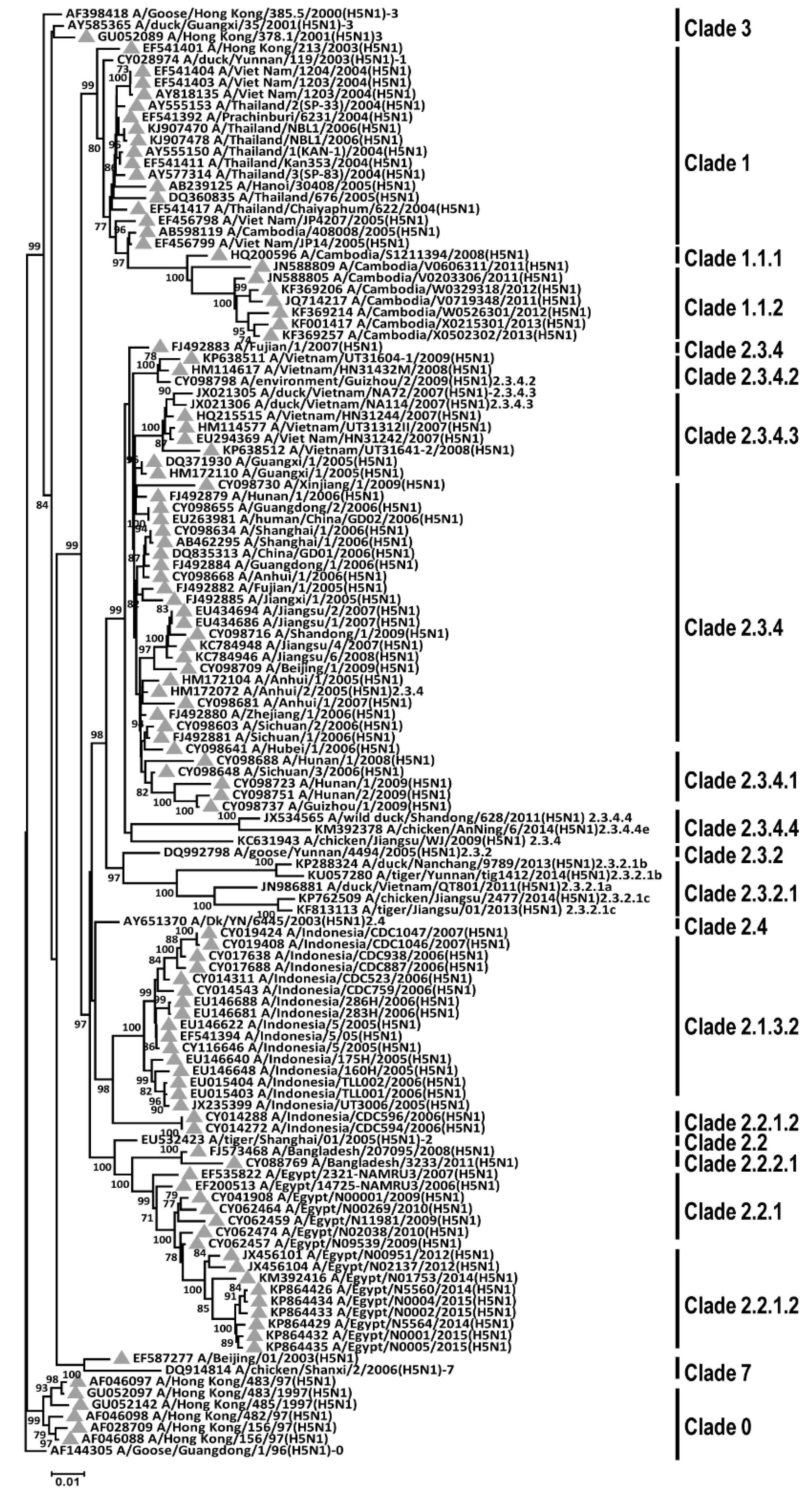
2.1. N-Linked Glycosylation Sites Are Conserved in HA of H5N1 AIVs among Different Clades Phylogenetically
2.2. Generation of Recombinant DC-SIGN Proteins for Interaction with H5N1 Virus
2.3. H5N1 Pseudotyped Lentiviruses Utilize DC-SIGN to Enhance Its Infectivity Compared to H1N1 and H3N2 Human Influenza A Viruses
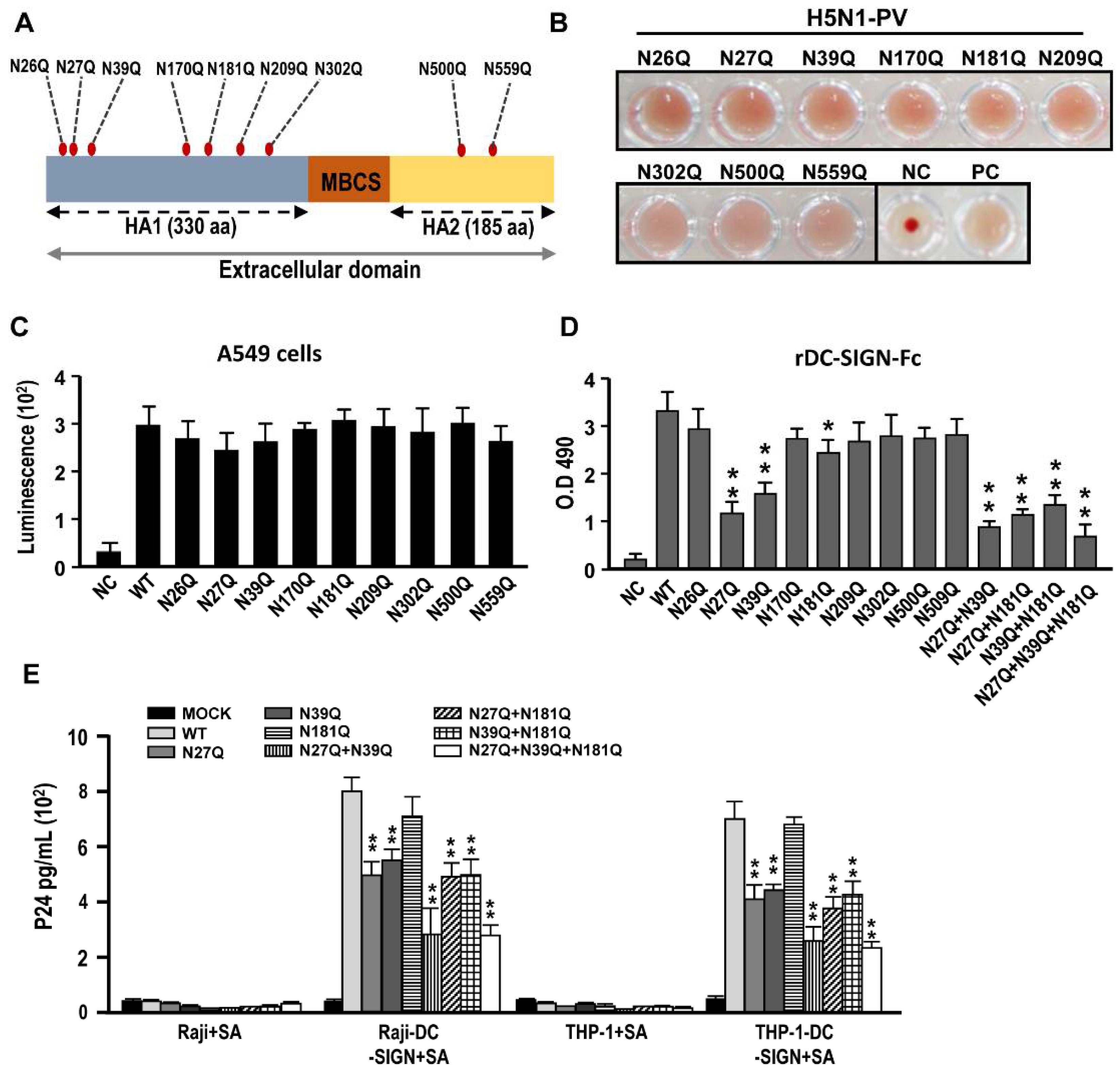
2.4. N-Glycosylation Sites N27, N39, N181 in Hemagglutinin Proteins of H5N1 AIVs Are Key in Interacting with DC-SIGN
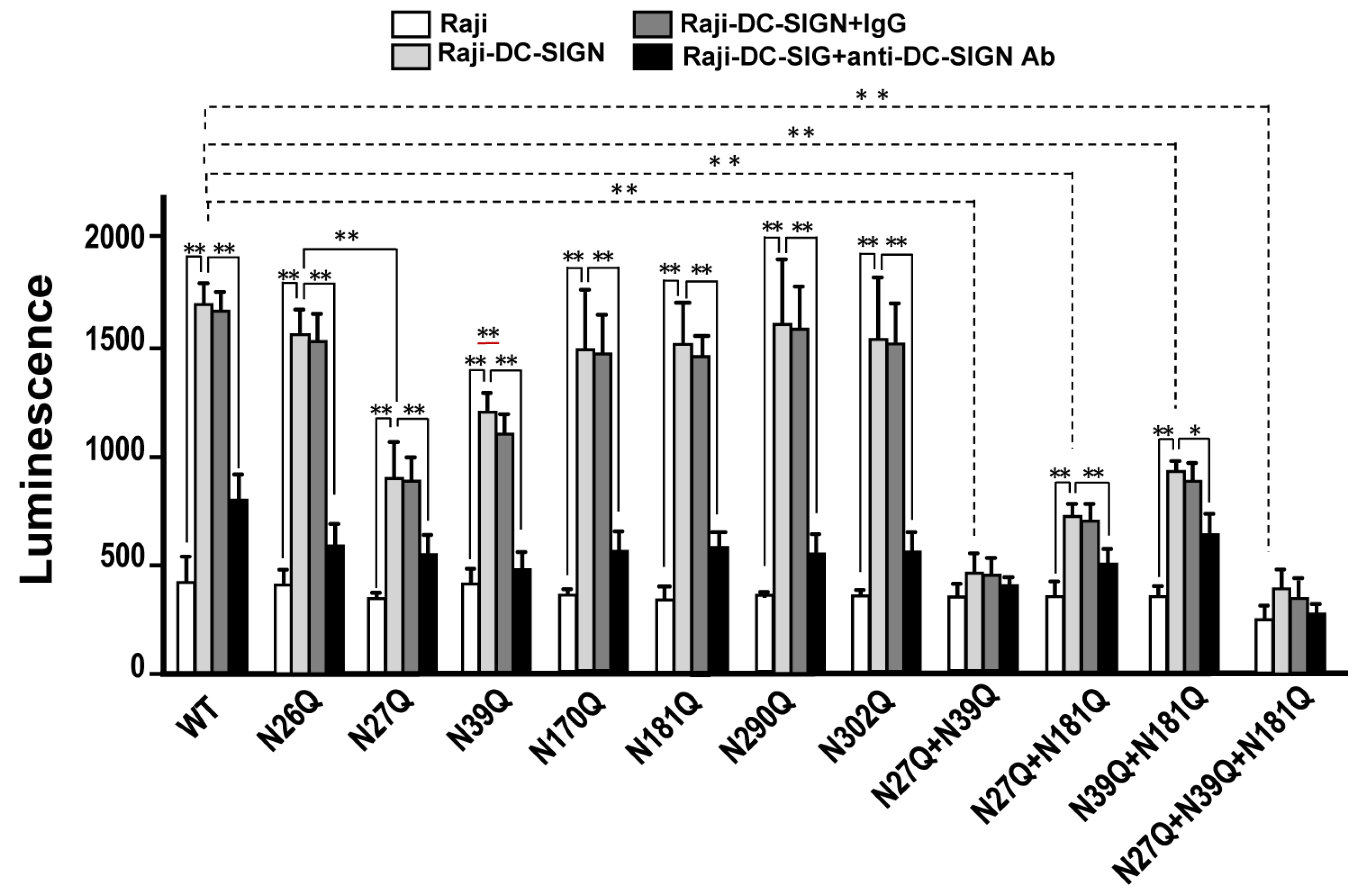
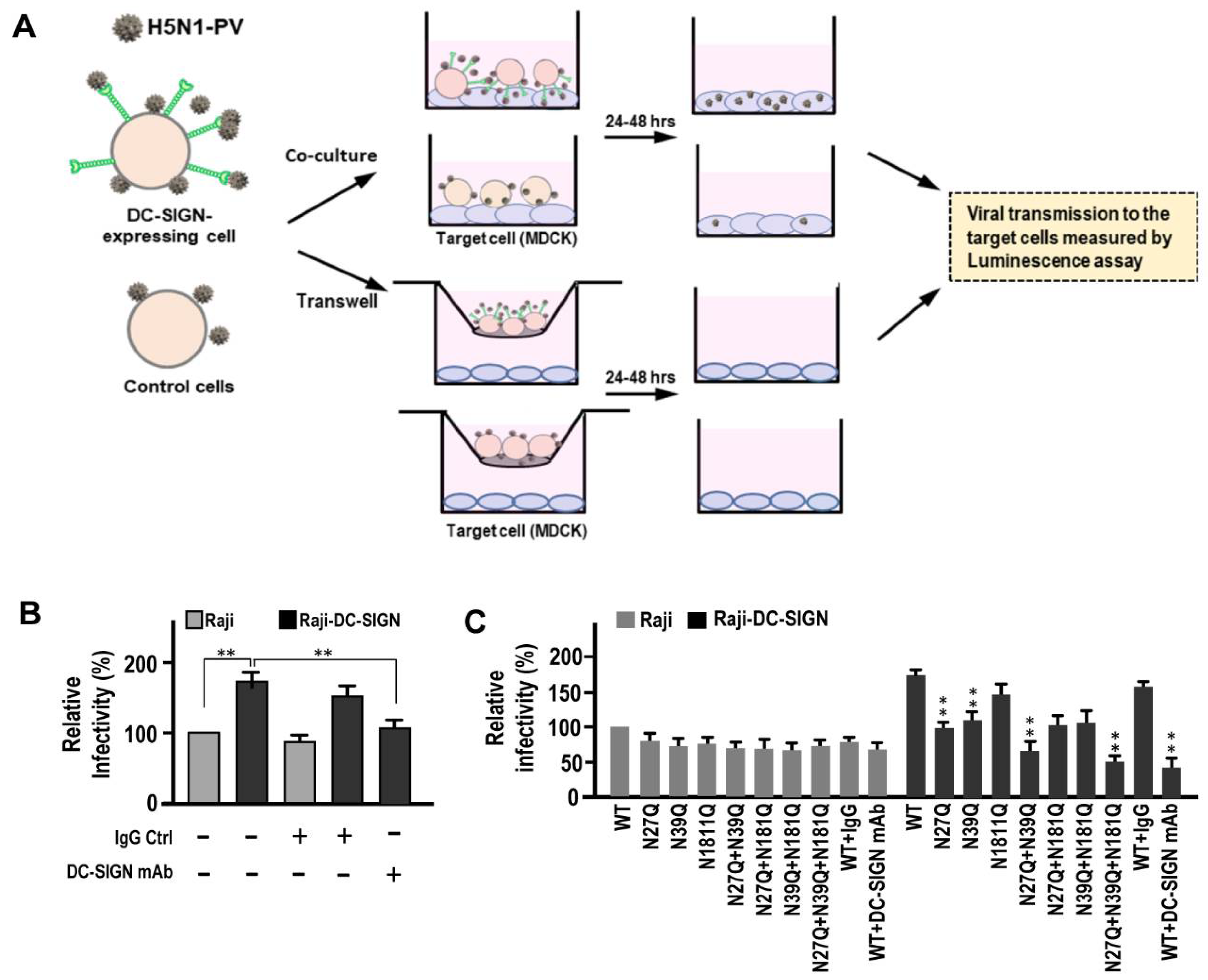
2.5. Mutation on N27 and N39 Ameliorate DC-SIGN Mediated H5N1 AIVs Infection in Cis and in Trans
2.6. Immature Dendritic Cells (DCs) with Higher DC-SIGN Expressions Display Higher Viral Susceptibility and Cell–Cell Transmission Abilities in Mediating H5N1 Infection
3. Discussion
4. Materials and Methods
4.1. Ethics Statement
4.2. Cell
4.3. H5N1 Pseudotype and Reverse-Genetic Virus Preparation
4.4. N-Linked Glycosylation Site Prediction
4.5. Mutagenesis of N-Linked Glycosylation Site of HA (H5) Protein
4.6. Immuno-Electron Microscopy (Immuno-EM)
4.7. ELISA
4.8. Immunofluorescent Staining
4.9. Cis Infectivity Assay
4.10. Trans Infectivity Assay
4.11. qRT-PCR
4.12. Generation of Monocyte-Derived Dendritic Cells (MDDCs)
4.13. Apoptosis Assays
4.14. Hemagglutination Assay (HA)
4.15. Statistical Analyses
Supplementary Materials
Author Contributions
Funding
Institutional Review Board Statement
Informed Consent Statement
Data Availability Statement
Acknowledgments
Conflicts of Interest
References
- Claas, E.C.; Osterhaus, A.D.; Van Beek, R.; De Jong, J.C.; Rimmelzwaan, G.F.A.; Senne, D.; Krauss, S.; Shortridge, K.F.; Webster, R.G. Human influenza A H5N1 virus related to a highly pathogenic avian influenza virus. Lancet 1998, 351, 472–477. [Google Scholar] [CrossRef]
- Chowdhury, S.; Hossain, M.E.; Ghosh, P.K.; Ghosh, S.; Hossain, M.B.; Beard, C.; Rahman, M.; Rahman, M.Z. The Pattern of Highly Pathogenic Avian Influenza H5N1 Outbreaks in South Asia. Trop. Med. Infect. Dis. 2019, 4, 138. [Google Scholar] [CrossRef] [Green Version]
- Peiris, J.S.M.; De Jong, M.D.; Guan, Y. Avian Influenza Virus (H5N1): A Threat to Human Health. Clin. Microbiol. Rev. 2007, 20, 243–267. [Google Scholar] [CrossRef] [Green Version]
- WHO/OIE/FAO. Continued evolution of highly pathogenic avian influenza A (H5N1): Updated nomenclature. Influenza Other. Respir. Virus. 2012, 6, 1–5. [Google Scholar] [CrossRef] [Green Version]
- Xu, L.; Bao, L.; Yuan, J.; Li, F.; Lv, Q.; Deng, W.; Xu, Y.; Yao, Y.; Yu, P.; Chen, H.; et al. Antigenicity and transmissibility of a novel clade 2.3.2.1 avian influenza H5N1 virus. J. Gen. Virol. 2013, 94, 2616–2626. [Google Scholar] [CrossRef] [PubMed] [Green Version]
- WHO. Cumulative Number of Confirmed Human Cases for Avian Influenza A(H5N1) Reported to WHO. 2020. Available online: https://www.who.int/influenza/human_animal_interface/H5N1_cumulative_table_archives/en/ (accessed on 23 October 2020).
- Wang, S.-F.; Lee, Y.-M.; Chan, Y.-J.; Liu, H.-F.; Yen, Y.-F.; Liu, W.-T.; Huang, J.C.; Chen, Y.-M.A. Influenza A virus in Taiwan, 1980–2006: Phylogenetic and antigenic characteristics of the hemagglutinin gene. J. Med. Virol. 2009, 81, 1457–1470. [Google Scholar] [CrossRef] [PubMed]
- Palese, P. Orthomyxoviridae: The Viruses and their Replication. Fields Virol. 2007, 2, 1647–1689. [Google Scholar]
- Bouvier, N.M.; Palese, P. The biology of influenza viruses. Vaccine 2008, 26, D49–D53. [Google Scholar] [CrossRef] [PubMed] [Green Version]
- Shinya, K.; Ebina, M.; Yamada, S.; Ono, M.; Kasai, N.; Kawaoka, Y. Avian flu: Influenza virus receptors in the human airway. Nature 2006, 440, 435–436. [Google Scholar] [CrossRef]
- Komar, N.; Olsen, B. Avian Influenza Virus (H5N1) Mortality Surveillance. Emerg. Infect. Dis. 2008, 14, 1176–1178. [Google Scholar] [CrossRef]
- Oslund, K.L.; Baumgarth, N. Influenza-induced innate immunity: Regulators of viral replication, respiratory tract pathology & adaptive immunity. Futur. Virol. 2011, 6, 951–962. [Google Scholar] [CrossRef]
- Zhang, Z.; Zhang, J.; Huang, K.; Li, K.-S.; Yuen, K.-Y.; Guan, Y.; Chen, H.; Ng, W.F. Systemic infection of avian influenza A virus H5N1 subtype in humans. Hum. Pathol. 2009, 40, 735–739. [Google Scholar] [CrossRef] [PubMed]
- Korteweg, C.; Gu, J. Pathology, Molecular Biology, and Pathogenesis of Avian Influenza A (H5N1) Infection in Humans. Am. J. Pathol. 2008, 172, 1155–1170. [Google Scholar] [CrossRef] [PubMed] [Green Version]
- Wang, S.-F.; Chen, K.-H.; Thitithanyanont, A.; Yao, L.; Lee, Y.-M.; Chan, Y.-J.; Liu, S.-J.; Chong, P.; Liu, W.T.; Huang, J.C.; et al. Generating and characterizing monoclonal and polyclonal antibodies against avian H5N1 hemagglutinin protein. Biochem. Biophys. Res. Commun. 2009, 382, 691–696. [Google Scholar] [CrossRef] [PubMed]
- Mason, C.P.; Tarr, A. Human Lectins and Their Roles in Viral Infections. Molecules 2015, 20, 2229–2271. [Google Scholar] [CrossRef] [Green Version]
- Chen, Y.-J.; Wang, S.-F.; Weng, I.-C.; Hong, M.-H.; Lo, T.-H.; Jan, J.-T.; Hsu, L.-C.; Chen, H.-Y.; Liu, F.-T. Galectin-3 Enhances Avian H5N1 Influenza A Virus–Induced Pulmonary Inflammation by Promoting NLRP3 Inflammasome Activation. Am. J. Pathol. 2018, 188, 1031–1042. [Google Scholar] [CrossRef] [Green Version]
- Wang, W.-H.; Yeh, C.-S.; Lin, C.-Y.; Yuan, R.-Y.; Urbina, A.N.; Lu, P.-L.; Chen, Y.-H.; Chen, Y.-M.A.; Liu, F.-T.; Wang, S.-F. Amino Acid Deletions in p6Gag Domain of HIV-1 CRF07_BC Ameliorate Galectin-3 Mediated Enhancement in Viral Budding. Int. J. Mol. Sci. 2020, 21, 2910. [Google Scholar] [CrossRef] [Green Version]
- Li, F.-Y.; Wang, S.-F.; Bernardes, E.S.; Liu, F.-T. Galectins in Host Defense against Microbial Infections. Cannabinoids Neuropsychiatr. Disord. 2020, 1204, 141–167. [Google Scholar] [CrossRef]
- Wang, W.-H.; Lin, C.-Y.; Chang, M.R.; Urbina, A.N.; Assavalapsakul, W.; Thitithanyanont, A.; Chen, Y.-H.; Liu, F.-T.; Wang, S.-F. The role of galectins in virus infection—A systemic literature review. J. Microbiol. Immunol. Infect. 2020, 53, 925–935. [Google Scholar] [CrossRef]
- Van Kooyk, Y.; Geijtenbeek, T.B.H. DC-SIGN: Escape mechanism for pathogens. Nat. Rev. Immunol. 2003, 3, 697–709. [Google Scholar] [CrossRef]
- Cunningham, A.L.; Harman, A.N.; Donaghy, H. DC-SIGN ’AIDS’ HIV immune evasion and infection. Nat. Immunol. 2007, 8, 556–558. [Google Scholar] [CrossRef] [PubMed]
- Poehlmann, S.; Zhang, J.; Baribaud, F.; Chen, Z.; Leslie, G.J.; Lin, G.; Granelli-Piperno, A.; Doms, R.W.; Rice, C.M.; McKeating, J.A. Hepatitis C Virus Glycoproteins Interact with DC-SIGN and DC-SIGNR. J. Virol. 2003, 77, 4070–4080. [Google Scholar] [CrossRef] [PubMed] [Green Version]
- Simmons, G.; Reeves, J.D.; Grogan, C.C.; Vandenberghe, L.H.; Baribaud, F.; Whitbeck, J.C.; Burke, E.; Buchmeier, M.J.; Soilleux, E.J.; Riley, J.L.; et al. DC-SIGN and DC-SIGNR Bind Ebola Glycoproteins and Enhance Infection of Macrophages and Endothelial Cells. Virology 2003, 305, 115–123. [Google Scholar] [CrossRef] [PubMed] [Green Version]
- Tassaneetrithep, B.; Burgess, T.H.; Granelli-Piperno, A.; Trumpfheller, C.; Finke, J.; Sun, W.; Eller, M.A.; Pattanapanyasat, K.; Sarasombath, S.; Birx, D.L.; et al. DC-SIGN (CD209) Mediates Dengue Virus Infection of Human Dendritic Cells. J. Exp. Med. 2003, 197, 823–829. [Google Scholar] [CrossRef] [Green Version]
- Shih, Y.-P.; Chen, C.-Y.; Liu, S.-J.; Chen, K.-H.; Lee, Y.-M.; Chao, Y.-C.; Chen, Y.-M.A. Identifying Epitopes Responsible for Neutralizing Antibody and DC-SIGN Binding on the Spike Glycoprotein of the Severe Acute Respiratory Syndrome Coronavirus. J. Virol. 2006, 80, 10315–10324. [Google Scholar] [CrossRef] [Green Version]
- Hillaire, M.L.B.; Nieuwkoop, N.J.; Boon, A.C.M.; De Mutsert, G.; Trierum, S.E.V.-V.; Fouchier, R.A.M.; Osterhaus, A.D.M.E.; Rimmelzwaan, G.F. Binding of DC-SIGN to the Hemagglutinin of Influenza A Viruses Supports Virus Replication in DC-SIGN Expressing Cells. PLoS ONE 2013, 8, e56164. [Google Scholar] [CrossRef] [Green Version]
- Londrigan, S.L.; Turville, S.G.; Tate, M.D.; Deng, Y.-M.; Brooks, A.G.; Reading, P.C. N-Linked Glycosylation Facilitates Sialic Acid-Independent Attachment and Entry of Influenza A Viruses into Cells Expressing DC-SIGN or L-SIGN. J. Virol. 2010, 85, 2990–3000. [Google Scholar] [CrossRef] [Green Version]
- Wang, S.-F.; Huang, J.C.; Lee, Y.-M.; Liu, S.-J.; Chan, Y.-J.; Chau, Y.-P.; Chong, P.; Chen, Y.-M.A. DC-SIGN mediates avian H5N1 influenza virus infection in cis and in trans. Biochem. Biophys. Res. Commun. 2008, 373, 561–566. [Google Scholar] [CrossRef]
- Hong, P.; Ninonuevo, M.R.; Lee, B.; Lebrilla, C.; Bode, L. Human milk oligosaccharides reduce HIV-1-gp120 binding to dendritic cell-specific ICAM3-grabbing non-integrin (DC-SIGN). Br. J. Nutr. 2009, 101, 482–486. [Google Scholar] [CrossRef] [Green Version]
- Geijtenbeek, T.B.; Kwon, D.S.; Torensma, R.; Van Vliet, S.J.; Van Duijnhoven, G.C.; Middel, J.; Cornelissen, I.L.; Nottet, H.S.; KewalRamani, V.N.; Littman, D.R.; et al. DC-SIGN, a Dendritic Cell–Specific HIV-1-Binding Protein that Enhances trans-Infection of T Cells. Cell 2000, 100, 587–597. [Google Scholar] [CrossRef] [Green Version]
- Wang, S.-F.; Su, M.-W.; Tseng, S.-P.; Li, M.-C.; Tsao, C.-H.; Huang, S.-W.; Chu, W.-C.; Liu, W.-T.; Chen, Y.-M.A.; Huang, J.C. Analysis of codon usage preference in hemagglutinin genes of the swine-origin influenza A (H1N1) virus. J. Microbiol. Immunol. Infect. 2016, 49, 477–486. [Google Scholar] [CrossRef] [Green Version]
- Kim, P.; Jang, Y.H.; Kwon, S.B.; Lee, C.M.; Han, G.; Seong, B.L. Glycosylation of Hemagglutinin and Neuraminidase of Influenza A Virus as Signature for Ecological Spillover and Adaptation among Influenza Reservoirs. Viruses 2018, 10, 183. [Google Scholar] [CrossRef] [PubMed] [Green Version]
- Chen, W.; Zhong, Y.; Qin, Y.; Sun, S.; Li, Z. The Evolutionary Pattern of Glycosylation Sites in Influenza Virus (H5N1) Hemagglutinin and Neuraminidase. PLoS ONE 2012, 7, e49224. [Google Scholar] [CrossRef] [Green Version]
- Na-Ek, P.; Thewsoongnoen, J.; Thanunchai, M.; Wiboon-Ut, S.; Sa-Ard-Iam, N.; Mahanonda, R.; Thitithanyanont, A. The activation of B cells enhances DC-SIGN expression and promotes susceptibility of B cells to HPAI H5N1 infection. Biochem. Biophys. Res. Commun. 2017, 490, 1301–1306. [Google Scholar] [CrossRef] [PubMed]
- Yu, L.; Shang, S.; Tao, R.; Wang, C.; Zhang, L.; Peng, H.; Chen, Y. High doses of recombinant mannan-binding lectin inhibit the binding of influenza A(H1N1)pdm09 virus with cells expressing DC-SIGN. APMIS 2017, 122, 136–664. [Google Scholar] [CrossRef] [PubMed]
- Van Breedam, W.; Pöhlmann, S.; Favoreel, H.W.; De Groot, R.J.; Nauwynck, H.J. Bitter-sweet symphony: Glycan–lectin interactions in virus biology. FEMS Microbiol. Rev. 2014, 38, 598–632. [Google Scholar] [CrossRef] [PubMed] [Green Version]
- Thompson, A.J.; Cao, L.; Ma, Y.; Wang, X.; Diedrich, J.K.; Kikuchi, C.; Willis, S.; Worth, C.; McBride, R.; Yates, J.R., 3rd; et al. Human Influenza Virus Hemagglutinins Contain Conserved Oligomannose N-Linked Glycans Allowing Potent Neutralization by Lectins. Cell. Host Microbe 2020, 27, 725–735.e5. [Google Scholar] [CrossRef]
- Feinberg, H.; Castelli, R.; Drickamer, K.; Seeberger, P.H.; Weis, W.I. Multiple Modes of Binding Enhance the Affinity of DC-SIGN for High Mannose N-Linked Glycans Found on Viral Glycoproteins. J. Biol. Chem. 2006, 282, 4202–4209. [Google Scholar] [CrossRef] [Green Version]
- Wanzeck, K.; Boyd, K.L.; McCullers, J.A. Glycan Shielding of the Influenza Virus Hemagglutinin Contributes to Immunopathology in Mice. Am. J. Respir. Crit. Care Med. 2011, 183, 767–773. [Google Scholar] [CrossRef] [Green Version]
- York, I.A.; Stevens, J.; Alymova, I.V. Influenza virus N-linked glycosylation and innate immunity. Biosci. Rep. 2019, 39, 1. [Google Scholar] [CrossRef] [PubMed] [Green Version]
- Su, S.V.; Hong, P.; Baik, S.S.W.; Negrete, O.A.; Gurney, K.B.; Lee, B. DC-SIGN Binds to HIV-1 Glycoprotein 120 in a Distinct but Overlapping Fashion Compared with ICAM-2 and ICAM-3. J. Biol. Chem. 2004, 279, 19122–19132. [Google Scholar] [CrossRef] [PubMed] [Green Version]
- Marzi, A.; Möller, P.; Hanna, S.L.; Harrer, T.; Eisemann, J.; Steinkasserer, A.; Becker, S.; Baribaud, F.; Poehlmann, S. Analysis of the Interaction of Ebola Virus Glycoprotein with DC-SIGN (Dendritic Cell–Specific Intercellular Adhesion Molecule 3–Grabbing Nonintegrin) and Its Homologue DC-SIGNR. J. Infect. Dis. 2007, 196, S237–S246. [Google Scholar] [CrossRef] [PubMed]
- Hong, P.W.-P.; Nguyen, S.; Young, S.; Su, S.V.; Lee, B. Identification of the Optimal DC-SIGN Binding Site on Human Immunodeficiency Virus Type 1 gp120. J. Virol. 2007, 81, 8325–8336. [Google Scholar] [CrossRef] [PubMed] [Green Version]
- Feinberg, H.; Mitchell, D.A.; Drickamer, K.; Weis, W.I. Structural Basis for Selective Recognition of Oligosaccharides by DC-SIGN and DC-SIGNR. Science 2001, 294, 2163–2166. [Google Scholar] [CrossRef] [Green Version]
- Mitchell, D.A.; Fadden, A.J.; Drickamer, K. A Novel Mechanism of Carbohydrate Recognition by the C-type Lectins DC-SIGN and DC-SIGNR. J. Biol. Chem. 2001, 276, 28939–28945. [Google Scholar] [CrossRef] [PubMed] [Green Version]
- Appelmelk, B.J.; van Die, I.; van Vliet, S.J.; Vandenbroucke-Grauls, C.M.J.E.; Geijtenbeek, T.B.H.; van Kooyk, Y. Cutting edge: Carbohydrate profiling identifies new pathogens that interact with dendritic cell-specific ICAM-3-grabbing nonintegrin on dendritic cells. J Immunol. 2003, 170, 1635–1639. [Google Scholar] [CrossRef] [Green Version]
- Menon, S.; Rosenberg, K.; Graham, S.A.; Ward, E.M.; Taylor, M.E.; Drickamer, K.; Leckband, D.E. Binding-site geometry and flexibility in DC-SIGN demonstrated with surface force measurements. Proc. Natl. Acad. Sci. USA 2009, 106, 11524–11529. [Google Scholar] [CrossRef] [Green Version]
- Guo, Y.; Feinberg, H.; Conroy, E.A.; Mitchell, D.; Alvarez, R.; Blixt, O.E.; Taylor, M.I.; Weis, W.; Drickamer, K. Structural basis for distinct ligand-binding and targeting properties of the receptors DC-SIGN and DC-SIGNR. Nat. Struct. Mol. Biol. 2004, 11, 591–598. [Google Scholar] [CrossRef]
- Medina, R.A.; Stertz, S.; Manicassamy, B.; Zimmermann, P.; Sun, X.; Albrecht, R.A.; Uusi-Kerttula, H.; Zagordi, O.; Belshe, R.B.; Frey, S.E.; et al. Glycosylations in the Globular Head of the Hemagglutinin Protein Modulate the Virulence and Antigenic Properties of the H1N1 Influenza Viruses. Sci. Transl. Med. 2013, 5, 187ra70. [Google Scholar] [CrossRef] [Green Version]
- Pokidysheva, E.; Zhang, Y.; Battisti, A.J.; Bator-Kelly, C.M.; Chipman, P.R.; Xiao, C.; Gregorio, G.G.; Hendrickson, W.A.; Kuhn, R.J.; Rossmann, M.G. Cryo-EM Reconstruction of Dengue Virus in Complex with the Carbohydrate Recognition Domain of DC-SIGN. Cell 2006, 124, 485–493. [Google Scholar] [CrossRef] [Green Version]
- Baribaud, F.; Pöhlmann, S.; Sparwasser, T.; Kimata, M.T.Y.; Choi, Y.-K.; Haggarty, B.S.; Ahmad, N.; Macfarlan, T.; Edwards, T.G.; Leslie, G.J.; et al. Functional and Antigenic Characterization of Human, Rhesus Macaque, Pigtailed Macaque, and Murine DC-SIGN. J. Virol. 2001, 75, 10281–10289. [Google Scholar] [CrossRef] [PubMed] [Green Version]
- Hoffmann, E.; Neumann, G.; Kawaoka, Y.; Hobom, G.; Webster, R.G. A DNA transfection system for generation of influenza A virus from eight plasmids. Proc. Natl. Acad. Sci. USA 2000, 97, 6108–6113. [Google Scholar] [CrossRef] [PubMed] [Green Version]
- Uchida, Y.; Takemae, N.; Saito, T. Application of Reverse Genetics for Producing Attenuated Vaccine Strains against Highly Pathogenic Avian Influenza Viruses. J. Veter. Med. Sci. 2014, 76, 1111–1117. [Google Scholar] [CrossRef] [PubMed] [Green Version]
- Liao, C.-F.; Wang, S.-F.; Lin, Y.-T.; Ho, D.D.; Chen, Y.-M.A. Identification of the DC-SIGN-Interactive Domains on the Envelope Glycoprotein of HIV-1 CRF07_BC. AIDS Res. Hum. Retrovir. 2011, 27, 831–839. [Google Scholar] [CrossRef] [PubMed]
- Wang, S.-F.; Tsao, C.-H.; Lin, Y.-T.; Hsu, D.K.; Chiang, M.-L.; Lo, C.-H.; Chien, F.-C.; Chen, P.; Chen, Y.-M.A.; Chen, H.-Y.; et al. Galectin-3 promotes HIV-1 budding via association with Alix and Gag p6. Glycobiology 2014, 24, 1022–1035. [Google Scholar] [CrossRef] [Green Version]
- Chang, Y.-F.; Wang, W.-H.; Hong, Y.-W.; Yuan, R.-Y.; Chen, K.-H.; Huang, Y.-W.; Lu, P.-L.; Chen, Y.-H.; Chen, Y.-M.A.; Su, L.-C.; et al. Simple Strategy for Rapid and Sensitive Detection of Avian Influenza A H7N9 Virus Based on Intensity-Modulated SPR Biosensor and New Generated Antibody. Anal. Chem. 2018, 90, 1861–1869. [Google Scholar] [CrossRef]
- Portolano, N.; Watson, P.J.; Fairall, L.; Millard, C.J.; Milano, C.P.; Song, Y.; Cowley, S.M.; Schwabe, J.W.R. Recombinant Protein Expression for Structural Biology in HEK 293F Suspension Cells: A Novel and Accessible Approach. J. Vis. Exp. 2014, 92, e51897. [Google Scholar] [CrossRef] [Green Version]
- Payungporn, S.; Chutinimitkul, S.; Chaisingh, A.; Damrongwantanapokin, S.; Buranathai, C.; Amonsin, A.; Theamboonlers, A.; Poovorawan, Y. Single step multiplex real-time RT-PCR for H5N1 influenza A virus detection. J. Virol. Methods 2006, 131, 143–147. [Google Scholar] [CrossRef]
- Crowley, L.C.; Marfell, B.J.; Scott, A.P.; Waterhouse, N. Quantitation of Apoptosis and Necrosis by Annexin V Binding, Propidium Iodide Uptake, and Flow Cytometry. Cold Spring Harb. Protoc. 2016, 2016. [Google Scholar] [CrossRef]
- WHO. WHO Manual on Animal Influenza Diagnosis and Surveillance; World Health Organization: Geneva, Switzerland, 2002. [Google Scholar]


| Mutation(s) in H5N1 Construct from Strain | Binding of HA5 to: | |||
|---|---|---|---|---|
| DC-SIGN | SA Receptor | |||
| None (wild-type) | + | +++ | ||
| N26Q | + | +++ | ||
| N27Q | + | ++ | ||
| N39Q | + | +++ | ||
| N170Q | + | +++ | ||
| N181Q | + | +++ | ||
| N209Q | + | +++ | ||
| N302Q | + | +++ | ||
| N500Q | + | +++ | ||
| N559Q | + | +++ | ||
| N26Q | N27Q | + | ++ | |
| N26Q | N181Q | + | +++ | |
| N27Q | N181Q | + | +++ | |
| N26Q | N27Q | N181Q | + | ++ |
Publisher’s Note: MDPI stays neutral with regard to jurisdictional claims in published maps and institutional affiliations. |
© 2021 by the authors. Licensee MDPI, Basel, Switzerland. This article is an open access article distributed under the terms and conditions of the Creative Commons Attribution (CC BY) license (http://creativecommons.org/licenses/by/4.0/).
Share and Cite
Yang, Z.-S.; Huang, S.-W.; Wang, W.-H.; Lin, C.-Y.; Wang, C.-F.; Urbina, A.N.; Thitithanyanont, A.; Tseng, S.-P.; Lu, P.-L.; Chen, Y.-H.; et al. Identification of Important N-Linked Glycosylation Sites in the Hemagglutinin Protein and Their Functional Impact on DC-SIGN Mediated Avian Influenza H5N1 Infection. Int. J. Mol. Sci. 2021, 22, 743. https://0-doi-org.brum.beds.ac.uk/10.3390/ijms22020743
Yang Z-S, Huang S-W, Wang W-H, Lin C-Y, Wang C-F, Urbina AN, Thitithanyanont A, Tseng S-P, Lu P-L, Chen Y-H, et al. Identification of Important N-Linked Glycosylation Sites in the Hemagglutinin Protein and Their Functional Impact on DC-SIGN Mediated Avian Influenza H5N1 Infection. International Journal of Molecular Sciences. 2021; 22(2):743. https://0-doi-org.brum.beds.ac.uk/10.3390/ijms22020743
Chicago/Turabian StyleYang, Zih-Syuan, Szu-Wei Huang, Wen-Hung Wang, Chih-Yen Lin, Chu-Feng Wang, Aspiro Nayim Urbina, Arunee Thitithanyanont, Sung-Pin Tseng, Po-Liang Lu, Yen-Hsu Chen, and et al. 2021. "Identification of Important N-Linked Glycosylation Sites in the Hemagglutinin Protein and Their Functional Impact on DC-SIGN Mediated Avian Influenza H5N1 Infection" International Journal of Molecular Sciences 22, no. 2: 743. https://0-doi-org.brum.beds.ac.uk/10.3390/ijms22020743


