Article

Kynurenine/Tryptophan Ratio as a Potential Blood-Based Biomarker in Non-Small Cell Lung Cancer

1
Section of Anatomic Pathology and Histology, Department of Medicine and Surgery, University of Perugia, 06129 Perugia, Italy
2
Section of Pharmacology, Department of Medicine and Surgery, University of Perugia, 06129 Perugia, Italy
3
Section of Thoracic Surgery, Department of Medicine and Surgery, University of Perugia, 06129 Perugia, Italy
4
Medical Oncology Division, Santa Maria della Misericordia Hospital, 06132 Perugia, Italy
5
Division of Medical Oncology, Ospedali Riuniti Padova Sud, 35043 Monselice, Italy
*
Author to whom correspondence should be addressed.
Int. J. Mol. Sci. 2021, 22(9), 4403; https://0-doi-org.brum.beds.ac.uk/10.3390/ijms22094403
Submission received: 16 March 2021 / Revised: 14 April 2021 / Accepted: 20 April 2021 / Published: 22 April 2021
(This article belongs to the Special Issue Decoding the Complex Crossroad of Tryptophan Metabolic Pathways)

Abstract

:
The enzyme indoleamine 2,3-dioxygenase 1 (IDO1) degrade tryptophan (Trp) into kynurenine (Kyn) at the initial step of an enzymatic pathway affecting T cell proliferation. IDO1 is highly expressed in various cancer types and associated with poor prognosis. Nevertheless, the serum Kyn/Trp concentration ratio has been suggested as a marker of cancer-associated immune suppression. We measured Kyn and Trp in blood samples of a wide cohort of non-small-cell lung cancer (NSCLC) patients, before they underwent surgery, and analyzed possible correlations of the Kyn/Trp ratio with either IDO1 expression or clinical–pathological parameters. Low Kyn/Trp significantly correlated with low IDO1 expression and never-smoker patients; while high Kyn/Trp was significantly associated with older (≥68 years) patients, advanced tumor stage, and squamous cell carcinoma (Sqcc), rather than the adenocarcinoma (Adc) histotype. Moreover, high Kyn/Trp was associated, among the Adc group, with higher tumor stages (II and III), and, among the Sqcc group, with a high density of tumor-infiltrating lymphocytes. A trend correlating the high Kyn/Trp ratio with the probability of recurrences from NSCLC was also found. In conclusion, high serum Kyn/Trp ratio, associated with clinical and histopathological parameters, may serve as a serum biomarker to optimize risk stratification and therapy of NSCLC patients.

1. Introduction

Lung cancer is the main cause of cancer-related death worldwide [1]. About 80% of the disease is classified into non-small-cell lung cancer (NSCLC). Among them, approximately 20% of cases are localized disease (stages I and II), 25% are locally advanced disease (stage III), and 55% of cases are metastatic disease (stage IV) [2]. Surgical resection is the preferred treatment option for patients with early-stage and some locally advanced NSCLC. Genomic profiling of resected tumors, allowing identification of potentially targetable alterations and/or eligibility for immune checkpoint inhibition, is now included in standard care and is considered an essential diagnostic tool to improve clinical outcomes in particular patient subgroups [3,4]. Biomarkers analyzed in such surgical samples, or traditional biopsy (particularly invasive in NSCLC patients), reflect tumor and disease status at a specific time; that of tissue removal. Nevertheless, to monitor tumor evolution over time, the dosage of cancer-associated markers in liquid biopsy is now considered an attractive means for the optimization of cancer therapeutic protocols. In a liquid biopsy, circulating tumor cells and cell-free DNA are principally investigated to improve lung cancer screening, detect minimal residual disease, monitor systemic treatment initiation and response, and determine the development of resistance [5]. However, metabolites of the kynurenine (Kyn) pathway also appear to be interesting candidates to be dosed in the blood to follow cancer evolution and control it by therapeutic intervention.
Kyn pathway, a tryptophan (Trp)-degrading enzymatic cascade controlled at the first rate-limiting step by indoleamine-2,3-dioxygenase 1 (IDO1), has been associated with a poorer prognosis in cancer patients [6,7,8]. IDO1 expression/activity has been observed in tumor cells as well as in the tumor-surrounding stroma, which is composed of endothelial cells, immune cells, fibroblasts, and mesenchymal cells [9]. Enhanced Trp breakdown occurring in the tumor microenvironment, reflected by decreased Trp and elevated Kyn concentrations in the peripheral blood, is often observed in cancer patients and related to tumor progression and poor clinical outcome [10]. In cancer immunoediting, IDO1 induction in malignant cells and their microenvironment is a key mechanism to modulate anti-tumor immune responses and thus escape immune surveillance [11,12]. Decreased Trp availability and accumulation of Trp metabolites, such as the kynurenines, directly affect immune T cell proliferation and functions, and induce their apoptosis [13,14]. Based on these mechanisms, IDO1 expression in tumor sites and circulating Trp and Kyn (evaluated as the Kyn/Trp ratio) have been hypothesized as possible markers of cancer progression [15,16]. In particular, recent studies have demonstrated that the Kyn/Trp ratio is a promising surveillance biomarker for several cancer types. Plasma and urinary Kyn/Trp ratio are higher in patients with bladder cancer [17]. In advanced melanoma and renal cell carcinoma patients treated with nivolumab, the serum Kyn/Trp ratio increases as an adaptive resistance mechanism associated with worse overall survival [18]. In serum of glioblastoma multiforme patients, the Kyn/Trp index seems to be a relevant clinical benchmark, providing prognostic value for enrolment in immunotherapeutic regimens [19]. In NSCLC patients with solid tumors treated with immunotherapy, both prognostic and predictive values of serum Kyn/Trp ratio have been proposed [20].
Implementing the use of non-invasive biomarkers (i.e., measured by liquid biopsy) is a relevant research target for both prognostic and therapeutic purposes. In the present study, we evaluated the Kyn/Trp ratio dosed in blood samples of a wide cohort of NSCLC resected patients and aimed to correlate this value with several clinical–pathological parameters and to patients’ overall survival (OS) and disease-free survival (DFS), to provide new insights contributing to define the Kyn/Trp ratio as a useful serum biomarker for better risk stratification and therapy improvement of NSCLC patients.

2. Results

2.1. Patients Series

One hundred and eighty patients were eligible for the study. Patients were all Caucasian; the median age was 68 years (range 38–84) with a median follow-up period of 51 months (range 1–107 months). One hundred and twenty-seven (71%) patients were males; most of the patients were either current smokers or former smokers (164; 91%). Sixty-nine (38%) patients relapsed after surgery and fifty-seven (32%) died from NSCLC.

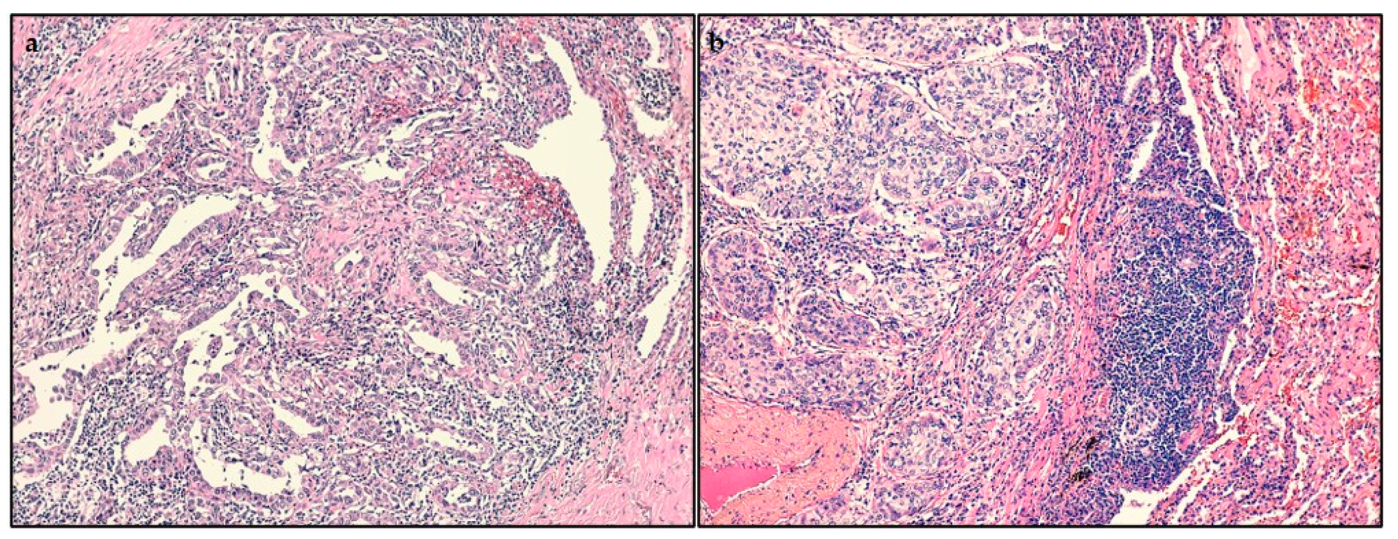
2.2. Histopathological Findings

Surgical specimens from radically resected tumors were analyzed by hematoxylin and eosin (H&E) staining for histopathological determination. One hundred and eighteen (66%) patients suffered from adenocarcinoma (Table 1), which often presented either a lepidic, acinar, papillary, or mucinous primary pattern (92; 78%, data not shown), less aggressive than solid pattern. The pathological stage of the disease was determined according to the classification of the 8th Edition for Cancer Staging by the American Joint Committee on Cancer (AJCC). Stage I was the most frequent (117; 65%). The majority of adenocarcinomas belonged to the stage I group (73; 62%), whereas most of the squamous cell carcinomas were in the stage II–III group (36; 58%).

2.3. Analysis of Tumor-Infiltrating Lymphocytes (TILs)

Through H&E staining, we analyzed the localization of TILs in surgical specimens. More frequently, it was intratumoral (83; 46%), regarding the entire series and the adenocarcinomas group (63; 53%) (Figure 1a). On the other hand, the mixed localization was the most observed among the squamous cell carcinomas (34; 55%) (Figure 1b).

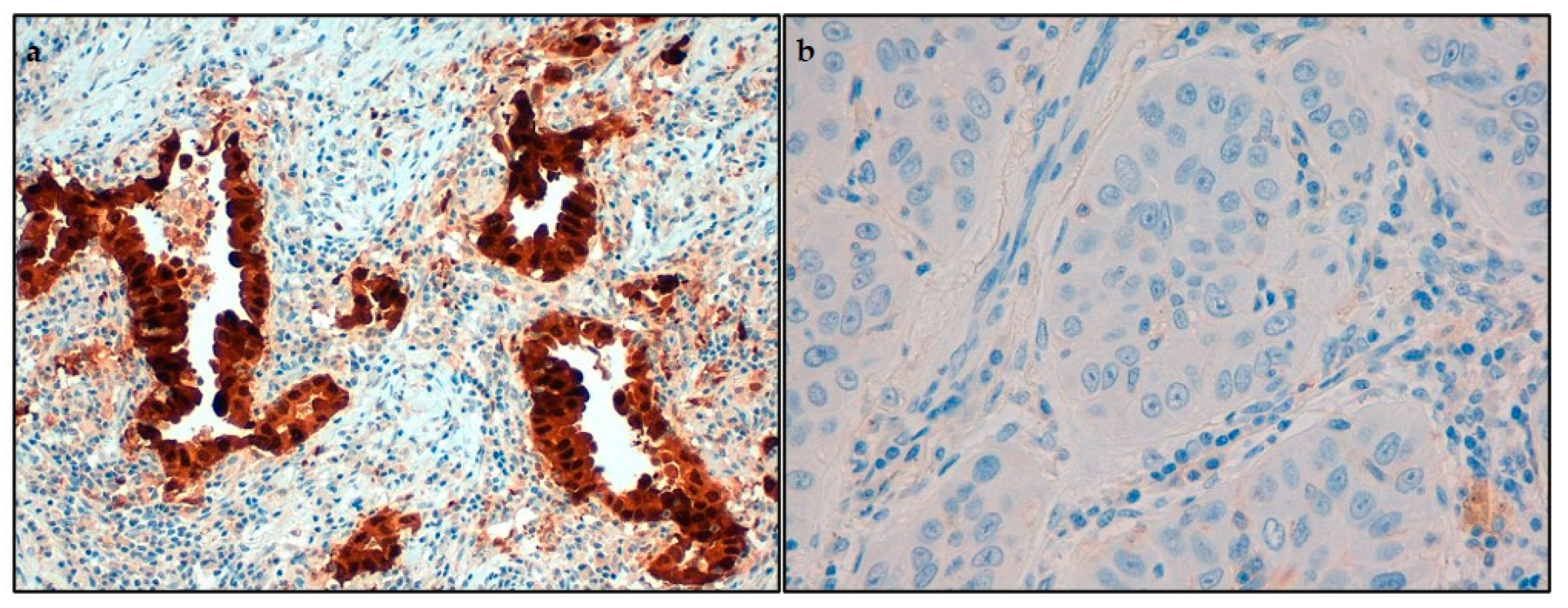
2.4. Immunohistochemical Analysis of IDO1 and Programmed Cell Death Ligand-1 (PD-L1)

Expression of IDO1, the tolerogenic enzyme converting Trp into Kyn, was investigated on neoplastic cells by immunohistochemical analysis. We found that most of the tumors presented a high expression of IDO1 molecule (101; 56%, Figure 2a), both among adenocarcinomas (65; 55%) and squamous cell carcinomas (36; 58%). On neoplastic cells, we also evaluated the immunostaining for PD-L1, a ligand inhibiting T cell proliferation and associated with increased tumor aggressiveness. With a trend opposite to IDO1, many of the neoplasms presented a low expression of PD-L1 (139; 77%, Figure 2b), both regarding adenocarcinomas (97; 82%) and squamous cell carcinomas (42; 68%).

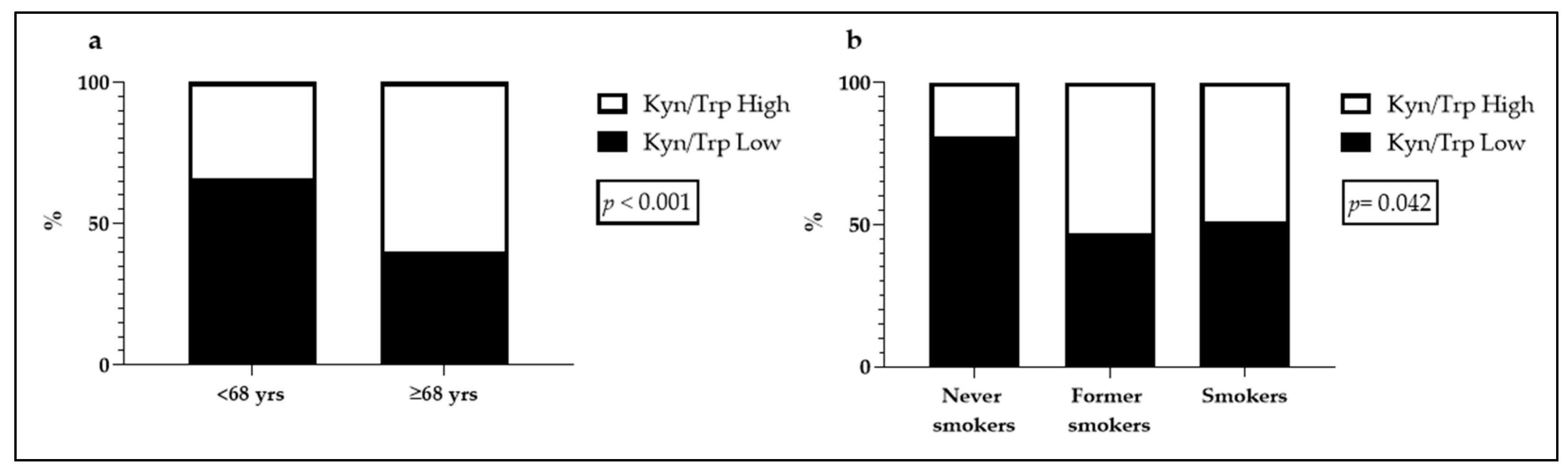
2.5. Kyn/Trp Ratio and Clinical Parameters Associations

Before the NSCLC patients underwent surgery, blood samples were collected for following HPLC analysis of Kyn and Trp relative concentration. Two groups of the Kyn/Trp ratio (low and high) were used for the subsequent evaluation of their associations with clinical–pathological parameters. The Kyn/Trp Low group encompassed the Kyn/Trp ratio values equal to or below the median value of 58, while the Kyn/Trp High group included the values above 58. If the serum concentration of Kyn was undetectable, the corresponding patient was excluded from the analyses. The associations between Kyn/Trp ratio and the clinical parameters are shown in Table 1. The analysis revealed that just over half of the patients presented a low Kyn/Trp ratio (93; 52%) and that Kyn/Trp ratio was higher in over 68-year-old patients (p < 0.001). On the other hand, there was a low Kyn/Trp ratio in patients who were never smokers (p = 0.042). The statistically significant associations found are summarized in Figure 3.

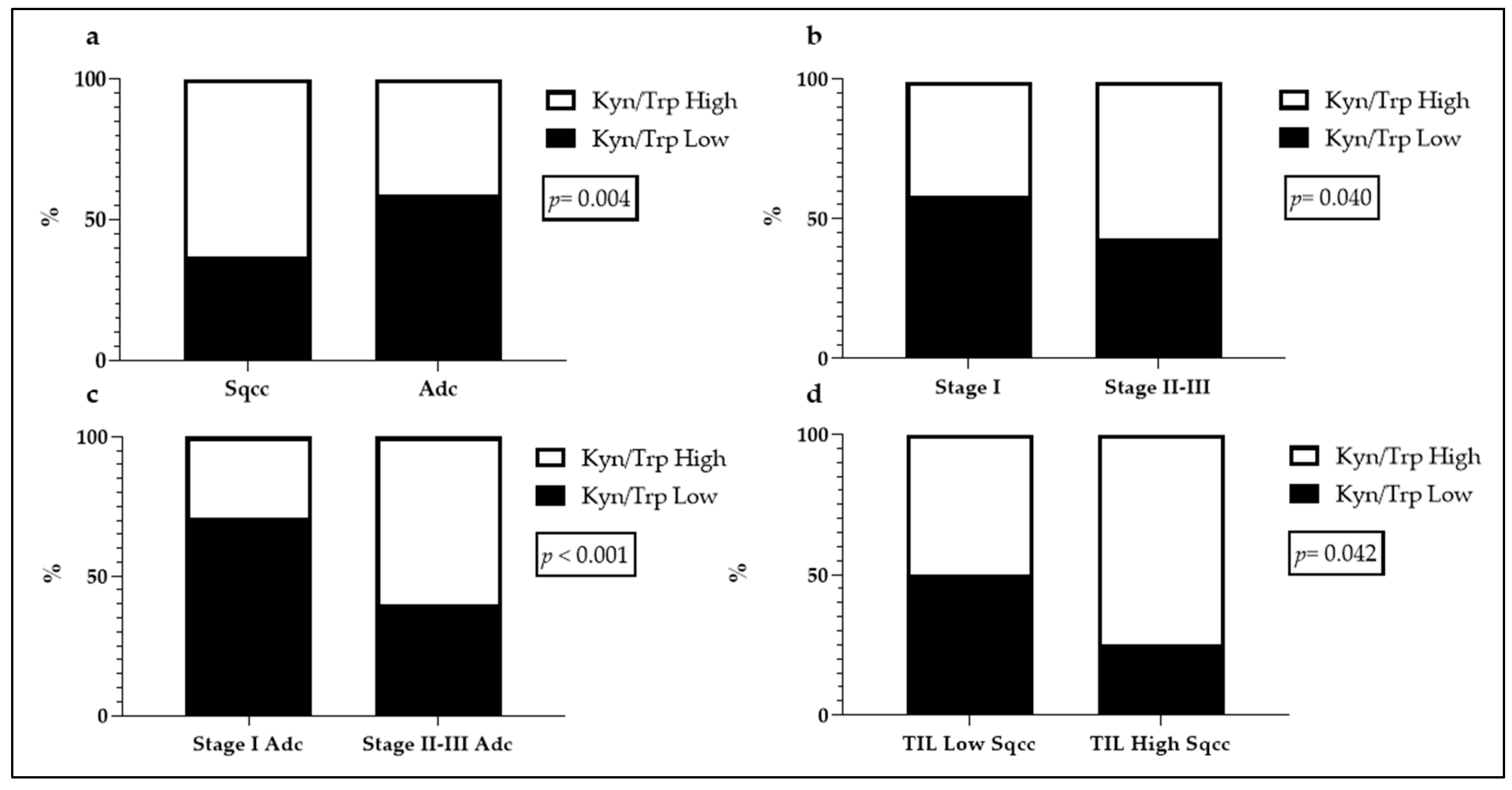
2.6. Kyn/Trp Ratio and Histopathological Findings Associations

Via HPLC, we analyzed Kyn and Trp serum concentration. We found values between 1.0240 and 30.3154 µM for Kyn (mean = 3.9772 ± 2.4368 µM; median = 3.6952 µM) and between 13.0968 and 592.2520 µM for Trp (mean = 65.0871 ± 43.4785 µM; median = 62.0500 µM).
We investigated possible correlations between Kyn/Trp ratio and the histopathological findings and summarized the analyzed associations in Table 2. A high Kyn/Trp ratio was found among squamous cell carcinoma histotype (p = 0.004) and, among this group, when the tumor presented a high TIL density (p = 0.042; Figure 4). Moreover, the higher the Kyn/Trp, the higher the stage of adenocarcinomas (p < 0.001; Figure 4).

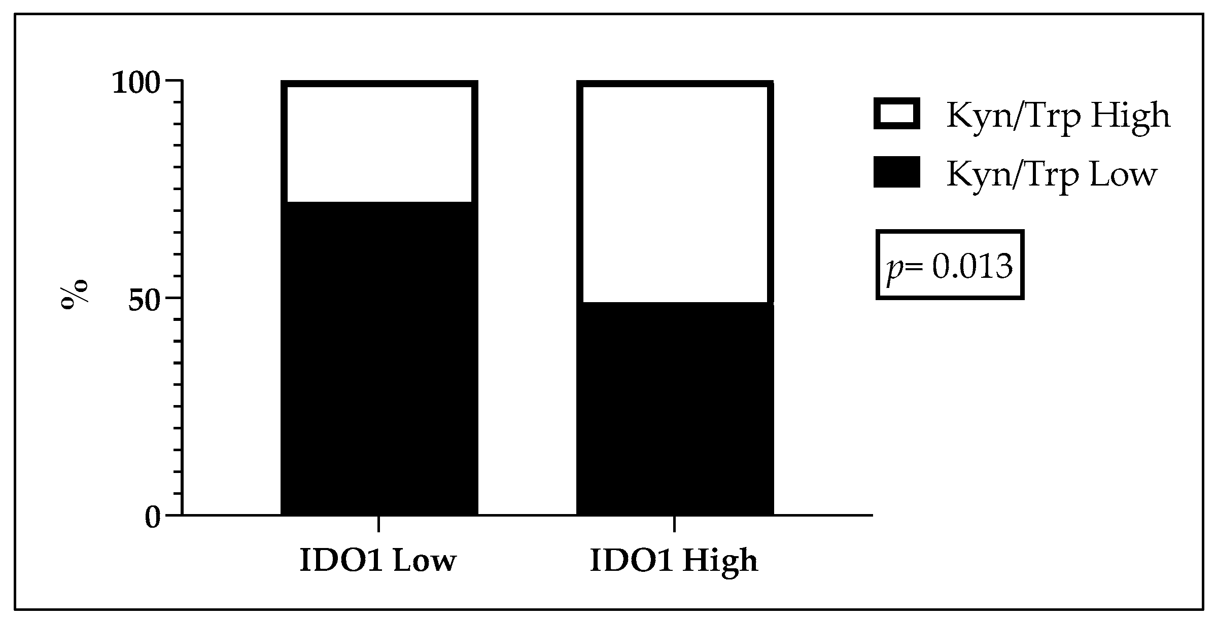
2.7. Kyn/Trp Ratio and Immunohistochemical Associations

The immunohistochemical analysis included the evaluation of either IDO1 or PD-L1 protein expressed by tumor cells in surgical specimens. For both IDO1 and PD-L1, low and high expression groups were identified according to a score assignment procedure, previously published [21] and here described in the Material and Methods. The only statistically significant association we found was between a low Kyn/Trp ratio and a low IDO1 expression in the adenocarcinoma group (p = 0.013; Table 3 and Figure 5).
We found no further statistically significant associations between Kyn/Trp ratio and the clinical–pathological parameters among those we explored (Table 1, Table 2 and Table 3).

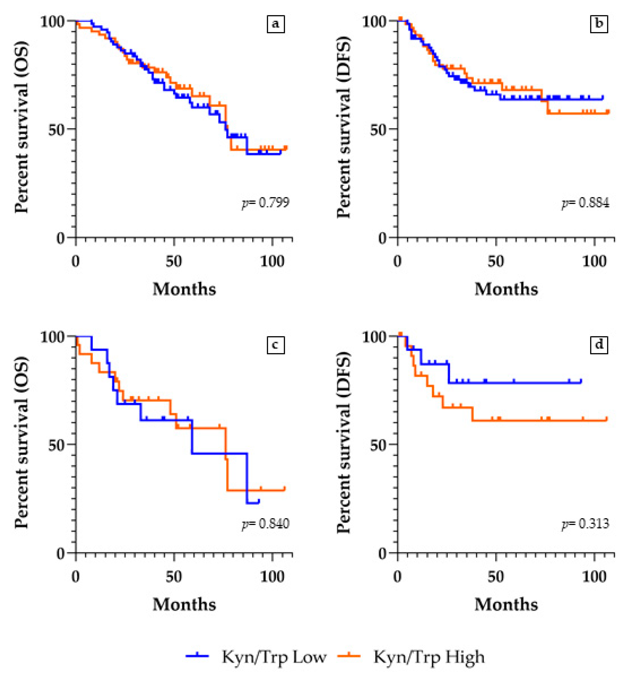
2.8. Kyn/Trp Ratio and Survivals Analyses

We conducted survival analyses in the whole cohort, in the subgroups of patients having adenocarcinoma or squamous cell carcinoma, and in the subgroups at stages II–III of disease. To investigate a possible prognostic value of the serum Kyn/Trp ratio, within each of the four groups, we compared OS and DFS curves of Kyn/Trp low vs. high groups. In the heterogeneous group of the whole NSCLC series, as in both the carcinoma histotype and advanced stage subgroups, OS analysis gave no statistically significant results, since the two groups of Kyn/Trp low and high were similarly distributed among the patients who died or survived during the follow-up period (Table 4). Similarly, among the two Kyn/Trp low and high groups, we did not find any statistically significant result by the DFS analysis on the patients who had a relapse and who did not have a recurrence from NSCLC (Table 4).
However, in a selected population of stage II–III patients who did not undergo adjuvant therapies, the DFS analysis showed a clear trend, even though not statistically significant (p = 0.313, HR 1.95, 95% CI 0.59–6.46), suggesting that the high Kyn/Trp ratio could correlate with a worse prognosis (Table 5 and Figure 6).

3. Discussion

Many studies demonstrate that solid tumors may escape immunosurveillance through upregulation of the IDO1 enzyme in cancer cells and tumor microenvironment, a condition associated with poor clinical outcomes [9,16]. IDO1 increased expression is reflected by the relative concentration of Kyn compared to Trp; hence, the Kyn/Trp ratio can be used as a prognostic clinicopathological marker to monitor cancer invasiveness and progression [22]. The involvement of IDO1 activity in cancer immunoediting and growth seems to be so relevant that targeting this enzyme represents an attractive approach for cancer immunotherapeutic intervention [23]. The inhibition of IDO1 catalytic activity is currently under investigation in several clinical trials and at least eight small molecule IDO1 inhibitors are evaluated as possible new anti-cancer drugs [24]. Lacking an assay allowing us to directly determine the activity of IDO1 enzyme in tumor tissues, most of the preclinical data provided by these IDO1-inhibition trials were derived from serum concentrations of Trp, Kyn, or the Kyn/Trp ratio, chosen as surrogate indicators for IDO1 activity evaluation [25,26,27,28]. Based on previous literature and to enlighten its effectiveness as a serum tumor biomarker, we looked for statistical associations between Kyn/Trp concentration ratio and several clinical, histopathological, and immunohistochemical parameters in a cohort of NSCLC patients who underwent surgical resection of tumors.
In our study, we assigned patients to the high or low Kyn/Trp group, depending on whether their Kyn/Trp ratio was above or below the median value of 58, respectively. Evaluating patients ranging between 38 and 84 years of age, with a median value of 68 years old, we found a positive and statistically significant correlation between the high Kyn/Trp ratio condition and the over-68-year-old patients’ age. The Kyn/Trp ratio, measured in blood, is documented to be robustly associated with aging in humans [29]. The more inflammation derived from age-related tissue damage increases, the more Kyn pathway, depleting tryptophan and producing tolerogenic metabolites, potentiated as a homeostatic response to control the so-called “inflammaging”. Thus, the Kyn/Trp ratio met the criteria for a biological age biomarker [30]. In our study, the presence of tumor tissue and relative inflamed tumor microenvironment could be a possible trigger for further increase of Trp catabolism, substantiating our finding of a higher Kyn/Trp ratio in older patients.
The smoking parameter appeared to be not related to Trp catabolism in our analysis, although a decreased serum activity of the IDO1 enzyme had been documented in a study on a representative cohort of smoking subjects not affected by cancer [31]. However, since we found that never-smoker status was statistically associated with the low Kyn/Trp ratio, Trp catabolism could be hypothesized as a cancerogenic mechanism in smoker NSCLC patients. To confirm this hypothesis, more data relative to the poorly represented population of never-smoker lung cancer patients are needed.
Another intriguing finding regards the association between the high Kyn/Trp ratio and the squamous cell histotype of NSCLC in our series. This could reflect the already established concept, according to which many suppressor gene alterations are specific features of squamous cell carcinoma, with consequent chromosomal instability and accumulation of somatic mutations. The latter increase the production of neoantigens, which can abruptly activate the innate immune response [32]. The resulting inflammatory microenvironment may ensue the overexpression of IDO1 [21], and, consequently, the activation of the kynurenine pathway, which leads to an increase of Kyn/Trp ratio in this kind of NSCLC. Moreover, recent studies have associated the role of IDO1 in developing immunotherapy resistance in squamous cell carcinomas, independently from the tumor site [20]. The typical hyperactivation of the IDO1 pathway in an inflammatory microenvironment, which is usually characterized by increased release of IFNγ [33], could also account for the statistically significant association between a high Kyn/Trp ratio and a high density of TILs at the tumor site among the squamous cell carcinoma group of the current study. In fact, as above mentioned, this tumor histotype usually elicits a higher immune stimulation because of its intrinsic higher antigenicity [32]. Furthermore, this finding seems partially to confirm data obtained from our recent study [21], which demonstrated that squamous cell lung cancers with a high TIL density presented a concomitant higher IDO1 immunohistochemical expression.
A recent study about cervical cancer found a correlation between the advanced stage of the disease and the higher Kyn/Trp ratio [34], and other authors described a relationship between an elevated Kyn/Trp ratio and known biologically aggressive neoplasms, such as glioblastoma [35]. These results, together with our statistically significant association between high Kyn/Trp ratio and more advanced NSCLC stages, among both the whole cohort of patients and the adenocarcinoma group, suggests the activation of the Kyn pathway in the tumor microenvironment as linked to poor clinicopathological parameters and, consequently, to more aggressive neoplasms. Furthermore, we confirmed the observation of Wang et al. [36], according to which a high level of IDO1 activity, indirectly measured as Kyn/Trp ratio, is typical of adenocarcinoma advanced stages rather than non-adenocarcinoma histotype of NSCLCs. This could lead to set up a multidisciplinary diagnostic workflow that would allow the early histopathological identification of NSCLC patients with adenocarcinoma, to specifically determine their serum Kyn/Trp ratio and identify the lung cancers which could benefit from therapies targeting the IDO1-kynurenine pathway axis. This potential additional therapeutic target would not only delay the tumor progression in the early stages of NSCLC, but also support other chemotherapeutic agents for the more advanced—and often resistant to currently available therapies—stages of this cancer type. Nevertheless, further studies, particularly including adequate patients’ follow-up periods and subsequent survival analysis, are needed to confirm our hypothesis.
In the series we analyzed in the present study, there was no statistically significant association between a high Kyn/Trp ratio and high immunohistochemical expression of IDO1, neither in the whole NSCLC population nor within the two histotype subgroups. To explain why a high Kyn/Trp ratio did not correlate with a high histochemical level of IDO1, we can speculate that not only tumor tissue but also other tumor microenvironment cell types [9] can importantly contribute to the tryptophan catabolism attempting to exhaust innate immune system activation in NSCLC. On the other hand, the finding of a low immunohistochemical expression of IDO1, statistically associated with a low Kyn/Trp ratio among NSCLC adenocarcinomas group, corroborated the need for preliminary histopathological screening to identify patients going to serum Kyn/Trp determination, to distinguish those that really could benefit from a blockade of Kyn pathway. As a matter of fact, we analyzed the immunohistochemical expression of IDO1 only at the tumor cell level, but the Kyn/Trp ratio could be the result of the catalytic enzyme overexpression in other cells of the tumor microenvironment, such as the dendritic cells [36]. Therefore, a better cellular stratification in NSCLCs microenvironment, particularly among adenocarcinomas, is needed in these patients.
Because of its relevance in triggering cancer-related tolerance in NSCLC and for its targetability in immunotherapy with immune-checkpoint inhibitors, which NSCLC is particularly responsive to [37], we chose to also investigate the immunohistochemical expression of the immune-checkpoint molecule PD-L1. The absence of statistically significant associations between a high or low Kyn/Trp ratio, and high or low immunohistochemical expression of PD-L1, in the whole NSCLC population, or within the two histotype subgroups, suggests that these two markers should be carefully considered in the stratification of patients to be treated with an immunotherapy combining the inhibition of IDO1 and the blockade of programmed cell death-1 (PD-1)/PD-L1 interaction. Such dual-agent therapy has been proposed for NSCLC [38,39] and is currently evaluated by oncological clinical trials [40] to maximize the clinical benefit of immune-checkpoint inhibitors, not effective on a significant fraction of NSCLC patients [37]. In evaluating the criteria for stratification of patients, not only the immunohistochemical expression of PD-L1 in tumor cells should be considered, but also the circulating component of soluble PD-L1, contributing to PD-1 receptor activation and, consequently, to the onset of cancer-permissive tolerance [41,42]. Thus, the absence of correlations between the serum Kyn/Trp ratio and PD-L1 immunohistochemical expression might suggest the importance of an integrated evaluation of histological (PD-L1 and IDO1) and serum (soluble PD-L1 and Kyn/Trp ratio) markers for specifically targeted NSCLC immunotherapy.
Though the Kyn/Trp ratio has been proposed as a useful prognostic clinicopathological marker [19,20,22], many aspects concerning its survival predictive value remain elusive. In our study, we verified that the Kyn/Trp ratio evaluated at the pre-surgical time in heterogeneous populations of NSCLC does not allow correlations between the Kyn/Trp serum parameter and patients’ OS or DFS to be defined. In such populations, different factors could have influenced the exitus occurrence, including the administration of adjuvant therapy, or the absence of it. Restricting the analysis to a subgroup of stage II–III NSCLC patients not treated with adjuvant therapy, a dichotomic trend can be enlightened, suggesting that a correlation between the high Kyn/Trp ratio and a poor prognosis could be established more successfully in the more advanced than in the early stages of the disease. This finding, obtained in our small subgroup of stage II–III NSCLC patients who did not undergo adjuvant therapy, needs to be statistically confirmed by further studies on a larger population. However, besides its possible use as a prognostic parameter in advanced-stage patients, we believe in the potentialities of the serum Kyn/Trp ratio as a non-invasive biomarker in the follow-up of NSCLC to monitor over time the level of IDO1 activity and its impact on immune responses, and to choose a new IDO1-based target therapy in addition to the currently available ones. Further investigations aimed to evaluate the modifications of the Kyn/Trp ratio in combination with other serum biomarkers would optimize the prognostic risk stratification of NSCLC patients.

4. Materials and Methods

4.1. Patients’ Recruitment

The current study was approved by the local ethics committee of Comitato Etico delle Aziende Sanitarie della Regione Umbria CEAS Umbria (15 November 2013), which assigned the protocol code N. 2216/13 to the present research. Patients were recruited from 2009 to 2015 at S. M. Misericordia Hospital, Perugia, Italy, selecting all the NSCLC cases, which underwent surgery with curative intent. Moreover, only the cases with both known clinical parameters were selected (Table 1).

4.2. Histopathological and Immunohistochemical Determinations

At the section of Anatomic Pathology and Histology, Department of Medicine and Surgery, University of Perugia, the surgical specimens were formalin-fixed (10% buffered formalin) and paraffin-embedded (FFPE). Sections of 4 μm were performed and placed on slides with a permanent positive charged surface for both H&E and immunohistochemical (IHC) stains. The H&E stain was carried out using a Leica ST5020 Multistainer (Leica Biosystems, Nußloch, Germany), using the kit ST Infinity H&E Staining System (Leica Biosystems, Richmond, IL, USA). The BOND-III fully automated immunohistochemistry stainer (LeicaBiosystems, Nußloch, Germany) performed the IHC stains (peroxidase immunoenzymatic reaction with development in diaminobenzidine). Proper positive and negative controls were included. A histological subtype was assigned according to 2015 World Health Organization (WHO) classification for lung tumors. Only tumors classified as adenocarcinomas or as squamous cell carcinomas were considered for the study.
The pathological stage was assigned according to the 8th Edition for Cancer Staging by the AJCC. According to this staging of disease, the NSCLCs were grouped into a stage I group, including stages from IA1 to IB, and a stage II–III group, encompassing the stages from IIA to IIIB.
The localization of TILs (absent; intratumoral = among tumor cells; peritumoral = at the interface between the neoplasia and healthy lung parenchyma; mixed = mixture of the last two localizations) and the density of TILs, according to the percentage of lymphocytes observed in a given localization (low < 20%; high ≥ 20%) were determined by experienced pathologists (G. Bellezza, M. Mandarano, and A. Sidoni) using H&E slides, as previously reported [21]. The same pathologists analyzed the immunohistochemical expression, on neoplastic cells, of IDO1 (courtesy of Professor Benoit J Van den Eynde, Ludwing Institute for Cancer Research, Louvain-la-Neuve, Belgium, clone 4.16H1 [43]; dilution 1:1000) and PD-L1 (Cell Signaling Technology, Danvers, MA, USA, Cat# 13684S, RRID:AB_2687655, dilution 1:200), as previously reported [21], obtaining an H score resulting from the sum of the intensity of the stain (evaluated as 0: Absent; 1+: Mild; 2+: Moderate; 3+: Intense) and the percentage of the tumor cells labeled (0:0%; 1: 1–25%; 2: 26–50%; 3: 51–75%; 4: 76–100%). Thereafter, two groups of staining were considered for the study: A low expression one—scores from 0 to 2—and a high expression one—scores from 3 to 7.

4.3. Determination of Kyn and Trp Serum Concentration

Blood samples were collected from patients before they underwent surgery and immediately centrifuged to obtain serum samples. After deproteinization, Kyn and Trp concentration was measured by HPLC. Briefly, a Perkin Elmer (series 200 HPLC) instrument and a Kinetex C18 column (250 × 4.6 mm, 5 μm, 100 Å, Phenomenex, Torrance, CA, USA) were used for the analysis. The column was maintained at a temperature of 25 °C and a pressure of 1800 PSI. Samples were eluted with a phase containing 10 mM NaH2PO4 (pH = 3.0; 99%) and methanol (1%) (Sigma–Aldrich, St. Louis, MO, USA), with a flow rate of 1.3 mL/min. Kyn was detected at 360 nm and Trp was detected at 220 nm by a UV detector. The software TURBOCHROM 4 was used for evaluating the concentration of Kyn and Trp in samples by mean of a calibration curve. Two groups of Kyn/Trp ratio were used for the subsequent evaluations of their associations with clinical–pathological parameters: The Kyn/Trp low, encompassing the value of the ratio ≤ 58, and the Kyn/Trp high, which included the value of ratio > 58. When the value of Kyn was undetectable, the corresponding patient was excluded from the analyses.

4.4. Statistical Analysis

The statistical analysis was performed using GraphPad Prism 8 (GraphPad Software Inc., San Diego, CA, USA). Categorical variables were presented as frequencies with row and column percentages. Categorical variables were compared between the groups using Chi-square test or Fisher’s exact test as appropriate. OS and DFS univariate analyses were performed for the subgroups determined by the Kyn/Trp ratio using a log-rank test and were represented by Kaplan–Meier curves. p-values (p) less than 0.05 were considered statistically significant.

5. Conclusions

In our study on resected NSCLC tumors, among other correlations, we found that the high Kyn/Trp ratio was significantly associated with the more aggressive squamous cell histotype and a more advanced tumor stage. These findings, confirming previous observations reported in several cancer types, importantly suggest the value of the non-invasive analysis of the serum Kyn/Trp ratio as a follow-up marker contributing to the indication of cancer progression. However, the serum determination of the Kyn/Trp ratio would be inadequate if used alone for the risk stratification of NSCLC patients and the identification of the best-personalized therapy. A more complex and complementary model of tumor biomarker analysis, both at histopathological and molecular/serum levels, should be considered for the best patients’ risk stratification and therapy.

Author Contributions

Conceptualization, G.B., M.L.B., J.V., V.L., R.C., F.P., A.S.; methodology, V.L.; serum sample collection and preparation S.B., F.R.T.; HPLC analysis, E.O.; investigation, R.C.; surgical resections J.V., F.P.; histological and immunohistochemical slides, E.L.; histopathological and immunohistochemical analyses, M.M., G.B., A.S.; data curation, M.M.; writing, M.L.B., M.M.; supervision, G.B., M.L.B.; funding acquisition, V.L. All authors have read and agreed to the published version of the manuscript.

Funding

This research was funded by Fondazione Cassa di Risparmio di Perugia, grant number 2315/16.

Institutional Review Board Statement

The study was conducted in accordance with the guidelines of the Declaration of Helsinki and approved by the Ethics Committee of Comitato Etico delle Aziende Sanitarie della Regione Umbria (CEAS Umbria) (protocol code N. 2216/13, date of approval: 15 November 2013).

Informed Consent Statement

According to the internal rules of both the University of Perugia and the Hospital of Perugia, we considered only the biological material which the patients expressed his/her specific informed consent to use, such as material for research purposes; we also observed all rules concerning confidentiality and protection of personal data, in accordance with European Union, International and National rules.

Data Availability Statement

The datasets analyzed in this article are not publicly available to respect the confidentiality and protection of patients’ data, in compliance with the processing of data covered and protected by the Italian Privacy Law and by the GDPR (General Data Protection Regulation, EU regulation no. 2016/679). However, requests for access to a properly anonymized dataset of the present article can be directed to M.M. ([email protected]).

Acknowledgments

We acknowledge Benoit J. Van den Eynde of Ludwig Institute for Cancer Research (de Duve Institute, Université catholique de Louvain, Louvain-la-Neuve, Belgium) for the kind donation of the anti-hIDO1 monoclonal antibody used in the immunohistochemical analysis of IDO1 expression.

Conflicts of Interest

The authors declare no conflict of interest. The funders had no role in the design of the study; in the collection, analyses, or interpretation of data; in the writing of the manuscript; or in the decision to publish the results.

References

  1. Barta, J.A.; Powell, C.A.; Wisnivesky, J.P. Global Epidemiology of Lung Cancer. Ann. Glob. Health 2019, 85. [Google Scholar] [CrossRef] [PubMed] [Green Version]
  2. Howlader, N.; Noone, A.M.; Krapcho, M.; Miller, D.; Brest, A.; Yu, M.; Ruhl, J.; Tatalovich, Z.; Mariotto, A.; Lewis, D.R.; et al. Cronin KA SEER Cancer Statistics Review, 1975–2017; National Cancer Institute: Bethesda, MD, USA, 2020.
  3. Lindeman, N.I.; Cagle, P.T.; Aisner, D.L.; Arcila, M.E.; Beasley, M.B.; Bernicker, E.H.; Colasacco, C.; Dacic, S.; Hirsch, F.R.; Kerr, K.; et al. Updated Molecular Testing Guideline for the Selection of Lung Cancer Patients for Treatment With Targeted Tyrosine Kinase Inhibitors: Guideline From the College of American Pathologists, the International Association for the Study of Lung Cancer, and the Association for Molecular Pathology. Arch. Pathol. Lab. Med. 2018, 142, 321–346. [Google Scholar]
  4. Planchard, D.; Popat, S.; Kerr, K.; Novello, S.; Smit, E.F.; Faivre-Finn, C.; Mok, T.S.; Reck, M.; Van Schil, P.E.; Hellmann, M.D.; et al. Metastatic non-small cell lung cancer: ESMO Clinical Practice Guidelines for diagnosis, treatment and follow-up. Ann. Oncol. 2018, 29, iv192–iv237. [Google Scholar] [CrossRef]
  5. Blackhall, F.; Frese, K.K.; Simpson, K.; Kilgour, E.; Brady, G.; Dive, C. Will liquid biopsies improve outcomes for patients with small-cell lung cancer? Lancet Oncol. 2018, 19, e470–e481. [Google Scholar] [CrossRef]
  6. Prendergast, G.C. Immune escape as a fundamental trait of cancer: Focus on IDO. Oncogene 2008, 27, 3889–3900. [Google Scholar] [CrossRef] [PubMed] [Green Version]
  7. Löb, S.; Königsrainer, A.; Rammensee, H.-G.; Opelz, G.; Terness, P. Inhibitors of indoleamine-2,3-dioxygenase for cancer therapy: Can we see the wood for the trees? Nat. Rev. Cancer 2009, 9, 445–452. [Google Scholar] [CrossRef]
  8. Munn, D.H.; Mellor, A.L. Indoleamine 2,3 dioxygenase and metabolic control of immune responses. Trends Immunol. 2013, 34, 137–143. [Google Scholar] [CrossRef] [Green Version]
  9. Meireson, A.; Devos, M.; Brochez, L. IDO Expression in Cancer: Different Compartment, Different Functionality? Front. Immunol. 2020, 11, 531491. [Google Scholar] [CrossRef]
  10. Huang, J.Y.; Larose, T.L.; Luu, H.N.; Wang, R.; Fanidi, A.; Alcala, K.; Stevens, V.L.; Weinstein, S.J.; Albanes, D.; Caporaso, N.E.; et al. Circulating markers of cellular immune activation in prediagnostic blood sample and lung cancer risk in the Lung Cancer Cohort Consortium (LC3). Int. J. Cancer 2020, 146, 2394–2405. [Google Scholar] [CrossRef]
  11. Beatty, G.L.; Gladney, W.L. Immune escape mechanisms as a guide for cancer immunotherapy. Clin. Cancer Res. 2015, 21, 687–692. [Google Scholar] [CrossRef] [PubMed] [Green Version]
  12. Timosenko, E.; Hadjinicolaou, A.V.; Cerundolo, V. Modulation of cancer-specific immune responses by amino acid degrading enzymes. Immunotherapy 2017, 9, 83–97. [Google Scholar] [CrossRef] [PubMed] [Green Version]
  13. Fallarino, F.; Grohmann, U.; Vacca, C.; Bianchi, R.; Orabona, C.; Spreca, A.; Fioretti, M.C.; Puccetti, P. T cell apoptosis by tryptophan catabolism. Cell Death Differ. 2002, 9, 1069–1077. [Google Scholar] [CrossRef] [PubMed]
  14. Zaher, S.S.; Germain, C.; Fu, H.; Larkin, D.F.P.; George, A.J.T. 3-Hydroxykynurenine Suppresses CD4+ T-Cell Proliferation, Induces T-Regulatory-Cell Development, and Prolongs Corneal Allograft Survival. Investig. Ophthalmol. Vis. Sci. 2011, 52, 2640–2648. [Google Scholar] [CrossRef] [PubMed] [Green Version]
  15. Hornyák, L.; Dobos, N.; Koncz, G.; Karányi, Z.; Páll, D.; Szabó, Z.; Halmos, G.; Székvölgyi, L. The Role of Indoleamine-2,3-Dioxygenase in Cancer Development, Diagnostics, and Therapy. Front. Immunol. 2018, 9, 151. [Google Scholar] [CrossRef]
  16. Yu, C.P.; Fu, S.F.; Chen, X.; Ye, J.; Ye, Y.; Kong, L.D.; Zhu, Z. The Clinicopathological and Prognostic Significance of IDO1 Expression in Human Solid Tumors: Evidence from a Systematic Review and Meta-Analysis. Cell Physiol. Biochem. 2018, 49, 134–143. [Google Scholar] [CrossRef]
  17. Lee, S.H.; Mahendran, R.; Tham, S.M.; Thamboo, T.P.; Chionh, B.J.; Lim, Y.X.; Tsang, W.C.; Wu, Q.H.; Chia, J.Y.; Tay, M.H.W.; et al. Tryptophan-kynurenine ratio as a biomarker of bladder cancer. BJU Int. 2021, 127, 445–453. [Google Scholar] [CrossRef]
  18. Li, H.; Bullock, K.; Gurjao, C.; Braun, D.; Shukla, S.A.; Bossé, D.; Lalani, A.A.; Gopal, S.; Jin, C.; Horak, C.; et al. Metabolomic adaptations and correlates of survival to immune checkpoint blockade. Nat. Commun. 2019, 10, 4346. [Google Scholar] [CrossRef] [PubMed] [Green Version]
  19. Zhai, L.; Dey, M.; Lauing, K.L.; Gritsina, G.; Kaur, R.; Lukas, R.V.; Nicholas, M.K.; Rademaker, A.W.; Dostal, C.R.; McCusker, R.H.; et al. The kynurenine to tryptophan ratio as a prognostic tool for glioblastoma patients enrolling in immunotherapy. J. Clin. Neurosci. 2015, 22, 1964–1968. [Google Scholar] [CrossRef] [PubMed] [Green Version]
  20. Botticelli, A.; Mezi, S.; Pomati, G.; Cerbelli, B.; Cerbelli, E.; Roberto, M.; Giusti, R.; Cortellini, A.; Lionetto, L.; Scagnoli, S.; et al. Tryptophan Catabolism as Immune Mechanism of Primary Resistance to Anti-PD-1. Front. Immunol. 2020, 11, 1243. [Google Scholar] [CrossRef] [PubMed]
  21. Mandarano, M.; Bellezza, G.; Belladonna, M.L.; Van den Eynde, B.J.; Chiari, R.; Vannucci, J.; Mondanelli, G.; Ludovini, V.; Ferri, I.; Bianconi, F.; et al. Assessment of TILs, IDO-1, and PD-L1 in resected non-small cell lung cancer: An immunohistochemical study with clinicopathological and prognostic implications. Virchows Arch. 2019, 474, 159–168. [Google Scholar] [CrossRef]
  22. Ala, M. The footprint of kynurenine pathway in every cancer: A new target for chemotherapy. Eur. J. Pharmacol. 2021, 896, 173921. [Google Scholar] [CrossRef] [PubMed]
  23. Brochez, L.; Chevolet, I.; Kruse, V. The rationale of indoleamine 2,3-dioxygenase inhibition for cancer therapy. Eur. J. Cancer 2017, 76, 167–182. [Google Scholar] [CrossRef] [PubMed]
  24. Yao, Y.; Liang, H.; Fang, X.; Zhang, S.; Xing, Z.; Shi, L.; Kuang, C.; Seliger, B.; Yang, Q. What is the prospect of indoleamine 2,3-dioxygenase 1 inhibition in cancer? Extrapolation from the past. J. Exp. Clin. Cancer Res. 2021, 40, 60. [Google Scholar] [CrossRef] [PubMed]
  25. Beatty, G.L.; O’Dwyer, P.J.; Clark, J.; Shi, J.G.; Bowman, K.J.; Scherle, P.A.; Newton, R.C.; Schaub, R.; Maleski, J.; Leopold, L.; et al. First-in-Human Phase I Study of the Oral Inhibitor of Indoleamine 2,3-Dioxygenase-1 Epacadostat (INCB024360) in Patients with Advanced Solid Malignancies. Clin. Cancer Res. 2017, 23, 3269–3276. [Google Scholar] [CrossRef] [PubMed] [Green Version]
  26. Suzuki, Y.; Suda, T.; Furuhashi, K.; Suzuki, M.; Fujie, M.; Hahimoto, D.; Nakamura, Y.; Inui, N.; Nakamura, H.; Chida, K. Increased serum kynurenine/tryptophan ratio correlates with disease progression in lung cancer. Lung Cancer 2010, 67, 361–365. [Google Scholar] [CrossRef]
  27. Weinlich, G.; Murr, C.; Richardsen, L.; Winkler, C.; Fuchs, D. Decreased serum tryptophan concentration predicts poor prognosis in malignant melanoma patients. Dermatology 2007, 214, 8–14. [Google Scholar] [CrossRef]
  28. de Jong, R.A.; Nijman, H.W.; Boezen, H.M.; Volmer, M.; Ten Hoor, K.A.; Krijnen, J.; van der Zee, A.G.; Hollema, H.; Kema, I.P. Serum tryptophan and kynurenine concentrations as parameters for indoleamine 2,3-dioxygenase activity in patients with endometrial, ovarian, and vulvar cancer. Int. J. Gynecol. Cancer 2011, 21, 1320–1327. [Google Scholar] [CrossRef]
  29. Sorgdrager, F.J.H.; Naudé, P.J.W.; Kema, I.P.; Nollen, E.A.; Deyn, P.P. Tryptophan Metabolism in Inflammaging: From Biomarker to Therapeutic Target. Front. Immunol. 2019, 10, 2565. [Google Scholar] [CrossRef]
  30. Bürkle, A.; Moreno-Villanueva, M.; Bernhard, J.; Blasco, M.; Zondag, G.; Hoeijmakers, J.H.; Toussaint, O.; Grubeck-Loebenstein, B.; Mocchegiani, E.; Collino, S.; et al. MARK-AGE biomarkers of ageing. Mech. Ageing Dev. 2015, 151, 2–12. [Google Scholar] [CrossRef]
  31. Pertovaara, M.; Heliövaara, M.; Raitala, A.; Oja, S.S.; Knekt, P.; Hurme, M. The activity of the immunoregulatory enzyme indoleamine 2,3-dioxygenase is decreased in smokers. Clin. Exp. Immunol. 2006, 145, 469–473. [Google Scholar] [CrossRef] [PubMed]
  32. Osoegawa, A.; Hiraishi, H.; Hashimoto, T.; Takumi, Y.; Abe, M.; Takeuchi, H.; Miyawaki, M.; Okamoto, T.; Sugio, K. The Positive Relationship Between γH2AX and PD-L1 Expression in Lung Squamous Cell Carcinoma. In Vivo 2018, 32, 171–177. [Google Scholar]
  33. Platten, M.; Wick, W.; Van den Eynde, B.J. Tryptophan catabolism in cancer: Beyond IDO and tryptophan depletion. Cancer Res. 2012, 72, 5435–5440. [Google Scholar] [CrossRef] [PubMed] [Green Version]
  34. Ferns, D.M.; Kema, I.P.; Buist, M.R.; Nijman, H.W.; Kenter, G.G.; Jordanova, E.S. Indoleamine-2,3-dioxygenase (IDO) metabolic activity is detrimental for cervical cancer patient survival. Oncoimmunology 2015, 4, e981457. [Google Scholar] [CrossRef]
  35. Lenzen, A.; Zhai, L.; Lauing, K.L.; Gritsina, G.; Ladomersky, E.; Genet, M.; James, C.D.; Bloch, O.; Wainwright, D.A. The Kynurenine/Tryptophan Ratio and Glioblastoma Patients Treated with Hsppc-96 Vaccine. Immunotherapy 2016, 2, 1–3. [Google Scholar] [CrossRef] [Green Version]
  36. Wang, Y.; Hu, G.F.; Wang, Z.H. The status of immunosuppression in patients with stage IIIB or IV non-small-cell lung cancer correlates with the clinical characteristics and response to chemotherapy. OncoTargets Ther. 2017, 10, 3557–3566. [Google Scholar] [CrossRef] [Green Version]
  37. Pradhan, M.; Chocry, M.; Gibbons, D.L.; Sepesi, B.; Cascone, T. Emerging biomarkers for neoadjuvant immune checkpoint inhibitors in operable non-small cell lung cancer. Transl. Lung Cancer Res. 2021, 10, 590–606. [Google Scholar] [CrossRef]
  38. Rocco, D.; Gravara, L.D.; Gridelli, C. The New Immunotherapy Combinations in the Treatment of Advanced Non-Small Cell Lung Cancer: Reality and Perspectives. Curr. Clin. Pharmacol. 2020, 15, 11–19. [Google Scholar] [CrossRef]
  39. Hellmann, M.D.; Gettinger, S.; Chow, L.Q.M.; Gordon, M.; Awad, M.M.; Cha, E.; Gong, X.; Zhou, G.; Walker, C.; Leopold, L.; et al. Phase 1 study of epacadostat in combination with atezolizumab for patients with previously treated advanced nonsmall cell lung cancer. Int. J. Cancer 2020, 147, 1963–1969. [Google Scholar] [CrossRef] [PubMed]
  40. Jung, K.H.; LoRusso, P.; Burris, H.; Gordon, M.; Bang, Y.J.; Hellmann, M.D.; Cervantes, A.; de Olza, M.O.; Marabelle, A.; Hodi, F.S.; et al. Phase I Study of the Indoleamine 2,3-Dioxygenase 1 (IDO1) Inhibitor Navoximod (GDC-0919) Administered with PD-L1 Inhibitor (Atezolizumab) in Advanced Solid Tumors. Clin. Cancer Res. 2019, 25, 3220–3228. [Google Scholar] [CrossRef] [Green Version]
  41. Murakami, S.; Shibaki, R.; Matsumoto, Y.; Yoshida, T.; Goto, Y.; Kanda, S.; Horinouchi, H.; Fujiwara, Y.; Yamamoto, N.; Ohe, Y. Association between serum level soluble programmed cell death ligand 1 and prognosis in patients with non-small cell lung cancer treated with anti-PD-1 antibody. Thorac. Cancer 2020, 11, 3585–3595. [Google Scholar] [CrossRef] [PubMed]
  42. Mildner, F.; Sopper, S.; Amann, A.; Pircher, A.; Pall, G.; Köck, S.; Naismith, E.; Wolf, D.; Gamerith, G. Systematic review: Soluble immunological biomarkers in advanced non-small-cell lung cancer (NSCLC). Crit. Rev. Oncol. Hematol. 2020, 153, 102948. [Google Scholar] [CrossRef] [PubMed]
  43. Théate, I.; van Baren, N.; Pilotte, L.; Moulin, P.; Larrieu, P.; Renauld, J.C.; Hervé, C.; Gutierrez-Roelens, I.; Marbaix, E.; Sempoux, C.; et al. Extensive profiling of the expression of the indoleamine 2,3-dioxygenase 1 protein in normal and tumoral human tissues. Cancer Immunol. Res. 2015, 3, 161–172. [Google Scholar] [CrossRef] [PubMed] [Green Version]
Figure 1. Histopathological analysis by hematoxylin and eosin (H&E) staining. (a) Adenocarcinoma of the lung, with a high intratumoral density of tumor-infiltrating lymphocytes (TILs), magnification: 100×; (b) squamous cell carcinoma of the lung, with a high mixed density of TILs, magnification: 100×.
Ijms 22 04403 g001
Figure 2. Immunohistochemical analysis of indoleamine-2,3-dioxygenase 1 (IDO1) and programmed cell death ligand-1 (PD-L1) expression. (a) Immunohistochemical high expression of IDO1 in adenocarcinoma; (b) immunohistochemical low expression of PD-L1 in squamous cell carcinoma. Original magnification: (a) 200×, (b) 400×.
Ijms 22 04403 g002
Figure 3. Statistically significant associations between kynurenine/tryptophan (Kyn/Trp) ratio and clinical parameters. (a) Association between Kyn/Trp ratio and patients’ age; (b) association between Kyn/Trp ratio and smoking status.
Ijms 22 04403 g003
Figure 4. Statistically significant associations between Kyn/Trp ratio and histopathological findings. (a) Association between Kyn/Trp and histotype; (b) association between Kyn/Trp and stage of disease; (c) association between Kyn/Trp and stage of disease, adenocarcinomas (Adc) group; (d) association between Kyn/Trp and TILs density, squamous cell carcinomas (Sqcc) group.
Ijms 22 04403 g004
Figure 5. Statistically significant association between low Kyn/Trp ratio and low immunohistochemical expression of IDO1, adenocarcinomas group.
Ijms 22 04403 g005
Figure 6. Overall survival (OS) and disease-free survival (DFS) curves according to the Kyn/Trp ratio. (a) OS and (b) DFS analysis of patients who did not undergo adjuvant therapies; (c) OS and (d) DFS analysis of patients in stages II–III of disease who did not undergo adjuvant therapies.
Ijms 22 04403 g006
Table 1. Serum Kyn/Trp ratio and clinical parameters associations.
ParameterKyn/Trp LowKyn/Trp Highp ValueTotal
N%N% N%
93528748 180100
Gender
Male624965510.23712771
Female315822425329
Age
<68 years54662834<0.0018246
≥68 years394059609854
Smokers
Current385737490.0427542
Former424747538949
Never1381319169
Table 2. Serum Kyn/Trp ratio and histopathological findings associations.
ParameterKyn/Trp LowKyn/Trp Highp ValueTotal
N%N% N%
93528748 180100
Histotype
Adc 1705948410.00411866
Sqcc 2233739636234
Stage
I585941410.0409955
II–III354346578145
Adc 1 stage 118100
I52712129<0.0017362
II–III184027604538
Sqcc 2 stage 62100
I62320770.0662642
II–III174719533658
TILs Density
Low505640440.2969050
High434847529050
Adc 1 118100
Low355825420.8246051
High356023405849
Sqcc 2 62100
Low155015500.0423048
High82524753252
TILs Localization
Intratumoral455458460.4188346
Peritumoral431969137
Mixed425337477944
Absent24036053
Adc 1 118100
Intratumoral375926410.4466353
Peritumoral35035065
Mixed296416364538
Absent12537544
Sqcc 2 62100
Intratumoral84012600.3412032
Peritumoral114686711
Mixed183821623455
Absent11000012
1 Adc: Adenocarcinoma. 2 Sqcc: Squamous cell carcinoma.
Table 3. Serum Kyn/Trp ratio and immunohistochemical findings associations.
ParameterKyn/Trp LowKyn/Trp Highp ValueTotal
N%N% N%
93528748 180100
IDO1
Low475932410.0637944
High4646555410156
Adc 1 118100
Low387215280.0135345
High324933516555
Sqcc 2 62100
Low93517650.7312642
High143922613658
PD-L1
Low735366470.67413977
High204921514123
Adc 1 118100
Low606237380.2299782
High104811522118
Sqcc 2 62100
Low133129690.1474268
High105010502032
1 Adc: Adenocarcinoma. 2 Sqcc: Squamous cell carcinoma.
Table 4. Survival analyses according to serum Kyn/Trp ratio, whole cohort of NSCLC patients.
Kyn/TrpExitus
(%)
p ValueHR 1
(95% CI 2)
Relapse
(%)
p ValueHR 1
(95% CI 2)
YesNo YesNo
NSCLCs
Low39(51)54(52)0.9411.02
(0.65–1.60)
36(52)57(51)0.9670.99
(0.62–1.59)
High37(49)50(48)33(48)5(49)
Adenocarcinomas
Low30(60)40(59)0.5951.16
(0.65–2.07)
31(58)39(60)0.7011.11
(0.64–1.93)
High20(40)28(41)22(42)26(40)
Squamous cell carcinomas
Low9(35)14(39)0.8370.92
(0.41–2.09)
5(31)18(39)0.7591.18
(0.42–3.29)
High17(65)22(61)11(69)28(61)
Stage II–III
Low16(41)19(45)0.8731.05
(0.56–1.99)
15(43)20(43)0.8871.05
(0.54–2.05)
High23(59)23(55)20(57)26(57)
1 HR: Hazard ratio; 2 CI: Confidence interval.
Table 5. Survival analyses according to serum Kyn/Trp ratio, subgroups of patients who did not undergo adjuvant therapy.
Kyn/TrpExitus (%)p ValueHR 1
(95% CI 2)
Relapse (%)p ValueHR 1
(95% CI 2)
YesNo YesNo
NSCLCs
Low31(57)42(52)0.7990.93
(0.55–1.60)
24(56)49(53)0.8840.96
(0.52–1.74)
High23(43)39(48)19(44)43(47)
Adenocarcinomas
Low25(69)35(60)0.9050.96
(0.47–1.94)
23(64)37(64)0.8591.06
(0.53–2.11)
High11(31)23(40)13(36)21(36)
Squamous cell carcinomas
Low6(33)7(30)0.6570.80
(0.29–2.23)
1(14)12(35)0.3982.41
(0.47–12.39)
High12(66)16(70)6(86)22(65)
Stage II–III
Low8(43)8(38)0.8400.91
(0.36–2.28)
3(27)13(45)0.3131.95
(0.59–6.46)
High11(58)13(62)8(73)16(55)
1 HR: Hazard ratio; 2 CI: Confidence interval.
Publisher’s Note: MDPI stays neutral with regard to jurisdictional claims in published maps and institutional affiliations.

