
Poly(ethylene-Co-vinyl Alcohol)/Titanium Dioxide Nanocomposite: Preparation and Characterization of Properties for Potential Use in Bone Tissue Engineering

 ,
,  ,
,  , and
, and
Abstract
:1. Introduction
2. Material and Methods
2.1. Chemicals
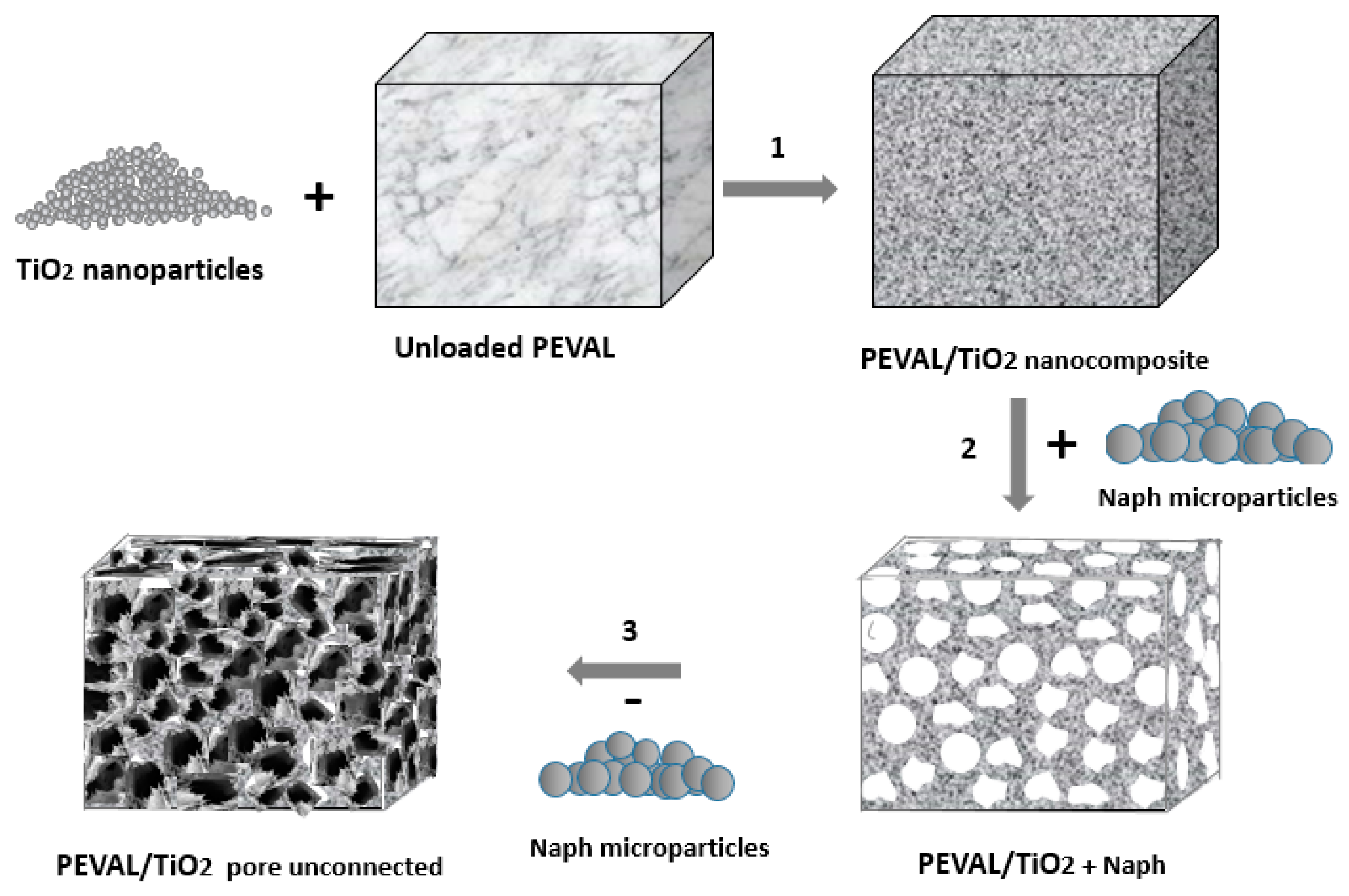
2.2. Preparation of the PEVAL/TiO2 Nanocomposite by the Solvent Casting Method
2.3. Preparation of PEVAL/TiO2 with Interconnected Pores
2.4. Characterization
2.4.1. FTIR Analysis
2.4.2. DSC Analysis
2.4.3. Thermogravimetric Analysis
2.4.4. XRD Analysis
2.4.5. SEM Analysis
2.4.6. Porosity and Pore Size Distribution
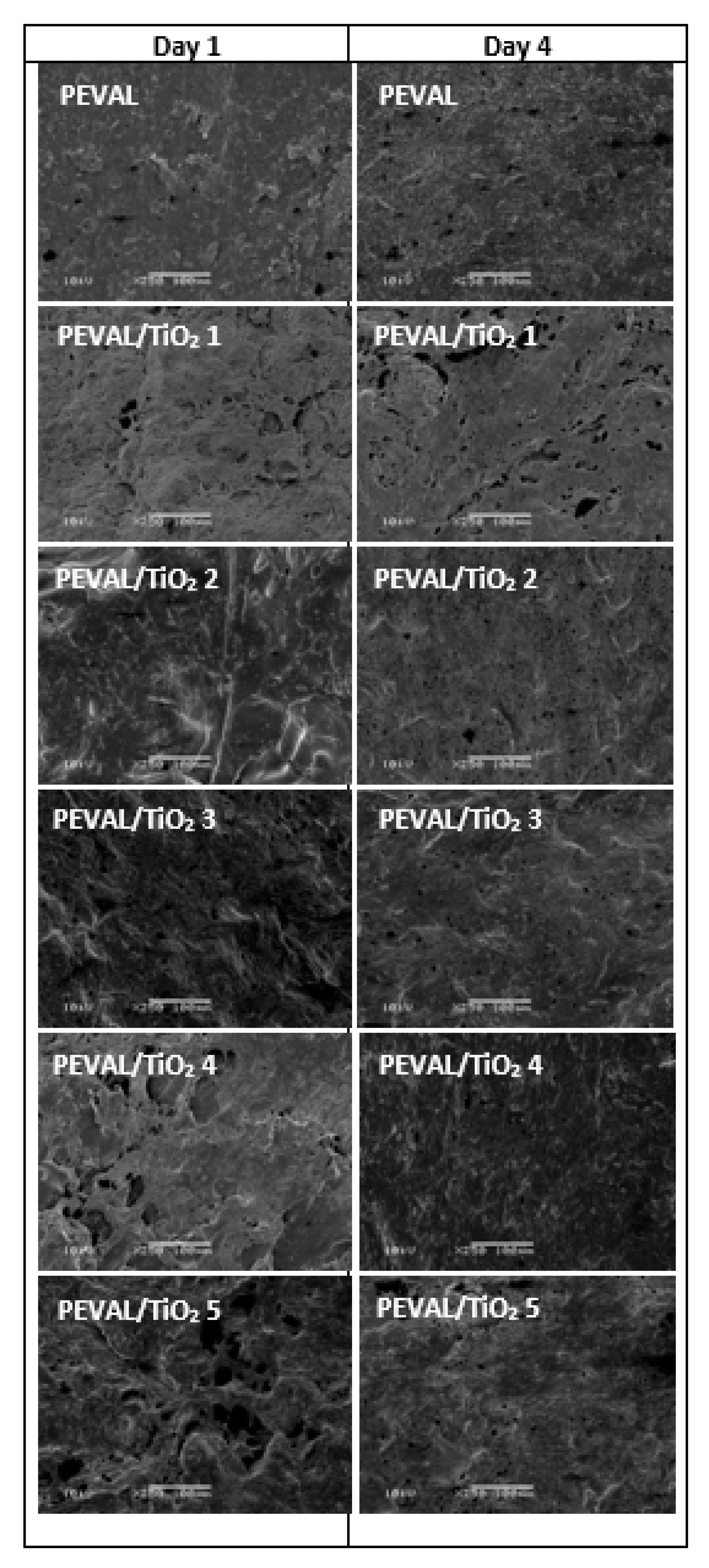
2.4.7. SEM of Seeded Scaffolds
2.4.8. Cellular Activity
3. Results and Discussion
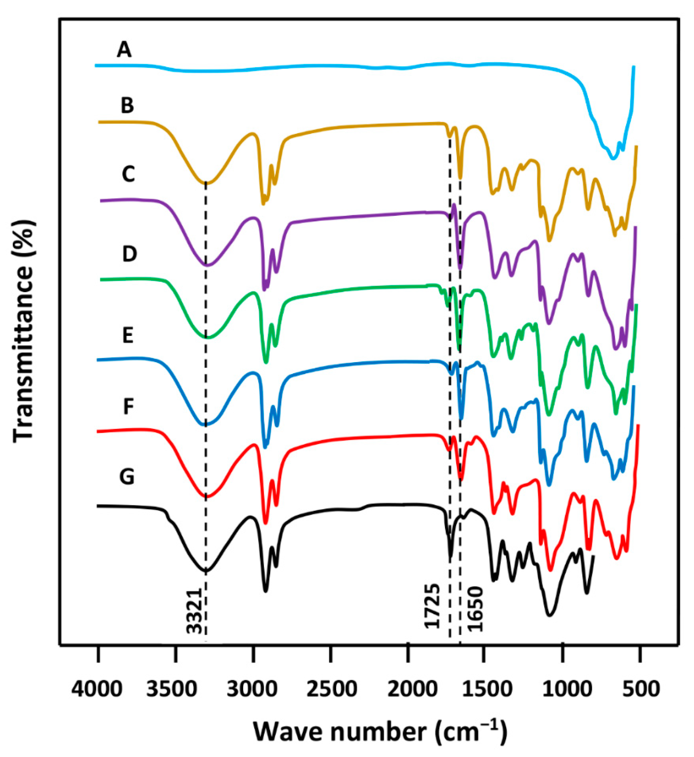
3.1. FTIR Analysis
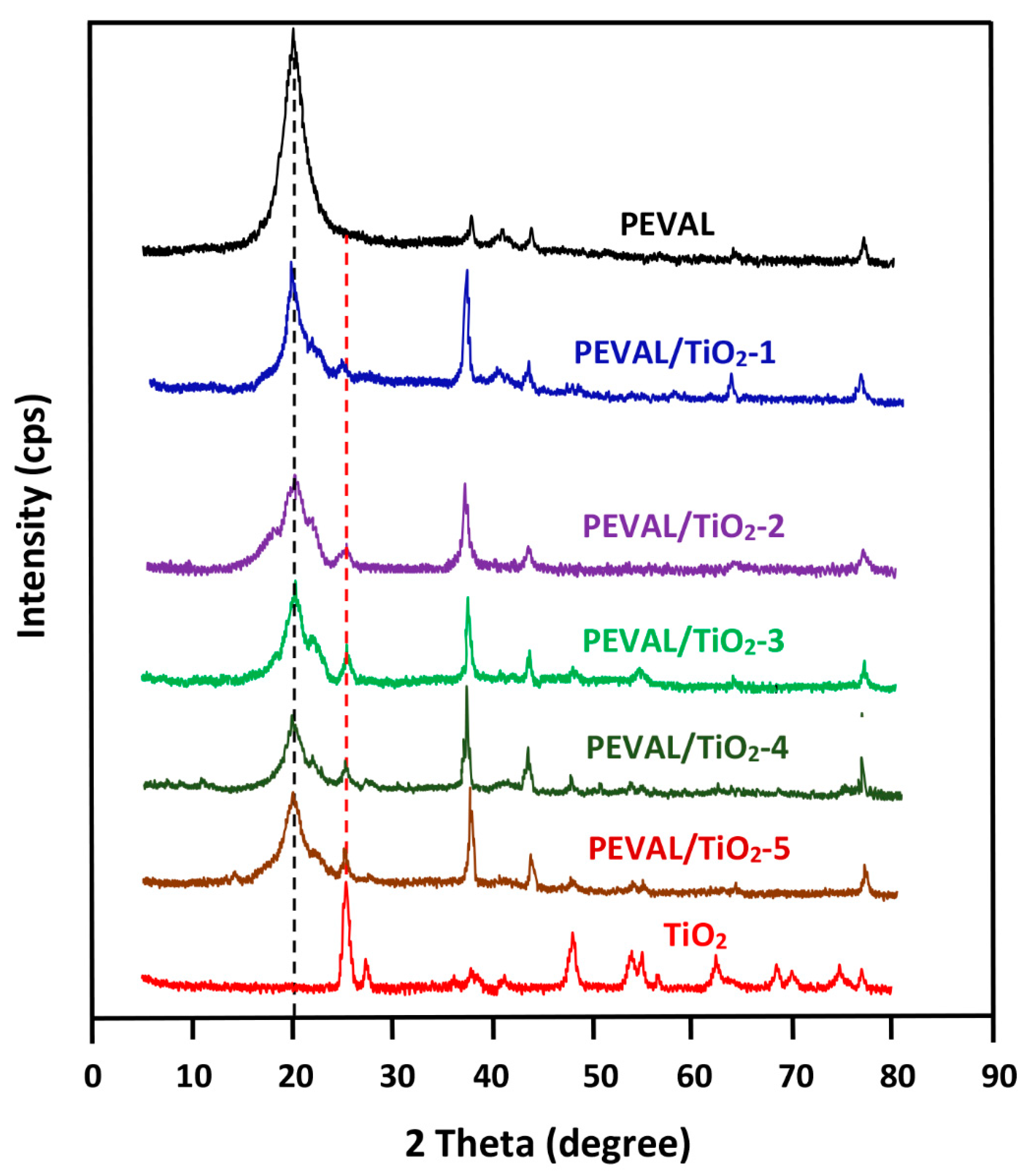
3.2. XRD Analysis
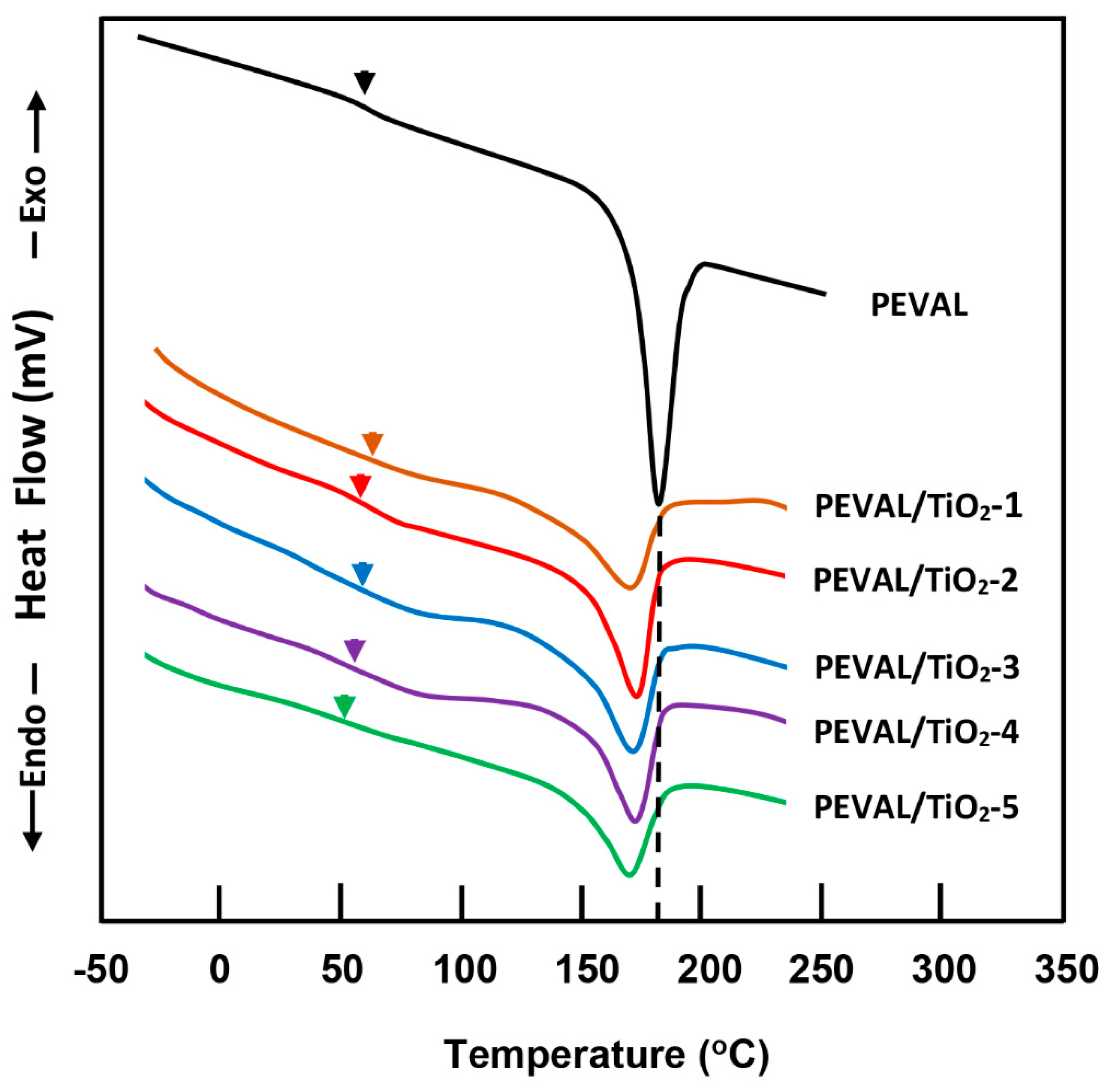
3.3. DSC Analysis
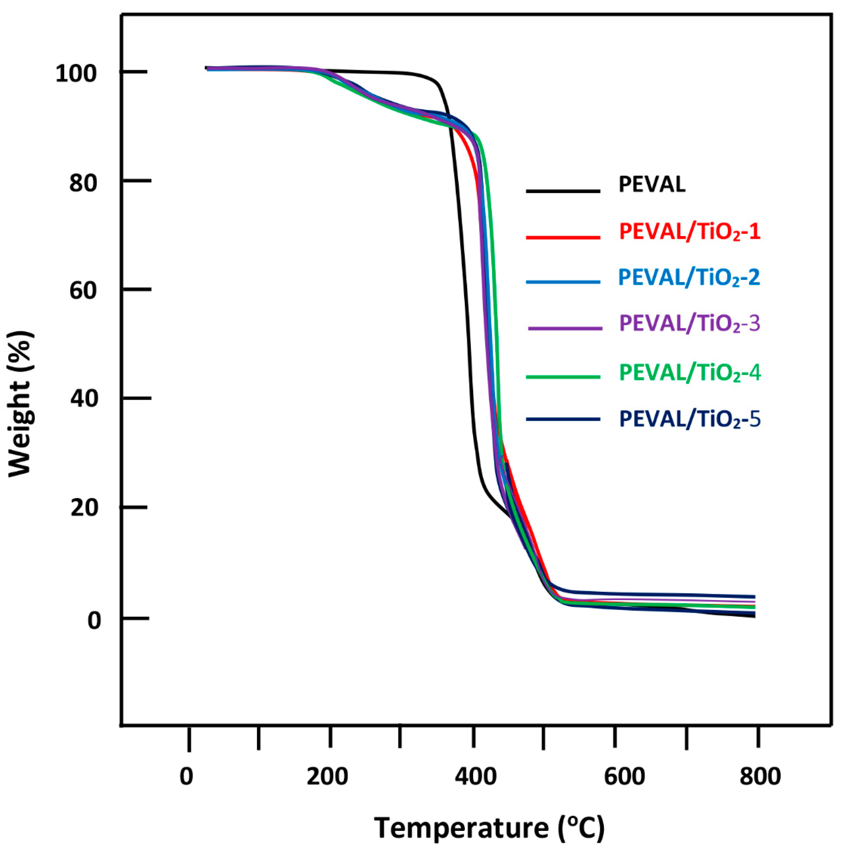
3.4. TGA
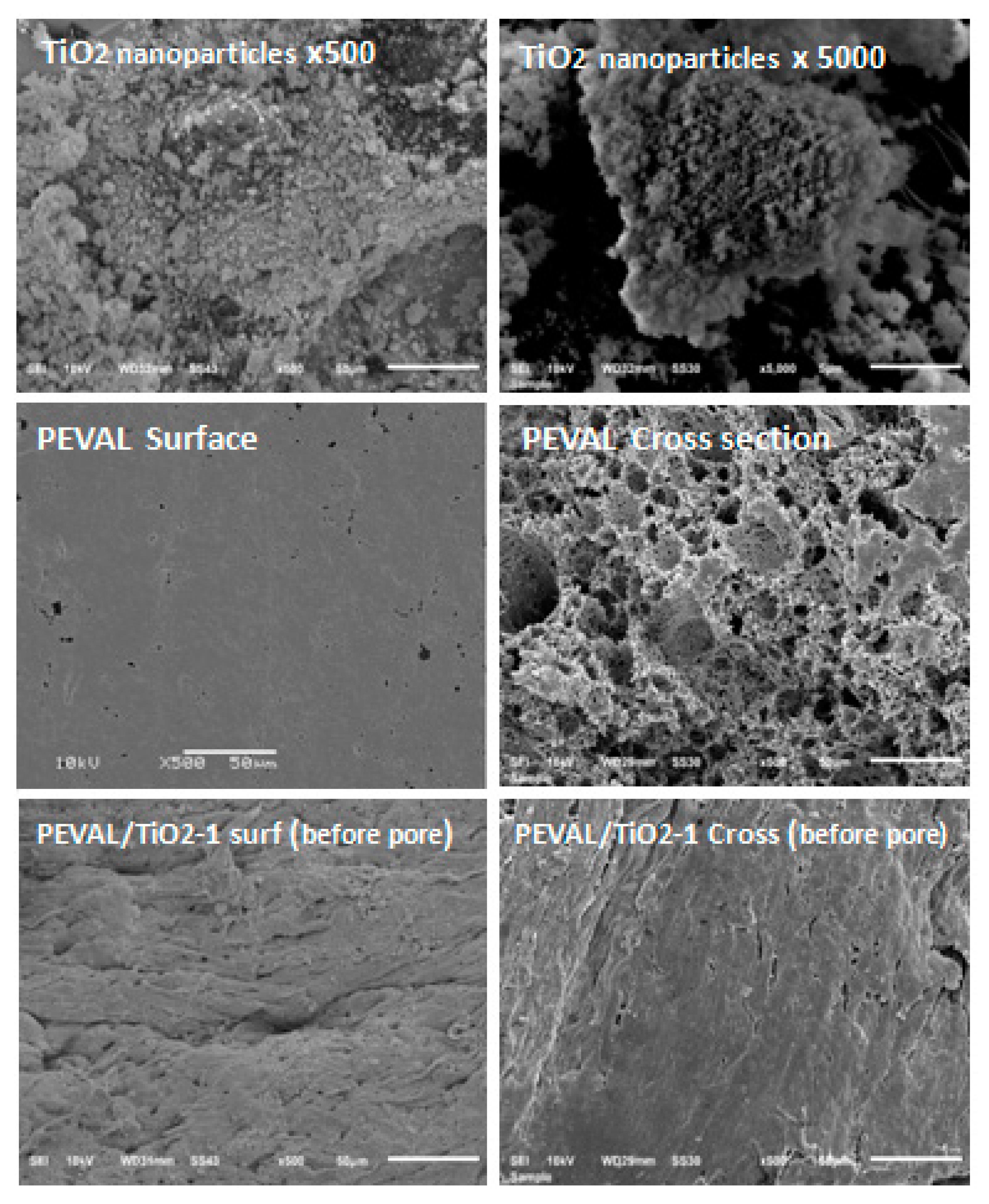
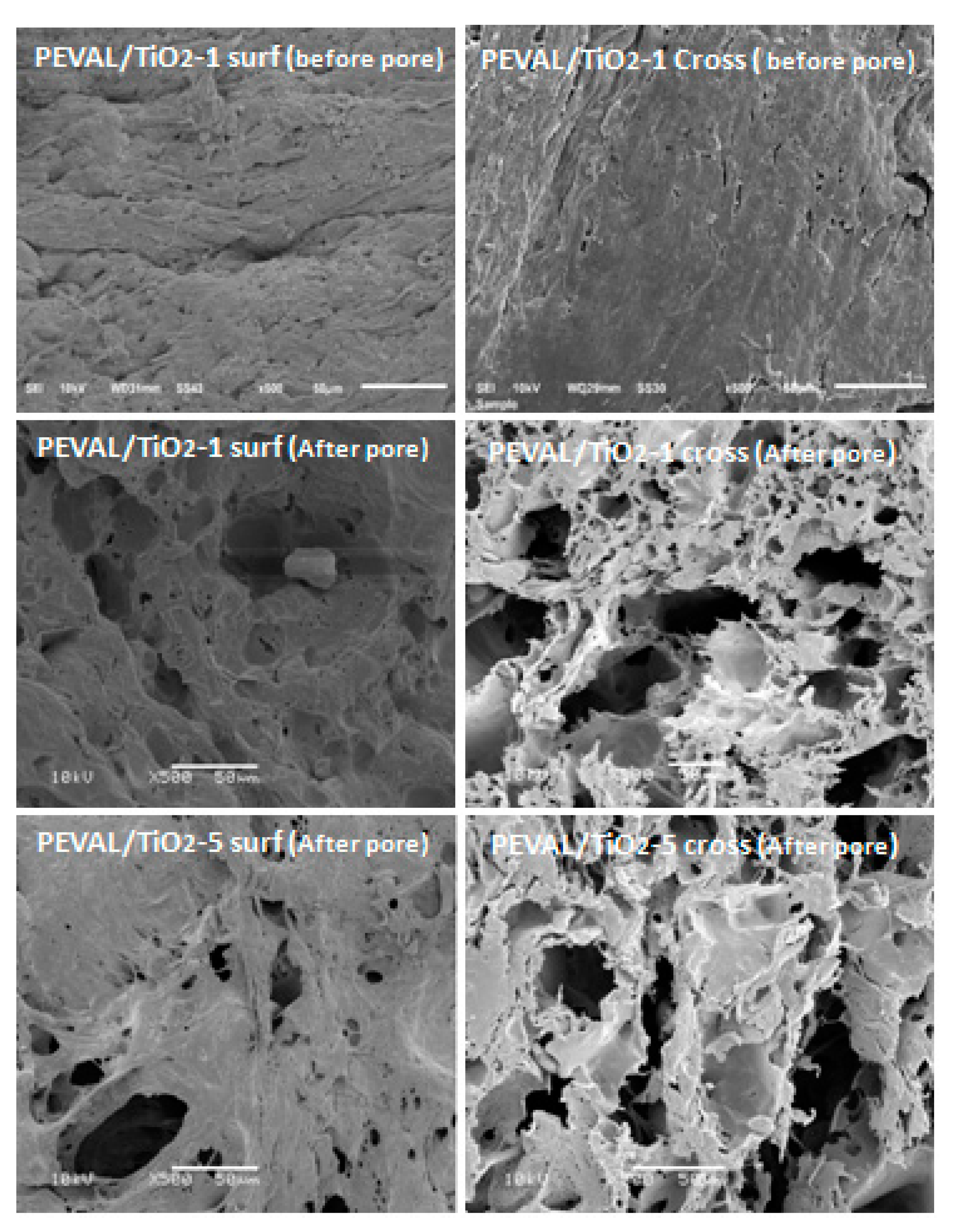
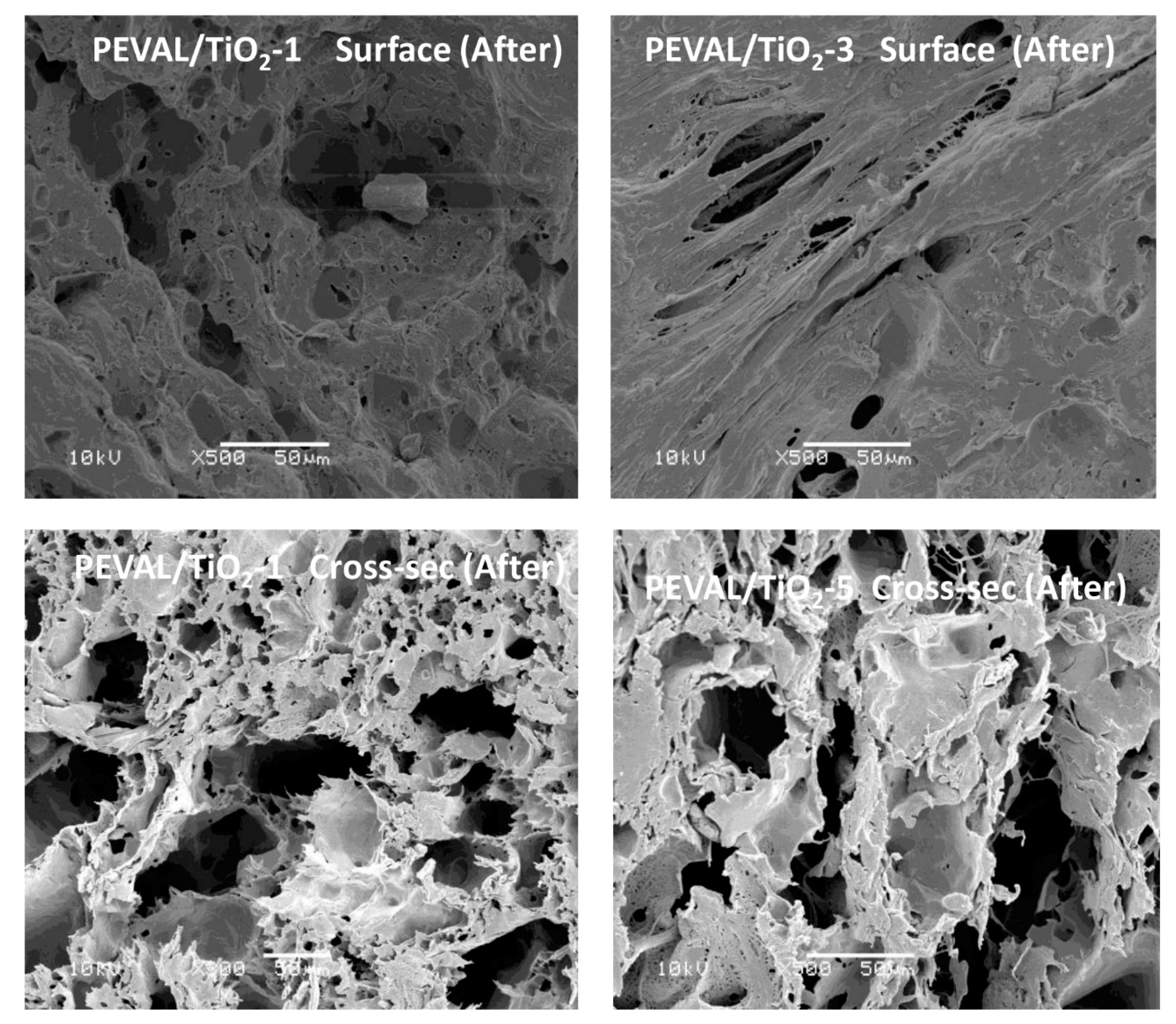
3.5. SEM Analysis
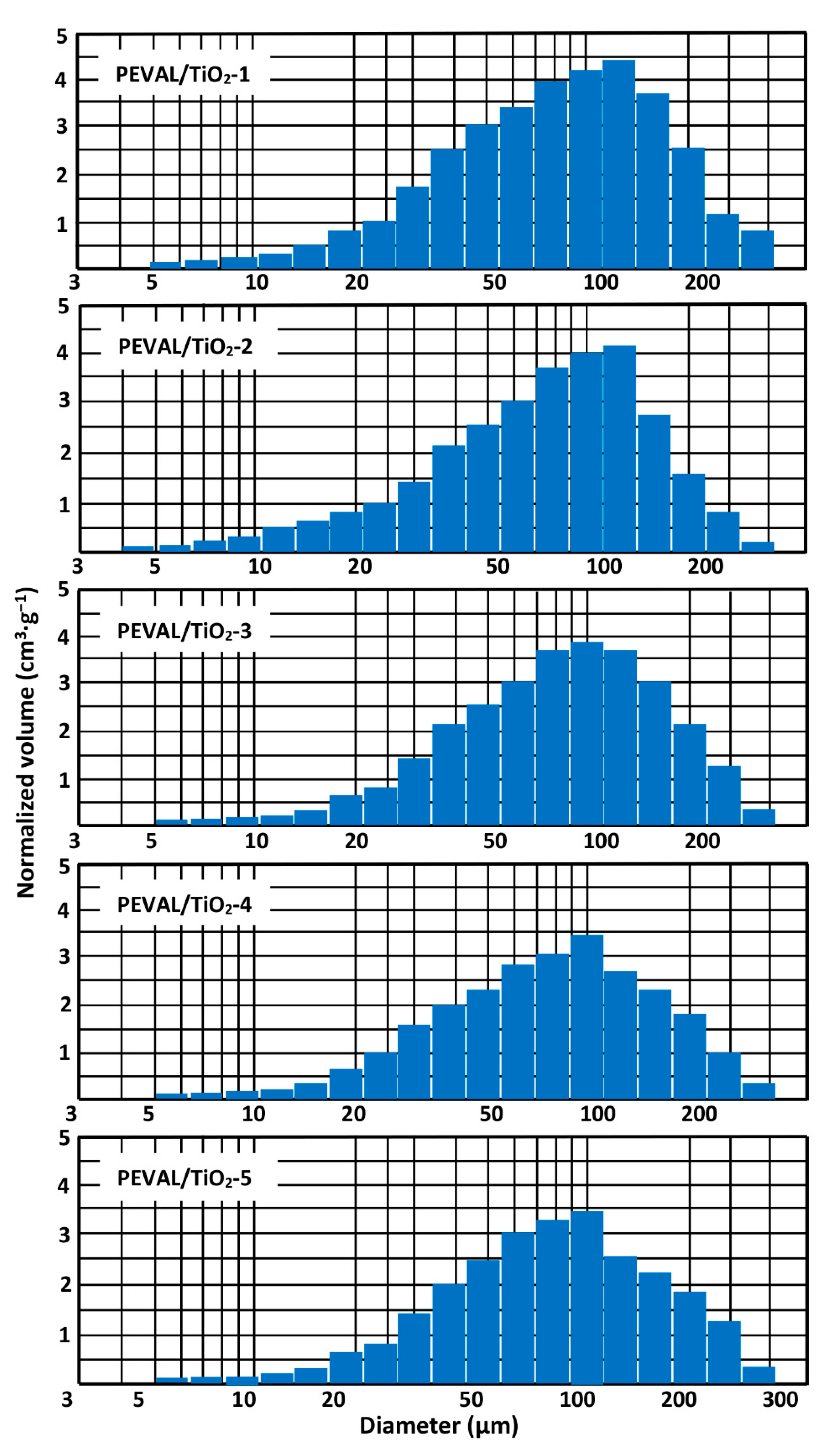
3.6. Porosity and Pore Size Distribution
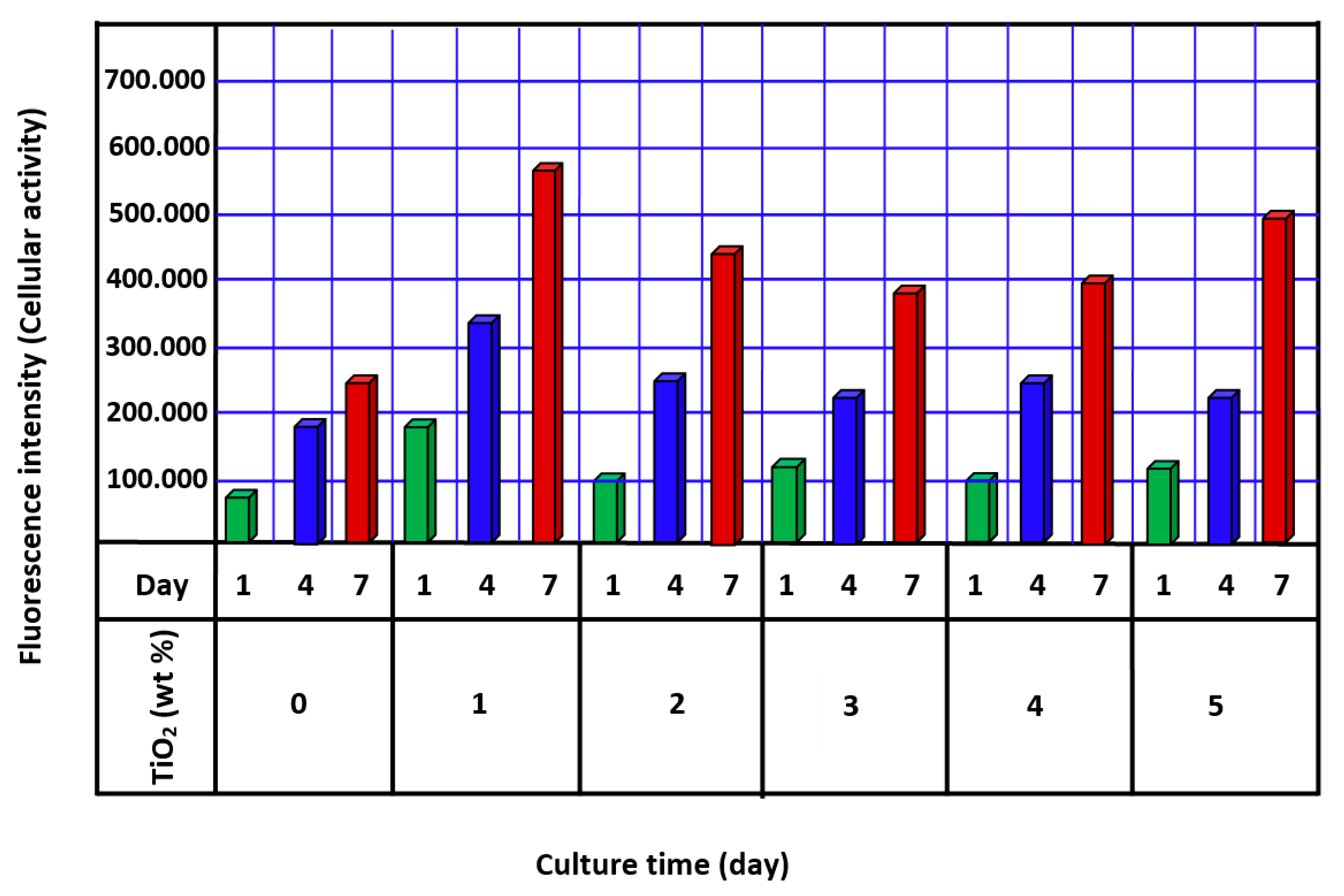
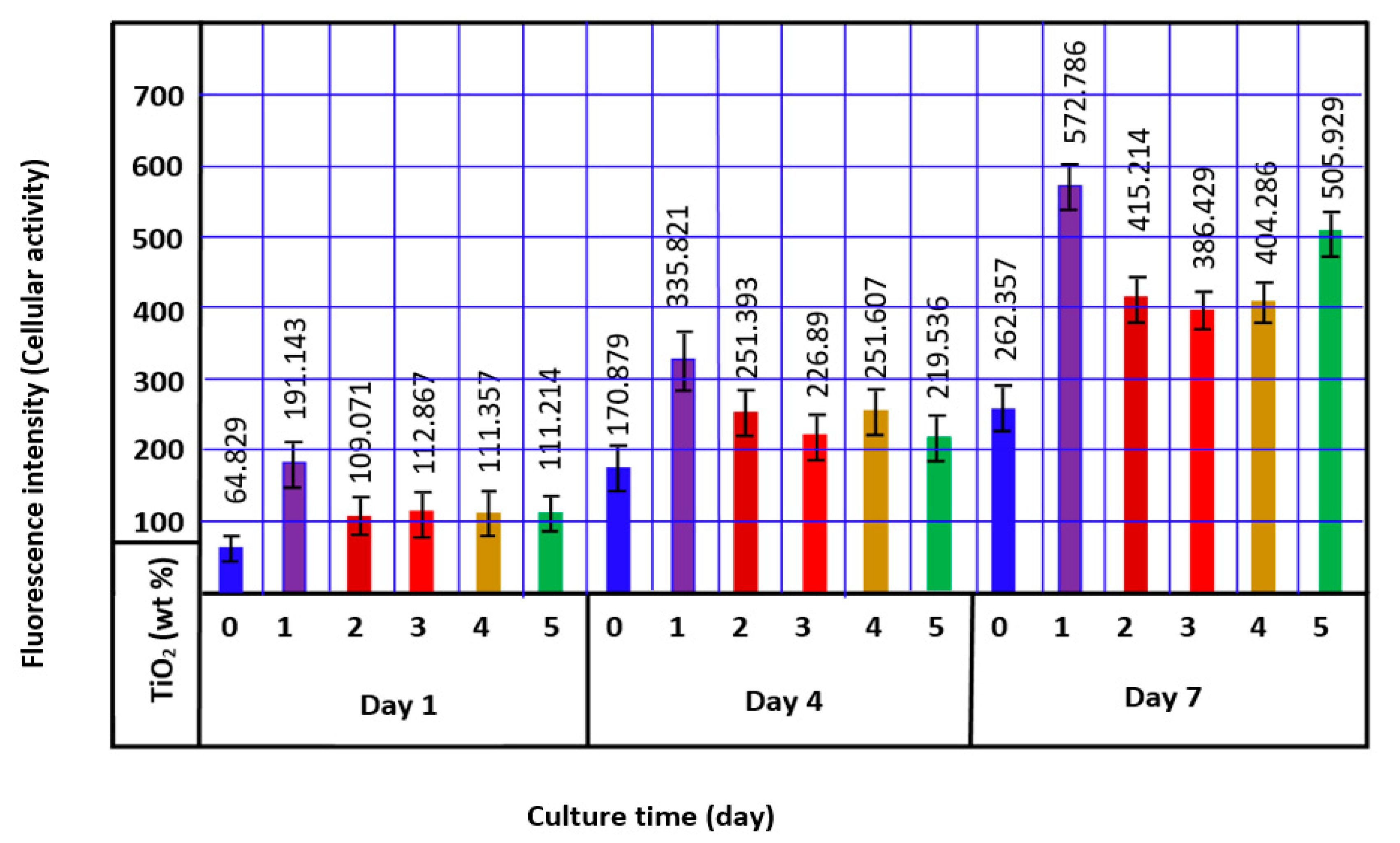
3.7. Cellular Activity
4. Conclusions
Author Contributions
Funding
Institutional Review Board Statement
Informed Consent Statement
Data Availability Statement
Acknowledgments
Conflicts of Interest
References
- Ikada, Y. Tissue Engineering: Fundamentals and Applications; Elsevier: Amsterdam, The Netherlands, 2011. [Google Scholar]
- Boccaccini, A.R.; Ma, P.X. Tissue Engineering Using Ceramics and Polymers; Elsevier: Amsterdam, The Netherlands, 2014. [Google Scholar]
- Gao, C.; Deng, Y.; Feng, P.; Mao, Z.; Li, P.; Yang, B.; Deng, J.; Cao, Y.; Shuai, C.; Peng, S. Current progress in bioactive ceramic scaffolds for bone repair and regeneration. Int. J. Mol. Sci. 2014, 15, 4714–4732. [Google Scholar] [CrossRef] [PubMed]
- Abbasi, N.; Hamlet, S.; Love, R.M.; Nguyen, N.-T. Porous scaffolds for bone regeneration. J. Sci. Adv. Mater. Devices 2020, 5, 1–9. [Google Scholar] [CrossRef]
- Tariverdian, T.; Sefat, F.; Gelinsky, M.; Mozafari, M. Scaffold for bone tissue engineering. In Handbook of Tissue Engineering Scaffolds: Volume One; Elsevier: Amsterdam, The Netherlands, 2019; pp. 189–209. [Google Scholar]
- Krieghoff, J.; Picke, A.-K.; Salbach-Hirsch, J.; Rother, S.; Heinemann, C.; Bernhardt, R.; Kascholke, C.; Möller, S.; Rauner, M.; Schnabelrauch, M. Increased pore size of scaffolds improves coating efficiency with sulfated hyaluronan and mineralization capacity of osteoblasts. Biomater. Res. 2019, 23, 26. [Google Scholar] [CrossRef] [PubMed] [Green Version]
- Dhandayuthapani, B.; Yoshida, Y.; Maekawa, T.; Kumar, D. Polymeric Scaffolds in Tissue Engineering Application: A Review. Int. J. Polym. Sci. 2011, 2011, 290602. [Google Scholar] [CrossRef]
- Neves, N.M.; Reis, R.L. Biomaterials from Nature for Advanced Devices and Therapies; John Wiley & Sons: Hoboken, NJ, USA, 2016. [Google Scholar]
- Nohynek, G.J.; Lademann, J.; Ribaud, C.; Roberts, M.S. Grey goo on the skin? Nanotechnology, cosmetic and sunscreen safety. Crit. Rev. Toxicol. 2007, 37, 251–277. [Google Scholar] [CrossRef]
- Appendini, P.; Hotchkiss, J.H. Review of antimicrobial food packaging. Innov. Food Sci. Emerg. Technol. 2002, 3, 113–126. [Google Scholar] [CrossRef]
- Jacoby, W.A.; Maness, P.C.; Wolfrum, E.J.; Blake, D.M.; Fennell, J.A. Mineralization of bacterial cell mass on a photocatalytic surface in air. Environ. Sci. Technol. 1998, 32, 2650–2653. [Google Scholar] [CrossRef]
- Brook, L.; Evans, P.; Foster, H.; Pemble, M.; Steele, A.; Sheel, D.; Yates, H. Highly bioactive silver and silver/titania composite films grown by chemical vapour deposition. J. Photochem. Photobiol. Chem. 2007, 187, 53–63. [Google Scholar] [CrossRef] [Green Version]
- Geetha, M.; Singh, A.K.; Asokamani, R.; Gogia, A.K. Ti based biomaterials, the ultimate choice for orthopaedic implants—A review. Prog. Mater. Sci. 2009, 54, 397–425. [Google Scholar] [CrossRef]
- Jeng, H.A.; Swanson, J. Toxicity of metal oxide nanoparticles in mammalian cells. J. Environ. Sci. Health Part A 2006, 41, 2699–2711. [Google Scholar] [CrossRef]
- Prasanna, A.; Niranjan, R.; Kaushik, M.; Devasena, T.; Kumar, J.; Chelliah, R.; Oh, D.-H.; Swaminathan, S. Metal oxide curcumin incorporated polymer patches for wound healing. Appl. Surf. Sci. 2018, 449, 603–609. [Google Scholar]
- Liu, H.; Slamovich, E.B.; Webster, T.J. Increased osteoblast functions on nanophase titania dispersed in poly-lactic-co-glycolic acid composites. Nanotechnology 2005, 16, S601. [Google Scholar] [CrossRef] [PubMed]
- Babushkina, E.A.; Belokopytova, L.V.; Grachev, A.M.; Meko, D.M.; Vaganov, E.A. Variation of the hydrological regime of Bele-Shira closed basin in Southern Siberia and its reflection in the radial growth of Larix sibirica. Reg. Environ. Chang. 2017, 17, 1725–1737. [Google Scholar] [CrossRef] [Green Version]
- Gerhardt, L.-C.; Jell, G.; Boccaccini, A. Titanium dioxide (TiO2) nanoparticles filled poly(D,L lactid acid) (PDLLA) matrix composites for bone tissue engineering. J. Mater. Sci. Mater. Med. 2007, 18, 1287–1298. [Google Scholar] [CrossRef] [Green Version]
- Chen, Q.; Roether, J.; Boccaccini, A. Tissue engineering scaffolds from bioactive glass and composite materials. Top. Tissue Eng. 2008, 4, 1–27. [Google Scholar]
- De Santis, R.; Catauro, M.; Di Silvio, L.; Manto, L.; Raucci, M.G.; Ambrosio, L.; Nicolais, L. Effects of polymer amount and processing conditions on the in vitro behaviour of hybrid titanium dioxide/polycaprolactone composites. Biomaterials 2007, 28, 2801–2809. [Google Scholar] [CrossRef]
- Chahal, R.P.; Mahendia, S.; Tomar, A.; Kumar, S. γ-Irradiated PVA/Ag nanocomposite films: Materials for optical applications. J. Alloys Compd. 2012, 538, 212–219. [Google Scholar] [CrossRef]
- Wu, W.; Liang, S.; Shen, L.; Ding, Z.; Zheng, H.; Su, W.; Wu, L. Preparation, characterization and enhanced visible light photocatalytic activities of polyaniline/Bi3NbO7 nanocomposites. J. Alloys Compd. 2012, 520, 213–219. [Google Scholar] [CrossRef]
- Zhu, Q.; Li, X.; Fan, Z.; Xu, Y.; Niu, H.; Li, C.; Dang, Y.; Huang, Z.; Wang, Y.; Guan, J. Biomimetic polyurethane/TiO2 nanocomposite scaffolds capable of promoting biomineralization and mesenchymal stem cell proliferation. Mater. Sci. Eng. C 2018, 85, 79–87. [Google Scholar] [CrossRef]
- Pelaseyed, S.S.; Hosseini, H.R.M.; Nokhbedehghan, Z.; Samadikuchaksaraei, A. PLGA/TiO2 nanocomposite scaffolds for biomedical applications: Fabrication, photocatalytic, and antibacterial properties. BioImpacts BI 2021, 11, 45. [Google Scholar] [CrossRef]
- Keskin, S.; Elliott, J.R. Binary interactions of poly(ethylene covinyl alcohol) with poly(4-vinyl pyridine) and poly(n-butyl methacrylate). Ind. Eng. Chem. Res. 2003, 42, 6331–6337. [Google Scholar] [CrossRef]
- Saito, A.; Kawanishi, H.; Yamashita, A.C.; Mineshima, M. (Eds.) High-Performance Membrane Dialyzers; Karger Medical and Scientific Publishers: Basel, Switzerland, 2011; Volume 173, pp. 1–10. [Google Scholar] [CrossRef]
- Tomita, K.; Kojoh, K.; Suzuki, A. Isolation of thermophiles assimilating poly(ethylene-co-vinyl alcohol). J. Ferment. Bioeng. 1997, 84, 400–402. [Google Scholar] [CrossRef]
- Mejia, G.; Lopez, O.; Sierra, L. Biodegradation of poly(vinylalcohol-co-ethylene) with the fungus Phanerochaete chrysosporium. Mater. Res. Innov. 2001, 4, 148–154. [Google Scholar] [CrossRef]
- Alotaibi, N.M.; Aouak, T. Preparation and non isothermal crystallization kinetic of acetylsalicylic acid-poly(vinylalcohol-co-ethylene) blend. Application in drug delivery domain. Macromol. Res. 2013, 21, 747–756. [Google Scholar]
- Mejía, G.A.I.; López, O.B.L.; Mulet, P.A. Biodegradation of poly(vinylalcohol) with enzymatic extracts of phanerochaete chrysosporium. In Macromolecular Symposia; WILEY-VCH Verlag GmbH & Co. KGaA: Weinheim, Germany, 1999; pp. 131–147. [Google Scholar]
- Bonomini, M.; Pavone, B.; Sirolli, V.; Del Buono, F.; Di Cesare, M.; Del Boccio, P.; Amoroso, L.; Di Ilio, C.; Sacchetta, P.; Federici, G. Proteomics characterization of protein adsorption onto hemodialysis membranes. J. Proteome Res. 2006, 5, 2666–2674. [Google Scholar] [CrossRef]
- Ishida, M. Blood compatibility of ethylene-vinyl alcohol copolymer dialyzers. Cells 2005, 37, 30–34. [Google Scholar]
- Itoh, S.; Susuki, C.; Tsuji, T. Platelet activation through interaction with hemodialysis membranes induces neutrophils to produce reactive oxygen species. J. Biomed. Mater. Res. Part A Off. J. Soc. Biomater. Jpn. Soc. Biomater. Aust. Soc. Biomater. Korean Soc. Biomater. 2006, 77, 294–303. [Google Scholar] [CrossRef]
- Sirolli, V.; Ballone, E.; Amoroso, L.; Di Liberato, L.; Di Mascio, R.; Cappelli, P.; Albertazzi, A.; Bonomini, M. Leukocyte adhesion molecules and leukocyte-platelet interactions during hemodialysis: Effects of different synthetic membranes. Int. J. Artif. Organs 1999, 22, 536–542. [Google Scholar] [CrossRef]
- Sirolli, V.; Ballone, E.; Di Stante, S.; Amoroso, L.; Bonomini, M. Cell activation and cellular-cellular interactions during hemodialysis: Effect of dialyzer membrane. Int. J. Artif. Organs 2002, 25, 529–537. [Google Scholar] [CrossRef]
- Sato, M.; Morita, H.; Ema, H.; Yamaguchi, S.; Amano, I. Effect of different dialyzer membranes on cutaneous microcirculation during hemodialysis. Clin. Nephrol. 2006, 66, 426–432. [Google Scholar] [CrossRef]
- Nakano, A. Ethylene vinyl alcohol co-polymer as a high-performance membrane: An EVOH membrane with excellent biocompatibility. High-Perform. Membr. Dialyzers 2011, 173, 164–171. [Google Scholar]
- Pertosa, G.; Simone, S.; Soccio, M.; Marrone, D.; Gesualdo, L.; Schena, F.P.; Grandaliano, G. Coagulation cascade activation causes CC chemokine receptor-2 gene expression and mononuclear cell activation in hemodialysis patients. J. Am. Soc. Nephrol. 2005, 16, 2477–2486. [Google Scholar] [CrossRef] [PubMed] [Green Version]
- Barth, R.H. Urea modeling and Kt/V: A critical appraisal. Kidney Int. Suppl. 1993, 41, S-252–S-260. [Google Scholar]
- Bruck, S. Medical applications of polymeric materials. Med. Prog. Through Technol. 1982, 9, 1–16. [Google Scholar]
- Jagur-Grodzinski, J. Polymers for tissue engineering, medical devices, and regenerative medicine. Concise general review of recent studies. Polym. Adv. Technol. 2006, 17, 395–418. [Google Scholar]
- Augustine, R.; Kalarikkal, N.; Thomas, S. Advancement of wound care from grafts to bioengineered smart skin substitutes. Prog. Biomater. 2014, 3, 103–113. [Google Scholar] [CrossRef] [Green Version]
- Augustine, R.; Dominic, E.A.; Reju, I.; Kaimal, B.; Kalarikkal, N.; Thomas, S. Electrospun poly(ε-caprolactone)-based skin substitutes: I n vivo evaluation of wound healing and the mechanism of cell proliferation. J. Biomed. Mater. Res. Part B Appl. Biomater. 2015, 103, 1445–1454. [Google Scholar] [CrossRef]
- Ketels, H.H.T.M. Synthesis, Characterization and Applications of Ethylene Vinylalcohol Copolymers. Ph.D. Thesis, Eindhoven University of Technology, Eindhoven, The Netherlands, 1989. [Google Scholar]
- Sodagar, A.; Akhoundi, M.S.A.; Bahador, A.; Jalali, Y.F.; Behzadi, Z.; Elhaminejad, F.; Mirhashemi, A.H. Effect of TiO2 nanoparticles incorporation on antibacterial properties and shear bond strength of dental composite used in Orthodontics. Dent. Press J. Orthod. 2017, 22, 67–74. [Google Scholar] [CrossRef] [Green Version]
- Sun, J.; Forster, A.M.; Johnson, P.M.; Eidelman, N.; Quinn, G.; Schumacher, G.; Zhang, X.; Wu, W.-L. Improving performance of dental resins by adding titanium dioxide nanoparticles. Dent. Mater. 2011, 27, 972–982. [Google Scholar] [CrossRef]
- Endogan Tanir, T.; Hasirci, V.; Hasirci, N. Preparation and characterization of Chitosan and PLGA-based scaffolds for tissue engineering applications. Polym. Compos. 2015, 36, 1917–1930. [Google Scholar] [CrossRef]
- Nakayama, G.R. Assessment of the Alamar Blue assay for cellular growth and viability in vitro. J. Immunol. Methods 1997, 204, 205–208. [Google Scholar] [CrossRef]
- Cabedo, L.; Lagarón, J.M.; Cava, D.; Saura, J.J.; Giménez, E. The effect of ethylene content on the interaction between ethylene-vinyl alcohol copolymers and water—II: Influence of water sorption on the mechanical properties of EVOH copolymers. Polym. Test. 2006, 25, 860–867. [Google Scholar] [CrossRef]
- Jipa, I.M.; Stoica, A.; Stroescu, M.; Dobre, L.-M.; Dobre, T.; Jinga, S.; Tardei, C. Potassium sorbate release from poly(vinyl alcohol)-bacterial cellulose films. Chem. Pap. 2012, 66, 138–143. [Google Scholar] [CrossRef]
- Rendón-Rivera, A.; Toledo-Antonio, J.; Cortés-Jácome, M.; Angeles-Chávez, C. Generation of highly reactive OH groups at the surface of TiO2 nanotubes. Catal. Today 2011, 166, 18–24. [Google Scholar] [CrossRef]
- Badwelan, M.; Alkindi, M.; Alghamdi, O.; Saeed, W.S.; Al-Odayni, A.-B.; Alrahlah, A.; Aouak, T. Poly(δ-valerolactone)/Poly(ethylene-co-vinylalcohol)/β-Tricalcium Phosphate Composite as Scaffolds: Preparation, Properties, and In Vitro Amoxicillin Release. Polymers 2021, 13, 46. [Google Scholar] [CrossRef] [PubMed]
- de Lima, J.A.; Felisberti, M.I. Poly(ethylene-co-vinyl alcohol) and poly(methyl methacrylate) blends: Phase behavior and morphology. Eur. Polym. J. 2008, 44, 1140–1148. [Google Scholar] [CrossRef]
- Ren, M.; Frimmel, F.H.; Abbt-Braun, G. Multi-cycle photocatalytic degradation of bezafibrate by a cast polyvinyl alcohol/titanium dioxide (PVA/TiO2) hybrid film. J. Mol. Catal. A Chem. 2015, 400, 42–48. [Google Scholar] [CrossRef]
- Gupta, S.; Sindhu, S.; Varman, K.A.; Ramamurthy, P.C.; Madras, G. Hybrid nanocomposite films of polyvinyl alcohol and ZnO as interactive gas barrier layers for electronics device passivation. RSC Adv. 2012, 2, 11536–11543. [Google Scholar] [CrossRef]
- Peng, Z.; Kong, L.X. A thermal degradation mechanism of polyvinyl alcohol/silica nanocomposites. Polym. Degrad. Stab. 2007, 92, 1061–1071. [Google Scholar] [CrossRef]
- Yang, H.; Xu, S.; Jiang, L.; Dan, Y. Thermal decomposition behavior of poly(vinyl alcohol) with different hydroxyl content. J. Macromol. Sci. Part B 2012, 51, 464–480. [Google Scholar] [CrossRef]
- Gomaa, M.M.; Hugenschmidt, C.; Dickmann, M.; Abdel-Hady, E.E.; Mohamed, H.F.; Abdel-Hamed, M.O. Crosslinked PVA/SSA proton exchange membranes: Correlation between physiochemical properties and free volume determined by positron annihilation spectroscopy. Phys. Chem. Chem. Phys. 2018, 20, 28287–28299. [Google Scholar] [CrossRef] [PubMed]
- Kumar, S.; Singh, R. Recovery of hydrocarbon liquid from waste high density polyethylene by thermal pyrolysis. Braz. J. Chem. Eng. 2011, 28, 659–667. [Google Scholar] [CrossRef] [Green Version]
- Saeed, W.S.; Al-Odayni, A.-B.; Alrahlah, A.; Alghamdi, A.A.; Aouak, T. Preparation and Characterization of Poly(δ-Valerolactone)/TiO2 Nanohybrid Material with Pores Interconnected for Potential Use in Tissue Engineering. Materials 2019, 12, 528. [Google Scholar] [CrossRef] [PubMed] [Green Version]


| System | TiO2 (g) | PEVAL (g) | TiO2 (wt%) |
|---|---|---|---|
| PEVAL/TiO2-1 | 0.01 | 0.99 | 1.0 |
| PEVAL/TiO2-2 | 0.02 | 0.98 | 2.0 |
| PEVAL/TiO2-3 | 0.03 | 0.97 | 3.0 |
| PEVAL/TiO2-4 | 0.04 | 0.96 | 4.0 |
| PEVAL/TiO2-5 | 0.05 | 0.95 | 5.0 |
| System | Tg (°C) | Tm (°C) | ∆Hm (J∙g−1) |
|---|---|---|---|
| PEVAL | 60 | 183 | 66.78 |
| PEVAL/TiO2-1 | 62 | 171 | 60.30 |
| PEVAL/TiO2-2 | 61 | 171 | 47.73 |
| PEVAL/TiO2-3 | 59 | 171 | 46.75 |
| PEVAL/TiO2-4 | 57 | 171 | 45.08 |
| PEVAL/TiO2-5 | 53 | 170 | 42.11 |
| System | Porosity (%) |
|---|---|
| PEVAL/TiO2-1 | 80.2 ± 3.8 |
| PEVAL/TiO2-2 | 78.3 ± 3.5 |
| PEVAL/TiO2-3 | 74.7 ± 3.4 |
| PEVAL/TiO2-4 | 72.8 ± 3.2 |
| PEVAL/TiO2-5 | 72.8 ± 3.2 |
Publisher’s Note: MDPI stays neutral with regard to jurisdictional claims in published maps and institutional affiliations. |
© 2022 by the authors. Licensee MDPI, Basel, Switzerland. This article is an open access article distributed under the terms and conditions of the Creative Commons Attribution (CC BY) license (https://creativecommons.org/licenses/by/4.0/).
Share and Cite
Saeed, W.S.; Alotaibi, D.H.; Al-Odayni, A.-B.; Haidyrah, A.S.; Al-Owais, A.A.; Khan, R.; De Vera, M.A.T.; Alrahlah, A.; Aouak, T. Poly(ethylene-Co-vinyl Alcohol)/Titanium Dioxide Nanocomposite: Preparation and Characterization of Properties for Potential Use in Bone Tissue Engineering. Int. J. Mol. Sci. 2022, 23, 3449. https://0-doi-org.brum.beds.ac.uk/10.3390/ijms23073449
Saeed WS, Alotaibi DH, Al-Odayni A-B, Haidyrah AS, Al-Owais AA, Khan R, De Vera MAT, Alrahlah A, Aouak T. Poly(ethylene-Co-vinyl Alcohol)/Titanium Dioxide Nanocomposite: Preparation and Characterization of Properties for Potential Use in Bone Tissue Engineering. International Journal of Molecular Sciences. 2022; 23(7):3449. https://0-doi-org.brum.beds.ac.uk/10.3390/ijms23073449
Chicago/Turabian StyleSaeed, Waseem Sharaf, Dalal H. Alotaibi, Abdel-Basit Al-Odayni, Ahmed S. Haidyrah, Ahmad Abdulaziz Al-Owais, Rawaiz Khan, Merry Angelyn Tan De Vera, Ali Alrahlah, and Taieb Aouak. 2022. "Poly(ethylene-Co-vinyl Alcohol)/Titanium Dioxide Nanocomposite: Preparation and Characterization of Properties for Potential Use in Bone Tissue Engineering" International Journal of Molecular Sciences 23, no. 7: 3449. https://0-doi-org.brum.beds.ac.uk/10.3390/ijms23073449