
Zirconia-Toughened Alumina Ceramic Wear Particles Do Not Elicit Inflammatory Responses in Human Macrophages
 , , , and
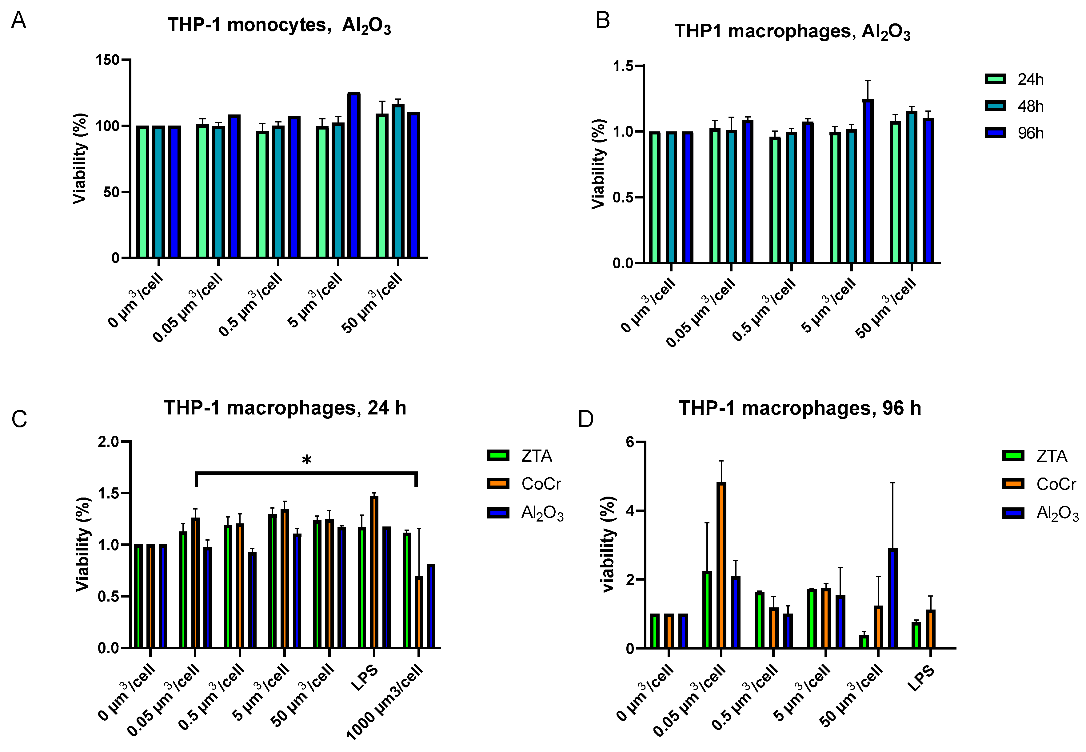
Abstract
:1. Introduction
2. Results
2.1. Al2O3 and ZTA Particles Do Not Affect THP-1 Macrophage Viability
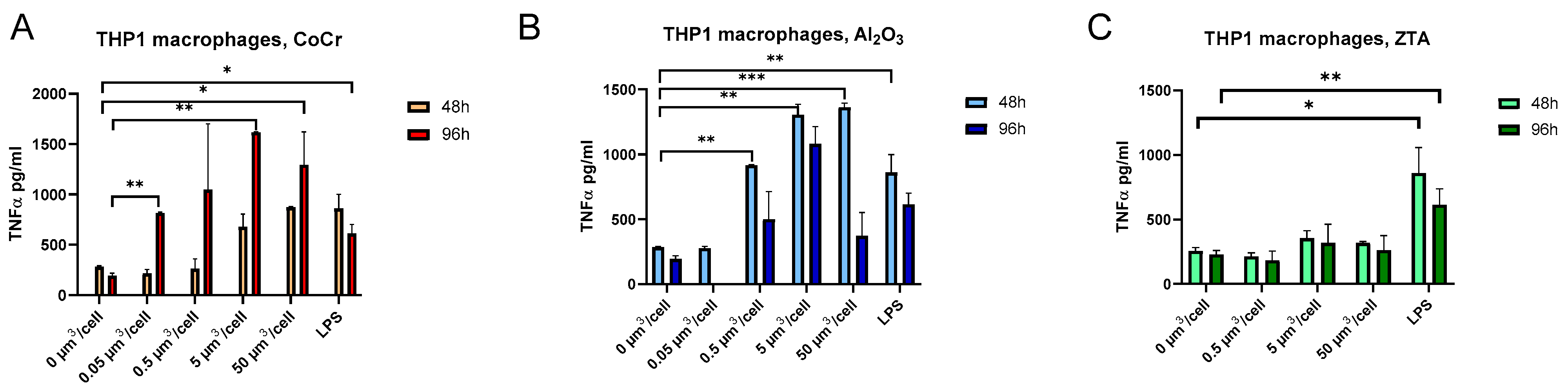
2.2. Treatment of THP-1 Macrophages with CoCr and Al2O3 Particles Caused an Increase in TNF-α Secretion
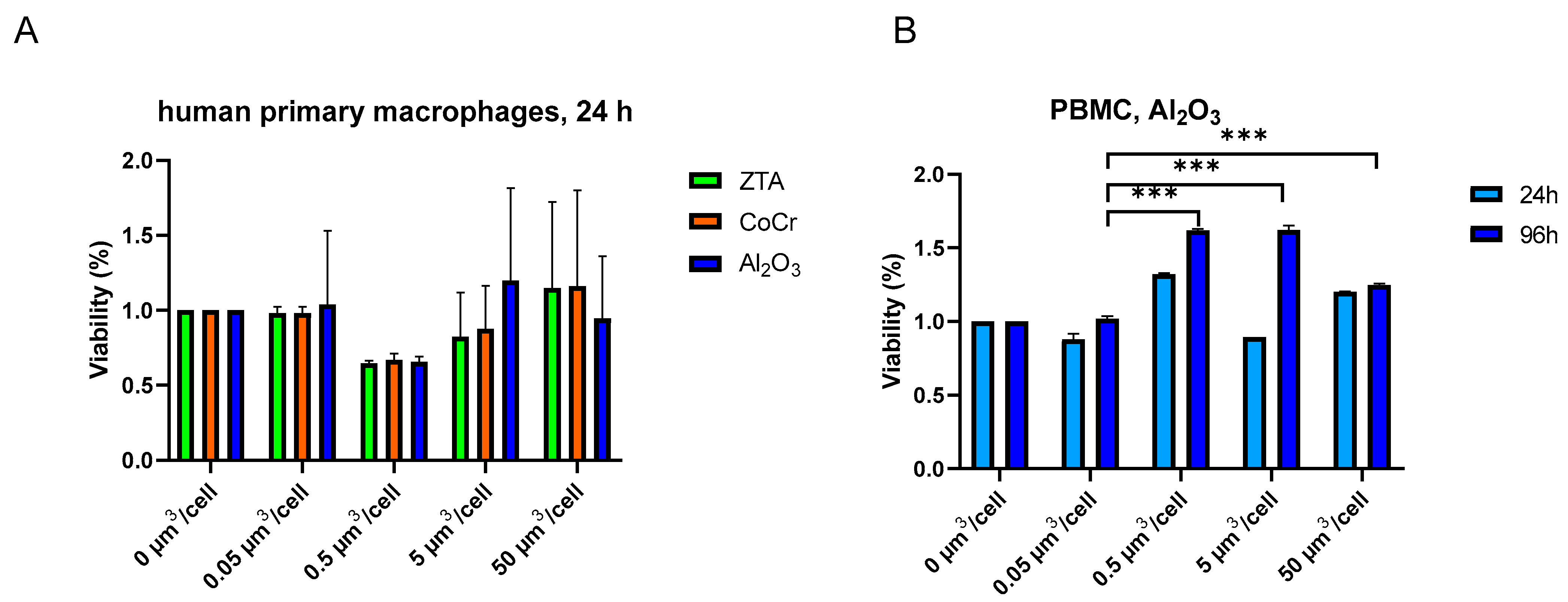
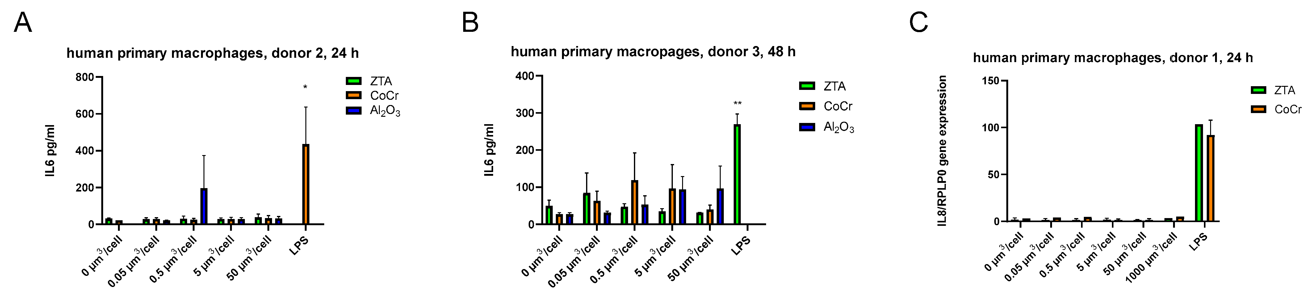
2.3. Al2O3, ZTA, and CoCr Particles Do Not Impact Cell Viability of Human Primary Macrophages and PBMCs
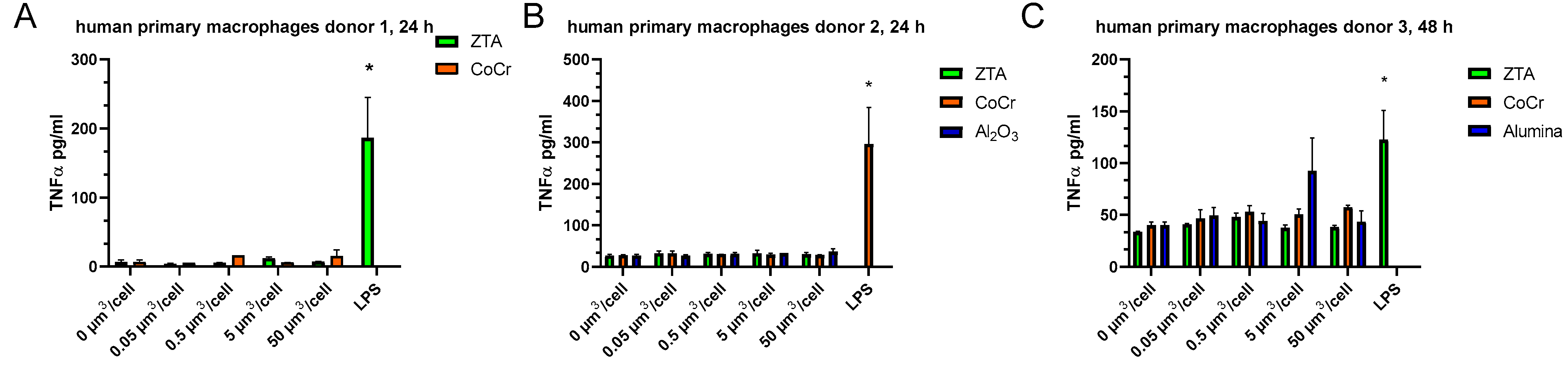
2.4. Al2O3, ZTA, and CoCr Particles Do Not Impact TNF-α Release of Human Primary Macrophages
3. Discussion
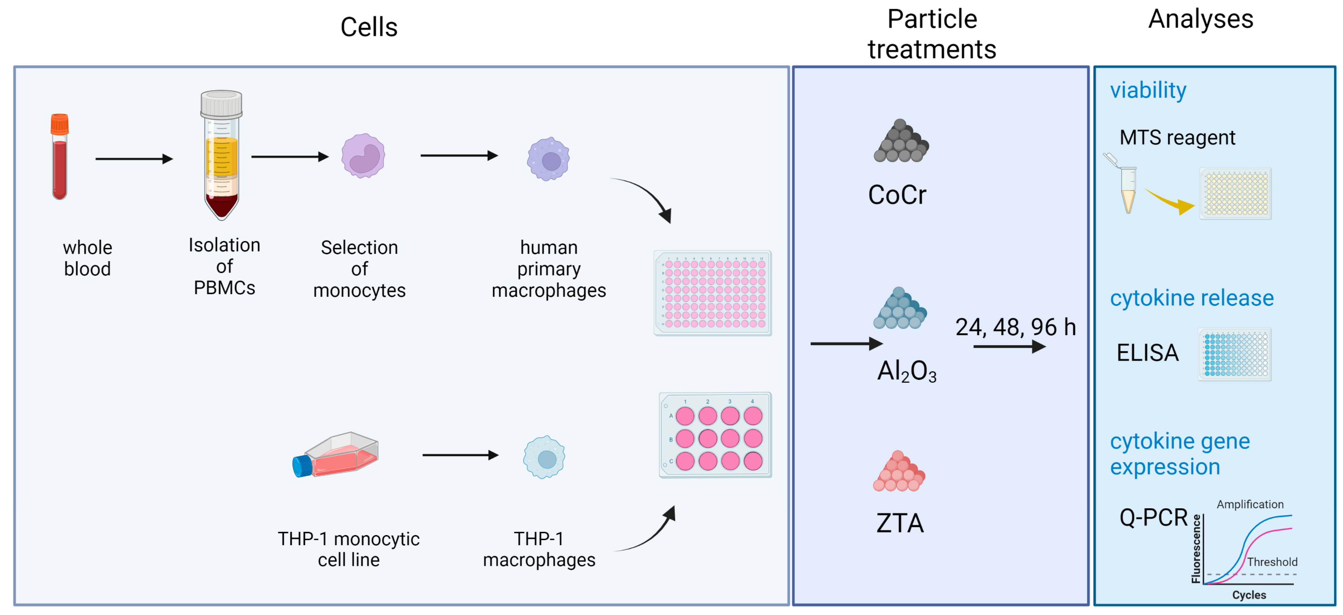
4. Materials and Methods
4.1. Ceramic and Metallic Test Particles
4.2. Cell Culturing and Differentiation of THP-1 Cell Line
4.3. Isolation of Human Peripheral Blood Mononuclear Cells (PBMCs)
4.4. Isolation of Human Primary Monocytes from PBMCs
4.5. Differentiation of Human Primary Monocytes to Macrophages
4.6. Treatment of Cells with Metallic and Ceramic Particles
4.7. RNA Isolation and Quantitative PCR
4.8. Enzyme-Linked Immunosorbent Assay
4.9. Viability Assay
4.10. Statistics
Supplementary Materials
Author Contributions
Funding
Institutional Review Board Statement
Informed Consent Statement
Data Availability Statement
Acknowledgments
Conflicts of Interest
References
- Kurtz, S.M.; Ong, K.L.; Schmier, J.; Mowat, F.; Saleh, K.; Dybvik, E.; Kärrholm, J.; Garellick, G.; Havelin, L.I.; Furnes, O.; et al. Future clinical and economic impact of revision total hip and knee arthroplasty. J. Bone Joint Surg. Am. 2007, 89 (Suppl. S3), 144–151. [Google Scholar]
- MacInnes, S.J.; Gordon, A.; Wilkinson, J.M. Risk Factors for Aseptic Loosening Following Total Hip Arthroplasty; IntechOpen: London, UK, 2012. [Google Scholar]
- National Joint Registry. 2017 14th Annual Report National Joint Registry. Available online: https://www.hqip.org.uk/resource/national-joint-registry-14th-annual-report-2017/ (accessed on 9 March 2023).
- Sundfeldt, M.; Carlsson, L.V.; Johansson, C.B.; Thomsen, P.; Gretzer, C. Aseptic loosening, not only a question of wear: A review of different theories. Acta Orthop. 2006, 77, 177–197. [Google Scholar] [CrossRef]
- Gallo, J.; Goodman, S.B.; Konttinen, Y.T.; Raska, M. Particle disease: Biologic mechanisms of periprosthetic osteolysis in total hip arthroplasty. Innate. Immun. 2013, 19, 213–224. [Google Scholar] [CrossRef] [PubMed] [Green Version]
- Mahendra, G.; Pandit, H.; Kliskey, K.; Murray, D.; Gill, H.S.; Athanasou, N. Necrotic and inflammatory changes in metal-on-metal resurfacing hip arthroplasties. Acta Orthop. 2009, 80, 653–659. [Google Scholar] [CrossRef] [Green Version]
- Brown, C.; Fisher, J.; Ingham, E. Biological effects of clinically relevant wear particles from metal-on-metal hip prostheses. Proc. Inst. Mech. Eng. Part H J. Eng. Med. 2006, 220, 355–369. [Google Scholar] [CrossRef]
- Patel, S.S.; Aruni, W.; Inceoglu, S.; Akpolat, Y.T.; Botimer, G.D.; Cheng, W.K.; Danisa, O.A. A comparison of Staphylococcus aureus biofilm formation on cobalt-chrome and titanium-alloy spinal implants. J. Clin. Neurosci. 2016, 31, 219–223. [Google Scholar] [CrossRef] [PubMed]
- Clarke, I.; Green, D.; Williams, P.; Kubo, K.; Pezzotti, G.; Lombardi, A.; Turnbull, A.; Donaldson, T. Hip-simulator wear studies of an alumina-matrix composite (AMC) ceramic compared to retrieval studies of AMC balls with 1–7 years follow-up. Wear 2009, 267, 702–709. [Google Scholar] [CrossRef]
- Warashina, H.; Sakano, S.; Kitamura, S.; Yamauchi, K.-I.; Yamaguchi, J.; Ishiguro, N.; Hasegawa, Y. Biological reaction to alumina, zirconia, titanium and polyethylene particles implanted onto murine calvaria. Biomaterials 2003, 24, 3655–3661. [Google Scholar] [CrossRef]
- Kurtz, S.M.; Kocagöz, S.; Arnholt, C.; Huet, R.; Ueno, M.; Walter, W.L. Advances in zirconia toughened alumina biomaterials for total joint replacement. J. Mech. Behav. Biomed. Mater. 2014, 31, 107–116. [Google Scholar] [CrossRef] [PubMed] [Green Version]
- Bos, I.; Willmann, G. Morphologic characteristics of periprosthetic tissues from hip prostheses with ceramic-ceramic couples: A comparative histologic investigation of 18 revision and 30 autopsy cases. Acta Orthop. 2001, 72, 335–342. [Google Scholar] [CrossRef]
- Bauer, T.W.; Campbell, P.A.; Hallerberg, G.; Grp, B.W. How Have New Bearing Surfaces Altered the Local Biological Reactions to Byproducts of Wear and Modularity? Clin. Orthop. Relat. Res. 2014, 472, 3687–3698. [Google Scholar] [CrossRef] [Green Version]
- Zais, I.E.; Sammali, S.; Pavan, M.; Chisari, E.; Krueger, C.A. The Local Toxicity of Cobalt Chrome implants: A Systematic Review of Preclinical Studies. J. Allergy Ther. 2022, 3. [Google Scholar] [CrossRef]
- Song, Z.; Borgwardt, L.; Høiby, N.; Wu, H.; Sørensen, T.S.; Borgwardt, A. Prosthesis infections after orthopedic joint replacement: The possible role of bacterial biofilms. Orthop. Rev. 2013, 5, 14–71. [Google Scholar] [CrossRef] [Green Version]
- Kempthorne, J.T.; Ailabouni, R.; Raniga, S.; Hammer, D.; Hooper, G. Occult Infection in Aseptic Joint Loosening and the Diagnostic Role of Implant Sonication. BioMed. Res. Int. 2015, 2015, 946215. [Google Scholar] [CrossRef] [PubMed] [Green Version]
- Moojen, D.J.F.; van Hellemondt, G.; Vogely, H.C.; Burger, B.J.; Walenkamp, G.H.I.M.; Tulp, N.J.; Schreurs, B.W.; Meulemeester, F.R.A.J.D.; Schot, C.S.; van de Pol, I.; et al. Incidence of low-grade infection in aseptic loosening of total hip arthroplasty. Acta Orthop. 2010, 81, 667–673. [Google Scholar] [CrossRef]
- Renz, N.; Mudrovcic, S.; Perka, C.; Trampuz, A. Orthopedic implant-associated infections caused by Cutibacterium spp.–A remaining diagnostic challenge. PLoS ONE 2018, 13, e0202639. [Google Scholar] [CrossRef] [Green Version]
- McNally, M.; Sousa, R.; Wouthuyzen-Bakker, M.; Chen, A.F.; Soriano, A.; Vogely, H.C.; Clauss, M.; Higuera, C.A.; Trebše, R. The EBJIS definition of periprosthetic joint infection. Bone Joint J. 2021, 103-B, 18–25. [Google Scholar] [CrossRef]
- Parisi, T.J.; Konopka, J.F.; Bedair, H.S. What is the Long-term Economic Societal Effect of Periprosthetic Infections After THA? A Markov Analysis. Clin. Orthop. Relat. Res. 2017, 475, 1891–1900. [Google Scholar] [CrossRef] [Green Version]
- Christel, P.S. Biocompatibility of surgical-grade dense polycrystalline alumina. Clin. Orthop. Relat. Res. 1992, 282, 10–18. [Google Scholar] [CrossRef]
- Nine, J.; Choudhury, D.; Hee, A.C.; Mootanah, R.; Abu Osman, N.A. Wear Debris Characterization and Corresponding Biological Response: Artificial Hip and Knee Joints. Materials 2014, 7, 980–1016. [Google Scholar] [CrossRef] [Green Version]
- Jamieson, S.; Mawdesley, A.; Deehan, D.; Kirby, J.; Holland, J.; Tyson-Capper, A. Inflammatory responses to metal oxide ceramic nanopowders. Sci. Rep. 2021, 11, 10531. [Google Scholar] [CrossRef]
- Lohmann, C.; Schwartz, Z.; Köster, G.; Jahn, U.; Buchhorn, G.; MacDougall, M.; Casasola, D.; Liu, Y.; Sylvia, V.; Dean, D.; et al. Phagocytosis of wear debris by osteoblasts affects differentiation and local factor production in a manner dependent on particle composition. Biomaterials 2000, 21, 551–561. [Google Scholar] [CrossRef] [PubMed]
- Bylski, D.; Wedemeyer, C.; Xu, J.; Sterner, T.; Loer, F.; von Knoch, M. Alumina ceramic particles, in comparison with titanium particles, hardly affect the expression of RANK-, TNF-alpha-, and OPG-mRNA in the THP-1 human monocytic cell line. J. Biomed. Mater Res. A 2009, 89, 707–716. [Google Scholar] [CrossRef] [PubMed]
- Radziun, E.; Wilczyńska, J.D.; Książek, I.; Nowak, K.; Anuszewska, E.; Kunicki, A.; Olszyna, A.; Ząbkowski, T. Assessment of the cytotoxicity of aluminium oxide nanoparticles on selected mammalian cells. Toxicol. Vitr. 2011, 25, 1694–1700. [Google Scholar] [CrossRef] [PubMed]
- Sterner, T.; Schutze, N.; Saxler, G.; Jakob, F.; Rader, C.P. Effects of clinically relevant alumina ceramic, zirconia ceramic and titanium particles of different sizes and concentrations on TNF-alpha release in a human macrophage cell line. Biomed. Tech. 2004, 49, 340–344. [Google Scholar] [CrossRef]
- Bertrand, J.; Delfosse, D.; Mai, V.; Awiszus, F.; Harnisch, K.; Lohmann, C.H. Ceramic prosthesis surfaces induce an inflammatory cell response and fibrotic tissue changes. Bone Jt. J. 2018, 100-B, 882–890. [Google Scholar] [CrossRef]
- Lawrence, H.; Deehan, D.J.; Holland, J.P.; Anjum, S.A.; Mawdesley, A.E.; Kirby, J.A.; Tyson-Capper, A.J. Cobalt ions recruit inflammatory cells in vitro through human Toll-like receptor 4. Biochem. Biophys. Rep. 2016, 7, 374–378. [Google Scholar] [CrossRef] [PubMed] [Green Version]
- Campbell, J.; Rajaee, S.; Brien, E.; Paiement, G.D. Inflammatory pseudotumor after ceramic-on-ceramic total hip arthroplasty. Arthroplast. Today 2017, 3, 83–87. [Google Scholar] [CrossRef] [PubMed] [Green Version]
- Hatton, A.; Nevelos, J.; Matthews, J.; Fisher, J.; Ingham, E. Effects of clinically relevant alumina ceramic wear particles on TNF-α production by human peripheral blood mononuclear phagocytes. Biomaterials 2003, 24, 1193–1204. [Google Scholar] [CrossRef]
- Daigneault, M.; Preston, J.A.; Marriott, H.M.; Whyte, M.K.B.; Dockrell, D.H. The Identification of Markers of Macrophage Differentiation in PMA-Stimulated THP-1 Cells and Monocyte-Derived Macrophages. PLoS ONE 2010, 5, e8668. [Google Scholar] [CrossRef]
- Schwende, H.; Fitzke, E.; Ambs, P.; Dieter, P. Differences in the state of differentiation of THP-1 cells induced by phorbol ester and 1,25-dihydroxyvitamin D3. J. Leukoc. Biol. 1996, 59, 555–561. [Google Scholar] [CrossRef]
- Germain, M.; Hatton, A.; Williams, S.; Matthews, J.; Stone, M.; Fisher, J.; Ingham, E. Comparison of the cytotoxicity of clinically relevant cobalt–chromium and alumina ceramic wear particles in vitro. Biomaterials 2003, 24, 469–479. [Google Scholar] [CrossRef] [PubMed]
- Wastesson, J.W.; Morin, L.; Tan, E.; Johnell, K. An update on the clinical consequences of polypharmacy in older adults: A narrative review. Expert Opin. Drug Saf. 2018, 17, 1185–1196. [Google Scholar] [CrossRef] [PubMed] [Green Version]
- Greenstein, A.S.; Gorczyca, J.T. Orthopedic Surgery and the Geriatric Patient. Clin. Geriatr. Med. 2019, 35, 65–92. [Google Scholar] [CrossRef] [PubMed]
- Fong, J.S.L.; Booth, M.A.; Rifai, A.; Fox, K.; Gelmi, A. Diamond in the Rough: Toward Improved Materials for the Bone−Implant Interface. Adv. Health Mater. 2021, 10, 2100007. [Google Scholar] [CrossRef] [PubMed]


Disclaimer/Publisher’s Note: The statements, opinions and data contained in all publications are solely those of the individual author(s) and contributor(s) and not of MDPI and/or the editor(s). MDPI and/or the editor(s) disclaim responsibility for any injury to people or property resulting from any ideas, methods, instructions or products referred to in the content. |
© 2023 by the authors. Licensee MDPI, Basel, Switzerland. This article is an open access article distributed under the terms and conditions of the Creative Commons Attribution (CC BY) license (https://creativecommons.org/licenses/by/4.0/).
Share and Cite
Porporati, A.A.; Mödinger, Y.; Fischer, S.; Polajžer, S.; Mettang, M.; Deisinger, U.; Podlogar, M.; Trebše, R.; Lovšin, N. Zirconia-Toughened Alumina Ceramic Wear Particles Do Not Elicit Inflammatory Responses in Human Macrophages. Int. J. Mol. Sci. 2023, 24, 6482. https://0-doi-org.brum.beds.ac.uk/10.3390/ijms24076482
Porporati AA, Mödinger Y, Fischer S, Polajžer S, Mettang M, Deisinger U, Podlogar M, Trebše R, Lovšin N. Zirconia-Toughened Alumina Ceramic Wear Particles Do Not Elicit Inflammatory Responses in Human Macrophages. International Journal of Molecular Sciences. 2023; 24(7):6482. https://0-doi-org.brum.beds.ac.uk/10.3390/ijms24076482
Chicago/Turabian StylePorporati, Alessandro Alan, Yvonne Mödinger, Sarah Fischer, Sara Polajžer, Melanie Mettang, Ulrike Deisinger, Matejka Podlogar, Rihard Trebše, and Nika Lovšin. 2023. "Zirconia-Toughened Alumina Ceramic Wear Particles Do Not Elicit Inflammatory Responses in Human Macrophages" International Journal of Molecular Sciences 24, no. 7: 6482. https://0-doi-org.brum.beds.ac.uk/10.3390/ijms24076482