Article

Establishment of a New Filamentous Cyanobacterial Genus, Microcoleusiopsis gen. nov. (Microcoleaceae, Cyanobacteria), from Benthic Mats in Open Channel, Jiangxi Province, China

1
College of Life and Environmental Sciences, Wenzhou University, Wenzhou 325039, China
2
Key Laboratory of Algal Biology, Institute of Hydrobiology, Chinese Academy of Sciences, Wuhan 430072, China
3
University of Chinese Academy of Sciences, Beijing 100039, China
4
Zhejiang Key Laboratory of Ecological and Environmental Monitoring, Forewarning and Quality Control, Zhejiang Environmental Monitoring Center, Hangzhou 310012, China
*
Authors to whom correspondence should be addressed.
Submission received: 29 September 2021 / Revised: 25 October 2021 / Accepted: 26 October 2021 / Published: 29 October 2021
(This article belongs to the Special Issue 2021 Feature Papers by Diversity’s Editorial Board Members)

Abstract

:
Cyanobacterial taxonomic studies performed by using the modern approaches always lead to creation of many new genera and species. During the field survey for cyanobacterial resources in China, a filamentous cyanobacterial strain was successfully isolated from a microbial mat attached to rock surfaces of the Ganfu Channel, Jiangxi Province, China. This strain was morphologically similar to the cyanobacterial taxa belonging to the genera Microcoleus and Phormidium. The phylogenetic analyses based on 16S rRNA gene sequences showed that this strain formed a well-supported clade, close to the filamentous genera Microcoleus, Tychonema, and Kamptonema. The maximum similarity of 16S rRNA gene sequence of this strain with the related genera was 95.04%, less than the threshold for distinguishing bacterial genus. The ITS secondary structures also distinguish this strain from the related cyanobacterial genera. Therefore, combined with morphology, 16S rRNA gene sequence, and ITS secondary structures, a novel cyanobacterial genus here as Microcoleusiopsis was established, with the species type as Microcoleusiopsis ganfuensis.

1. Introduction

In the past decade, the molecular biological methods have shown a powerful solution to taxonomic problems in many cyanobacterial categories [1]. The polyphasic approach—based on the combination of morphological, cytomorphological, ecological, and molecular characteristics—has been widely used in characterization and integrated to solve the taxonomic problems of cyanobacteria have been accepted by more and more cyanobacterial researchers, leading to much progress in studies on cyanobacterial diversity [2,3]. The classification criteria based on only morphological observation gradually lost their original utility, and the morphological boundaries among many related genera became even more blurred. The problem that morphological characteristics could not be well integrated with phylogeny was so evident that it became urgent to revise the existing classification system of cyanobacteria from a more phylogenetic perspective. Thus, based on the polyphasic method, Komárek et al. proposed the eight-order system, later the ten-order system, resolving some phylogenetic issues [2,4,5,6,7,8].
The genus Microcoleus Desmazières ex Gomont was first described in 1892 [9], and this genus contains a group of filamentous cyanobacteria widely existing in various ecological niches, and was considered as one of the largest genera in the family Microcoleaceae. The type species Microcoleus vaginatus (Vaucher) Gomont ex Gomont, was characterized with many bright blue-green trichomes per colorless and unlamellated sheath, with specific ecology (soil biotope) [10,11]. As currently defined, there are 112 species of Microcoleus including aquatic and terrestrial species in all database, only 55 species have been accepted taxonomically based on the Algaebase Database up to 2017 (www.algaebase.org, accessed on 13 May 2021). Most species of this genus have typical characteristics of usually simple filaments, densely packed trichomes, isodiametric vegetative cells, strongly constricted cross walls, no calyptra, end cells typically longer than wide, sheaths open at the apex, and crosswise cell division [12].
For a long time, the phylogenetic evidence has indicated the genus Microcoleus to be polyphyletic. Its taxonomic revisions were continuously performed, mainly by separating several species in the genus away from the type species M. vaginatus. Boyer et al. (2002) summarized the 31 strains of Microcoleus as two morphological species (M. vaginatus and M. steenstrupii) falling into two distinct clades which were regarded as two genera [13]. Similarly, Siegesmund et al. (2008) proposed another important species within Microcoleus, M. chthonoplastes, in the new genus/species Coleofasciculus chthonoplastes based on its genetic distance to the type species [14]. Strunecky et al. (2013) targeted the morphological and molecular criteria for the revision of the genus Microcoleus through extensive examination of 92 strains of M. vaginatus and Phormidium autumnale from a wide range of regions and biotopes, and they further established the new family of Microcoleaceae and more than 10 new combination species of Microcoleus by transferring from species formerly placed in the genus Phormidium and Oscillatoria [15]. Niiyama and Tuji (2019) also described a new species, Microcoleus pseudautumnalis, producing both 2-methylisoborneol (2-MIB) and geosmin based on the polyphasic approach [16]. Similarly, Kimberly et al. (2020) also proposed a novel anatoxin-a and dihydroanatoxin-a producing species, M. anatoxicus, and these two recent studies even provided some new clues revealing the new species of Microcoleus related to some environmental issues [17]. However, the further revisions for the taxonomy of the genus Microcoleus are required, which will lead to more new genera and species during the revisionary course.
In recent years, the construction of water diversion projects has become an important measure in China to solve the problems for the increasing demand of water resources in water shortage areas, leading to a large number of new artificial channel with flowing water biotopes. Filamentous cyanobacteria accounting for a large proportion of microbial mats growing on both sides of the channels are mainly composed of Oscillatorean cyanobacteria such as Microcoleus, Oscillatoria, Phormidium, Lyngbya, and Tychonema. In this study, one filamentous cyanobacterial strain with Microcoleus-like morphology was isolated from the Ganfu Channel in Jiangxi Province, China. The polyphasic method based on morphological and molecular and phylogenetic analyses was used to characterize this new isolated cyanobacterium, and results revealed that it represents a novel genus of the family Microcoleaceae. Thus, the new genus as Microcoleusiopsis gen. nov and type species as Microcoleusiopsis ganfuensis sp. nov. were described.

2. Materials and Methods

2.1. Sampling and Cultivation

Benthic mat samples were separated in August 2019 from Ganfu channel, Jiangxi Province, China (28°33′7.48″ N, 115°56′44.62″ E). For strain isolation, mats were scraped off using a circular knife and live material was washed thoroughly in sterile liquid CT medium [18]. Sub-samples were coated onto the surface of sterile solid CT plate and the Pasteur pipette washing method was used to obtain unialgal filaments or single cells under 40× microscope (Olympus CKX31, Tokyo, Japan), kept at 25 °C under cool white fluorescence light on a 12:12 h L:D photoperiod with a photon flux density of 40 μmol m−2 s−1. Finally, a filamentous strain (named as CHAB 4138) was isolated and transferred into several 25 mL flasks containing 15 mL of CT medium. These strains were stored in the culture collection of Harmful Algae Biology laboratory (CHAB) in the Institute of Hydrobiology, Wuhan, China.

2.2. Morphological and Ultrastructural Characterization

Cell morphological observation was investigated with a Nikon Eclipse 80i microscope (Nikon, Japan). Filaments and vegetative cells were measured more than 100 individuals with a DS-Ri1 digital camera (Nikon, Japan). Microphotographs taken at 400 times were analyzed by using Nikon software NIS-Elements D 3.2. For ultrastructure examination, fresh samples were fixed using 2.5% glutaraldehyde in 0.1 M phosphate buffer at a pH 7.2 and 4 °C for three days. Then, these samples were washed using 0.1 M phosphate buffer after which they were post-fixed using 1% osmium tetroxide for 2 h, and washed again using 0.1 M phosphate buffer to remove osmium tetroxide after which they were dehydrated using a sequential ethanol gradient (30, 50, 70, 90, and 100%) and embedded in Spurr’s resin [19]. Uranyl acetate (2%) and lead citrate were used to stain the sections. Finally, the specimens were examined with an HT7700 (Japan) transmission electron microscope under 80 kV on Hitachi TEM system control (Hitachi, Tokyo, Japan).

2.3. DNA Extraction and PCR Amplification

To avoid extra bacteria contamination, fresh material of strain CHAB 4138 was collected by filtering onto Millipore filter (3.0 μm aperture, Merck Millipore, Darmstadt, Germany) and was further cleaned with sterile CT medium for two to three times, collected in clean EP tubes. Total genomic DNA from this strain was extracted using the modified cetyltrimethylammonium bromide (CTAB) method [20]. DNA was quantified using a NanoDrop™ 1000 Spectrophotometer (Thermo Scientific, Waltham, MA, USA).
The primers PA [21] and B23S [22] were used to amplify segments including the 16S rRNA gene and the 16S–23S internal transcribed spacer (ITS). Each PCR amplification was performed using a BIO-RAD Thermal Cycler (Bio-Rad, Hercules, CA, USA) with total PCR reaction volume of 20 μL consisted of 1 μL of genomic DNA (100 ng μL−1), 0.5 μL of each primer (10 μmol L−1), 8 μL of sterile water and 10 μL of 2× PCR mix with Taq polymerase (Beijing Tsingke Biotech Co., Ltd., Beijing, China). The program for 16S rRNA gene ran for one cycle of 3 min at 94 °C; 34 cycles of 30 s at 94 °C, 30 s at 58 °C (30 s at 55 °C for ITS), and 1 min at 72 °C (30 s for ITS) and then a final elongation step at 72 °C for 5 min. The PCR products were purified by the Qiaquik PCR purification columns (Qiagen, Germany) using TSINGKE DNA Gel Extraction Kit (Beijing Tsingke Biotech Co., Ltd., Beijing, China), cloned to the pMDTM18-T vector (TaKaRa, TaKaRa BioInc., Otsu, Japan) and inserted into Escherichia coli trans5α cells. Finally, the positive clones including target fragment were sequenced bidirectionally using an ABI 3730 Automated Sequencer (PerkinElmer, Waltham, MA, USA). At least three clones were sequenced for each target fragment.

2.4. Detection for Cyanotoxin Synthesis Genes

Genomic DNA from strain CHAB 4138 was detected for the cyanotoxin synthesis genes such as microcystins, paralytic shellfish toxins, cylindrospermopsin, and anatoxin-a. The corresponding primers and PCR procedures refer to the methods of previous studies by Jungblut and Neilan [23], Al-Tebrineh et al. [24], McGregor and Sendall [25], and Rantala-Ylinen et al. [26], respectively.

2.5. Phylogenetic Analyses

The 16S rRNA gene sequences obtained from a single clone of strain CHAB 4138 were initially screened at the NCBI Website (BLAST), and higher similar reference sequences were downloaded from GenBank database to construct the molecular phylogeny of these two strains. Using MAFFT v7.312 software we obtained a matrix of 162 sequences with 1237 nucleotide sites [27] after multiple sequence alignment. The standard selection nucleic acid substitution model (GTR+I+G) based on the Akaike information criterion (AIC) for Bayesian analysis (BI) and maximum likelihood analysis (ML) were selected to analyzed the alignments, and then particular parameters were individually estimated by MrBayes v3.2.6 [28] and PhyML 3.0 [29]. The Kimura–2 model was selected with 1000 bootstrap replicates to perform neighbor joining (NJ) analysis using MEGA software v7.0 [30]. Both ML and Bayesian phylogenetic trees were viewed and edited in FigTree v1.4.3 (http://tree.bio.ed.ac.uk/software/figtree/), and all obtained phylogenetic trees were edited by Tree View 1.6.6 software [31]. Similarity matrix of the 16S rRNA was established via MEGA software v7.0 to calculate p-distance with pairwise deletion of gaps.

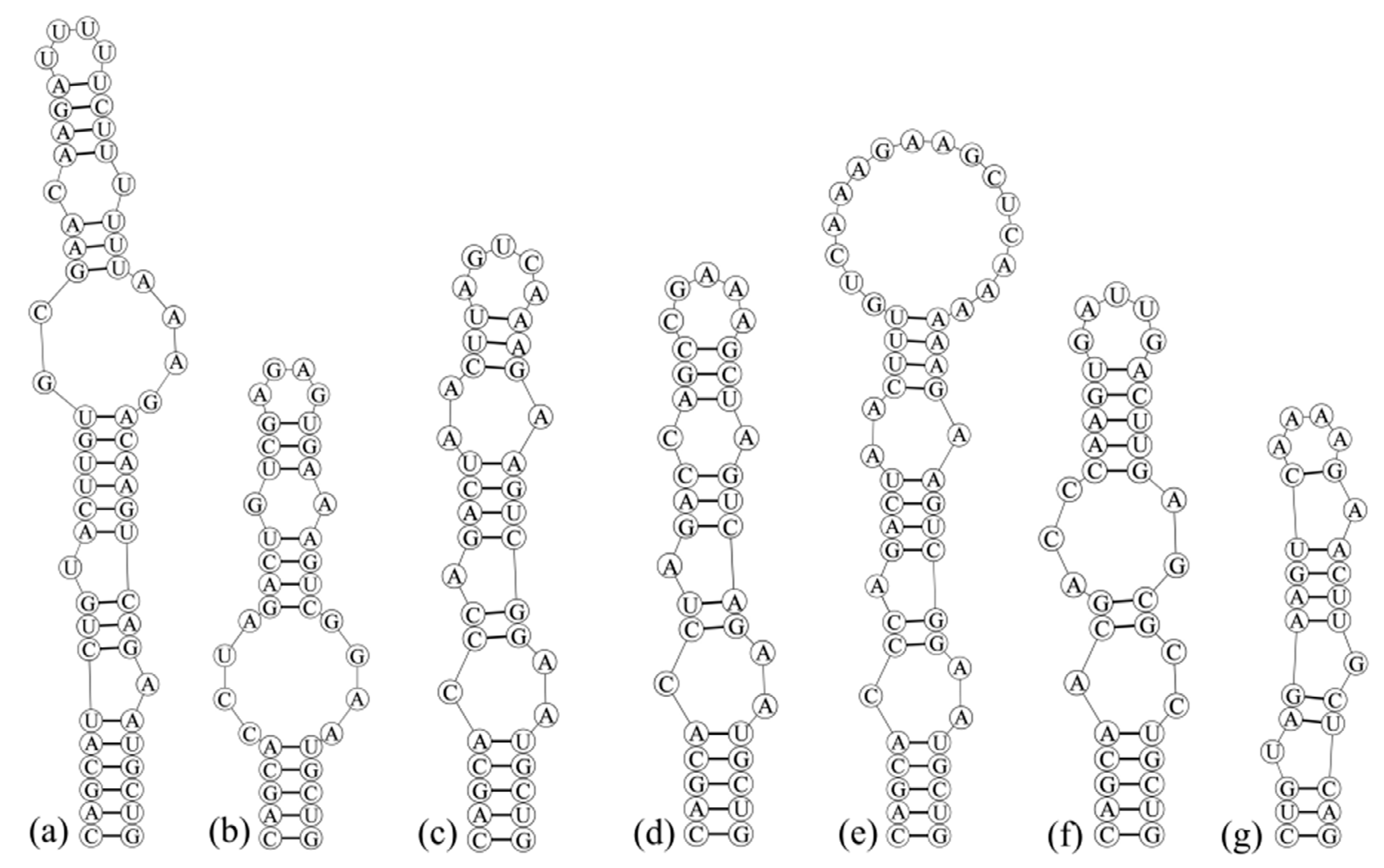
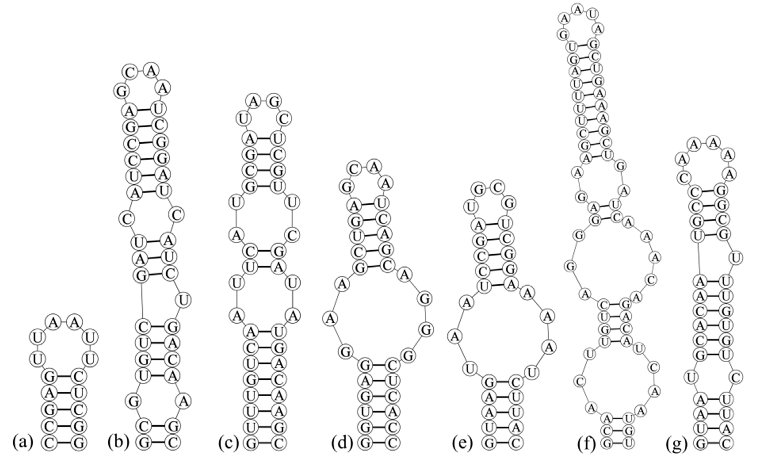
2.6. Construction of Secondary Structure of 16S–23S Internal Transcribed Spacer (ITS)

The 16S–23S rRNA ITS secondary structures of D1–D1′, Box–B and V3 helices of this strain and other closed species were determined using RNA structure, version 5.6 [32]. The 16S–23S rRNA gene nucleotide sequences obtained in this study have been deposited in the GenBank database, and the accession numbers are OK422506 and OK422507.

3. Results

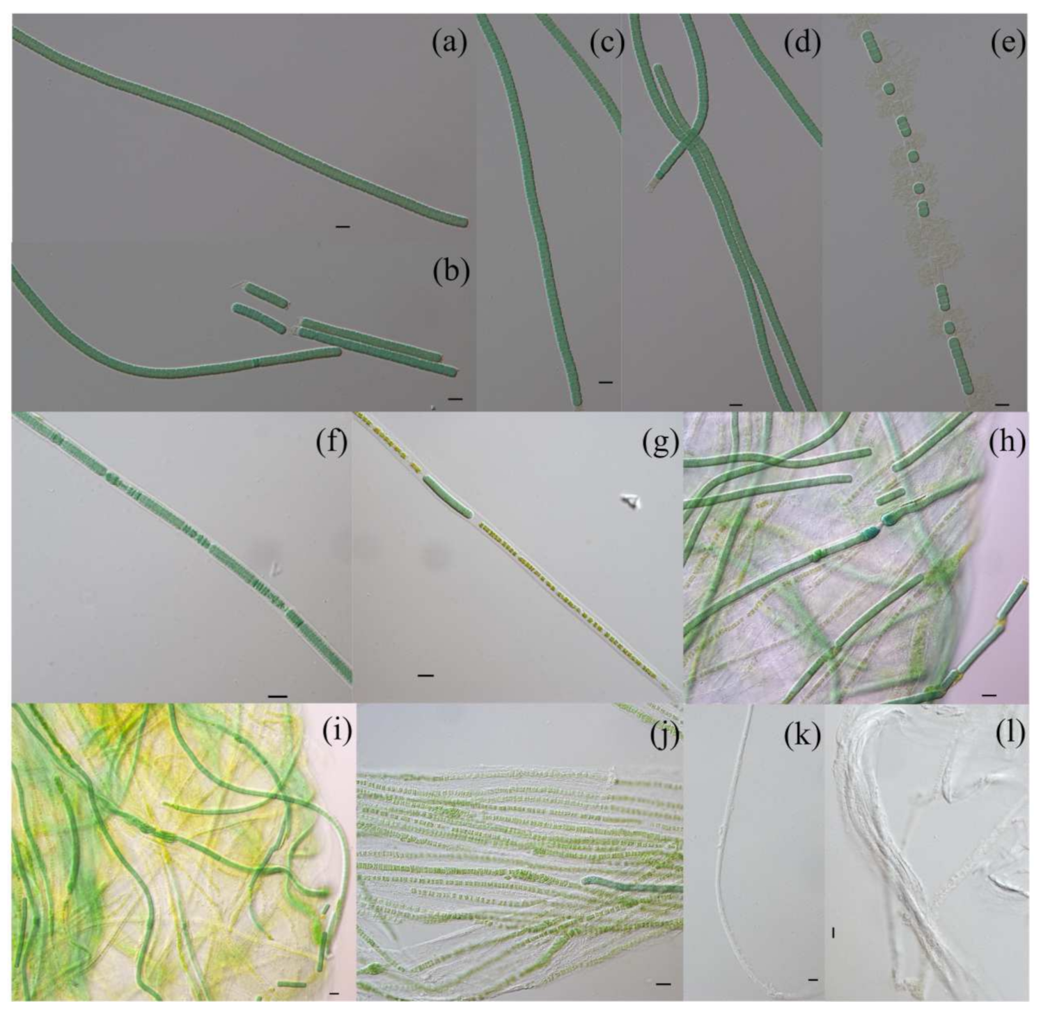
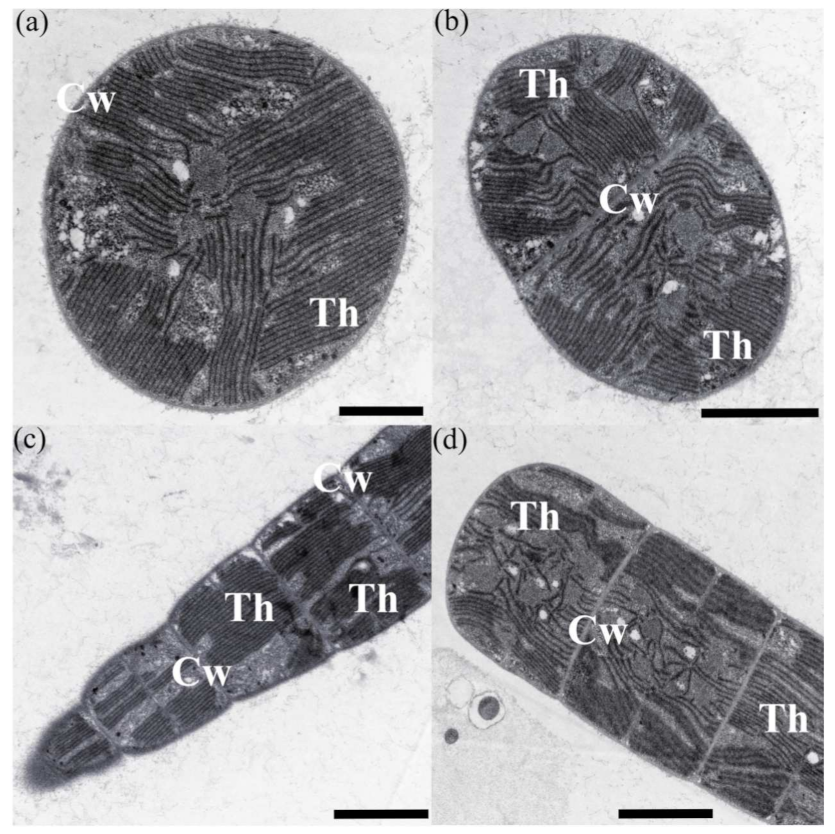
3.1. Morphological Description

Microcoleusiopsis R. Geng et G. Yu gen. nov.
Diagnosis: This genus appears morphologically similar to the genera of Microcoleus and Phormidium. The phylogenetic relationship was close to members of the family Microcoleaceae.
Description: In nature, colonies macroscopic, usually forming algal mats attached to the rock surface on freshwater rivers. Filaments long, straight, or slightly curved, blue-green to yellow-brown, surrounded by hyaline, colorless envelopes. Trichomes cylindrical, isopolar, not attenuated toward ends. Vegetative cells discoid, isopolar, always broader than long. Reproduction by motile hormogonia formed by necridia. Thylakoids radially arranged.
Type species: Microcoleusiopsis ganfuensis R. Geng et G. Yu sp. nov.
Etymology: The name of new genus “Microcoleusiopsis” was chosen because it was closely related to genus Microcoleus.
Microcoleusiopsis ganfuensis R. Geng et G. Yu sp. nov. (Figure 1).
Diagnosis: This species appears morphologically similar to the genera of Microcoleus-like. Filaments are long, not attenuated towards ends, and not or slightly constricted at the cross-walls. Apical cell rounded, without calyptra or thickened outer cell wall. Phylogenetic analysis suggested that this species formed a separated clade which was close to members of the families Microcoleaceae, such as Microcoleus, Tychonema, and Kamptonema.
Description: In nature, colonies usually form cyanobacterial mats attached to the surface of wet rocks on freshwater rivers and channels. Filaments long, unbranched, straight or slightly curved, blue-green, green when young, and yellow-brown when old, surrounded by hyaline, colorless sheaths. Trichomes isopolar, cylindrical, not attenuated towards ends, not or slightly constricted at the cross-walls. Vegetative cells usually discoid, isopolar, 2.28–(3.09)–4.27 μm long, 4.52–(5.69)–6.18 μm broad, width: length ratio 1.8, with granular content, not aerotopes. Apical cell rounded, without calyptra or thickened outer cell wall. Sheath finer, colorless, hyaline, not diffluent, and always open at the apex. Reproduction by motile hormogonia formed by necridia. Heterocytes and akinetes were not observed. Thylakoids radially arranged (Figure 2).
Reference strain: CHAB 4138.
Type locality: In Ganfu open channel, Jiangxi Province, China. (August 2019, 28°33′7.48′′ N, 115°56′44.62′′ E).
Holotype here designated: Dry material of this strain CHAB 4138 with no. JXGF201902, stored at Freshwater Algae Biology Herbarium (HBI), Institute of Hydrobiology, Chinese Academy of Science, Wuhan, Hubei Province, China.
Etymology: The name of species “ganfuensis” was chosen because this strain was separated from the Ganfu open channel.
Habitat: Attached on wet rock surfaces.

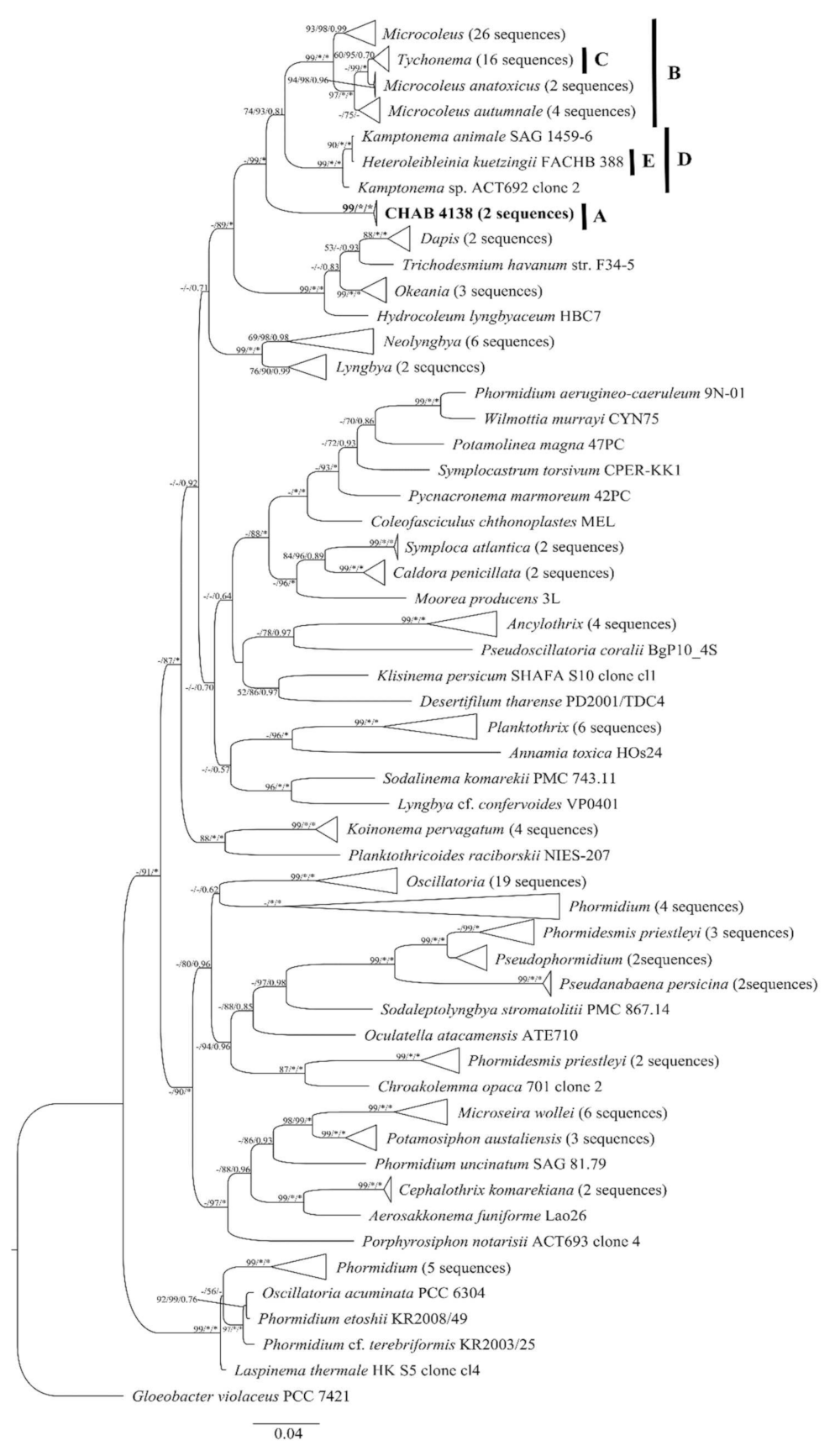
3.2. Molecular and Phylogeny Analyses

Through single sequencing, we obtained two 16S rRNA gene clones (1494bp) of strain CHAB 4138 which shared 99.91% similarities with each other. The 16S rRNA gene phylogenetic trees based on NJ, ML, and Bayesian methods with 162 sequences of family Microcoleaceae and Oscillatoriaceae strains downloaded from the NCBI database (Figure 3) indicated that the two clones of CHAB 4138 clustered a well-supported independent cluster (cluster A), supported by NJ/ML/BI approaches with high bootstrap values of 99%/100%/1.00. This unique clade was close to those formed by the filamentous genera Microcoleus (cluster B), Tychonema (cluster C), Kamptonema (cluster D), and Heteroleibleinia (cluster E), with a maximum similarity as 95.04%, probably representing a novel genus of filamentous cyanobacteria (sharing similarities to Microcoleus, Kamptonema, Heteroleibleinia, Tychonema, NeoLyngbya, Lyngbya, Okeania, Hydrocoleum, Dapis, Moorea, Symploca, Caldora, Wilmottia, Laspinema, Trichodesmium, Coleofasciculus, Oscillatoria, Aerosakkonema, and Phormidium were 94.09–95.04%, 94.43–94.52%, 94.43–94.52%, 94.26–94.35%, 94.09–94.17%, 93.83–93.91%, 93.13–93.22%, 92.96–93.04%, 92.78–92.87%, 92.70–92.78%, 92.61–92.70%, 92.52–92.61%, 92.43–92.52%, 92.43–92.52%, 92.09–92.17%, 92.09–92.17%, 92.00–92.09%, 92.00–92.09%, and 91.74–91.83%, respectively) (Table 1).
Besides, four type of cyanotoxin genes were not detected in Microcoleusiopsis ganfuensis CHAB 4138 and we did not obtain any PCR products by using the primers responsible for the synthesis genes for these toxins (microcystins, cylindrospermopsin, paralytic shellfish toxins and anatoxins).

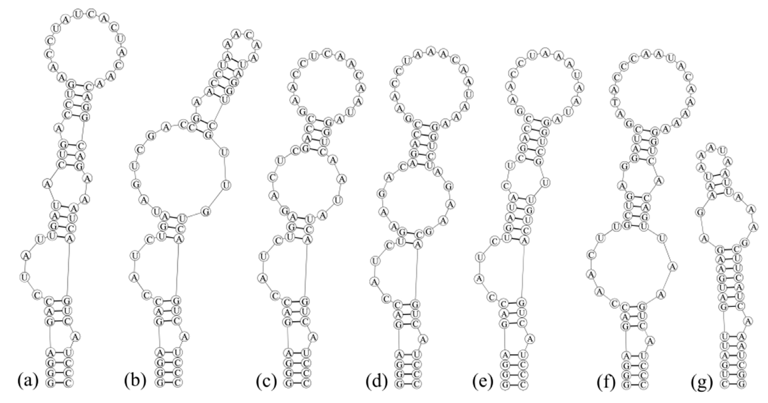
3.3. Analyses of ITS between 16S and 23S rRNA Gene and Secondary Structures

The partial 16S–23S ITS sequences of Microcoleusiopsis ganfuensis CHAB 4138 were obtained with a total length of 761 bp in this study (Table 2), and they were used, together with seven species clones from three genera including Microcoleus, Oscillatoria and Coleofasciculus downloaded from NCBI, to construct the ITS secondary structures. In general, all sequences contained both tRNA Ile and tRNA Ala (Table 2). As the most conserved structure, the D1–D1′ helix (Figure 4) of strain CHAB 4138 was similar to those of several species of close genera like Microcoleus vaginatus CSU-U-KK1, Microcoleus vaginatus PTRS-2, Microcoleus autumnale SAG 78.79, and Oscillatoria princeps CCALA 1115 in basal and apical stem–loop, but significantly different from those of Microcoleus pseudautumnalis Ak1609 and Coleofasciculu chthonoplastes MEL. In the basal stem of strain CHAB 4138 and other six species mentioned above, there was a 4-bp helix (a 6-bp helix in C. chthonoplastes MEL), followed by a small unidirectional bulge, and the apical structures contained a 4-bp helix (5-bp in M. pseudautumnalis Ak1609, M. vaginatus PTRS-2 and M. autumnale SAG 78.79; 3-bp in C. chthonoplastes MEL) with a 15-bp loop (5-bp in M. pseudautumnalis Ak1609 and C. chthonoplastes MEL; 14-bp in M. vaginatus CSU-U-KK1 and M. autumnale SAG 78.79; 16-bp and 17-bp in M. vaginatus PTRS-2 and O. princeps CCALA 1115, respectively).
The Box–B (Figure 5) and V3 (Figure 6) helices of CHAB 4138 were conspicuously different from those of other related genera in sequence length and stem–loop structures (Table 2). CHAB 4138 had its own unique Box–B helix, consisting of one 4-bp helix, two 3-bp helices, two 6-bp helices, two small unidirectional bulges, one 1:1 bp base bilateral bulge, one 2:4 bp base bilateral bulge, and one 4-bp apical loop. Whereas the other six related species had five Box–B helices types, especially the genus Microcoleus could be divided into three types, represented by M. pseudautumnalis Ak1609, M. vaginatus CSU-U-KK1, and M. vaginatus PTRS-2 with M. autumnale SAG 78.79, respectively. No regular patterns were found for V3 helices between CHAB 4138 and other seven filamentous species. The studied strain CHAB 4138 only had a 5-bp helix followed by a 6-bp apical loop, which significantly differed from other species.

4. Discussion

Benthic cyanobacteria can grow in patches on the attached substrates to form algal mats, and they are important primary producers in river and lake communities. Those mat-forming taxa mainly include Chroococcus-like cyanobacteria containing single cell and colonies with mucilage, a considerable number of filamentous Oscillatoria-like cyanobacteria without cell differentiation, Nostoc-like cyanobacteria with cell differentiation, Stigonema-like cyanobacteria with true branches, and Chamaesiphon-like cyanobacteria forming endospores [33,34]. During the field investigation, Oscillatorean cyanobacteria were found to be the main dominant species in the algal mats, and their characterization and correct identification based on the modern taxonomic system should be emphasized. It is expected that the ideal cyanobacteria genera and species in the current cyanobacterial taxonomy should be monophyletic, which means the need to make constant revisions to have this goal achieved [8,14,35,36,37,38].
Previous taxonomy of cyanobacteria was morphology based system, especially at a high rank, by using morphological characteristics such as the size of filaments and cells, polarity and branch types of filaments [34]. However, with the introduction of molecular biology methods, typical characteristics were proven to appear and lose many times during evolutionary process of cyanobacteria, making the distinction between some species of critical morphological characteristics increasingly blurred [34,39]. In this study, the benthic filamentous cyanobacterium isolated from the algal mats of the Ganfu Channel was difficult to be accurately classified based on morphological characteristics only such as the shapes of cells and filaments, types of end cells, and facultative presence of sheaths. Therefore, the polyphasic approach exhibited its power to determine the correct taxonomic attribution and phylogenetic relationship of this novel filamentous cyanobacterium.
The phylogenetic tree based on the 16S rRNA gene sequences indicated that the position of Microcoleusiopsis ganfuensis CHAB 4138 was close to the filamentous genera Microcoleus, Tychonema, Kamptonema, and Heteroleibleinia; however, the strains within the genus Microcoleus formed three small clades, and further clustered into a large clade with the strains of Tychonema (Figure 3 clade B). Kamptonema, originally described as “Oscillatoria animalis” (Figure 3 clade D), is a newly established filamentous cyanobacterial genus of family Microcoleaceae, by separating from genus Phormidium in recent years [40]. In addition, a geosmin producer [41]—Heteroleibleinia kuetzingii FACHB 388 (one filamentous strain originally identified as Lyngbya kuetzingii at the FACHB Culture Collection)—was shown to be clustered with Kamptonema strains in family Microcoleaceae (Figure 3 clade E), supported by NJ/ML/BI approaches as 99%/100%/1.00, and such a result implied that this strain may need to be re-identified as belonging to the genus Kamptonema. Comparison of 16S rRNA sequences showed that the two clones of CHAB 4138 clustered a well-supported independent cluster (cluster A), with a maximum similarity of 16S rRNA sequences as 95.04% to the existing cyanobacterial taxa, below the threshold of bacterial genus; therefore, this strain probably represents a new cyanobacterial taxon [42,43,44].
As one of the effective tools to distinguish cyanobacterial species, the secondary structures of ITS including D1–D1′, Box–B, and V3 helices can also distinguish Microcoleusiopsis ganfuensis from other filamentous cyanobacteria [45,46,47,48,49]. The D1–D1′ (Figure 4), Box–B (Figure 5), and V3 (Figure 6) helices of M. ganfuensis were significantly different from other related genera (Microcoleus, Oscillatoria, and Coleofasciculus) in stem–loop structures. It is worth mentioning that there were three configurations of the stem–loop structure of Box–B helix in multiple strains of the genus Microcoleus in this study, one as M. pseudautumnalis Ak1609, one as M. vaginatus CSU-U-KK1, and the third as M. vaginatus PTRS-2 and M. autumnale SAG 78.79—implying some relationship between ITS secondary structures and the ability of secondary metabolites.
Nowadays, the biological proliferation dominated by benthic filamentous cyanobacteria in rivers, lakes, and channels worldwide is frequently increasing, and the harmful effects caused by benthic cyanobacteria has gradually become a problem which cannot be ignored [50,51,52]. Microcoleus and Tychonema species were widely reported as toxigenic cyanobacteria since they were found to produce neurotoxic anatoxin-a/homoanatoxin-a in USA [17,53], Italy [54,55], and Germany [56]. Species Kamptonema formosum, a member of the newly established genus, was even found to form microcystins, anatoxin-a/homoanatoxin-a, and other anatoxin congeners in a recent published paper [57]. However, in this study, Microcoleusiopsis ganfuensis was shown to lack the synthesis genes of four type of cyanotoxins, indicating that it may not be a potential producer of cyanotoxins. Furthermore, the morphological observation based on both field sample and the cultured strain showed no bundle formation of trichomes covered by a sheath, confirming the distinction of M. ganfuensis from the type species of Microcoleus. Thus, the establishment of the new genus/species Microcoleusiopsis ganfuensis was well supported by the combination of morphology, 16S rRNA gene sequence, and 16S–23S ITS secondary structures.

Author Contributions

Conceptualization, R.G. and W.L.; methodology, R.G.; software, R.G.; formal analysis, W.L. and X.G.; investigation, R.G.; resources, A.C.; data curation, R.G.; writing—original draft preparation, R.G.; writing—review and editing, R.L.; visualization, H.L.; funding acquisition, G.Y. And R.L. All authors have read and agreed to the published version of the manuscript.

Funding

This study is funded by the NSFC projects (NSFC no. 51779247 and NSFC no. 31970219).

Institutional Review Board Statement

Not applicable.

Data Availability Statement

Not applicable.

Acknowledgments

We would like to thank the calculating resources supported by the Wuhan Branch, Supercomputing Center, Chinese Academy of Sciences, China for the computing resources. The first author thanks Zhenfei Xing and Yuan Xiao for technical assistance with TEM images.

Conflicts of Interest

The authors declare no conflict of interest.

References

  1. Garcia–Pichel, F.; Lopez–Cortes, A.; Nubel, U. Phylogenetic and morphological diversity of cyanobacteria in soil desert crusts from the Colorado plateau. Appl. Environ. Microbiol. 2001, 67, 1902–1910. [Google Scholar] [CrossRef] [PubMed] [Green Version]
  2. Komárek, J. Several problems of the polyphasic approach in the modern cyanobacterial system. Hydrobiologia 2018, 811, 7–17. [Google Scholar] [CrossRef]
  3. Mareš, J.; Strunecký, O.; Bučinská, L.; Wiedermannová, J. Evolutionary patterns of thylakoid architecture in cyanobacteria. Front. Microbiol. 2019, 10, 277. [Google Scholar] [CrossRef]
  4. Comte, K.; Holland, D.P.; Walsby, A.E. Changes in cell turgor pressure related to uptake of solutes by Microcystis sp. strain 8401. FEMS Microbiol. Ecol. 2007, 61, 399–405. [Google Scholar] [CrossRef] [PubMed]
  5. Sciuto, K.; Rascio, N.; Andreoli, C.; Moro, I. Polyphasic characterization of ITD–01, a cyanobacterium isolated from the Ischia Thermal District (Naples, Italy). Fottea. 2011, 11, 31–39. [Google Scholar] [CrossRef]
  6. Komárek, J.; Kaštovský, J.; Mareš, J.; Johansen, J.R. Taxonomic classification of cyanoprokaryotes (Cyanobacterial genera) 2014, using a polyphasic approach. Preslia 2014, 86, 295–335. [Google Scholar]
  7. Dvořák, P.; Poulíčková, A.; Hašler, P.; Belli, M.; Casamatta, D.A.; Papini, A. Species concepts and speciation factors in cyanobacteria, with connection to the problems of diversity and classification. Biodivers. Conserv. 2015, 24, 739–757. [Google Scholar] [CrossRef] [Green Version]
  8. Komárek, J.; Johansen, J.R.; Šmarda, J.; Strunecký, O. Phylogeny and taxonomy of Synechococcus-like cyanobacteria. Fottea 2020, 20, 171–191. [Google Scholar] [CrossRef]
  9. Gomont, M. Monographie des Oscillariées (Nostocacées homocystées). Annal. Sci. Nat. Bot. 1892, 16, 91–264. [Google Scholar]
  10. Geitler, L. Schizophyta (Klasse Schizophyceae). In Natürliche Pflanzenfamilien; Engler, A., Prantl, K., Eds.; Duncker & Humblot: Berlin, Germany, 1942; pp. 1–232. [Google Scholar]
  11. Drouet, F. Revision of the classifification of the Oscillatoriaceae. Monogr. Acad. Nat. Sci. Phil. 1968, 15, 1–370. [Google Scholar]
  12. Anagnostidis, K.; Komárek, J. Modern approach to the classification system of cyanophytes 3-Oscillatoriales. Algol. Stud. 1988, 50–53, 327–478. [Google Scholar]
  13. Boyer, S.L.; Johansen, J.R.; Flechtner, V.R. Phylogeny and Genetic Variance in Terrestrial Microcoleus (Cyanophyceae) Species Based on Sequence Analysis of the 16S rRNA Gene and Associated 16S–23S ITS Region. J. Phycol. 2002, 38, 1222–1235. [Google Scholar] [CrossRef]
  14. Siegesmund, M.A.; Johansen, J.R.; Karsten, U.; Friedl, T. Coleofasciculus gen. nov. (Cyanobacteria): Morphological and molecular criteria for revision of the genus Microcoleus Gomont. J. Phycol. 2008, 44, 1572–1585. [Google Scholar] [CrossRef] [PubMed]
  15. Strunecký, O.; Komárekjeffrey, J.; Johansenjeffrey, R. Molecular and morphological criteria for revision of the genus Microcoleus (Oscillatoriales, Cyanobacteria). J. Phycol. 2013, 49, 1167–1180. [Google Scholar] [CrossRef] [PubMed]
  16. Niiyama, Y.; Tuji, A. Microcoleus pseudautumnalis sp. nov. (Cyanobacteria, Oscillatoriales) producing 2–methylisoborneol. Bull. Natl. Mus. Nat. Sci. Ser. B 2019, 45, 93–101. [Google Scholar]
  17. Conklin, K.Y.; Stancheva, R.; Otten, T.G. Molecular and morphological characterization of a novel dihydroanatoxin-a producing Microcoleus species (cyanobacteria) from the Russian River, California, USA. Harmful Algae 2020, 93, 101767. [Google Scholar] [CrossRef]
  18. Ichimura, T. Isolation and culture methods of algae. Methods Phycol. Stud. 1979, 2, 294–305. [Google Scholar]
  19. Spurr, A.R. A low–viscosity epoxy resin embedding medium for electron microscopy. J. Ultrastruct. Res. 1969, 26, 31–43. [Google Scholar] [CrossRef]
  20. Neilan, B.A.; Jacobs, D.; Goodman, A.E. Genetic diversity and phylogeny of toxic cyanobacteria determined by DNA polymorphisms within the phycocyanin locus. Appl. Environ. Microbiol. 1995, 61, 3875–3883. [Google Scholar] [CrossRef] [Green Version]
  21. Edwards, U.; Rogall, T.; Blöcker, H.; Emde, M.; Böttger, E.C. Isolation and direct complete nucleotide determination of entire genes. Characterization of a gene coding for 16S ribosomal RNA. Nucleic Acids Res. 1989, 17, 7843–7853. [Google Scholar] [CrossRef] [Green Version]
  22. Gkelis, S.; Rajaniemi, P.; Vardaka, E.; Moustaka–gouni, M.; Lanaras, T.; Sivonen, K. Limnothrix redekei (Van Goor) Meffert (Cyanobacteria) strains from Lake Kastoria, Greece form a separate phylogenetic group. Microb. Ecol. 2005, 49, 176–182. [Google Scholar] [CrossRef] [PubMed]
  23. Jungblut, A.D.; Neilan, B.A. Molecular identification and evolution of the cyclic peptide hepatotoxins, microcystin and nodularin, synthetase genes in three orders of cyanobacteria. Arch. Microbiol. 2006, 185, 107–114. [Google Scholar] [CrossRef]
  24. Al-Tebrineh, J.; Pearson, L.A.; Yasar, S.A.; Neilan, B.A. A multiplex qPCR targeting hepato- and neurotoxigenic cyanobacteria of global significance. Harmful Algae 2012, 15, 19–25. [Google Scholar] [CrossRef]
  25. Mcgregor, G.B.; Sendall, B.C. Phylogeny and toxicology of Lyngbya wollei (Cyanobacteria, Oscillatoriales) from north-eastern Australia, with a description of Microseira gen. nov. J. Phycol. 2015, 51, 109–119. [Google Scholar] [CrossRef]
  26. Rantala-Ylinen, A.; Känä, S.; Wang, H. Anatoxin-a synthetase gene cluster of the cyanobacterium Anabaena sp. strain 37 and molecular methods to detect potential producers. Appl. Environ. Microbiol. 2011, 77, 7271–7278. [Google Scholar] [CrossRef] [Green Version]
  27. Katoh, K.; Standley, D.M. MAFFT multiple sequence alignment software version 7: Improvements in performance and usability. Mol. Biol. Evol. 2013, 30, 772–780. [Google Scholar] [CrossRef] [PubMed] [Green Version]
  28. Ronquist, F.; Teslenko, M.; Van Der Mark, P.; Ayres, D.L.; Darling, A.; Höhna, S.; Huelsenbeck, J.P. MrBayes 3.2: Efficient Bayesian phylogenetic inference and model choice across a large model space. Syst. Biol. 2012, 61, 539–542. [Google Scholar] [CrossRef] [Green Version]
  29. Guindon, S.; Dufayard, J.F.; Lefort, V.; Anisimova, M.; Hordijk, W.; Gascuel, O. New Algorithms and Methods to Estimate Maximum–Likelihood Phylogenies: Assessing the Performance of PhyML 3.0. Syst. Biol. 2010, 59, 307–321. [Google Scholar] [CrossRef] [Green Version]
  30. Kumar, S.; Stecher, G.; Tamura, K. MEGA7: Molecular Evolutionary Genetics Analysis version 7.0 for bigger datasets. Mol. Biol. Evol. 2016, 33, 1870–1874. [Google Scholar] [CrossRef] [Green Version]
  31. Page, R.D.M. TreeView: An application to display phylogenetic trees on personal computers. Comput. Appl. Biosci. 1996, 12, 357–358. [Google Scholar] [PubMed] [Green Version]
  32. Mathews Lab. RNAstructure Version 5.6. Available online: http://rna.urmc.rochester.edu/RNAstructure.html (accessed on 13 May 2021).
  33. Rippka, R.; Deruelles, J.; Waterbury, J.B.; Herdman, M.; Stanier, R.Y. Generic assignments, strain histories and properties of pure cultures of cyanobacteria. J. Gen. Microbiol. 1979, 111, 1–61. [Google Scholar] [CrossRef] [Green Version]
  34. Komárek, J. Cyanoprokaryota—3.Teil/ 3rd Part: Heterocytous Genera. In Süsswasserflora von Mitteleuropa Freshwater Flora of Central Europe 19/2; Büdel, B., Krienitz, L., Gärtner, G., Schagerl, M., Eds.; Springer/Spektrum: Heidelberg, Germany, 2013. [Google Scholar]
  35. Anagnostidis, K.; Komárek, J. Modern approach to the classification system of cyanophytes. 1-Introduction. Algol. Stud. Arch. Für Hydrobiol. 1985, 38–39, 291–302. [Google Scholar]
  36. Johansen, J.R.; Casamatta, D.A. Recognizing cyanobacterial diversity through adoption of a new species paradigm. Algol. Studies 2005, 117, 71–93. [Google Scholar] [CrossRef]
  37. Casamatta, D.A.; Johansen, J.R.; Vis, M.L. Molecular and morphological characterization of ten polar and near–polar strains with the Oscillatoriales (Cyanobacteria). J. Phycol. 2010, 41, 421–438. [Google Scholar] [CrossRef]
  38. Iii, R.; Johansen, J.R.; Kovácik, L. A unique Pseudanabaenalean (cyanobacteria) genus Nodosilinea gen. nov. based on morphological and molecular data1. J. Phycol. 2011, 47, 1397–1412. [Google Scholar]
  39. Gugger, M.; Molica, R.; Le, B.B. Genetic diversity of Cylindrospermopsis strains (Cyanobacteria) isolated from four continents. Appl. Environ. Microbiol. 2005, 71, 1097–1100. [Google Scholar] [CrossRef] [PubMed] [Green Version]
  40. Strunecký, O.; Komárek, J.; Šmarda, J. Kamptonema (Microcoleaceae, Cyanobacteria), a new genus derived from the polyphyletic Phormidium on the basis of combined molecular and cytomorphological markers. Preslia 2014, 86, 193–207. [Google Scholar]
  41. Wang, Z.J.; Song, G.F.; Li, Y.G. The diversity, origin, and evolutionary analysis of geosmin synthase gene in cyanobacteria. Sci. Total. Environ. 2019, 689, 789–796. [Google Scholar] [CrossRef] [PubMed]
  42. Wayne, L.G.; Brenner, D.J.; Colwell, R.R.; Grimont, P.A.D.; Kandler, O.; Krichevsky, M.I.; Starr, M.P. Report of the ad hoc committee on reconciliation of approaches to bacterial systematics. Int. J. Syst. Evol. Microbiol. 1987, 37, 463–464. [Google Scholar] [CrossRef] [Green Version]
  43. Stackebrandt, E.; Goebel, B.M. Taxonomic note: A placefor DNA–DNA reassociation and 16S rRNA sequence analysisin the present species definition in bacteriology. Int. J. Syst. Bacteriol. 1994, 44, 846–849. [Google Scholar] [CrossRef] [Green Version]
  44. Stackebrandt, E.; Ebers, J. Taxonomic parameters revisited: Tarnished gold standards. Microbiology 2006, 33, 152–155. [Google Scholar]
  45. Iteman, I.; Rippka, R.; Marsac, N.T.D.; Herdman, M. Comparison of conserved structural and regulatory domains within divergent 16s rRNA–23s rRNA spacer sequences of cyanobacteria. Microbiology 2000, 146, 1275–1286. [Google Scholar] [CrossRef] [PubMed] [Green Version]
  46. Vaccarino, M.A.; Johansen, J.R. Brasilonema angustatum sp. nov. (Nostocales), a new filamentous cyanobacterial species from the Hawaiian Islands. J. Phycol. 2012, 48, 1178–1186. [Google Scholar] [CrossRef] [PubMed]
  47. Osorio–Santos, K.; Pietrasiak, N.; Bohunická, M.; Miscoe, L.H.; Kováčik, L.; Martin, M.P.; Johansen, J.R. Seven new species of Oculatella (Pseudanabaenales, Cyanobacteria): Taxonomically recognizing cryptic diversification. Eur. J. Phycol. 2014, 49, 450–470. [Google Scholar] [CrossRef] [Green Version]
  48. Pietrasiak, N.; Mühlsteinová, R.; Siegesmund, M.A.; Johansen, J.R. Phylogenetic placement of Symplocastrum (Phormidiaceae, Cyanophyceae) with a new combination S. californicum and two new species: S. flechtnerae and S. torsivum. Phycologia 2014, 53, 529–541. [Google Scholar] [CrossRef]
  49. Mareš, J. Multilocus and SSU rRNA gene phylogenetic analyses of available cyanobacterial genomes, and their relation to the current taxonomic system. Hydrobiologia 2018, 811, 19–34. [Google Scholar] [CrossRef]
  50. Quiblier, C.; Wood, S.; Echenique-Subiabre, I.; Heath, M.; Villeneuve, A.; Humbert, J.-F. A review of current knowledge on toxic benthic freshwater cyanobacteria—Ecology, toxin production and risk management. Water Res. 2013, 47, 5464–5479. [Google Scholar]
  51. Fetscher, A.E.; Howard, M.D.; Stancheva, R.; Kudela, R.M.; Stein, E.D.; Sutula, M.A.; Busse, L.B.; Sheath, R.G. Wadeable streams as widespread sources of benthic cyanotoxins in California, USA. Harmful Algae 2015, 49, 105–116. [Google Scholar] [CrossRef]
  52. Echenique-Subiabre, I.; Dalle, C.; Duval, C.; Heath, M.W.; Coute, A.; Wood, S.A.; Humbert, J.-F.; Quiblier, C. Application of a spectrofluorimetric tool (bbe BenthoTorch) for monitoring potentially toxic benthic cyanobacteria in rivers. Water Res. 2016, 101, 341–350. [Google Scholar] [CrossRef]
  53. Bouma-Gregson, K.; Olm, M.R.; Probst, A.J.; Anantharaman, K.; Power, M.E.; Banfield, J.F. Impacts of microbial assemblage and environmental conditions on the distribution of anatoxin-a producing cyanobacteria within a river network. ISME J. 2019, 13, 1618–1634. [Google Scholar] [CrossRef] [PubMed] [Green Version]
  54. Shams, S.; Capelli, C.; Cerasino, L.; Ballot, A.; Dietrich, D.R.; Sivonen, K.; Salmaso, N. Anatoxin-a producing Tychonema (cyanobacteria) in European waterbodies. Water Res. 2015, 69, 68–79. [Google Scholar] [CrossRef] [PubMed] [Green Version]
  55. Nico, S.; Leonardo, C.; Adriano, B.; Camilla, C. Planktic Tychonema (cyanobacteria) in the large lakes south of the alps: Phylogenetic assessment and toxigenic potential. Fems Microbiol. Ecol. 2016, 92, fiw155. [Google Scholar]
  56. Jutta, F.; Camilla, B.; Britta, G.; Anja, H.; Roswitha, K.; Kinga, T. Fatal neurotoxicosis in dogs associated with tychoplanktic, anatoxin-a producing Tychonema sp. in mesotrophic lake tegel, Berlin. Toxins 2018, 10, 60. [Google Scholar]
  57. Blahova, L.; Sehnal, L.; Lepsova-Skacelova, O.; Szmucova, V.; Babica, P.; Hilscherova, K.; Teikari, J.; Sivonen, K.; Blaha, L. Occurrence of cylindrospermopsin, anatoxin-a and their homologs in the southern Czech Republic–Taxonomical, analytical, and molecular approaches. Harmful Algae 2021, 108, 102101. [Google Scholar] [CrossRef] [PubMed]
Figure 1. Light microscopy of Microcoleusiopsis ganfuensis strains. (ae) Immature filaments without sheaths. (f) Immature filaments with colorless sheaths. (g,h) Trichome fragmentation and formation of necridia. (i,j) Old filaments of 3–month–old. (k,l) Decline filaments with lamellated sheaths. Scale bars: 10 μm.
Diversity 13 00548 g001
Figure 2. Ultrastructure of Microcoleusiopsis ganfuensis strains. (Cw, cell wall, Th, thylakoids). (a) Transverse section. (bd) Longitudinal sections. Scale bars: (ad), 2 μm.
Diversity 13 00548 g002
Figure 3. Bayesian inference (BI) phylogenetic tree of Microcoleusiopsis ganfuensis CHAB 4138 based on 16S rRNA gene sequences. Bootstrap values greater than 50% are showed on the BI tree for NJ/ML methods and Bayesian posterior probabilities. AE represent Microcoleusiopsis ganfuensis strains, Microcoleus strains, Tychonema strains, Kamptonema strains and Heteroleibleinia strain, respectively. “*” indicates bootstrap values of 100 in ML and NJ and BI posterior probabilities of 1.00. The novel filamentous strains of this study indicate in bold. Bar, 0.04.
Diversity 13 00548 g003
Figure 4. D1–D1′ helix in Microcoleusiopsis ganfuensis and other six related species. (a). Microcoleusiopsis ganfuensis CHAB 4138. (b). Microcoleus pseudautumnalis AK1609. (c). Microcoleus vaginatus CSU-U-KK1. (d). Microcoleus vaginatus PTRS-2. (e). Microcoleus autumnale SAG 78.79. (f) Oscillatoria princeps CCALA 1115. (g). Coleofasciculu chthonoplastes MEL.
Diversity 13 00548 g004
Figure 5. Box–B helix in Microcoleusiopsis ganfuensis and other six related species. (a). Microcoleusiopsis ganfuensis CHAB 4138. (b). Microcoleus pseudautumnalis AK1609. (c). Microcoleus vaginatus CSU-U-KK1. (d). Microcoleus vaginatus PTRS-2. (e). Microcoleus autumnale SAG 78.79. (f) Oscillatoria princeps CCALA 1115. (g). Coleofasciculu chthonoplastes MEL.
Diversity 13 00548 g005
Figure 6. V3 helix in Microcoleusiopsis ganfuensis and other six related species. (a). Microcoleusiopsis ganfuensis CHAB 4138. (b). Microcoleus pseudautumnalis AK1609. (c). Microcoleus vaginatus CSU-U-KK1. (d). Microcoleus vaginatus PTRS-2. (e). Microcoleus autumnale SAG 78.79. (f) Oscillatoria princeps CCALA 1115. (g). Coleofasciculu chthonoplastes MEL.
Diversity 13 00548 g006
Table 1. Sequence similarity comparison of the 16S rRNA gene between Microcoleusiopsis ganfuensis strains and its closed species and genera. Similarity = [1 − (p-distance)] * 100.
Strains123456789101112131415161718192021222324
1.Microcoleusiopsis ganfuensisCHAB 4138 clone 1
2.Microcoleusiopsis ganfuensisCHAB 4138 clone 299.91
3. Microcoleus pseudautumnalis Ak160994.7094.78
4. Microcoleus vaginatus CSU-U-KK194.9695.0499.39
5. Microcoleus anatoxicus PTRS-294.2694.3598.7098.35
6. Microcoleus autumnale SAG 78.7994.0994.1798.5298.0099.13
7. Oscillatoria princeps CCALA 1115 clone F392.0092.0993.0492.6192.7892.52
8. Phormidium etoshii KR2008/4991.7491.8392.7092.7892.7092.7892.61
9. Tychonema bourrellyi FEM GT52994.2694.3598.6198.0999.7499.2292.7892.52
10. Tychonema bornetii NIVA-CYA 6094.2694.3598.6198.0999.7499.2292.7892.52100.00
11. Kamptonema animale SAG 1459-694.4394.5296.1795.7495.9195.5793.3991.5795.7495.74
12. Heteroleibleinia kutzingii FACHB 38894.4394.5296.1795.7495.9195.5793.3991.5795.7495.74100.00
13. Lyngbya hieronymusii CN4-393.8393.9193.3993.6593.3093.6591.9193.3093.3093.3093.3093.30
14. Neolyngbya granulosa ALCB 11439394.0994.1793.1393.3992.3592.5291.2292.7892.1792.1792.6192.6196.26
15. Coleofasciculus chthonoplastes MEL92.0992.1792.5292.7892.2692.2692.2691.4892.0992.0993.8393.8393.3993.39
16. Hydrocoleum lyngbyaceum HBC792.9693.0493.7493.4892.9693.1391.5791.7492.9692.9693.5793.5793.3993.3092.52
17. Okeania plumata NAC8-4593.1393.2294.0993.6593.8394.0090.8791.7493.8393.8394.3594.3593.3992.9692.4397.30
18. Symploca atlantica PCC 800292.6192.7093.3092.9693.1392.6192.6191.3992.8792.8793.4893.4892.8792.5294.0092.0992.70
19. Wilmottia murrayi CYN7592.4392.5293.0493.2293.2292.8791.9190.7892.9692.9693.2293.2292.5292.2694.0991.6592.7093.74
20. Aerosakkonema funiforme Lao2692.0092.0993.0493.3092.0992.2692.7090.7892.0092.0092.7892.7892.0992.0992.8792.3591.6592.4392.09
21. Dapis pnigousa BCBC12-1292.7892.8793.9193.4893.6593.8391.2291.1393.6593.6593.9193.9193.1392.1792.7097.0497.0492.0992.0991.39
22. Moorea producens 3L92.7092.7891.4891.0491.3091.3091.6590.8791.3091.3093.0493.0492.1792.0093.5791.7492.1794.2692.1790.7891.57
23. Caldora penicillata HMC13-992.5292.6192.7892.3592.2692.2692.8792.0992.1792.1793.3093.3092.5292.7894.8792.0992.2696.0993.6592.1792.4394.43
24. Laspinema thermale HK S5 clone cl492.4392.5293.3093.5793.0493.1393.2298.9692.8792.8792.2692.2693.6593.4891.5792.5292.0991.9191.1391.3991.3991.5792.52
25. Trichodesmium havanum str. F34-592.0992.1793.6593.2293.0493.3990.7091.5793.0493.0493.1393.1392.3592.3591.5796.5297.2291.5791.5790.8797.2290.7891.8391.83
Table 2. Analyses on ITS of 16S–23S region for Microcoleusiopsis ganfuensis strains.
OrganismsGenBankITS Total
Length (nt)
D1–D1′
Helix
Length
(nt)
D2 RegiontRNAIletRNAAlaBox B Helix
Length (nt)
Box A SpacerV3 Helix Length
(nt)
Microcoleusiopsis ganfuensisCHAB 4138OK42250676160CTTTCAAACTAG++58GAACCTTGAAAA16
Microcoleus pseudautumnalis Ak1609LC48630254558CTTTCAAACTAT++38GAACCTTGAAAA39
Microcoleus vaginatus CSU-U-KK1EF66796258660CTTTCAAACTAT++40GAACCTTGAAAA40
Microcoleus anatoxicus PTRS-2MT01320854863CTTTCAAACTAT++37GAACCTTGAAAA33
Oscillatoria princeps CCALA 1115 clone F3MG25527774660CTTTCAAACTAA++37GAACCTTGAAAA62
Microcoleus autumnale SAG 78.79AM77871757358CTTTCAAACTAT++53GAACCTTGAAAA31
Coleofasciculus chthonoplastes MELEF65403852644CTTTCAAACTGG++27GAACCTTGAAAA37
Publisher’s Note: MDPI stays neutral with regard to jurisdictional claims in published maps and institutional affiliations.

