Article

Dissemination of Aerosol and Splatter in Clinical Environment during Cavity Preparation: An In Vitro Study

by
Muhammad Adeel Ahmed
* and
Rizwan Jouhar
Department of Restorative Dentistry and Endodontics, College of Dentistry, King Faisal University, Al-Ahsa 31982, Saudi Arabia
*
Author to whom correspondence should be addressed.
Int. J. Environ. Res. Public Health 2021, 18(7), 3773; https://0-doi-org.brum.beds.ac.uk/10.3390/ijerph18073773
Submission received: 10 March 2021 / Revised: 1 April 2021 / Accepted: 1 April 2021 / Published: 4 April 2021

Abstract

:
Dental health care workers around the world are in a constant state of fear and anxiety because they work in a constrained space of the dental practice. During routine dental procedures, they are exposed to aerosol and splatter. These airborne particles pose a great risk of transmitting contagious infections to health care workers and patients, especially in an era of social distancing due to COVID-19. The current study was conducted to evaluate contamination amount, duration, the distance of aerosol, and splatter produced after cavity preparation using a two-hole and four-hole handpiece. The study was performed on a dental manikin in a dental simulation laboratory at the College of Dentistry, King Faisal University Al Ahsa. The dental manikin was set to a reclined position to simulate the clinical operatory position of the patient for dental restorative procedures. Aerosol and splatter were collected on Grade 1 qualitative cotton cellulose filter paper. These were placed on adhesive tape extending from the headrest of the dental manikin in six different directions (2, 4, 6, 8, 10, and 12 o’clock) for up to 60 inches and on certain positions of the operator and assistant such as the chest, head, forearms, upper leg, and inside facemask. Class V cavity preparation was done by the principal investigator at a specific time of 3 min on tooth #11 using a two-hole high-speed handpiece, then on the next day, Class V cavity preparation was performed on tooth #21 by a four-hole handpiece. High volume suction was used throughout the cavity preparation. Immediately after cavity preparation, the first filter paper disc was replaced with new ones in all positions. The second set of filter papers was removed after 30 min. Transparent grids were used to count the contamination area on the filter paper disc. No statistically significant difference was found in the mean amount of aerosol and splatter produced by both handpieces, however, a statistically significant difference was found in an amount of aerosol and splatter produced at a 12, 24, and 36 inches distance immediately after cavity preparation and 30 min after cavity preparation, regardless of the type of handpiece used. It is advisable to refrain from removing the personal protective barriers immediately after the procedure within the vicinity of the dental practice. The use of other adjuncts such as high volume suction to reduce the spread of aerosol and splatter is also recommended.

1. Introduction

The majority of the dental health care workers are working in the constrained space of dental practice [1]. During routine dental procedures, they are exposed to aerosol and splatter [2]. Aerosol is defined as very tiny particles of less than 50 micrometers in diameter and has the potential ability to remain suspended in the air for a considerable time until they settle on the environmental surface or enter into the respiratory tract [3]. On the other hand, splatter is larger particles of more than 50 micrometers in diameter and is believed to stay in the air for a short time due to its size [4]. These airborne particles pose a great risk of carrying and transmitting contagious infections to health care workers and patients. Several diseases such as tuberculosis, measles, severe acute respiratory syndrome, and herpetic viral infection have been reported to be transmitted through the airborne route [2,5]. Studies have shown concern that the highly contaminated breathing zone in a dental practice could be the reason for the increased prevalence of the respiratory disease among dentists [5,6].
Dental procedures involving the use of a high- or low-speed handpiece, ultrasonic scaler, and air-water simultaneous spray from a triple syringe are liable to produce a huge amount of aerosols in the dental environment contaminated with saliva and/or blood [7,8]. A high-speed handpiece for cavity preparation is usually available as two-hole and three or four-hole. A two-hole high-speed handpiece is designed to have one hole for water flow and the other for airflow. In this handpiece, the bur continues to rotate even after the clinician removes their foot from the foot pedal, while in the three-hole handpiece, the third hole serves as a vent and allows excessive air collected in the head of the handpiece to exit, thus stopping the bur earlier than the two-hole handpiece. Similarly, the four-hole handpiece is designed to have an additional hole for fiber optics [9]. Presumably, it is a common finding that clinicians usually withdraw the handpiece from the mouth once the foot pedal is released, especially when working on the labial side of anterior teeth to avoid accidental tissue damage [10]. If the bur is moving outside the mouth, as in the case of the two-hole handpiece, there are likely chances that it will generate more aerosol in the air.
Dental rubber dam isolation reduces microbial contamination by controlling the dissemination of aerosol and splatter in clinical ambiance. Studies have also highlighted the significant role of high volume suction in mitigating the production of aerosol and splatter generated by the high-speed handpiece during dental procedures to more than 90% [11,12].
Recently, the airborne route of infection has received great attention due to the COVID-19 pandemic [13]. The impact of the COVID-19 pandemic entails the Centers for Disease Control and Prevention (CDC) and American Dental Association (ADA) to change their guidelines for cross infection control in dentistry (source: ADA Coronavirus (COVID-19) Center for Dentists). Fear and anxiety among dentists prevailed during the COVID-19 pandemic. Several studies have reported that dentists around the globe are reluctant to perform routine dental procedures due to psychological distress, lack of coordination between health care services, the emergence of new variants of COVID-19, and fear of acquiring and transmitting the infection to their family [14,15,16,17,18,19]. Working in dental (teaching) hospitals with multiple operators, staff and patients pose a higher risk of acquiring COVID-19 infection. The paucity of strong clinical evidence on aerosol and splatter contamination distance, duration may be a barrier for the implementation of quality dental services and dental education, which in turn are likely to influence the quality of care provided to patients and affect the learning of students, if not addressed promptly.
Considering the aforementioned rationale, the current study was conducted to evaluate contamination amount, duration, the distance of aerosol, and splatter produced after the cavity preparation using a two-hole and four-hole high-speed handpiece. In addition, it may help clinicians to propose guidelines to design adequate space for safe dental operations.

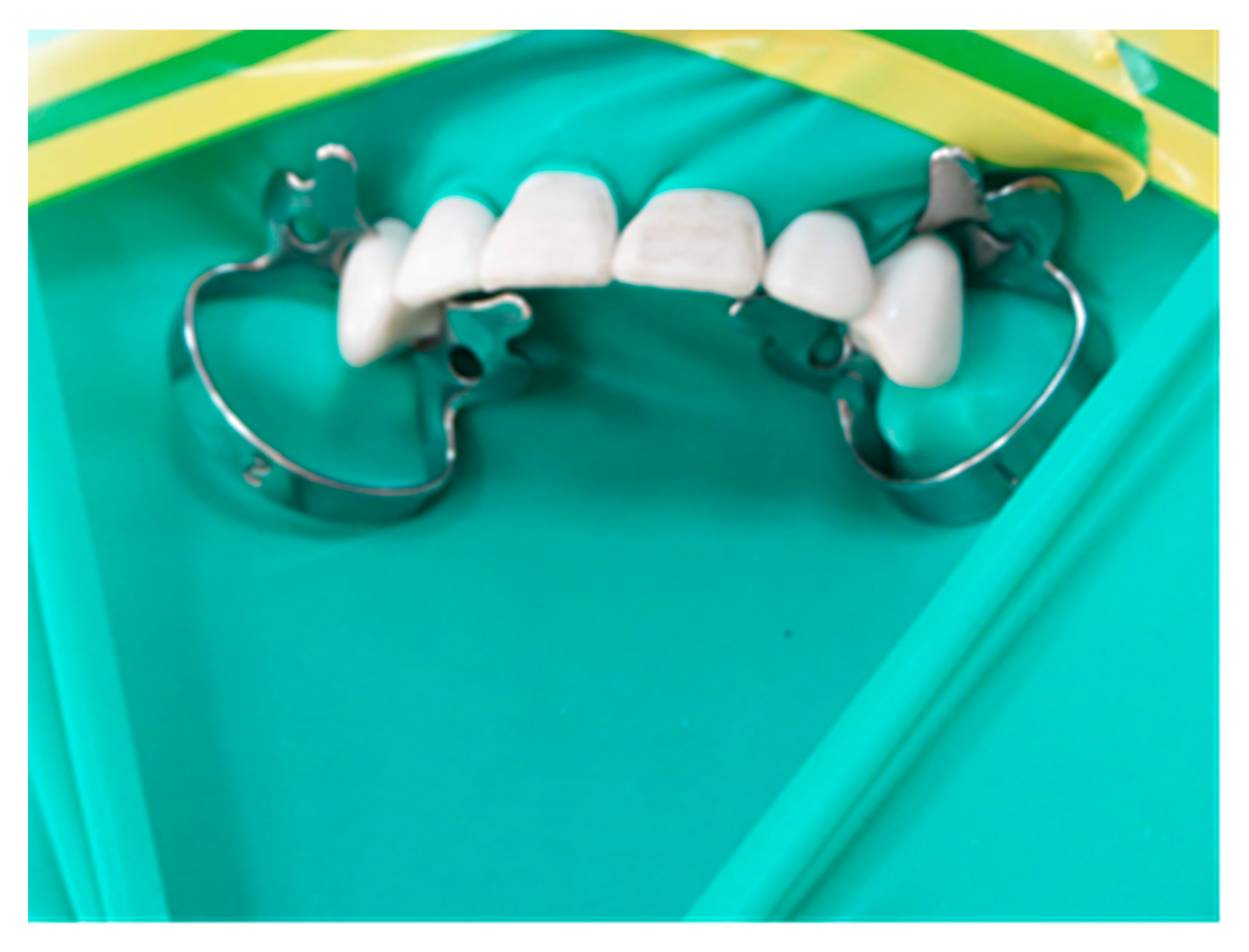
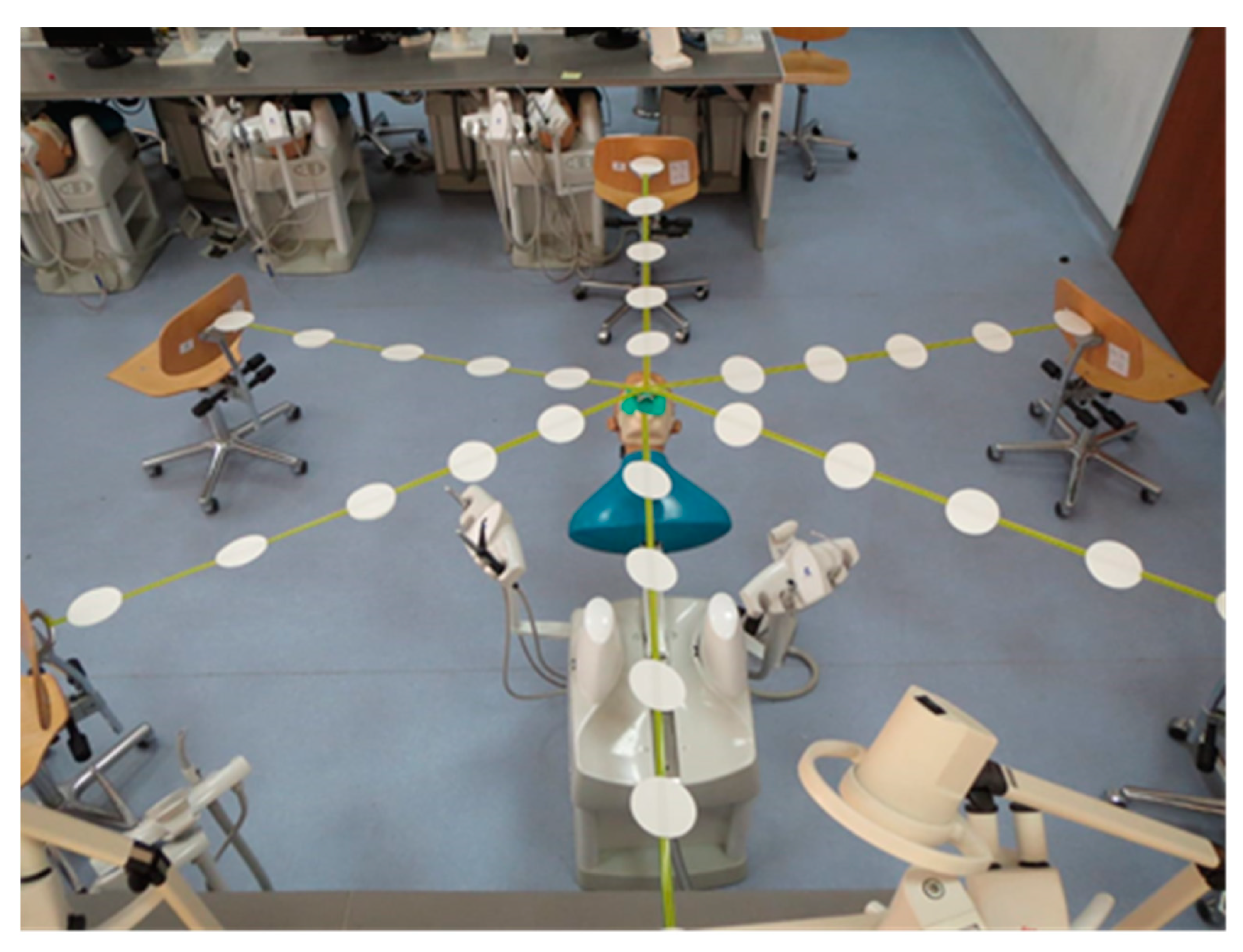
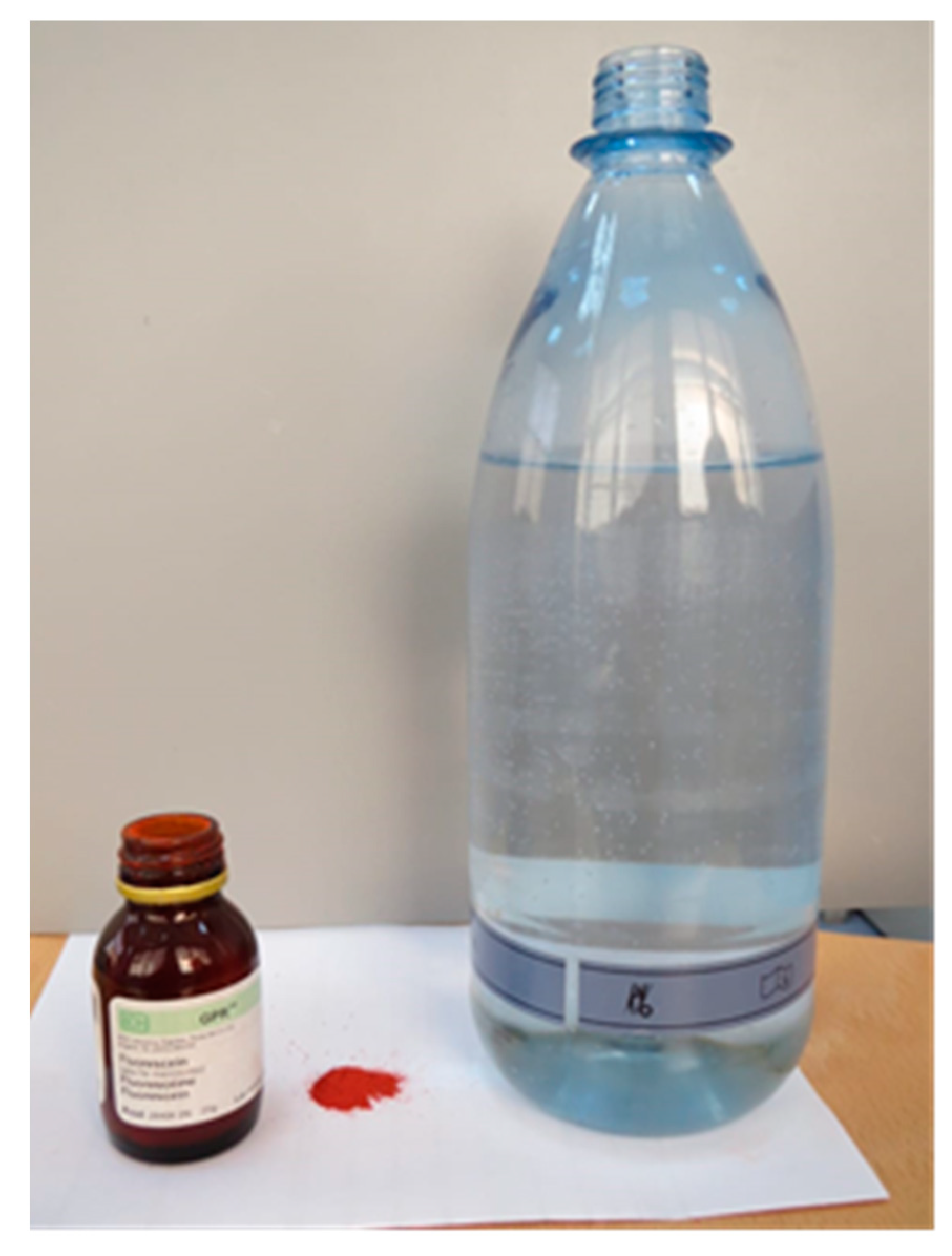
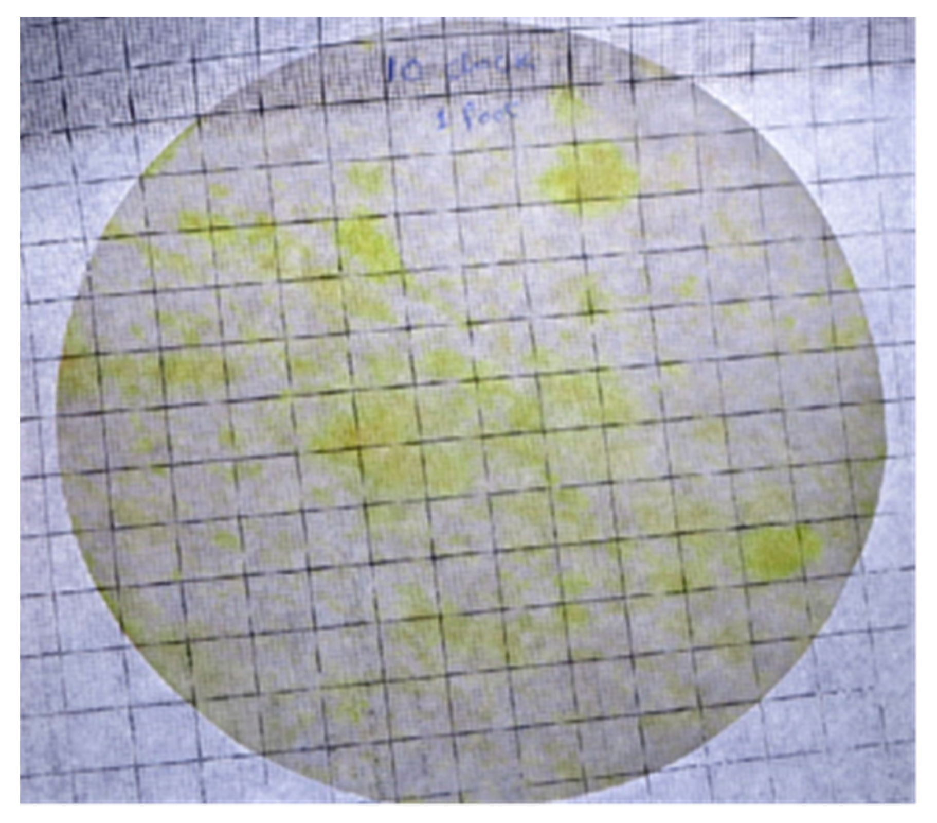
2. Materials and Methods

The study was performed on a dental manikin in the dental simulation laboratory at the College of Dentistry, King Faisal University Al Ahsa. The manikin was set to a reclined position to simulate the clinical operatory position of the patient for dental restorative procedures. Rubber dam isolation was achieved for the upper anterior quadrant from tooth #13 to #23, as shown in Figure 1. Adhesive tape was placed in six different directions from the head of the manikin at 2, 4, 6, 8, 10, and 12 o’clock positions (Figure 2). Grade 1 qualitative cotton cellulose filter paper (Whatman; Maidstone, England) was attached on adhesive tape at a distance of 12-inches from each other up to 60-inches around the dental training manikin. Filter papers were also placed at certain positions of the operator and assistant such as the chest, head, forearms, upper leg, and inside facemask.
One gram of ultra-filtrate containing fluorescent dye (Fluorescein, BDH, Poole, England) was mixed with one liter of water and filtered (Figure 3). This mixture was filled in a reservoir water bottle attached to the dental manikin.
Class V cavity preparation was done by the principal investigator in a specific time of 3 min on tooth #11 using a two-hole high-speed handpiece (PANA MAX, NSK, Tochigi, Japan) then on the next day, Class V cavity preparation was performed on tooth #21 by a four-hole handpiece (KAVO, Biberach, Germany). High volume suction attached to the dental manikin (DSEplus Type 5193, KAVO, Warthausen, Germany) was used with a vented tip throughout the cavity preparation, which was standardized by testing the liquid uptake of 1 L per minute. Immediately after cavity preparation, the first filter paper discs were replaced with new ones in all positions. The second set of filter papers were removed after 30 min. The splatter and aerosol contaminated area on the filter papers were calculated by using transparent grids containing a 1 cm2 box (Figure 4). Each filter paper covered 184 boxes maximum. Even a small amount in a square box was taken as a positive finding.

Statistical Analysis

Data were analyzed using SPSS version 23.0. Mean and standard deviation was calculated for aerosol and splatter produced during cavity preparation at different positions and distances. An independent t-test was used to compare differences in the aerosol and splatter produced using two holes and four holes handpiece. Differences in the amount of splatter produced immediately after cavity preparation and 30 min after cavity preparation were assessed using a paired t-test. A p-value ≤ 0.05 was considered statistically significant.

3. Results

Immediately after the cavity preparation, the greatest amount of splatter of 74 cm2 and 70 cm2 were recorded using the two- and four-hole handpiece, respectively, at the 10 o’clock position and a distance of 12 inches. The amount of splatter decreased with an increase in distance and was found to be the lowest at a maximum distance of 60 inches away from the patient (Table 1). Most splatters were produced at the 10 o’clock position followed by 2 o’clock, 12 o’clock, 9 o’clock, 6 o’clock, and least at the 4 o’clock position.
The assessment of the contaminated surface area on the body revealed that the operator area was more contaminated (254 cm2) compared to the assistant area (197 cm2). The operator’s right hand and the assistant’s left hand were most prone to contamination by the splatter during cavity preparation, followed by the head, chest, and the least contamination was inside the mask for both the operator and assistant (Table 2).
Comparing the two and four-hole handpieces, no significant difference was found in the mean amount of aerosol and splatter produced by both handpieces (Table 3). The amount of aerosol and splatter produced was greatly reduced after 30 min of cavity preparation and only a meager amount was found at a distance of 12 inches and almost no splatter at farther distances (Table 4). Statistically, significant differences were found in the amount of aerosol and splatter produced at distances of 12, 24, and 36 inches immediately after cavity preparation and 30 min after cavity preparation, regardless of the type of handpiece used.

4. Discussion

This study was conducted to evaluate the amount and duration of aerosol and splatter produced during cavity preparation using a two-hole and four-hole high-speed handpiece, at various positions and distances. The result of the current study showed no statistically significant difference in the mean amount of aerosol and splatter produced by both handpieces, however, a statistically significant difference was found in the amount of aerosol and splatter produced at distances of 12, 24, and 36 inches immediately after cavity preparation and 30 min after cavity preparation, regardless of the type of handpiece used.
In this study, maximum splatter and aerosol produced immediately after the procedure were found at the operator zone, followed by the assistant zone. This finding contradicts the previous study in which investigators found more splatter in the assistant zone compared to the operator zone [4]. This difference in findings could be due to different types of dental procedures used for the generation of aerosol and splatter. Furthermore, higher splatter was found on the right arm of the operator and left arm of the assistant followed by the head, chest, and inside the face mask. This result is partially in line with the previous study [4] in which the operator’s right arm and assistant’s left arm were the highest spread area, while the chest was the second most affected area.
Indubitably, various contagious diseases such as HIV and HBV are transmitted through blood, saliva, and the gingival fluid of patients with known or unknown infection; therefore, it is wise to consider all patients potentially infective, irrespective of their medical history [20,21]. Additionally, arm protection is necessary as minor skin scratches are often unrecognized and can be exposed to blood and saliva splatter during the procedure [22]. However, the risk from exposure to impaired skin or mucous membrane is far less likely than that from parenteral contact such as needle prick injury [23].
Recently, COVID-19 infection has received great attention due to the easy transmission through the respiratory route. Ahmed et al. [14] reported fear and anxiety among dentists from different countries of acquiring COVID-19 infection during practice and unintentionally causing harm to their families. Considering the current situation, this study was designed to follow the recommendations from the ADA (American Dental Association) interim guidance to curb the transmission of COVID-19 [24]. The measures included were the use of a rubber dam, four-handed dentistry, and high volume suction. In this study, the penetration of fluorescent dye on the filter paper inside the facemask was found, regardless of the type of handpiece. This result was similar to the previous study in [23], despite the use of a three-layered facemask in our study compared to a one-layered facemask. This finding further endorsed the importance of using a facemask and other protective barriers such as a face shield and external oral suction device to reduce the chance of spreading infection, especially during the time of COVID-19.
The two-hole handpiece is also called Borden’s handpiece, after its successful development by Dr. John Borden’s in 1950. The turbine and body are the main components of the air-driven handpiece. Stainless steel and brass are the most common materials used to manufacture handpieces. Stainless steel handpieces are heavier and expensive than brass handpieces. However, titanium-based handpieces are preferable nowadays by clinicians and the manufacturer due to their lighter weight and greater resistance to repeated sterilization cycles compared to stainless steel [9]. In the present study, although no significant difference was found between the two-hole and four-hole handpiece, however, overall splatter was noticed more in the two-hole handpiece at all positions and distances. A possible explanation for this finding in the two-hole handpiece could be related to its working mechanism, during handpiece working, air traps in the head lead to the continuous rotation of the bur, even after the release of the air-water spray foot pedal; this phenomenon is called the coast speed. With time, manufacturers developed three-hole handpieces to reduce coast speed in which trapped air at the head of the handpiece easily comes out from the third hole that acts as a vent, and thus the bur comes to a pause at the earliest [9,10]. Likewise, the four-hole handpiece has been marketed with an additional hole for fiber optics.
It has been noticed that the whole circumference around the manikin showed splatter contamination for up to 36 inches immediately after the procedure except at the 4 o’clock position with both handpieces and 12 o’clock with the four-hole handpiece. This result is similar to Llandro et al. [25] in which they found a one-meter (39 inches) circumferential spread of splatter in a positive control group (crown preparation with high-speed handpiece). In contrast, Veena et al. [4] found the circumferential spread of splatter up to 12-inches around the dental manikin; which may be due to differences in the procedure (ultrasonic scaling) and technique used for the study.
Bennet et al. [26] reported that aerosol remains in the practice for around 10 to 30 min following scaling. This finding conforms to our study in which the amount of aerosol was found mostly at a distance of 12-inches after 30 min and almost no aerosol at farther distances, although a significant reduction was noticed after 30 min compared to the time of immediate cavity preparation. Therefore, to reduce the risk of contamination from airborne pathogens, especially in this era of the COVID-19 pandemic, it is recommended that practitioners keep wearing their personal protective barriers after the completion of the procedure for a certain period [27].
Considering the findings of the current study, aerosol and splatter can spread up to 36 inches; therefore it is desirable to make an arbitrary “red zone” around the dental unit. This red zone will require thorough cleaning and disinfection by antiviral or antimicrobial disinfectant after every patient. No one should be allowed to enter the red zone during the procedure, except for the dentist and the assistant.
Clinic ventilation plays a major role in mitigating the spread of airborne infections. Sarapultseva et al. [28] reported that the use of aspirating systems installed with HEPA (high-efficiency particulate air) filters can help to evacuate and dissipate aerosols into a specialized area away from the dental practice, thus providing a safe ambiance to the dental staff, especially in the era of the COVID-19 pandemic. However, these are expensive equipment and are not cost-effective. Another reasonable alternative in hand is external high volume suction. In a high-volume evacuator, the use of a large-bore tip diameter (8 mm and above) can efficiently reduce aerosol and splatter up to 93–96% by removing air at 100 cubic feet per minute [29].
This study has some limitations that include the use of liquid volume uptake to measure suction flow due to the unavailability of a flowcheck device. Moreover, the concentration of splatter was not assessed and even a single drop was counted as positive in measuring grid squares. Since the tooth preparation was done only on anterior teeth by the high-speed handpiece, the results should be interpreted cautiously and cannot be generalized, as the results might be different for posterior teeth and with the use of some other devices such as a low-speed handpiece or ultrasonic scaler. Finally, a complete study was conducted on the dental manikin, which does not have saliva and blood, and hence may not produce a real-life work scenario.

5. Conclusions

This study emphasizes that the dissemination of contaminated aerosol and splatter after cavity preparation remains a major threat to dental staff in the era of the COVID-19 pandemic. Therefore, it is advisable to use surgical disposable gowns, caps, face shields, or safety glasses, and refrain from removing the personal protective barriers immediately after the procedure within the vicinity of the dental practice. The use of other adjuncts such as high volume suction to reduce the spread of aerosol and splatter is also recommended.

Author Contributions

Conceptualization, R.J.; Methodology, M.A.A.; Software, M.A.A.; Validation, R.J.; Formal analysis, M.A.A.; Investigation, M.A.A. and R.J.; Resources, R.J.; Data curation, R.J.; Writing—original draft preparation, M.A.A. and R.J.; Writing—review and editing, M.A.A.; Visualization, M.A.A.; Supervision and project administration, M.A.A.; Funding acquisition, M.A.A. All authors have read and agreed to the published version of the manuscript.

Funding

The authors extend their appreciation to the Deanship of Scientific Research, King Faisal University, Al-Ahsa for funding this project, under the Nasher’s Track 206096.

Institutional Review Board Statement

The study was conducted according to the guidelines of the Declaration of Helsinki, and approved by the Research Ethics Committee, Deanship of Scientific Research, King Faisal University (KFU-REC/2021-02-02) on 3 February 2021.

Informed Consent Statement

Not applicable.

Data Availability Statement

Not applicable.

Acknowledgments

The authors are grateful to the Deanship of Scientific Research, King Faisal University, Al-Ahsa for facilitating this project.

Conflicts of Interest

The authors declare no conflict of interest.

References

  1. Shimoji, S.; Ishihama, K.; Yamada, H.; Okayama, M.; Yasuda, K.; Shibutani, T.; Ogasawara, T.; Miyazawa, H.; Furusawa, K. Occupational safety among dental health-care workers. Adv. Med. Educ. Pract. 2010, 1, 41–47. [Google Scholar] [CrossRef] [PubMed] [Green Version]
  2. Harrel, S.K.; Molinari, J. Aerosols and splatter in dentistry: A brief review of the literature and infection control implications. J. Am. Dent. Assoc. 2004, 135, 429–437. [Google Scholar] [CrossRef]
  3. Matys, J.; Grzech-Leśniak, K. Dental aerosol as a hazard risk for dental workers. Materials 2020, 13, 5109. [Google Scholar] [CrossRef]
  4. Veena, H.R.; Mahantesha, S.; Joseph, P.A.; Patil, S.R.; Patil, S.H. Dissemination of aerosol and splatter during ultrasonic scaling: A pilot study. J. Infect. Public Health 2015, 8, 260–265. [Google Scholar] [CrossRef] [PubMed]
  5. Ahmed, M.A.; Jouhar, R.; Adnan, S.; Ahmed, N.; Ghazal, T.; Adanir, N. Evaluation of Patient’s Knowledge, Attitude, and Practice of Cross-Infection Control in Dentistry during COVID-19 Pandemic. Eur. J. Dent. 2020, 14, S1–S6. [Google Scholar] [CrossRef] [PubMed]
  6. Rautemaa, R.; Nordberg, A.; Wuolijoki-Saaristo, K.; Meurman, J.H. Bacterial aerosols in dental practice—A potential hospital infection problem? J. Hosp. Infect. 2006, 64, 76–81. [Google Scholar] [CrossRef] [PubMed]
  7. Yang, M.; Chaghtai, A.; Melendez, M.; Hasson, H.; Whitaker, E.; Badi, M.; Sperrazza, L.; Godel, J.; Yesilsoy, C.; Tellez, M.; et al. Mitigating saliva aerosol contamination in a dental school clinic. BMC Oral Health 2021, 21. [Google Scholar] [CrossRef]
  8. Azim, A.A.; Shabbir, J.; Khurshid, Z.; Zafar, M.S.; Ghabbani, H.M.; Dummer, P.M.H. Clinical endodontic management during the COVID-19 pandemic: A literature review and clinical recommendations. Int. Endod. J. 2020, 53, 1461–1471. [Google Scholar] [CrossRef]
  9. Bhandary, N.; Desai, A.; Shetty, B.Y. High-Speed Handpiece. J. Am. Dent. Assoc. 1993, 124, 16. [Google Scholar] [CrossRef]
  10. Hailu, K.; Lawoyin, D.; Glascoe, A.; Jackson, A. Unexpected Hazards with Dental High Speed Drill. Dent. J. 2017, 5, 10. [Google Scholar] [CrossRef] [Green Version]
  11. Ravenel, T.D.; Kessler, R.; Comisi, J.C.; Kelly, A.; Renne, W.G.; Teich, S.T. Evaluation of the spatter-reduction effectiveness and aerosol containment of eight dry-field isolation techniques. Quintessence Int. 2020, 51, 2–12. [Google Scholar] [CrossRef]
  12. Jacks, M.E. A laboratory comparison of evacuation devices on aerosol reduction. J. Dent. Hyg. 2002, 76, 202–206. [Google Scholar] [PubMed]
  13. Setti, L.; Passarini, F.; De Gennaro, G.; Barbieri, P.; Perrone, M.G.; Borelli, M.; Palmisani, J.; Di Gilio, A.; Piscitelli, P.; Miani, A. Airborne transmission route of covid-19: Why 2 meters/6 feet of inter-personal distance could not be enough. Int. J. Environ. Res. Public Health 2020, 17, 2932. [Google Scholar] [CrossRef] [PubMed] [Green Version]
  14. Ahmed, M.A.; Jouhar, R.; Ahmed, N.; Adnan, S.; Aftab, M.; Zafar, M.S.; Khurshid, Z. Fear and practice modifications among dentists to combat novel coronavirus disease (COVID-19) outbreak. Int. J. Environ. Res. Public Health 2020, 17, 2821. [Google Scholar] [CrossRef] [PubMed]
  15. Consolo, U.; Bellini, P.; Bencivenni, D.; Iani, C.; Checchi, V. Epidemiological aspects and psychological reactions to COVID-19 of dental practitioners in the Northern Italy districts of modena and reggio emilia. Int. J. Environ. Res. Public Health 2020, 17, 3459. [Google Scholar] [CrossRef] [PubMed]
  16. Tysiąc-Miśta, M.; Dziedzic, A. The attitudes and professional approaches of dental practitioners during the COVID-19 outbreak in poland: A cross-sectional survey. Int. J. Environ. Res. Public Health 2020, 17, 4703. [Google Scholar] [CrossRef] [PubMed]
  17. Mijiritsky, E.; Hamama-Raz, Y.; Liu, F.; Datarkar, A.N.; Mangani, L.; Caplan, J.; Shacham, A.; Kolerman, R.; Mijiritsky, O.; Ben-Ezra, M.; et al. Subjective overload and psychological distress among dentists during COVID-19. Int. J. Environ. Res. Public Health 2020, 17, 5074. [Google Scholar] [CrossRef]
  18. Sarapultseva, M.; Zolotareva, A.; Kritsky, I.; Nasretdinova, N.; Sarapultsev, A. Psychological distress and post-traumatic symptomatology among dental healthcare workers in Russia: Results of a pilot study. Int. J. Environ. Res. Public Health 2021, 18, 708. [Google Scholar] [CrossRef]
  19. Kumar, A.R.N.; Karumaran, S.C.; Kattula, D.; Thavarajah, R.; Anusa, A.M. Perceived stress and psychological (Dis)stress among indian endodontists during COVID19 pandemic lock down. MedRxiv 2020, 311, 1–19. [Google Scholar] [CrossRef]
  20. Dahiya, P.; Kamal, R.; Sharma, V.; Kaur, S. “Hepatitis”—Prevention and management in dental practice. J. Educ. Health Promot. 2015, 4, 33. [Google Scholar] [CrossRef]
  21. Sebastiani, F.R.; Dym, H.; Kirpalani, T. Infection Control in the Dental Office. Dent. Clin. North Am. 2017, 61, 435–457. [Google Scholar] [CrossRef]
  22. Kohn, W.G.; Harte, J.A.; Malvitz, D.M.; Collins, A.S.; Cleveland, J.L.; Eklund, K.J. Guidelines for infection control in dental health care settings—2003. J. Am. Dent. Assoc. 2004, 135, 33–47. [Google Scholar] [CrossRef] [PubMed]
  23. Bentley, C.D.; Burkhart, N.W.; Crawford, J.J. Evaluating spatter and aerosol contamination during dental procedures. J. Am. Dent. Assoc. 1994, 125, 579–584. [Google Scholar] [CrossRef] [PubMed]
  24. ADA Releases Interim Guidance on Minimizing COVID-19 Transmission Risk when Treating Dental Emergencies. Available online: https://www.ada.org/en/publications/ada-news/2020-archive/april/ada-releases-interim-guidance-on-minimizing-covid-19-transmission-risk-when-treating-emergencies (accessed on 3 April 2021).
  25. Llandro, H.; Allison, J.R.; Currie, C.C.; Edwards, D.C.; Bowes, C.; Durham, J.; Jakubovics, N.; Rostami, N.; Holliday, R. Evaluating splatter and settled aerosol during orthodontic debonding: Implications for the COVID-19 pandemic. Br. Dent. J. 2021. [Google Scholar] [CrossRef]
  26. Bennett, A.M.; Fulford, M.R.; Walker, J.T.; Bradshaw, D.J.; Martin, M.V.; Marsh, P.D. Microbial aerosols in general dental practice. Br. Dent. J. 2000, 189, 664–667. [Google Scholar] [CrossRef] [PubMed]
  27. Gandolfi, M.G.; Zamparini, F.; Spinelli, A.; Sambri, V.; Prati, C. Risks of aerosol contamination in dental procedures during the second wave of COVID-19—experience and proposals of innovative IPC in dental practice. Int. J. Environ. Res. Public Health 2020, 17, 8954. [Google Scholar] [CrossRef]
  28. Sarapultseva, M.; Hu, D.; Sarapultsev, A. SARS-CoV-2 Seropositivity among Dental Staff and the Role of Aspirating Systems. JDR Clin. Transl. Res. 2021, 6, 132–138. [Google Scholar] [CrossRef]
  29. Aurangjeb, A.M.; Zaman, T.; Badruddoza, M. Practice of Dental Surgeons about Dental Splatter and Aerosol. City Dent. Coll. J. 2013, 10, 10–16. [Google Scholar] [CrossRef] [Green Version]
Figure 1. Upper anterior quadrant isolation by the rubber dam.
Ijerph 18 03773 g001
Figure 2. Distribution of filter papers around the dental manikin at different positions and distances.
Ijerph 18 03773 g002
Figure 3. Dispersion of the ultra-filtrate containing the fluorescent dye.
Ijerph 18 03773 g003
Figure 4. Filter paper disc with a transparent grid to count the contamination area.
Ijerph 18 03773 g004
Table 1. Distribution of aerosol and splatter at different positions and distances by the two- and four-hole handpiece.
DistanceContaminated Surface Area (cm2)
Two-Hole HandpieceFour-Hole Handpiece
12 o’clock2 o’clock4 o’clock6 o’clock8 o’clock10 o’clock12 o’clock2 o’clock4 o’clock6 o’clock8 o’clock10 o’clock
12 inches637141556074596036485570
24 inches495819382853405410293045
36 inches1530-191322-23-111227
48 inches-11-4-10-5---7
60 inches-4---3------
Mean25.434.81223.220.232.419.828.49.217.619.429.8
St. Dev29.0229.0918.1723.225.0630.1227.9327.5515.5920.7123.3828.56
Table 2. Collection of aerosol and splatter on different body parts of the operator and assistant.
Body Part Contaminated Surface Area (cm2)
Operator
Contaminated Surface Area (cm2)
Assistant
Head6752
Chest5539
Right arm9119
Left arm3485
Inside mask72
Total254197
Table 3. Comparison of the mean aerosol and splatter produced by two-hole and four-hole handpiece.
Distancep-ValueMean DifferenceStd. Error Difference
12 Inches0.3986.000006.79379
24 Inches0.5006.166678.81318
36 Inches0.4984.333336.16532
48 Inches0.4012.166672.46869
60 Inches0.1501.166670.74907
Table 4. Comparison of the mean aerosol and splatter produced immediately after cavity preparation and 30 min after cavity preparation using a two-hole and four-hole handpiece.
HandpiecesDistanceTimeMeanSt. Deviationp-Value
Two holes12 inchesPost filling60.666711.910780.000
After 30 min10.175.707
24 inchesPost filling40.833315.223880.001
After 30 min0.501.225
36 inchesPost filling16.500010.054850.010
After 30 min0.000.000
48 inchesPost filling4.16675.154290.105
After 30 min0.000.000
60 inchesPost filling1.16671.834850.180
After 30 min0.000.000
Four holes12 inchesPost filling54.666711.621820.000
After 30 min3.173.817
24 inchesPost filling34.666715.305770.003
After 30 min0.000.000
36 inchesPost filling12.166711.267950.046
After 30 min0.000.000
48 inchesPost filling2.00003.162280.182
After 30 min0.000.000
60 inchesPost filling0.00000.00000-
After 30 min0.000.000
Publisher’s Note: MDPI stays neutral with regard to jurisdictional claims in published maps and institutional affiliations.

