Article

The In Silico Identification of Potential Members of the Ded1/DDX3 Subfamily of DEAD-Box RNA Helicases from the Protozoan Parasite Leishmania infantum and Their Analyses in Yeast

1
Expression Génétique Microbienne, UMR8261 CNRS, Université de Paris, Institut de Biologie Physico-Chimique, 13 rue Pierre et Marie Curie, 75005 Paris, France
2
PSL Research University, 75005 Paris, France
3
Laboratory of Molecular Epidemiology and Experimental Pathology (LR16IPT04), Institut Pasteur de Tunis, Université de Tunis El Manar, 13 Place Pasteur, BP74 Tunis-Belvédère 1002, Tunisia
4
Institut National des Sciences Appliquées et Technologies, Université de Carthage, CEDEX, Tunis 1080, Tunisia
*
Authors to whom correspondence should be addressed.
Submission received: 27 November 2020 / Revised: 27 January 2021 / Accepted: 29 January 2021 / Published: 1 February 2021
(This article belongs to the Special Issue Microbial Genomics and Evolution)

Abstract

:
DEAD-box RNA helicases are ubiquitous proteins found in all kingdoms of life and that are associated with all processes involving RNA. Their central roles in biology make these proteins potential targets for therapeutic or prophylactic drugs. The Ded1/DDX3 subfamily of DEAD-box proteins is of particular interest because of their important role(s) in translation. In this paper, we identified and aligned the protein sequences of 28 different DEAD-box proteins from the kinetoplast-protozoan parasite Leishmania infantum, which is the cause of the visceral form of leishmaniasis that is often lethal if left untreated, and compared them with the consensus sequence derived from DEAD-box proteins in general, and from the Ded1/DDX3 subfamily in particular, from a wide variety of other organisms. We identified three potential homologs of the Ded1/DDX3 subfamily and the equivalent proteins from the related protozoan parasite Trypanosoma brucei, which is the causative agent of sleeping sickness. We subsequently tested these proteins for their ability to complement a yeast strain deleted for the essential DED1 gene. We found that the DEAD-box proteins from Trypanosomatids are highly divergent from other eukaryotes, and consequently they are suitable targets for protein-specific drugs.

1. Introduction

Leishmaniases are parasitic diseases caused by the protozoan parasite of the genus Leishmania. There are currently 54 recognized Leishmania species of which at least 21 are human pathogens that are transmitted by the numerous species of female sandflies belonging to the subfamily Phlebotominae [1,2,3]. These parasites have a complex, dimorphic, life cycle that involves a number of metabolic transitions and differentiated forms. The sandflies inject the flagellated Leishmania metacyclic promastigotes into the bloodstream while feeding on their hosts. The promastigotes are eventually engulfed by macrophages (and other cell types) through phagocytosis whereupon the parasites transform into immobile amastigotes in the acidic environment of the modified phagolysosome (the parasitophorous vacuole; reviewed by [4]) where they feed and reproduce. The replicating amastigotes can infect other macrophages and other cells when the macrophages are lysed or by other poorly defined mechanisms. Infected macrophages are eventually taken back up by the sandfly during the blood meal whereupon the amastigotes differentiate into extracellular, flagellated, procyclic promastigotes in the midgut of the sandflies. They subsequently divide into metacyclic promastigotes that migrate to the pharyngeal valve of the sandfly for the next round of infection [4,5,6].
Leishmania are endemic in more than 98 countries creating serious health problems in many countries around the world. They are manifested in several clinical forms ranging from self-healing skin lesions of cutaneous leishmaniasis (CL) to the more severe visceral leishmaniasis (VL) [7,8,9]. Around 700,000 to 1 million new cases and some 26,000 to 65,000 deaths occur annually (WHO, 2020, [10]). Despite tremendous efforts, no effective vaccine has yet been developed [11,12,13]. Mainstay treatment regimes for leishmaniases are based on chemotherapy involving the use of the pentavalent antimonials. Commonly used second-line drugs are miltefosine, amphotericin B, liposomal amphotericin B and paromomycin. All these treatments require long treatment courses, are toxic and costly, and have adverse effects; moreover, there is the risk of the parasites developing drug resistance [14,15,16]. Thus, there is need to identify new specific targets in order to develop new drugs to control the disease. Due to the availability of the genomic sequence of most Leishmania species during the last few years, there are extensive efforts to identify new genes involved in the pathogenicity or that are essential for survival of the parasites that could be the targets for therapeutic or prophylactic compounds.
The DEAD-box family of RNA helicases are ubiquitous proteins found in all kingdoms of life that are involved in all processes involving RNA from transcription to decay (reviewed by [17,18,19]). They are part of the DExD/H-box superfamily 2 (SF2) of putative DNA and RNA helicases that are named after the amino acid sequence of the Walker B motif (motif II) that is involved in NTP binding and hydrolysis. They contain catalytic cores consisting of two, linked, RecA-like domains that have the conserved motifs associated with ligand binding and enzymatic activity, and they have highly variable amino- and carboxyl-terminal sequences and insertions within the catalytic core (reviewed by [20,21]). The DEAD-box proteins are ATP-dependent RNA binding proteins and RNA-dependent ATPases that are capable of unwinding short duplexes in a nonprocessive fashion in vitro, but typically at very high protein to substrate ratios. In addition, they can remodel RNA and ribonucleoprotein (RNP) complexes, but they generally have little or no substrate specificity. In contrast, in vivo the proteins show a high degree of specificity; for example, the yeast Saccharomyces cerevisiae encodes 25 different DEAD-box proteins that are mostly essential and that are not interchangeable—even when overexpressed—with the exception of a single paralog resulting from gene duplication [22,23]. Humans have 37 different proteins [21]. Thus, DEAD-box proteins, and DExD/H-box proteins in general, provide a rich source of potential drug targets (reviewed by [24,25,26,27,28,29]).
The Ded1/DDX3 subfamily of DEAD-box proteins is of particular interest (reviewed by [30,31,32,33]). They are considered translation-initiation factors that are important for 43S ribosome scanning to the initiation complex, but they also shuttle between the nucleus and cytoplasm using the XpoI/CrmI and Mex67/TAP nuclear pore complexes, and they are found in cytoplasmic foci (P-bodies or stress granules) containing translation-inactive mRNAs under stress conditions [34,35,36,37,38]. The yeast DED1 is an essential gene that is rescued by the plasmid-borne expression of its orthologs from other eukaryotes, including mammalian DDX3X, which demonstrates a high degree of functional conservation ([34] and references therein). Due to the important roles these proteins play in the cell, the Ded1/DDX3 subfamily of proteins are considered important targets for therapeutic or prophylactic drugs (reviewed by [25,26,39,40,41,42]). However, it is not clear whether the Ded1/DDX3 subfamily of proteins is actually functionally conserved throughout all the eukaryotes. Moreover, it is not clear what distinguishes the Ded1/DDX3 subfamily from closely related subfamilies such as Vasa/DDX4, which is involved in developmental regulation (reviewed by [43,44,45,46]).
The Kinetoplastida trypanosomatids belong to the Euglenozoa phylum that separated very early from other eukaryotes during evolution [47,48]. Therefore, their genes are expected to be highly divergent from other organisms. Moreover, Trypanosoma and Leishmania are obligate parasites, which means their genes are subjected to additional divergent forces. Thus, we expect that their encoded proteins will have unique features that will make them particularly useful as targets for the development of protein-specific drugs. In the same vein, we expect comparisons with the homologs from other organisms will reveal highly conserved features that characterize the functionality of the proteins and that will facilitate their identification in other organisms. Moreover, the majority of the Leishmania and Trypanosoma genes are constitutively expressed as polycistronic RNAs that are subsequently processed into separate capped and polyadenylated mRNAs; thus, translation, and probably DDX3-like proteins, plays a particularly important role in the control of gene expression, especially for the differentiation into the different parasitic forms (reviewed by [49,50,51]). Indeed, we previously showed that the eIF4A-like translation protein, LieIF4A (LINF_010012800/LINF_010012900), is a viable target for therapeutic drugs [52,53].
In this work, we aligned the RecA-like core sequences of 28 of the 29 identified DEAD-box proteins from L. infantum and compared them to the consensus sequences of DEAD-box proteins, in general, and to the consensus sequence of the Ded1/DDX3 subfamily in particular. We further analyzed the amino- and carboxyl-terminal sequences for motifs that are characteristic of the Ded1/DDX3 proteins. We then compared the identified candidate proteins to their homologs from T. brucei. Finally, we tested the identified proteins from Leishmania and Trypanosoma for their capacity to complement a yeast strain deleted for the endogenous Ded1 protein at different expression levels and as chimeras of the Ded1 protein. Our results reveal not only the conservation of certain features in these proteins but also the high degree of divergence from other eukaryotes.

2. Materials and Methods

2.1. Data Acquisition and Analyses

Sequences for L. infantum JPCM5 were obtained from the TriTrypDB website (Release 46; https://tritrypdb.org/tritrypdb/) using DEAD-box and helicase as a search terms [54]. These sequences were then compared with those obtained with a Blast of the TriTrypDB website using Ded1 as bait (E-value ≤ 4 × 10⁻⁴). The sequences were subsequently screened for the presence of the conserved motifs that are characteristic of DEAD-box family proteins and that distinguished them from related helicase families. We obtained 29 different genes. The derived protein sequences were then aligned along with representative Ded1/DDX3 proteins on the EMBL-EBI T-coffee server (https://www.ebi.ac.uk/Tools/msa/tcoffee/) using ClustalW with the default setting for the initial alignments [55]. These alignments were subsequently refined by eye to maximize the sequence homology and conserved distances of the motifs using our previous characterizations of DEAD-box proteins as a basis [56]. Phylogenetic trees were derived from the EMBL-EBI T-coffee server using ClustalW and the default settings.
To identify Ded1/DDX3-specific features, we did a Blast search (PMID: 9254694) of the UniProtKB web site (https://www.uniprot.org/) of the Swiss-Prot (release 54.6) and TrEMBL (release 37.6) entries using yeast Ded1 (P06634) as bait [57]. We obtained 250 sequences from various organisms (E-value ≤ 4 × 10−78), most of which were classified as Ded1/DDX3 or Vasa/DDX4 in the annotations, that were refined to eliminate duplicates, fragments, and proteins that did not conform to the DEAD-box motifs to yield 229 unique sequences. These sequences were aligned as described above. We used the Bock coot-alignment-consensus Perlscript to derive the consensus sequence, which is unfortunately no longer available on the EMBL-Heidelberg server. The consensus sequence yielded most but not all the known motifs of the Ded1/DDX3 subfamily; for example, the eIF4E binding motif was not recovered. Hence, we further refined the alignments using Blast sequences with E-values equal to or less than 10−113 to yield 96 unique sequences. These sequences were used to obtain the final consensus sequence, but they do not represent the full diversity of sequences of the Ded1/DDX3 subfamily.
The equivalent T. brucei 427 and 927 genes were identified by Blasts against the trypanosome genome using the selected LINF genes as bait. The proteins identified were equivalent to those listed on the TriTrypDB website as homologs of the corresponding LINF genes. The Tb427.05.3600 protein (the long form of Tb927.5.3600) as listed on TriTrypDB had three unidentified amino acids; we sequenced the gene and identified the amino acids as X107V, X280V, and X529C. These sequences were aligned as described above.
The selected proteins were analyzed with PredictProtein (https://www.predictprotein.org) and Pfam (https://pfam.xfam.org/), and they were compared with the results for Ded1 and DDX3 [58,59]. The presence of leucine-rich nuclear export signals (NES) was analyzed with the NetNES 1.1 Server (http://www.cbs.dtu.dk/services/NetNES/) [60]. We further modeled the selected proteins with Swiss-Model using the default setting; the resulting different models were visualized with Swiss-PdbViewer 4.1.0 [61,62,63].

2.2. Preparation of L. infantum and T. brucei DNA

We used L. infantum LV50 strain (MHOM/TN/94/LV50) that was isolated from a patient suffering from visceral leishmaniasis [64]. The promastigote form was cultured in RPMI-1640/GlutaMAX medium (Gibco BRL, Darmstadt, Germany) complemented with 100 U/mL penicillin, 100 µg/mL streptomycin, and 10% heat-inactivated fetal bovine serum (FBS; Gibco BRL) at 22 °C as previously described [64]. The production of flagellated promastigotes was monitored with a microscope. Parasites at the stationary phase were collected by centrifugation at 4 °C at 3500 rpm (2380 g) in a JOUAN CR3.12 swinging bucket centrifuge for 15 min and stored at −80 °C until needed.
The total genomic DNA was extracted from stationary phase cells. The cell pellet was resuspended in a lysis buffer containing 50 mM NaCl, 10 mM EDTA, 50 mM Tris–HCl, pH 7.5, 100 μg/mL Proteinase K, and 0.5% SDS, and then it was incubated overnight at 55 °C to lyse the cells. The DNA was extracted with phenol and chloroform:isoamyl alcohol (24:1), and then it was precipitated with ethanol as previously described [65]. The concentration of DNA was quantified using the Qubit 2.0 Fluorometer (Invitrogen, Singapore City, Singapore). The T. brucei 427 strain DNA was a kind gift from Lucy Glover (Pasteur Institute, Paris, France).

2.3. Cloning and Yeast Manipulations

All manipulations of yeast including media preparation, growth conditions, transformation, and 5-fluoroorotic acid (5-FOA) selection, were done according to standard procedures [66]. We used the W303 and BY4742 wildtype yeast strains for all the protein expression experiments. The ∆ded1 yeast strain (ded1::HIS3) containing the YCplac33-DED1 plasmid used in the complementation assays was as previously described [67].
The oligonucleotides used in the cloning are shown in Supplementary Materials Table S1. The selected LINF genes were PCR amplified using the indicated oligonucleotides with the purified L. infantum or T. brucei DNA. PCR reactions were done in a Bio-Rad T100 Thermal Cycler with Q5 High-Fidelity DNA polymerase (New England Biolabs, Évry-Courcouronnes, France) according to the manufacturer’s recommendations. In brief, the amplifications were performed with 1 µL of DNA (20 ng/µL) in a 50 µL volume containing 0.02 U/µL Q5 High-Fidelity DNA polymerase, 1 × buffer, 200 µM dNTPs, 1 × GC enhancer, and 2 µM from each oligonucleotide for 30 cycles. The amplified DNA was digested with SpeI and XhoI for most of the constructs, with SpeI and SalI for LINF08, XbaI and SalI for TRYP08, and with SpeI and SmaI for TRYP35. The digested fragments were purified on a 0.8% agarose gel, eluted with a NucleoSpin Gel and PCR Clean-Up kit (Macherey-Nagel, Düren, Germany) according to the manufacturer’s instructions and cloned into the equivalent sites of the Bluescript plasmid (Stratagene, La Jolla, CA, USA). Final constructs were verified by sequencing and are shown in Supplementary Materials Table S2.
The constructs were subsequently subcloned into the ADH-2HA_p415, ADH-2HA_p424, and GPD-2HA_p424 yeast expression plasmids, and transformed into either the wildtype or ded1::HIS yeast strains. Cells were grown in synthetic defined (SD) medium lacking leucine (for p415) or tryptophan (for p424). Cultures of the ded1::HIS yeast strain were subsequently serially diluted by a factor of 10 and spotted on SD plates containing 5-FOA and grown at 18 °C, 30 °C, and 36 °C. Proteins from cultures of the wildtype strains were extracted for Western blot analyses.

2.4. Synthetic Gene Construction

The synthetic sequence constructs were made by first optimizing the codons to yeast DED1 using the COOL software that was previously available on the cool.syncti.org server [68]. The proposed sequences were then visually modified to eliminate long strings of homogeneous nucleotides and conflicting restriction sites. The genes were synthesized by Eurofin Scientific (Luxembourg) with 5’ SpeI-NdeI and 3’ XhoI restriction sites. The DNAs were digested with SpeI and XhoI and cloned into the equivalent sites of the 2HA-p415 and 2HA-p424 plasmids. Final constructs were all verified by sequencing. The LINF08Lsyn and LINF32syn had the correct protein sequence. The LINF35syn construct introduced an E408K mutation, which was in the amino-terminal sequence outside the catalytic core. The LINF08Ssyn was constructed by PCR amplification of the LINF08Lsyn with LinJ08_up3 and LinJ08_low2 oligonucleotides containing SpeI, NdeI, and XhoI sites (Supplementary Materials Table S1). The PCR product was digested with SpeI and XhoI, and then cloned into the equivalent sites of the above plasmids. The final constructs were all verified by sequencing.

2.5. Ded1 Chimeras of the LINF and TRYP Proteins

The protein chimeras contained the LINF or TRYP RecA-like cores as previously defined [22], using the optimized LINFsyn and wildtype T. brucei genes, and the amino- and carboxyl-terminal sequences of Ded1. The design took advantage of a conserved aspartic acid (or asparagine in the case of DDX3X) that occurred 7–8 residues upstream of the isolated conserved aromatic group (generally an F) of the Q motif, and a conserved aliphatic group 29–31 residues downstream of motif VI. The final constructs contained the entire RecA-like cores of the LINF and TRYP proteins and the corresponding flanking sequences of Ded1 (1–136, 525–604). Constructs were made with the Gibson Assembly Cloning kit (New England Biolabs) according to the manufacturer’s instructions. Oligonucleotides were designed using the NEBuilder Assembly Tool provided on the New England Biolabs web site and are shown in Supplementary Materials Table S1. As a positive control, we also made a Ded1-DDX3X chimera. All constructs were verified by sequencing.

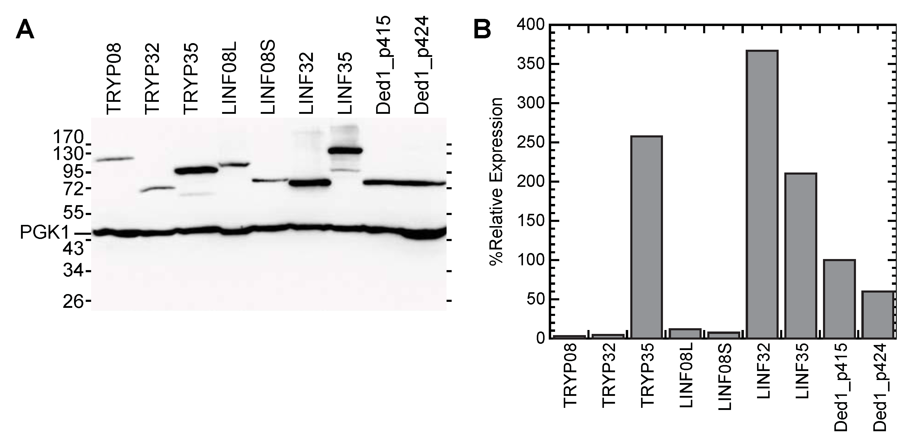
2.6. Western Blot Analyses

The W303 yeast strain was transformed with the above described plasmids or with the empty plasmids as negative controls. Cells were grown with shaking at 30 °C in SD-LEU medium for cultures transformed with the p415 plasmid and in SD-TRP medium for cultures with the p424 plasmid, and they were harvested by centrifugation at an OD600 of 1–2. Proteins were extracted from the cell pellets by alkaline hydrolysis as previously described [69]. The extracted proteins were separated on a 10% SDS-Laemmli-polyacrylamide gel, electrophoretically transferred to nitrocellulose membranes (Amersham Protran, GE Healthcare Life Science, Chicago, IL, USA), probed with anti-HA antibodies (Covalab, Bron, France) and the signals were revealed with a Clarity Western ECL Substrate kit (Bio-Rad, Hercules, CA, USA) using a Bio-Rad ChemiDoc XRS+ and Image Lab 5.2 software.

3. Results

3.1. Bioinformatic Analyses

3.1.1. Identification of L. infantum DEAD-Box Proteins

We were primarily interested in L. infantum because it is the cause of visceral leishmaniasis, which is the most severe form of the disease, and consequently the proteins were of interest as potential targets for therapeutic drugs. Out of 102 sequences listed as helicases on the TriTryp website, we found 29 genes that contained the conserved motifs that were characteristic of DEAD-box proteins in L. infantum. We compared these sequences with those of other Leishmania species and discovered that LINF_080005700 was much longer in most other species of Leishmania and other trypanosomatids than for L. infantum. We inspected the upstream DNA sequence and found another ATG start codon in the same open reading frame that would yield a protein product of similar size to those of the other species. It contained an amino-terminus that was 181 amino acids longer than that listed on the TriTryp database for L. infantum, and it was possible that both long and short forms of the protein were expressed. Moreover, the T. brucei equivalent, Tb927.5.3600, was predicted to be expressed as both a long and short form [70]. Consequently, we used both forms for all subsequent analyses.
We compared our results with the previous in silico analyses of RNA helicases in trypanosomatids [71]. L. major and L. infantum are closely related, and the majority of the identified DEAD-box proteins were the same [72]. LmjF.05.0140 did not originally appear to exist in L. infantum but very recent work shows that the L. infantum homolog (LINF_050006300) was misclassified as a rRNA on the TriTrypDB database [73]. Consequently, it was not included in these alignments, but it encodes a DEAD-box protein that functions as a putative nucleolar RNA helicase II [74]. We found three more DEAD-box proteins in both organisms: LINF_200013600 (LmjF.20.0870), LINF_110006800 (LmjF.11.0190), and LINF_160005500 (LmjF.16.0050). Finally, the L. infantum equivalent of LmjF.10.0140, LINF_100006300, lacked the motifs characteristic of DEAD-box proteins. Thus, we identified 30 different DEAD-box genes in L. infantum, although the eIF4A-like LINF_010012800 and LINF_010012900 encoded the same protein. Thus, there were 29 different DEAD-box proteins, which includes LINF_050006300.

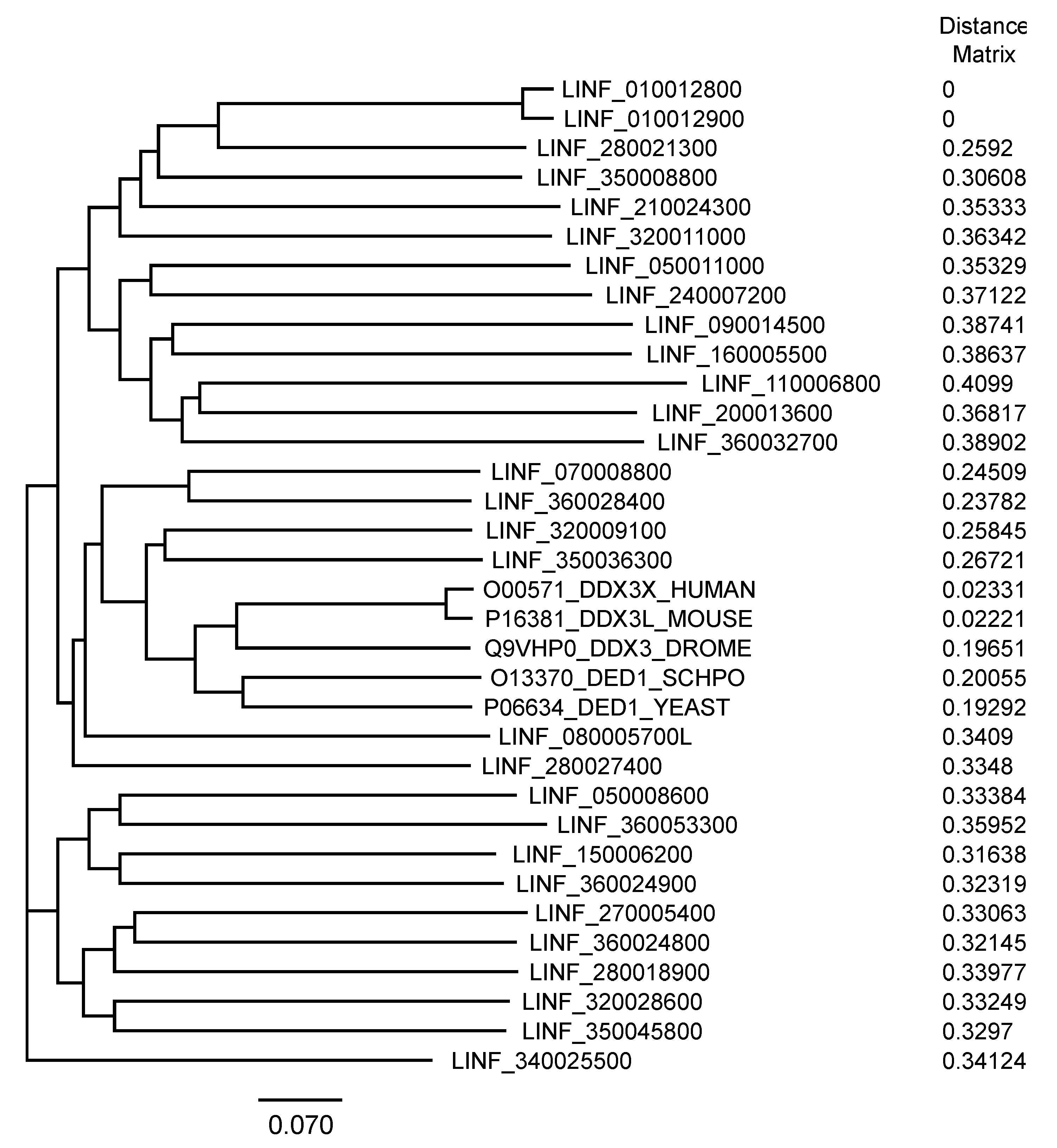
3.1.2. Phylogenetic Relationship of the L. infantum Proteins and the Ded1/DDX3 Subfamily

We used T-coffee to align these sequences against yeast Ded1 and four DDX3-like proteins that are the functional homologs—orthologs—of Ded1; which is, the four proteins are able to complement a yeast strain deleted for the essential DED1 gene ([34] and reference therein). The phylogenetic tree result is shown in Figure 1. These data showed that the Leishmania proteins were highly divergent from the known Ded1/DDX3 proteins, but that the previously identified LINF_320009100 and to a lesser extent LINF_350036300 were the most closely related [75,76]. However, DEAD-box proteins are characterized by highly conserved RecA-like cores and highly variable flanking sequences [22]. It was unclear whether the former, the latter or both contained the distinguishing features of the Ded1/DDX3 subfamily.
Consequently, we repeated the T-coffee alignment with the core sequences alone starting from the highly conserved, but isolated, amino-terminal aromatic residue to the carboxyl-terminal end of motif VI. As expected, the phylogenetic tree was similar, but it showed less divergence (Supplementary Materials Figure S1A). The Ded1/DDX3 subfamily of proteins are characterized by amino-terminal, leucine-rich, nuclear-export signal (NES), an eIF4E binding motif, and a conserved GINF sequence, and they contain a conserved carboxyl-terminal RDYR sequence [77,78]. The flanking sequences of the LINF DEAD-box proteins varied enormously in length; nevertheless, we repeated the T-coffee alignments with the flanking sequences alone (Supplementary Materials Figure S1B). The results showed that LINF_320009100 again was the most closely related to the Ded1/DDX3 subfamily. The LINF_350036300 protein was more distantly related after LINF_070008800. Thus, both the RecA-like cores and flanking sequences provided distinguishing features, but the flanking sequences were more pronounced, probably in part because of their more variable sizes.

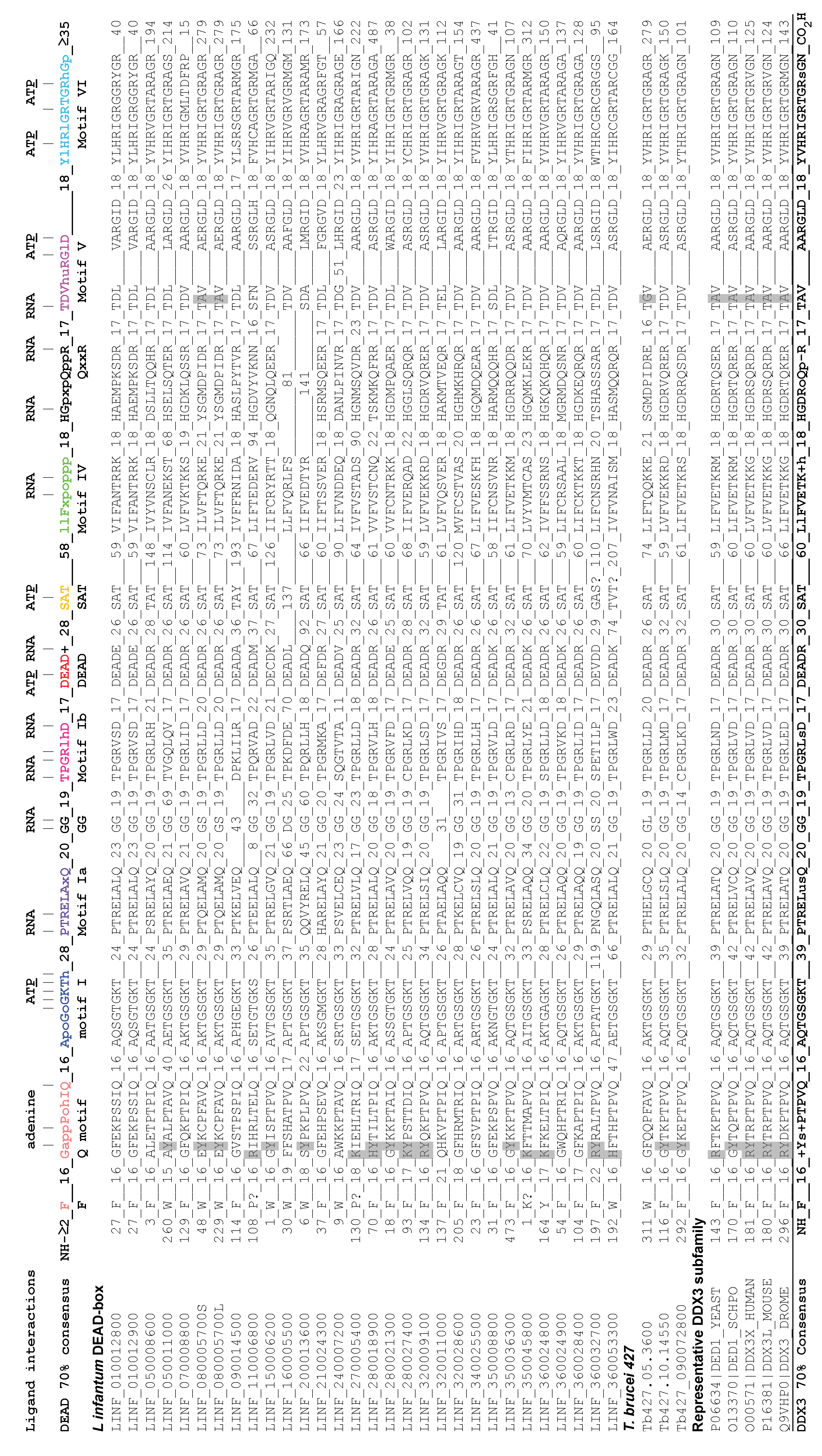
3.1.3. Sequence Alignments of the DEAD-Box Proteins

We aligned the RecA-like core sequences according to the parameters that we previously determined [56]. The conserved motifs generally appear at fixed distances from one another due to the steric constraints of the RecA-like core. This was used to refine alignments with divergent motifs. The results are shown in Figure 2. Most of the proteins had conserved motifs that conformed well to the consensus sequence that was previously determined for DEAD-box proteins in general [56]. These motifs form highly conserved interactions with the ligands, as determined from the solved crystal structures of a number of different DEAD-box proteins; these interactions are shown in Figure 2 ([56,79] and references therein). However, a number of proteins had poorly defined or missing motifs, which was most pronounced for LINF_160005500 and LINF_200013600. The QxxR motif appeared to be particularly divergent in the Leishmania proteins. However, it was possible that some of the differences were due to sequencing errors or strain variability. This may explain the 51 amino-acid insertion in motif V of LINF_240007200, but we very occasionally see an insertion at this position in other DEAD-box proteins as well. This region of motif V may be particularly vulnerable to insertions as the insert occurred between the regions that interacted with the RNA and ATP. We otherwise noted that the distances between motifs V, VI, and QxxR were conserved in this protein.

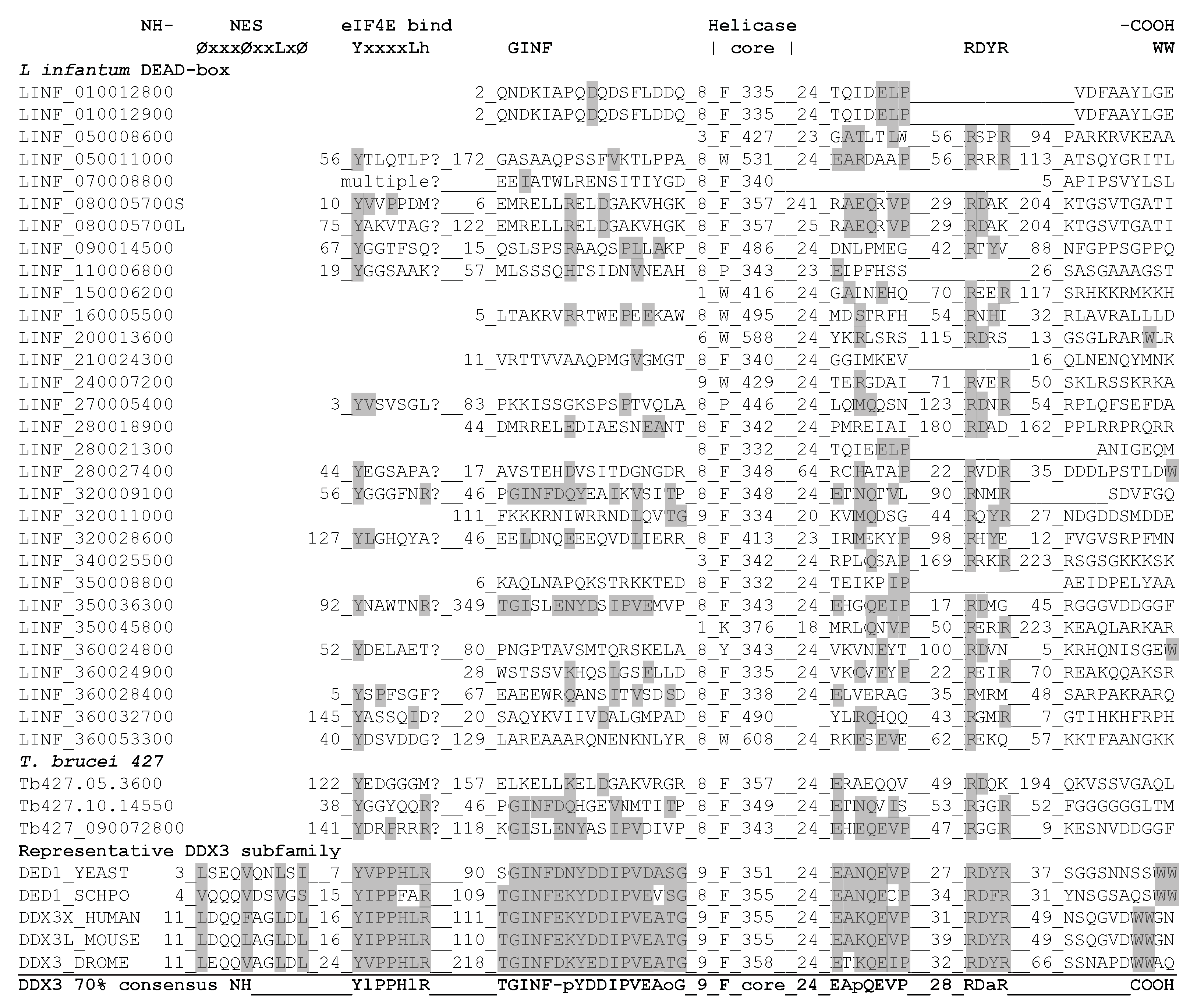
3.1.4. Identification of Ded1/DDX3-Specific Characteristics

To facilitate comparisons, we determined the consensus sequence of 229 Ded1/DDX3 proteins that were identified from different organisms on the UniProtKB web site (https://www.uniprot.org/) using yeast Ded1 as bait and aligned using CLUSTAL W [81,82]. We further refined these alignments by using only those sequences with an E-value of 10−113 or less to yield 96 unique sequences that largely had the characteristics (defined above) of the Ded1/DDX3 subfamily. We used an arbitrary 70% consensus as a reference to further compensate for misidentified or ambiguous protein sequences (Figure 2). Consequently, the consensus sequences shown do not take into account the full diversity of sequences of potential members of this subfamily.
The core motifs of the Ded1/DDX3 subfamily were similar to those of other DEAD-box proteins in general, but there were significant differences. The Q motif consists of a loop-helix-loop-helix structure on the amino-terminus of the RecA-like core [83]. It contains an isolated aromatic group, which is often a phenylalanine, that is generally 17 residues upstream of the Q motif sequence GappPohIQ, where the glutamine is absolutely conserved, a is an aromatic group that is often a phenylalanine, p is a polar residue, o is an alcohol, and h is hydrophobic. The isolated phenylalanine forms stacking interactions with the proline, and the loop-helix-loop-helix forms a “cap” on RecA-like domain 1 [83]. The conserved glutamine interacts with the N6 and N7 positions of the adenine residue of ATP, and it is the only residue that can do so in the context of the DEAD-box protein structure. A glycine or small hydrophobic residue before the aromatic group is present in over 75% of the Q motifs of the DEAD-box sequences. The glycine occurs at a helix-loop transition, which is probably facilitated by a small residue. In contrast, we found that the Ded1/DDX3 subfamily had a positively charged residue, generally arginine, in over 85% of the sequences. This was true for a number of the LINF proteins as well, but the LINF_320009100 protein was the most similar.
Likewise, motif V is characterized by an aspartic acid in the second position in over 75% of the sequences and by a polar group in over 90%. This aspartic acid interacts with the first arginine of motif VI, which also makes highly conserved cation–π interactions with the conserved phenylalanine of motif IV that are important for the cooperative binding of RNA and ATP [84]. The Ded1/DDX3 subfamily had an alanine or small hydrophobic residue in over 75% of the sequences that would be expected to eliminate this interaction between motifs V and VI. Among the LINF proteins, only LINF_080005700 shared this characteristic. The Ded1/DDX3 subfamily also is thought to be distinguished by an insert between motif I and Ia that was proposed to affect RNA binding [31,85]. However, this region was highly variable in the LINF proteins and Ded1/DDX3 alignments, and regardless the insert is far from the RNA binding site in the solved crystal structures. It is not well conserved in plants and invertebrates [31]. Therefore, it did not appear to be a distinguishing feature. Likewise, other differences of the core sequences did not appear to be distinctive for the Ded1/DDX3 subfamily.

3.1.5. Identification of Ded1/DDX3-Specific Motifs outside the RecA-Like Core

The Ded1/DDX3 subfamily of proteins are cap-associated factors involved in translation initiation that actively shuttle between the nucleus and cytoplasm using the Mex67/TAP and XpoI/CrmI nuclear pore complexes (reviewed by [34,35]). The XpoI/CrmI-dependent export involves a leucine-rich NES near the amino-terminus with the sequence ØxxxØxxLxØ, where Ø denotes amino acids M, V, I, L, F, or W [86]. This region was clearly visible in the alignments of the five verified Ded1/DDX3 proteins (where V and L are interchangeable), but it was not found in the T-coffee alignments of the Ded1/DDX3 subfamily in general, probably because it lacked sufficient sequence conservation (Figure 3). Indeed, the NetNES 1.1 server was only able to predict the metazoan amino-terminal NES and not the yeast sequences [60]. The trypanosomatids contain the Ran-GTPase associated with the CrmI nuclear pore, but the sequence elements recognized are still poorly characterized [87]. Hence, it was unclear whether the leucine-rich NES was present in any of the LINF sequences.
Proteins involved in translation initiation often have one or more eIF4E binding motif; in the case of the Ded1/DDX3 subfamily, this motif is close to the amino terminus [78,88,89]. The motif is defined by the sequence YxxxxLh, where x is any amino acid [88]. The Ded1/DDX3 subfamily differed from this motif by having an arginine or positively charged residue as the terminal residue in over 80% of the sequences instead of a hydrophobic group (Figure 3). Leishmania are known to have often cryptic eIF4E binding motifs that are difficult to identify [90]. Nevertheless, we could find partial potential eIF4E binding motifs on the amino terminus for most of the LINF proteins, but LINF_080005700S, the short version, showed the best fit at a relevant distance from the amino terminus (Figure 3). Interestingly, LINF_070008800 showed multiple repeats of potential eIF4E binding motifs. However, it should be noted that trypanosomatids have at least six variant eIF4E proteins that are structurally different from those of other organisms due to the unusual cap-4 structure on their mRNAs (reviewed by [49,91,92]). Indeed, none of the tested Leishmania eIF4E proteins complement a yeast strain deleted for the essential eIF4E gene [93].
Shih et al. and Floor et al. found an amino-terminal motif consisting of the GINF sequence and a carboxyl-terminal motif consisting of RDYR that is characteristic of the Ded1/DDX3 subfamily through an alignment of six proteins [77,78]. We were able to expand on these results with our more extensive collection of sequences and compare them with our previous DEAD-box alignments [56]. The results are shown in Figure 3. The conserved residues of the GINF motif were characteristic of the Ded1/DDX3 subfamily, and they were more extensive than previously noted. The motif occurred at a fixed distance from the isolate aromatic group of the Q motif, which made its identification largely unambiguous. It is predicted to form a short α-helix [77]. None of the LINF sequences showed a strong conservation in this sequence, but LINF_320009100 and LINF_350036300 were the most similar.
The RDYR motif appeared at a more variable distance from motif VI, and it is thought to be important for the oligomerization of the protein [77]. Most of the LINF proteins had potential variants of this sequence as did the DEAD-box proteins in general, and consequently it did not appear to be a distinguishing feature of this subfamily (Figure 3). Moreover, it has been noted that a pair of tryptophans were often present at the carboxyl-terminus of the Ded1/DDX3 subfamily [77,78]. We found a tryptophan doublet within the last five residues of the carboxyl-terminus in about 70% of our aligned Ded1/DDX3 proteins and less often a tryptophan-aspartic acid (20%). The role(s) of these residues is unknown. Tryptophans were often present in our LINF alignments, but they were scattered at different positions and rarely appeared as doublets. Finally, we noted an additional motif with the sequence EApQEVP that appeared at a conserved distance after motif VI that existed in the Ded1/DDX3 subfamily but not in DEAD-box proteins in general (Figure 3). This sequence was most evident in LINF_080005700, LINF_320009100, and LINF_350036300.
From these data, and based on its similar size, we concluded that LINF_320009100 was the most likely ortholog of the Ded1/DDX3 subfamily. LINF_350036300 appeared to belong to the related DDX4 subfamily of proteins that includes Drosophila Vasa. Vasa has very occasionally shown partial complementation of a yeast strain deleted for the DED1 gene, but only after very long incubation times, which also reflects the remarkable adaptability of yeast [94]. LINF_080005700 appeared to be a potential paralog of the Ded1/DDX3 subfamily because of its unusual motif V and Q motif, and because of the potential eIF4E binding and EApQEVP motifs. LINF_070008800 and LINF_360028400 lacked most of the distinguishing features of the Ded1/DDX3 subfamily despite their proximity in the phylogenetic tree.

3.1.6. Comparisons between the L. infantum and T. brucei Ded1/DDX3-Like Proteins

To validate the selection of these proteins, we analyzed the equivalent genes from T. brucei. The Tb427.05.3600 protein was similar to LINF_080005700 in that it contained an unusual glycine in the second position of motif V (not a D or polar group; Figure 2), but it lacked the other characteristic features. In contrast, motif Ia in both proteins contained either a glutamine or a histidine in place of the highly conserved arginine in the third position; this residue makes conserved interactions with the RNA substrate, and it is an arginine in over 95% of the aligned DEAD-box and Ded1/DDX3-subfamily proteins (Figure 2). Both the glutamine and histidine substitutions would be expected to lose or significantly reduce the interactions with the RNA at this position. Indeed, a similar R276K mutation in human DDX3X is associated with medulloblastoma cancer [95]. Finally, both Tb427.10.14550 and Tb427_090072800 had partial conservation of the GINF and EApQEVP motifs, which was consistent with them being homologs of LINF_320009100 and LINF_350036300, respectively (Figure 3).
The nuclear localization signal (NLS) is poorly defined in the Ded1/DDX3 subfamily, but nuclear proteins of trypanosomatids are often characterized by a KRxR motif [96]. This motif was present in Tb427.05.3600 and LINF_080005700, but was not apparent in the other proteins. Moreover, Tb427.05.3600 has a predicted NES as well [96]. Nevertheless, Tb427.05.3600, Tb427.10.14550, and Tb427_090072800 are all enriched in the nuclear fractions of the tsetse fly midgut-form of T. brucei extracts [96]. These results were consistent with the identified proteins shuttling between the cytoplasm and nucleus and re-enforced their identification as potential Ded1/DDX3 homologs.
To facilitate subsequent analyses and comparisons, LINF_080005700 will be henceforth named LINF08L/S (long/short), LINF_320009100 is LINF32, LINF_350036300 is LINF35, Tb427.05.3600 is TRYP08, Tb427.10.14550 is TRYP32, and Tb427_090072800 is TRYP35.

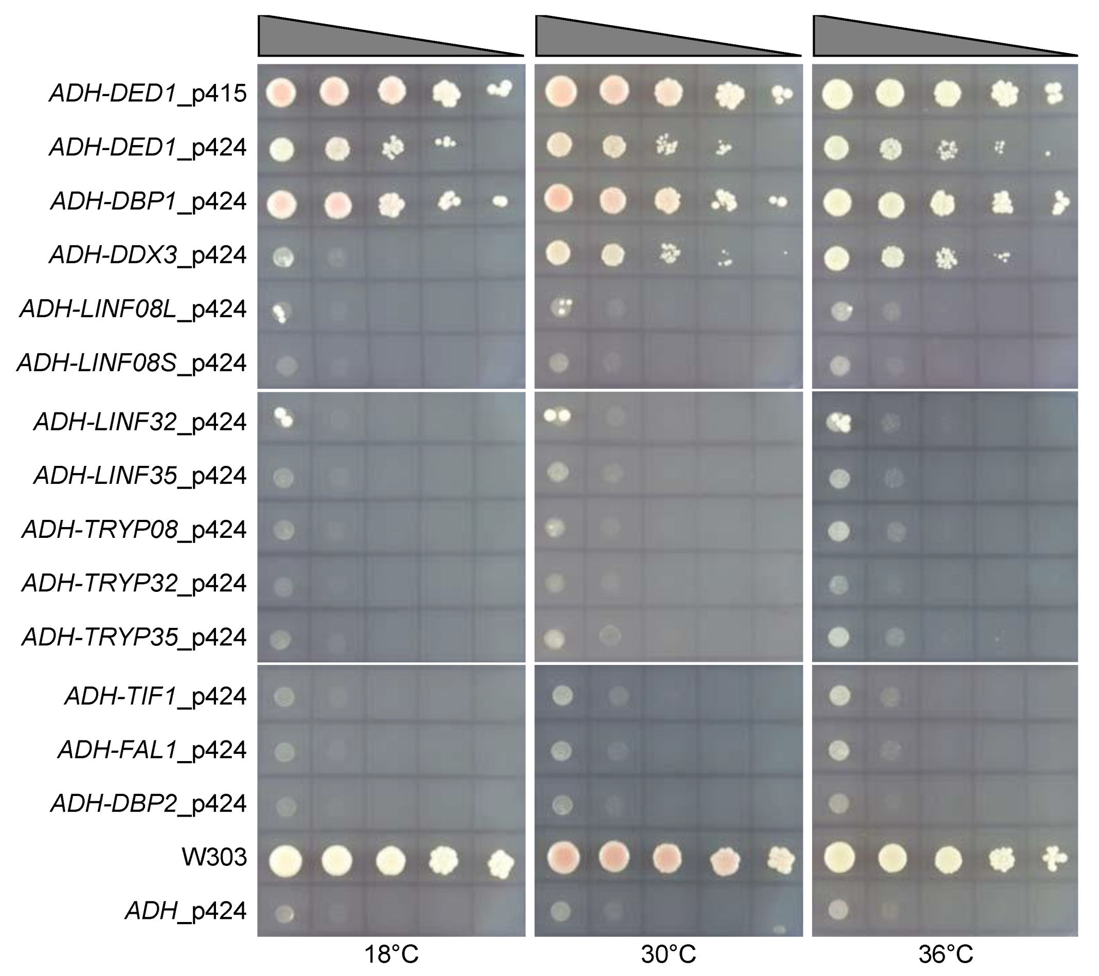
3.2. Yeast Complementation

The best evidence for a functional conservation of the Ded1/DDX3 subfamily is the ability to complement the deletion of the equivalent gene in yeast. Indeed, others and we have shown that proteins from organisms as diverse as Drosophila, mammals, and S. pombe are able to support growth in yeast strains deleted for the essential DED1 gene ([34] and references therein). Therefore, we cloned the selected genes in yeast expression vectors with two, amino-terminal, HA tags under the control of the strong ADH promoter to test if the selected proteins were the orthologs of the Ded1/DDX3 subfamily [97]. The various constructs were then transformed into a yeast strain deleted for the chromosomal copy of the DED1 gene (ded1::HIS) but that expressed Ded1 off a plasmid containing a URA marker [67]. Only cells that had lost the URA plasmid can grow when plated on agar medium containing 5-FOA, but only if the corresponding TRP or LEU plasmids expressed functional homologs of Ded1. As positive controls, we used the human DDX3X gene and a yeast paralog of DED1, DBP1, which partially compensates for the deletion of the DED1 gene when overexpressed [98]. As negative controls, we used TIF1 (eIF4A), FAL1, and DBP2 genes, all of which encode unrelated yeast DEAD-box proteins. The results are shown in Figure 4.
Overexpression of the yeast Ded1 protein results in a dominant-negative growth phenotype [22,99]. This was apparent by the reduced growth of cultures expressing the ADH-DED1_p424, 2µ-multicopy, plasmid relative to the wildtype growth of W303 or the centromeric p415 plasmid (Figure 4). As expected, both human DDX3X and yeast DBP1 partially compensated for the loss of the DED1 gene, but the cells transformed with the LINF or TRYP genes showed no growth, although we did see a few isolated colonies. We recovered the plasmids from these isolated colonies and sequenced the genes; they all contained copies of DED1. This most likely resulted because the URA plasmid encoding DED1 developed a mutation in the URA gene that inactivated its expression, which resulted in isolated cell growth on the 5-FOA plates. Therefore, none of the expressed LINF or TRYP genes complemented the ded1 deletion. This was in contradiction with a previous publication that showed that the equivalent of LINF32 and LINF35 in L. major, LmjF.32.0400, and LmjF.35.3200, respectively, showed nearly wildtype growth [75]. However, these cultures were restreaked a second time on 5-FOA plates, and it was likely that the growth resulted from the above mentioned DED1-URA plasmids. Indeed, the expression of the tagged L. major LmjF32.0400 was not detected in yeast [75].

3.3. Protein Expression

A trivial explanation for the lack of complementation by the LINF or TRYP genes was that the proteins were poorly expressed or rapidly degraded. Therefore, we prepared cell cultures expressing the HA-tagged proteins in the wildtype W303 strain, extracted the proteins, separated them on SDS-PAGE, transferred the proteins to nitrocellulose membranes, and then probed with IgG specific for the HA tag. As a control, we probed the same membranes with IgG against the endogenously expressed PGK1 protein (YCR012W). The results are shown in Figure 5.
LINF32 and TRYP08 showed some variable expression levels between different preparations, but LINF32 was generally expressed in excess over Ded1 while TRYP08 was poorly expressed. The other proteins showed more consistent expression levels. Both LINF08S and LINF08L were very poorly expressed. The long form of LINF08, which is about 24% larger than the short form, seemed to be expressed slightly better. The much larger LINF35 was 2-fold more highly expressed than Ded1_p415 while the TRYP35 was 2.5-fold more expressed. Similar results were obtained when the proteins were expressed off the p415 or p424 plasmids. Thus, it was unlikely that the absence of complementation was due to the poor expression of LINF35 and TRYP35, but the other proteins were more ambiguous.

3.4. Synthetic Genes

Yeast genes tend to be rich in adenines and thymidines. Indeed, Ded1 has a G/C content of 46% (Table 1). In contrast, both Trypanosoma and Leishmania have much higher G/C content in their genes, and the proteins of interest had over 50% and 60%, respectively. This high G/C content could have interfered with the transcription or translation of the genes, although LINF35 was well expressed even with the highest G/C content of over 65% (Table 1). However, gene expression depends on the context of the G-C base pairs and on the presence of rare codons. Thus, it was possible that the variable or poor expression was caused both by the G/C content and by rare codons. Thus, we synthesized the Leishmania genes with a codon usage optimized based on DED1, and we then tested for their ability to complement the ded1 deletion strain. We cloned the genes into plasmids with the strong ADH and the very strong GPD promoters; the latter has up to 10-fold higher expression levels than the ADH promoter [97]. The results are shown in Supplementary Materials Figure S2.
We obtained a few isolated colonies with ADH-LINF08syn and a more significant number of colonies with GPD-LINF32syn; we recovered the plasmids from these colonies and sequenced them as described above. In all cases we recovered the DED1 gene. Hence, none of the LINF genes complemented the yeast strain deleted for the endogenous DED1. It remained possible that the proteins were not well expressed even off the strong ADH and very strong GPD promoters. Therefore, we prepared yeast extracts of the W303 strain transformed with genes as described above. The results are shown in Supplementary Materials Figure S3.
The synthetic LINF genes had very high expression levels of LINF08Lsyn and LINF32syn that was 3.5- and 5.4-fold higher, respectively, than for DED1. LINF35syn showed only 63% of the expression while LINF08Ssyn was expressed at about the same level as DED1 (1.1-fold). Consequently, the LINF genes with the codons optimized for expression in yeast showed very different expression levels than the wildtype genes. Constructs under the control of the GPD promoter showed similar results. Nevertheless, none of the genes were able to complement the deletion of DED1 even at these high levels of expression. The relative reduced expression of the LINF35 synthetic gene was probably a result of increased protein degradation that resulted from very high expression [100].

3.5. Ded1 Chimeras

Our alignments indicated that both the catalytic cores and the amino- and carboxyl-terminal flanking sequences contained specific features characteristic of the Ded1/DDX3 subfamily. However, it seemed likely that the flanking sequences were further adapted for their cellular environment. For example, the amino-terminus of human DDX3X interacts with innate, immune, signaling factors that are not present in yeast Ded1 (reviewed by [101]). It was possible that the LINF and TRYP genes were similarly modified to optimize their functionality in the environment of the parasites. In contrast, we expected the catalytic cores to retain the same functionality as the enzymatic activities would be expected to be similar in all organisms. We previously showed that the catalytic cores of yeast DEAD-box proteins were only interchangeable between proteins with similar functionalities [22]. We reasoned that this might be the case for the trypanosomatids as well.
We based our constructions on sequence alignments, 3D modeling of the proteins and on our previous experience [22]. We used the catalytic cores of the wildtype TRYP genes and the optimized, synthetic, LINF genes, and we used the amino- and carboxyl-terminal sequences of Ded1. As a positive control, we made a chimera between human DDX3X and yeast DED1, as the DDX3X protein showed partial complementation in yeast (Figure 4). To push the system, we incubated the plates for over a week, and to facilitate comparisons we used the same series of cultures as shown in Figure 4. The results are shown in Supplementary Materials Figure S4.
At very long incubation times even the empty plasmid showed slight growth at 30 °C and 36 °C. This might reflect an adaptation by the yeast and by the weak expression of the endogenous DBP1 gene [98]. The negative controls TIF1, FAL1, and DBP2 similarly showed weak growth. The Ded1-DDX3 positive control showed slightly less growth then the intact DDX3X protein, which was probably the result of altered interactions between the core and flanking sequences. Except for the DED1-TRYP32 and DED1-TRYP35 chimeras, all of the LINF and TRYP DED1 chimeras showed a very slight enhancement of growth that oddly was most apparent at 36 °C. The optimum growth temperature for yeast is around 30 °C [66]. Similarly, the wildtype LINF32, LINF35, TRYP08, and TRYP35 constructs showed slightly better growth that was most apparent at 36 °C after long incubation times. Slight growth was previously observed with the DDX4 protein Vasa but at 30 °C [94]. Thus, the catalytic cores of the LINF and TRYP proteins seem to be optimized for higher temperatures than the human and yeast proteins. While the weak growth was indicative of a homology between the proteins, it was too weak to demonstrate that they were functional orthologs of the Ded1/DDX3 subfamily.

4. Discussion

Our sequence alignments indicate that LINF32 and TRYP32, also known as HEL67 [102], are the most likely orthologs of DDX3, LINF35 and TRYP35 the homologs of DDX4-like proteins, and LINF08 and TRYP08 are probably more distantly related paralogs of DDX3. The DDX4 proteins have only been characterized in metazoans, and they are involved in developmental regulation ([33] and references therein). They probably evolved after the branching of metazoans [103]; however, it remains possible that such proteins have emerged in trypanosomatids for similar reasons. This nevertheless remains conjuncture because none of the proteins complement a yeast strain deleted for DED1. Moreover, although the Leishmania genomes are highly conserved at the content and synteny levels, the encoded genes have a high divergence of functionality even between species (reviewed by [104]). Thus, it is possible that all the identified proteins are paralogs of DDX3 that have evolved other cellular functions. Alternatively, the different proteins may be optimized, and expressed, for the different cellular environments that exist within the insect or mammalian hosts, but they nevertheless retain the same functionality. Indeed, mammals encode at least two to three different DDX3 proteins that are not interchangeable (reviewed by [30,31,33]). Human DDX3Y is needed for normal spermatogenesis, and it is translated only in the testes; DDX3X is ubiquitously expressed. It is interesting to note that the plant Nicotiana benthamiana contains six DDX3-like homologs, which perhaps reflects the divergent types of cells (roots, leaves, flowers, etc.; [105]).
The Ded1/DDX3 subfamily is characterized by specific sequence motifs in the amino- and carboxyl-terminal domains involving the NES and eIF4E binding motif, and other motifs of undefined functionality. Only LINF32, LINF35, TRYP32, TRYP35, and to a lesser extent LINF08 partially share these characteristics. However, it should be emphasized that this is based on the consensus sequence of a selected subset of Ded1/DDX3 proteins that does not reflect the full diversity of sequences. In addition, the Ded1/DDX3 proteins have RecA-like cores with a noncanonical Q motif and motif V. Typically, the first amino acid of the Q motif is a glycine or other small residue, which is probably needed to facilitate the helix-loop transition in the structure. In the Ded1/DDX3 subfamily this residue is often an arginine followed by a tyrosine. Only LINF_360032700 and LINF32 share this characteristic. However, this position appears flexible as S. pombe has a more typical glycine as the first amino acid. Moreover, changing the arginine to glycine in yeast Ded1 has no apparent phenotype (our unpublished data). The aspartic acid to alanine change in the second position of motif V is more profound as it would be expected to eliminate an important interaction with the first arginine of motif VI and thereby introduce instability in the interactions between motifs, which may be important for the functionality of the Ded1/DDX3 proteins. Only LINF08 (an alanine) and TRYP08 (a glycine) shared this characteristic. However this change may not be critical as the alanine to arginine mutation in yeast yields only a slight growth phenotype (our unpublished data). However, we expect that the arginine to alanine change would alter the cooperative binding of ATP and RNA [84].
The L. major equivalents of LINF32 and LINF35 and LmjF32.0400 and LmjF35.3100, respectively, have a weak interaction with the eIF4E proteins LeishIF4E-1 and LeishIF4E-4 that is probably mediated by the mRNA in both the promastigotes and axenic amastigote-like cells, which is consistent with a role in translation [75,106]. However, in contrast to yeast Ded1, no direct contact was noted between the proteins and the Leishmania eIF4A, eIF4E, and eIF4G proteins [34,75]. LmjF35.3100 showed more variable interactions between the different eIF4E proteins and Leishmania forms that are consistent with it having a developmental role. However, pronounced defects in translation and cell growth were only noted when both of the equivalent T. brucei proteins were silenced in procyclic cells of Trypanosoma ([75] and reference therein). Consistent with this, the double knockout of L. infantum LINF32 was viable although it was sensitive to heat stress and acidic pH, and consequently it was unable to undergo axenic amastigote differentiation [76,102]. LmjF35.3100 is more abundant in promastigotes while LmjF32.0400 is more prevalent in amastigotes [75]. LINF32 was likewise more prevalent in axenic amastigotes [107]. The equivalent proteins in L. donovani, LdBPK_080080 (LINF08), LdBPK_320410 (LINF32), and LdBPK_353150 (LINF35) were recovered in both promastigotes and amastigotes, but LdBPK_080080 showed a weak signal [108]. This might explain why only LINF32 and LINF35 were found in L. infantum promastigotes [109].
LINF32 also has been implicated in blocking antisense-dependent rRNA fragmentation in L. infantum and thereby reducing cell death under stress conditions [76]. Similarly, LINF32 plays a central role in mitochondrial proteostasis under conditions of stress [102]. Under these conditions, the LINF32 knockout mutant has depolarized mitochondrial membranes that lead to mitochondrial fragmentation and cell death. Pull-down experiments reveal interactions with proteins involved in antioxidant and unfolded protein response, but also interactions with LINF35 [102]. The L. donovani null (knockout) mutations of LdBPK_320410 impair the infectivity and induce protective immunity against visceral leishmaniasis, and as with L. infantum they were unable to differentiate as axenic amastigotes [110]. Thus, the Leishmania DDX3-like proteins show significant functional divergence from their counterparts in other organisms.
LINF08 and TRYP08 remain an enigma, as there is little information in the literature. Both proteins appear to be derived from DDX3-like proteins, but they have amino acid substitutions in motif Ia that would be expected to reduce RNA binding. Consistent with this, the equivalent proteins of LINF08, LINF32, and LINF35 in L. mexicana (LmxM.08.0080, LmxM.31.0400, and LmxM.34.3100, respectively) are expressed in both amastigotes and promastigotes, but only LmxM.31.0400 and LmxM.34.3100 are recovered as proteins prominently crosslinked to mRNAs [111]. However, other LINF proteins have altered RNA-binding motifs as well, including LINF_090014500, LINF_110006800, LINF_200013600, LINF_240007200, LINF_320011000, LINF_320028600, and LINF_360032700. The LINF proteins LINF_090014500, LINF_110006800, LINF_160005500, and LINF_240007200 appear to be particularly divergent from the canonical DEAD-box consensus motifs. While this may partially reflect sequencing errors and strain variability, it also reflects the high degree of divergence of the LINF proteins from other eukaryotes. It remains to be seen whether they still function as ATP-dependent RNA binding proteins and RNA-dependent ATPases.
The functional roles of LINF08 and TRYP08 remain unclear, but RNA interference studies in T. brucei show that TRYP08 is of critical importance for the viability of the bloodstream form but not for the other forms [112]. Interestingly, the bloodstream loss-of-fitness group was strongly overrepresented by proteins associated with flagellar motility, which was not the case for the flagellated, procyclic, loss-of-fitness group [112]. Thus, TRYP08 plays a critical role at only specific phases of the life cycle. In contrast, TRYP35 is important throughout the lifecycle while TRYP32 plays a more minor role [112]. Double and triple knockdowns are required to determine if these three proteins have overlapping activities.
Further work is needed to characterize these proteins in order to determine their cellular role(s) and whether they are true homologs of the Ded1/DDX3 subfamily. This includes the enzymatic characterization of the purified proteins and deletions and mutations of the endogenous genes to reveal their functional importance at the different phases of the parasite. Nevertheless, it is clear that the Leishmania and Trypanosoma DEAD-box proteins are highly divergent from their eukaryote counterparts in other organisms, and thus they provide a rich source of potential, protein-specific, drug targets.

Supplementary Materials

The following are available online at https://0-www-mdpi-com.brum.beds.ac.uk/2073-4425/12/2/212/s1, Table S1: Oligonucleotides used in this study, Table S2: Constructs used in this study, Figure S1: Phylogenetic tree of LINF and Ded1/DDX3 proteins, Figure S2: Complementation of synthetic LINF genes optimized for expression in yeast, Figure S3: Expression of synthetic LINF genes optimized for yeast, Figure S4: Complementation of the yeast ded1::HIS strain.

Author Contributions

Conceptualization: N.K.T., M.B., I.G.; methodology: N.K.T., I.G.; investigation: M.M., Y.Z.A., J.B., E.H., H.Y.-A., R.O., N.K.T.; data curation: M.M., N.K.T.; writing—original draft preparation: N.K.T., M.M.; writing—review and editing: M.M., Y.Z.A., N.K.T., M.B., I.G.; visualization; supervision: M.B., I.G., N.K.T.; project administration, M.B., I.G., N.K.T.; funding acquisition, M.B., I.G., N.K.T. All authors have read and agreed to the published version of the manuscript.

Funding

This research was funded by PHC Utique project N° 37063TG/N.K.T.; PHC Utique projet N° 17G 0820/M.B.; the Centre National de la Recherche Scientifique; and Initiative d’Excellence program from the French State grant DYNAMO [grant number ANR-11-LABX-0011-01]. The study received support from the Ministry of Higher Education and Scientific Research (LR16IPT04 contract program/I.G.).

Institutional Review Board Statement

Not applicable.

Informed Consent Statement

Not applicable.

Data Availability Statement

Not applicable.

Acknowledgments

We thank Caroline Lacoux and Stéphanie Ørum for helpful discussions; The human DDX3X clone was a kind gift of Giovanni Maga. We thank Marine Pasquier for help with the cloning of the TRYP genes, and we thank Lucy Glover for the kind gift of the Trypanosoma DNA.

Conflicts of Interest

The authors declare no conflict of interest. The funders had no role in the design of the study; in the collection, analyses, or interpretation of data; in the writing of the manuscript, or in the decision to publish the results.

References

  1. Akhoundi, M.; Kuhls, K.; Cannet, A.; Votýpka, J.; Marty, P.; Delaunay, P.; Sereno, D. Correction: A Historical Overview of the Classification, Evolution, and Dispersion of Leishmania Parasites and Sandflies. PLoS Negl. Trop. Dis. 2016, 10, e0004770. [Google Scholar] [CrossRef] [Green Version]
  2. Akhoundi, M.; Kuhls, K.; Cannet, A.; Votýpka, J.; Marty, P.; Delaunay, P.; Sereno, D. A Historical Overview of the Classification, Evolution, and Dispersion of Leishmania Parasites and Sandflies. PLoS Negl. Trop. Dis. 2016, 10, e0004349. [Google Scholar] [CrossRef]
  3. Akhoundi, M.; Downing, T.; Votýpka, J.; Kuhls, K.; Lukeš, J.; Cannet, A.; Ravel, C.; Marty, P.; Delaunay, P.; Kasbari, M.; et al. Leishmania infections: Molecular targets and diagnosis. Mol. Aspects Med. 2017, 57, 1–29. [Google Scholar] [CrossRef] [PubMed]
  4. Uribe-Querol, E.; Rosales, C. Control of Phagocytosis by Microbial Pathogens. Front. Immunol. 2017, 8, 1368. [Google Scholar] [CrossRef] [Green Version]
  5. Liévin-Le Moal, V.; Loiseau, P.M. Leishmania hijacking of the macrophage intracellular compartments. FEBS J. 2016, 283, 598–607. [Google Scholar] [CrossRef] [PubMed]
  6. Arango Duque, G.; Descoteaux, A. Leishmania survival in the macrophage: Where the ends justify the means. Curr. Opin. Microbiol. 2015, 26, 32–40. [Google Scholar] [CrossRef] [PubMed]
  7. Alvar, J.; Vélez, I.D.; Bern, C.; Herrero, M.; Desjeux, P.; Cano, J.; Jannin, J.; den Boer, M. Leishmaniasis worldwide and global estimates of its incidence. PLoS ONE 2012, 7, e35671. [Google Scholar] [CrossRef]
  8. Guizani, I.; Mukhtar, M.; Alvar, J.; Ben Abderrazak, S.; Shaw, J. Encyclopedia of Environmental Health; Nriagu, J.O., Ed.; Elsevier: Burlington, VT, USA, 2011; pp. 453–480. [Google Scholar]
  9. Herwaldt, B.L. Leishmaniasis. Lancet 1999, 354, 1191–1199. [Google Scholar] [CrossRef] [Green Version]
  10. World Health Organization. 2020. Available online: http://www.who.int/news-room/fact-sheets/detail/leishmaniasis (accessed on 23 November 2020).
  11. Didwania, N.; Shadab, M.; Sabur, A.; Ali, N. Alternative to Chemotherapy-The Unmet Demand against Leishmaniasis. Front. Immunol. 2017, 8, 1779. [Google Scholar] [CrossRef] [Green Version]
  12. Kumar, R.; Engwerda, C. Vaccines to prevent leishmaniasis. Clin. Transl. Immunol. 2014, 3, e13. [Google Scholar] [CrossRef]
  13. Seyed, N.; Peters, N.C.; Rafati, S. Translating Observations From Leishmanization Into Non-Living Vaccines: The Potential of Dendritic Cell-Based Vaccination Strategies Against Leishmania. Front. Immunol. 2018, 9, 1227. [Google Scholar] [CrossRef] [PubMed]
  14. Ghorbani, M.; Farhoudi, R. Leishmaniasis in humans: Drug or vaccine therapy? Drug Des. Dev. Ther. 2018, 12, 25–40. [Google Scholar] [CrossRef] [PubMed] [Green Version]
  15. Hefnawy, A.; Berg, M.; Dujardin, J.C.; De Muylder, G. Exploiting Knowledge on Leishmania Drug Resistance to Support the Quest for New Drugs. Trends Parasitol. 2017, 33, 162–174. [Google Scholar] [CrossRef] [PubMed] [Green Version]
  16. Roatt, B.M.; de Oliveira Cardoso, J.M.; De Brito, R.C.F.; Coura-Vital, W.; de Oliveira Aguiar-Soares, R.D.; Reis, A.B. Recent advances and new strategies on leishmaniasis treatment. Appl. Microbiol. Biotechnol. 2020, 104, 8965–8977. [Google Scholar] [CrossRef] [PubMed]
  17. Cordin, O.; Banroques, J.; Tanner, N.K.; Linder, P. The DEAD-box protein family of RNA helicases. Gene 2006, 367, 17–37. [Google Scholar] [CrossRef]
  18. Linder, P.; Jankowsky, E. From unwinding to clamping—The DEAD box RNA helicase family. Nat. Rev. Mol. Cell Biol. 2011, 12, 505–516. [Google Scholar] [CrossRef] [Green Version]
  19. Putnam, A.A.; Jankowsky, E. DEAD-box helicases as integrators of RNA, nucleotide and protein binding. Biochim. Biophys. Acta 2013, 1829, 884–893. [Google Scholar] [CrossRef] [Green Version]
  20. Byrd, A.K.; Raney, K.D. Superfamily 2 helicases. Front. Biosci. 2012, 17, 2070–2088. [Google Scholar] [CrossRef]
  21. Fairman-Williams, M.E.; Guenther, U.P.; Jankowsky, E. SF1 and SF2 helicases: Family matters. Curr. Opin. Struct. Biol. 2010, 20, 313–324. [Google Scholar] [CrossRef] [Green Version]
  22. Banroques, J.; Cordin, O.; Doère, M.; Linder, P.; Tanner, N.K. Analyses of the functional regions of DEAD-box RNA “helicases” with deletion and chimera constructs tested in vivo and in vitro. J. Mol. Biol. 2011, 413, 451–472. [Google Scholar] [CrossRef] [Green Version]
  23. de la Cruz, J.; Kressler, D.; Linder, P. Unwinding RNA in Saccharomyces cerevisiae: DEAD-box proteins and related families. Trends Biochem. Sci. 1999, 24, 192–198. [Google Scholar] [CrossRef]
  24. Cencic, R.; Pelletier, J. Throwing a monkey wrench in the motor: Targeting DExH/D box proteins with small molecule inhibitors. Biochim. Biophys. Acta 2013, 1829, 894–903. [Google Scholar] [CrossRef] [PubMed]
  25. Bol, G.M.; Xie, M.; Raman, V. DDX3, a potential target for cancer treatment. Mol. Cancer 2015, 14, 188. [Google Scholar] [CrossRef] [PubMed] [Green Version]
  26. Heerma van Voss, M.R.; van Diest, P.J.; Raman, V. Targeting RNA helicases in cancer: The translation trap. Biochim. Biophys. Acta Rev. Cancer 2017, 1868, 510–520. [Google Scholar] [CrossRef]
  27. Kwong, A.D.; Rao, B.G.; Jeang, K.T. Viral and cellular RNA helicases as antiviral targets. Nat. Rev. Drug Discov. 2005, 4, 845–853. [Google Scholar] [CrossRef]
  28. Shadrick, W.R.; Ndjomou, J.; Kolli, R.; Mukherjee, S.; Hanson, A.M.; Frick, D.N. Discovering new medicines targeting helicases: Challenges and recent progress. J. Biomol. Screen. 2013, 18, 761–781. [Google Scholar] [CrossRef] [Green Version]
  29. Abdelkrim, Y.Z.; Banroques, J.; Kyle Tanner, N. Known Inhibitors of RNA Helicases and Their Therapeutic Potential. Methods Mol. Biol. 2021, 2209, 35–52. [Google Scholar] [CrossRef]
  30. Sharma, D.; Jankowsky, E. The Ded1/DDX3 subfamily of DEAD-box RNA helicases. Crit Rev. Biochem. Mol. Biol. 2014, 49, 343–360. [Google Scholar] [CrossRef]
  31. Chang, T.C.; Liu, W.S. The molecular evolution of PL10 homologs. BMC Evol. Biol. 2010, 10, 127. [Google Scholar] [CrossRef] [Green Version]
  32. Tarn, W.Y.; Chang, T.H. The current understanding of Ded1p/DDX3 homologs from yeast to human. RNA Biol. 2009, 6, 17–20. [Google Scholar] [CrossRef] [Green Version]
  33. Rosner, A.; Rinkevich, B. The DDX3 subfamily of the DEAD box helicases: Divergent roles as unveiled by studying different organisms and in vitro assays. Curr. Med. Chem. 2007, 14, 2517–2525. [Google Scholar] [CrossRef] [PubMed]
  34. Senissar, M.; Le Saux, A.; Belgareh-Touze, N.; Adam, C.; Banroques, J.; Tanner, N.K. The DEAD-box helicase Ded1 from yeast is an mRNP cap-associated protein that shuttles between the cytoplasm and nucleus. Nucleic Acids Res. 2014, 42, 10005–10022. [Google Scholar] [CrossRef] [Green Version]
  35. Soto-Rifo, R.; Ohlmann, T. The role of the DEAD-box RNA helicase DDX3 in mRNA metabolism. Wiley Interdiscip. Rev. RNA 2013, 4, 369–385. [Google Scholar] [CrossRef] [PubMed]
  36. Ivanov, P.; Kedersha, N.; Anderson, P. Stress Granules and Processing Bodies in Translational Control. Cold Spring Harb. Perspect. Biol. 2019, 11. [Google Scholar] [CrossRef]
  37. Guzikowski, A.R.; Chen, Y.S.; Zid, B.M. Stress-induced mRNP granules: Form and function of processing bodies and stress granules. Wiley Interdiscip Rev. RNA 2019, 10. [Google Scholar] [CrossRef] [PubMed]
  38. Hondele, M.; Sachdev, R.; Heinrich, S.; Wang, J.; Vallotton, P.; Fontoura, B.M.A.; Weis, K. DEAD-box ATPases are global regulators of phase-separated organelles. Nature 2019, 573, 144–148. [Google Scholar] [CrossRef] [PubMed]
  39. Zhao, L.; Mao, Y.; Zhou, J.; Zhao, Y.; Cao, Y.; Chen, X. Multifunctional DDX3: Dual roles in various cancer development and its related signaling pathways. Am. J. Cancer Res. 2016, 6, 387–402. [Google Scholar] [PubMed]
  40. Kukhanova, M.K.; Karpenko, I.L.; Ivanov, A.V. DEAD-box RNA Helicase DDX3: Functional Properties and Development of DDX3 Inhibitors as Antiviral and Anticancer Drugs. Molecules 2020, 25, 1015. [Google Scholar] [CrossRef] [PubMed] [Green Version]
  41. Brai, A.; Fazi, R.; Tintori, C.; Zamperini, C.; Bugli, F.; Sanguinetti, M.; Stigliano, E.; Esté, J.; Badia, R.; Franco, S.; et al. Human DDX3 protein is a valuable target to develop broad spectrum antiviral agents. Proc. Natl. Acad. Sci. USA 2016, 113, 5388–5393. [Google Scholar] [CrossRef] [Green Version]
  42. Garbelli, A.; Radi, M.; Falchi, F.; Beermann, S.; Zanoli, S.; Manetti, F.; Dietrich, U.; Botta, M.; Maga, G. Targeting the human DEAD-box polypeptide 3 (DDX3) RNA helicase as a novel strategy to inhibit viral replication. Curr. Med. Chem. 2011, 18, 3015–3027. [Google Scholar] [CrossRef]
  43. Abdelhaleem, M. RNA helicases: Regulators of differentiation. Clin. Biochem. 2005, 38, 499–503. [Google Scholar] [CrossRef] [PubMed]
  44. Lasko, P. The DEAD-box helicase Vasa: Evidence for a multiplicity of functions in RNA processes and developmental biology. Biochim. Biophys. Acta 2013, 1829, 810–816. [Google Scholar] [CrossRef] [PubMed]
  45. Noce, T.; Okamoto-Ito, S.; Tsunekawa, N. Vasa homolog genes in mammalian germ cell development. Cell Struct. Funct. 2001, 26, 131–136. [Google Scholar] [CrossRef] [Green Version]
  46. Poon, J.; Wessel, G.M.; Yajima, M. An unregulated regulator: Vasa expression in the development of somatic cells and in tumorigenesis. Dev. Biol. 2016, 415, 24–32. [Google Scholar] [CrossRef] [PubMed]
  47. Cavalier-Smith, T. A revised six-kingdom system of life. Biol Rev. Camb. Philos. Soc. 1998, 73, 203–266. [Google Scholar] [CrossRef]
  48. Fernandes, A.P.; Nelson, K.; Beverley, S.M. Evolution of nuclear ribosomal RNAs in kinetoplastid protozoa: Perspectives on the age and origins of parasitism. Proc. Natl. Acad. Sci. USA 1993, 90, 11608–11612. [Google Scholar] [CrossRef] [Green Version]
  49. Karamysheva, Z.N.; Gutierrez Guarnizo, S.A.; Karamyshev, A.L. Regulation of Translation in the Protozoan Parasite Leishmania. Int. J. Mol. Sci. 2020, 21, 2981. [Google Scholar] [CrossRef]
  50. Clayton, C.E. Gene expression in Kinetoplastids. Curr. Opin. Microbiol. 2016, 32, 46–51. [Google Scholar] [CrossRef]
  51. De Pablos, L.M.; Ferreira, T.R.; Walrad, P.B. Developmental differentiation in Leishmania lifecycle progression: Post-transcriptional control conducts the orchestra. Curr. Opin. Microbiol. 2016, 34, 82–89. [Google Scholar] [CrossRef]
  52. Abdelkrim, Y.Z.; Harigua-Souiai, E.; Barhoumi, M.; Banroques, J.; Blondel, A.; Guizani, I.; Tanner, N.K. The steroid derivative 6-aminocholestanol inhibits the DEAD-box helicase eIF4A (LieIF4A) from the Trypanosomatid parasite Leishmania by perturbing the RNA and ATP binding sites. Mol. Biochem. Parasitol. 2018, 226, 9–19. [Google Scholar] [CrossRef] [Green Version]
  53. Harigua-Souiai, E.; Abdelkrim, Y.Z.; Bassoumi-Jamoussi, I.; Zakraoui, O.; Bouvier, G.; Essafi-Benkhadir, K.; Banroques, J.; Desdouits, N.; Munier-Lehmann, H.; Barhoumi, M.; et al. Identification of novel leishmanicidal molecules by virtual and biochemical screenings targeting Leishmania eukaryotic translation initiation factor 4A. PLoS Negl. Trop. Dis. 2018, 12, e0006160. [Google Scholar] [CrossRef] [PubMed] [Green Version]
  54. Aslett, M.; Aurrecoechea, C.; Berriman, M.; Brestelli, J.; Brunk, B.P.; Carrington, M.; Depledge, D.P.; Fischer, S.; Gajria, B.; Gao, X.; et al. TriTrypDB: A functional genomic resource for the Trypanosomatidae. Nucleic Acids Res. 2010, 38, D457–D462. [Google Scholar] [CrossRef] [PubMed] [Green Version]
  55. Madeira, F.; Park, Y.M.; Lee, J.; Buso, N.; Gur, T.; Madhusoodanan, N.; Basutkar, P.; Tivey, A.R.N.; Potter, S.C.; Finn, R.D.; et al. The EMBL-EBI search and sequence analysis tools APIs in 2019. Nucleic Acids Res. 2019, 47, W636–W641. [Google Scholar] [CrossRef] [Green Version]
  56. Banroques, J.; Tanner, N.K. Bioinformatics and biochemical methods to study the structural and functional elements of DEAD-box RNA helicases. Methods Mol. Biol. 2015, 1259, 165–181. [Google Scholar] [CrossRef] [PubMed]
  57. Altschul, S.F.; Madden, T.L.; Schäffer, A.A.; Zhang, J.; Zhang, Z.; Miller, W.; Lipman, D.J. Gapped BLAST and PSI-BLAST: A new generation of protein database search programs. Nucleic Acids Res. 1997, 25, 3389–3402. [Google Scholar] [CrossRef] [Green Version]
  58. Yachdav, G.; Kloppmann, E.; Kajan, L.; Hecht, M.; Goldberg, T.; Hamp, T.; Hönigschmid, P.; Schafferhans, A.; Roos, M.; Bernhofer, M.; et al. PredictProtein--an open resource for online prediction of protein structural and functional features. Nucleic Acids Res. 2014, 42, W337–W343. [Google Scholar] [CrossRef] [Green Version]
  59. El-Gebali, S.; Mistry, J.; Bateman, A.; Eddy, S.R.; Luciani, A.; Potter, S.C.; Qureshi, M.; Richardson, L.J.; Salazar, G.A.; Smart, A.; et al. The Pfam protein families database in 2019. Nucleic Acids Res. 2019, 47, D427–D432. [Google Scholar] [CrossRef]
  60. La Cour, T.; Kiemer, L.; Mølgaard, A.; Gupta, R.; Skriver, K.; Brunak, S. Analysis and prediction of leucine-rich nuclear export signals. Protein Eng. Des. Sel. 2004, 17, 527–536. [Google Scholar] [CrossRef] [Green Version]
  61. Waterhouse, A.; Bertoni, M.; Bienert, S.; Studer, G.; Tauriello, G.; Gumienny, R.; Heer, F.T.; de Beer, T.A.P.; Rempfer, C.; Bordoli, L.; et al. SWISS-MODEL: Homology modelling of protein structures and complexes. Nucleic Acids Res. 2018, 46, W296–W303. [Google Scholar] [CrossRef] [Green Version]
  62. Bienert, S.; Waterhouse, A.; de Beer, T.A.; Tauriello, G.; Studer, G.; Bordoli, L.; Schwede, T. The SWISS-MODEL Repository-new features and functionality. Nucleic Acids Res. 2017, 45, D313–D319. [Google Scholar] [CrossRef] [Green Version]
  63. Guex, N.; Peitsch, M.C.; Schwede, T. Automated comparative protein structure modeling with SWISS-MODEL and Swiss-PdbViewer: A historical perspective. Electrophoresis 2009, 30 (Suppl. 1), S162–S173. [Google Scholar] [CrossRef] [PubMed]
  64. Oualha, R.; Barhoumi, M.; Marzouki, S.; Harigua-Souiai, E.; Ben Ahmed, M.; Guizani, I. Infection of Human Neutrophils with Leishmania infantum or Leishmania major Strains Triggers Activation and Differential Cytokines Release. Front. Cell Infect. Microbiol. 2019, 9, 153. [Google Scholar] [CrossRef] [PubMed] [Green Version]
  65. Guizani, I.; Van Eys, G.J.; Ismail, R.B.; Dellagi, K. Use of recombinant DNA probes for species identification of Old World Leishmania isolates. Am. J. Trop Med. Hyg. 1994, 50, 632–640. [Google Scholar] [CrossRef] [PubMed]
  66. Guthrie, C.; Fink, G.R. (Eds.) Guide to Yeast Genetics and Molecular Biology; Academic Press: San Diego, CA, USA, 1991. [Google Scholar]
  67. Iost, I.; Dreyfus, M.; Linder, P. Ded1p, a DEAD-box protein required for translation initiation in Saccharomyces cerevisiae, is an RNA helicase. J. Biol. Chem. 1999, 274, 17677–17683. [Google Scholar] [CrossRef] [Green Version]
  68. Yu, K.; Ang, K.S.; Lee, D.Y. Synthetic Gene Design Using Codon Optimization On-Line (COOL). Methods Mol. Biol. 2017, 1472, 13–34. [Google Scholar] [CrossRef]
  69. Horvath, A.; Riezman, H. Rapid protein extraction from Saccharomyces cerevisiae. Yeast 1994, 10, 1305–1310. [Google Scholar] [CrossRef]
  70. Berriman, M.; Ghedin, E.; Hertz-Fowler, C.; Blandin, G.; Renauld, H.; Bartholomeu, D.C.; Lennard, N.J.; Caler, E.; Hamlin, N.E.; Haas, B.; et al. The genome of the African trypanosome Trypanosoma brucei. Science 2005, 309, 416–422. [Google Scholar] [CrossRef] [Green Version]
  71. Gargantini, P.R.; Lujan, H.D.; Pereira, C.A. In silico analysis of trypanosomatids’ helicases. FEMS Microbiol. Lett. 2012, 335, 123–129. [Google Scholar] [CrossRef] [Green Version]
  72. Rochette, A.; Raymond, F.; Ubeda, J.M.; Smith, M.; Messier, N.; Boisvert, S.; Rigault, P.; Corbeil, J.; Ouellette, M.; Papadopoulou, B. Genome-wide gene expression profiling analysis of Leishmania major and Leishmania infantum developmental stages reveals substantial differences between the two species. BMC Genom. 2008, 9, 255. [Google Scholar] [CrossRef] [Green Version]
  73. Requena, J.M. LINF_050006300. Medeley Data 2020, V1. [Google Scholar] [CrossRef]
  74. González-de la Fuente, S.; Peiró-Pastor, R.; Rastrojo, A.; Moreno, J.; Carrasco-Ramiro, F.; Requena, J.M.; Aguado, B. Resequencing of the Leishmania infantum (strain JPCM5) genome and de novo assembly into 36 contigs. Sci. Rep. 2017, 7, 18050. [Google Scholar] [CrossRef] [PubMed]
  75. Zinoviev, A.; Akum, Y.; Yahav, T.; Shapira, M. Gene duplication in trypanosomatids—Two DED1 paralogs are functionally redundant and differentially expressed during the life cycle. Mol. Biochem. Parasitol. 2012, 185, 127–136. [Google Scholar] [CrossRef] [PubMed]
  76. Padmanabhan, P.K.; Samant, M.; Cloutier, S.; Simard, M.J.; Papadopoulou, B. Apoptosis-like programmed cell death induces antisense ribosomal RNA (rRNA) fragmentation and rRNA degradation in Leishmania. Cell Death Differ. 2012, 19, 1972–1982. [Google Scholar] [CrossRef] [PubMed] [Green Version]
  77. Floor, S.N.; Condon, K.J.; Sharma, D.; Jankowsky, E.; Doudna, J.A. Autoinhibitory Interdomain Interactions and Subfamily-specific Extensions Redefine the Catalytic Core of the Human DEAD-box Protein DDX3. J. Biol. Chem. 2016, 291, 2412–2421. [Google Scholar] [CrossRef] [Green Version]
  78. Shih, J.W.; Tsai, T.Y.; Chao, C.H.; Wu Lee, Y.H. Candidate tumor suppressor DDX3 RNA helicase specifically represses cap-dependent translation by acting as an eIF4E inhibitory protein. Oncogene 2008, 27, 700–714. [Google Scholar] [CrossRef] [Green Version]
  79. Ozgur, S.; Buchwald, G.; Falk, S.; Chakrabarti, S.; Prabu, J.R.; Conti, E. The conformational plasticity of eukaryotic RNA-dependent ATPases. FEBS J. 2015, 282, 850–863. [Google Scholar] [CrossRef]
  80. Sengoku, T.; Nureki, O.; Nakamura, A.; Kobayashi, S.; Yokoyama, S. Structural basis for RNA unwinding by the DEAD-box protein Drosophila Vasa. Cell 2006, 125, 287–300. [Google Scholar] [CrossRef] [Green Version]
  81. The-UniProt-Consortium. UniProt: A worldwide hub of protein knowledge. Nucleic Acids Res. 2019, 47, D506–D515. [Google Scholar] [CrossRef] [Green Version]
  82. Chenna, R.; Sugawara, H.; Koike, T.; Lopez, R.; Gibson, T.J.; Higgins, D.G.; Thompson, J.D. Multiple sequence alignment with the Clustal series of programs. Nucleic Acids Res. 2003, 31, 3497–3500. [Google Scholar] [CrossRef] [Green Version]
  83. Tanner, N.K.; Cordin, O.; Banroques, J.; Doere, M.; Linder, P. The Q motif: A newly identified motif in DEAD box helicases may regulate ATP binding and hydrolysis. Mol. Cell 2003, 11, 127–138. [Google Scholar] [CrossRef]
  84. Banroques, J.; Cordin, O.; Doère, M.; Linder, P.; Tanner, N.K. A conserved phenylalanine of motif IV in superfamily 2 helicases is required for cooperative, ATP-dependent binding of RNA substrates in DEAD-box proteins. Mol. Cell Biol. 2008, 28, 3359–3371. [Google Scholar] [CrossRef] [PubMed] [Green Version]
  85. Garbelli, A.; Beermann, S.; Di Cicco, G.; Dietrich, U.; Maga, G. A motif unique to the human DEAD-box protein DDX3 is important for nucleic acid binding, ATP hydrolysis, RNA/DNA unwinding and HIV-1 replication. PLoS ONE 2011, 6, e19810. [Google Scholar] [CrossRef] [PubMed]
  86. Askjaer, P.; Bachi, A.; Wilm, M.; Bischoff, F.R.; Weeks, D.L.; Ogniewski, V.; Ohno, M.; Niehrs, C.; Kjems, J.; Mattaj, I.W.; et al. RanGTP-regulated interactions of CRM1 with nucleoporins and a shuttling DEAD-box helicase. Mol. Cell Biol. 1999, 19, 6276–6285. [Google Scholar] [CrossRef] [PubMed] [Green Version]
  87. Casanova, M.; Portalès, P.; Blaineau, C.; Crobu, L.; Bastien, P.; Pagès, M. Inhibition of active nuclear transport is an intrinsic trigger of programmed cell death in trypanosomatids. Cell Death Differ. 2008, 15, 1910–1920. [Google Scholar] [CrossRef] [PubMed]
  88. Mader, S.; Lee, H.; Pause, A.; Sonenberg, N. The translation initiation factor eIF-4E binds to a common motif shared by the translation factor eIF-4 γ and the translational repressors 4E-binding proteins. Mol. Cell Biol. 1995, 15, 4990–4997. [Google Scholar] [CrossRef] [Green Version]
  89. Hilliker, A.; Gao, Z.; Jankowsky, E.; Parker, R. The DEAD-box protein Ded1 modulates translation by the formation and resolution of an eIF4F-mRNA complex. Mol. Cell 2011, 43, 962–972. [Google Scholar] [CrossRef] [Green Version]
  90. Dhalia, R.; Reis, C.R.; Freire, E.R.; Rocha, P.O.; Katz, R.; Muniz, J.R.; Standart, N.; de Melo Neto, O.P. Translation initiation in Leishmania major: Characterisation of multiple eIF4F subunit homologues. Mol. Biochem. Parasitol. 2005, 140, 23–41. [Google Scholar] [CrossRef]
  91. Ross-Kaschitza, D.; Altmann, M. eIF4E and Interactors from Unicellular Eukaryotes. Int. J. Mol. Sci. 2020, 21, 2170. [Google Scholar] [CrossRef] [Green Version]
  92. Freire, E.R.; Sturm, N.R.; Campbell, D.A.; de Melo Neto, O.P. The Role of Cytoplasmic mRNA Cap-Binding Protein Complexes in Trypanosoma brucei and Other Trypanosomatids. Pathogens 2017, 6, 55. [Google Scholar] [CrossRef]
  93. Yoffe, Y.; Zuberek, J.; Lerer, A.; Lewdorowicz, M.; Stepinski, J.; Altmann, M.; Darzynkiewicz, E.; Shapira, M. Binding specificities and potential roles of isoforms of eukaryotic initiation factor 4E in Leishmania. Eukaryot. Cell 2006, 5, 1969–1979. [Google Scholar] [CrossRef] [Green Version]
  94. Banroques, J.; Doère, M.; Dreyfus, M.; Linder, P.; Tanner, N.K. Motif III in superfamily 2 “helicases” helps convert the binding energy of ATP into a high-affinity RNA binding site in the yeast DEAD-box protein Ded1. J. Mol. Biol. 2010, 396, 949–966. [Google Scholar] [CrossRef] [PubMed]
  95. Robbins, C.J.; Bou-Dargham, M.J.; Sanchez, K.; Rosen, M.C.; Sang, Q.A. Decoding Somatic Driver Gene Mutations and Affected Signaling Pathways in Human Medulloblastoma Subgroups. J. Cancer 2018, 9, 4596–4610. [Google Scholar] [CrossRef] [PubMed]
  96. Goos, C.; Dejung, M.; Janzen, C.J.; Butter, F.; Kramer, S. The nuclear proteome of Trypanosoma brucei. PLoS ONE 2017, 12, e0181884. [Google Scholar] [CrossRef] [PubMed] [Green Version]
  97. Mumberg, D.; Müller, R.; Funk, M. Yeast vectors for the controlled expression of heterologous proteins in different genetic backgrounds. Gene 1995, 156, 119–122. [Google Scholar] [CrossRef]
  98. Jamieson, D.J.; Beggs, J.D. A suppressor of yeast spp81/ded1 mutations encodes a very similar putative ATP-dependent RNA helicase. Mol. Microbiol. 1991, 5, 805–812. [Google Scholar] [CrossRef]
  99. Beckham, C.; Hilliker, A.; Cziko, A.M.; Noueiry, A.; Ramaswami, M.; Parker, R. The DEAD-box RNA helicase Ded1p affects and accumulates in Saccharomyces cerevisiae P-bodies. Mol. Biol. Cell 2008, 19, 984–993. [Google Scholar] [CrossRef] [Green Version]
  100. Mattanovich, D.; Gasser, B.; Hohenblum, H.; Sauer, M. Stress in recombinant protein producing yeasts. J. Biotechnol. 2004, 113, 121–135. [Google Scholar] [CrossRef]
  101. Fullam, A.; Schröder, M. DExD/H-box RNA helicases as mediators of anti-viral innate immunity and essential host factors for viral replication. Biochim. Biophys. Acta 2013, 1829, 854–865. [Google Scholar] [CrossRef] [Green Version]
  102. Padmanabhan, P.K.; Zghidi-Abouzid, O.; Samant, M.; Dumas, C.; Aguiar, B.G.; Estaquier, J.; Papadopoulou, B. DDX3 DEAD-box RNA helicase plays a central role in mitochondrial protein quality control in Leishmania. Cell Death Dis. 2016, 7, e2406. [Google Scholar] [CrossRef]
  103. Mochizuki, K.; Nishimiya-Fujisawa, C.; Fujisawa, T. Universal occurrence of the vasa-related genes among metazoans and their germline expression in Hydra. Dev. Genes Evol. 2001, 211, 299–308. [Google Scholar] [CrossRef]
  104. Cruz, A.K.; Freitas-Castro, F. Genome and transcriptome analyses of Leishmania spp.: Opening Pandora’s box. Curr. Opin. Microbiol. 2019, 52, 64–69. [Google Scholar] [CrossRef] [PubMed]
  105. Sulkowska, A.; Auber, A.; Sikorski, P.J.; Silhavy, D.N.; Auth, M.; Sitkiewicz, E.; Jean, V.; Merret, R.M.; Bousquet-Antonelli, C.C.; Kufel, J. RNA Helicases from the DEA(D/H)-Box Family Contribute to Plant NMD Efficiency. Plant. Cell Physiol. 2020, 61, 144–157. [Google Scholar] [CrossRef] [PubMed]
  106. Zinoviev, A.; Léger, M.; Wagner, G.; Shapira, M. A novel 4E-interacting protein in Leishmania is involved in stage-specific translation pathways. Nucleic Acids Res. 2011, 39, 8404–8415. [Google Scholar] [CrossRef] [PubMed] [Green Version]
  107. Brotherton, M.C.; Racine, G.; Foucher, A.L.; Drummelsmith, J.; Papadopoulou, B.; Ouellette, M. Analysis of stage-specific expression of basic proteins in Leishmania infantum. J. Proteome Res. 2010, 9, 3842–3853. [Google Scholar] [CrossRef] [PubMed]
  108. Nirujogi, R.S.; Pawar, H.; Renuse, S.; Kumar, P.; Chavan, S.; Sathe, G.; Sharma, J.; Khobragade, S.; Pande, J.; Modak, B.; et al. Moving from unsequenced to sequenced genome: Reanalysis of the proteome of Leishmania donovani. J. Proteom. 2014, 97, 48–61. [Google Scholar] [CrossRef] [Green Version]
  109. Sanchiz, Á.; Morato, E.; Rastrojo, A.; Camacho, E.; González-de la Fuente, S.G.; Marina, A.; Aguado, B.; Requena, J.M. The Experimental Proteome of Leishmania infantum Promastigote and Its Usefulness for Improving Gene Annotations. Genes 2020, 11, 1036. [Google Scholar] [CrossRef]
  110. Pandey, S.C.; Pande, V.; Samant, M. DDX3 DEAD-box RNA helicase (Hel67) gene disruption impairs infectivity of Leishmania donovani and induces protective immunity against visceral leishmaniasis. Sci Rep. 2020, 10, 18218. [Google Scholar] [CrossRef]
  111. de Pablos, L.M.; Ferreira, T.R.; Dowle, A.A.; Forrester, S.; Parry, E.; Newling, K.; Walrad, P.B. The mRNA-bound Proteome of Leishmania mexicana: Novel Genetic Insight into an Ancient Parasite. Mol. Cell Proteom. 2019, 18, 1271–1284. [Google Scholar] [CrossRef] [Green Version]
  112. Alsford, S.; Turner, D.J.; Obado, S.O.; Sanchez-Flores, A.; Glover, L.; Berriman, M.; Hertz-Fowler, C.; Horn, D. High-throughput phenotyping using parallel sequencing of RNA interference targets in the African trypanosome. Genome Res. 2011, 21, 915–924. [Google Scholar] [CrossRef] [Green Version]
Figure 1. Phylogenetic tree of LINF and Ded1/DDX3 proteins. A neighbor-joining tree is shown with the branch lengths and distances as shown. The UniProt identifying numbers are shown for the Ded1 and DDX3 proteins, and the TriTryp numbers for L. infantum. The DDX3 proteins were from humans, mice and Drosophila melanogaster, and the Ded1 proteins from Schizosaccharomyces pombe and S. cerevisiae.
Genes 12 00212 g001
Figure 2. Alignment of the helicase cores of DEAD-box proteins of Leishmania. The protein sequence of the 28 different proteins is shown with representative members of the Ded1/DDX3 subfamily of proteins. Note that LINF_010012800 and LINF_010012900 encode the same protein, and both the long and short forms of LINF_080005700 are shown. LINF_050006300 was found after this work was completed. The DEAD-box consensus was derived from 700 sequences from different organisms as previously described [56]. The capital case letters are conserved amino acids and the lowercase letters indicate functional conservation, where a is an aromatic group, l is an aliphatic residue, h is hydrophobic, o is an alcohol, + is positively charged, p is polar, u is tiny, and x is any residue. The numbers signify the typical distances between motifs. The interactions with the RNA and ATP are as shown, based on the solved crystal structures of the ligand-bound DEAD-box protein Vasa (PDB# 2DB3; [80]). However, these interactions are largely conserved in all the solved crystal structures of DEAD-box proteins. The underlined P shows interactions with the phosphates of ATP, while the adenine is from ATP. The helicase core is defined by 2–3 residues that extend from the amino terminus of the conserved phenylalanine of the Q motif and about 33–35 residues that extend from the carboxyl terminus of motif VI. Similarly, the Ded1/DDX3 subfamily consensus was derived from 96 of the original 229 unique sequences with the closest homology to yeast Ded1. Residues distinctive for this subfamily are shown in gray.
Genes 12 00212 g002
Figure 3. Alignment of the helicase flanking sequences of DEAD-box proteins of Leishmania. The consensus for the Ded1/DDX3 subfamily is as described in Figure 2, where it signifies a negatively charged residue. Aliphatic groups (I, L, V) are largely interchangeable, and we considered them equivalent in this figure. Ø denotes amino acids M, V, I, L, F, or W. The conserved sequences listed as characteristic of this subfamily (top) was based on previous work [77,78]. Distinctive residues for this subfamily are shown in gray. Note that the DDX3 70% consensus was based on a subset of the original sequences as indicated in Figure 2; hence it is not representative of the full diversity of annotated Ded1/DDX3 sequences obtained in Blasts.
Genes 12 00212 g003
Figure 4. Complementation of the genes in a ded1-deletion strain. The listed genes in the HA-p415 centromeric or HA-p424 2µ plasmids were constitutively expressed in the ded1::HIS yeast strain from the strong ADH promoter, and then cultures were serially diluted by factors of 10 and spotted on SD plates containing 5-FOA that were subsequently incubated at the indicated temperatures. Plates were incubated three days at 30 °C and 36 °C, and for 7 days at 18 °C. W303 is a wildtype strain expressing endogenous DED1. TIF1, FAL1, and DBP2 encode yeast DEAD-box proteins that are unrelated to the Ded1/DDX3 subfamily. DDX3X is the human protein and DBP1 is the yeast paralog of DED1 that suppresses the DED1 deletion when overexpressed. ADH_p424 is the empty plasmid. Similar results were obtained with proteins expressed off the p415 plasmid.
Genes 12 00212 g004
Figure 5. Expression of the proteins in yeast. The HA-tagged proteins in the p424 plasmid were expressed off the ADH promoter in the W303 yeast strain. (A) The proteins from the extracted cells were separated on a 10% SDS-PAGE, the separated proteins transferred to nitrocellulose membranes, and then visualized with IgG specific to the HA tag or PGK1. (B) The quantified values of the gels shown in (A). Variations in loading were adjusted relative to the PGK1, and then the values were normalized relative to the expression of HA-Ded1_p415.
Genes 12 00212 g005
Table 1. Properties of selected DDX3-like genes and expressed proteins a.
ProteinIdentification bMW (Da)Size (aa)%Identity cpKi d%G/C
Ded1_S.c.P0663465,550605100.08.2246.1
Ded1_S.p.O1337069,75663668.08.8045.8
DDX3X_HuO0057173,24166262.07.1745.9
DDX3L_MuP1638173,13866061.07.1750.7
DDX3_D.m.Q9VHP085,07879861.07.7056.4
LINF08SLINF_08000570075,34568631.06.7061.2
LINF08LLINF_08000570093,67086731.06.1461.4
LINF32LINF_32000910066,80661551.09.0464.6
LINF35LINF_350036300100,53392546.06.4065.4
TRYP08Tb427.05.3600103,32094933.06.2050.2
TRYP32Tb427.10.1455066,56261951.09.2555.2
TRYP35Tb427_09007280082,64173649.08.7853.2
a Data taken from DNAStrider 2.0. b Identification of UniProtKB or TriTrypDB. c Identity was based on 30 residues upstream of the isolated aromatic group of the Q motif, the entire RecA-like core, and 35 residues beyond motif VI. The flanking amino- and carboxyl-terminal sequences were not considered as their lengths were highly variable between proteins. d Log of the ionization constant.
Publisher’s Note: MDPI stays neutral with regard to jurisdictional claims in published maps and institutional affiliations.

