Article

Choroidal Vascularity Index in Adult-Onset Foveomacular Vitelliform Dystrophy: A Pilot Study

by
Solmaz Abdolrahimzadeh
*,†,
Serena Fragiotta
,
Chiara Ciacimino
,
Mariachiara Di Pippo
and
Gianluca Scuderi
Ophthalmology Unit, Neurosciences, Mental Health, and Sensory Organs (NESMOS) Department, Sapienza University of Rome, St. Andrea Hospital, Via di Grottarossa 1035/1039, 00189 Rome, Italy
*
Author to whom correspondence should be addressed.
These authors contributed equally to the paper.
Submission received: 10 October 2021 / Revised: 3 November 2021 / Accepted: 4 November 2021 / Published: 8 November 2021
(This article belongs to the Special Issue Optical Coherence Tomography: From OCT-A to All Latest Advances)

Abstract

:
This pilot study aims to investigate choroidal vascular status in eyes with adult-onset foveomacular vitelliform dystrophy (AOFVD), early age-related macular degeneration (AMD), and age-matched controls. In this retrospective study, choroidal thickness (CT) was measured manually using spectral domain optical coherence tomography images of the fovea, and 500 and 1500 µm from the nasal and temporal regions in the fovea. The horizontal B-scan was imported into Fiji software. Choroidal vascularity index (CVI) and luminal and stromal areas were calculated. A total of 36 eyes from 36 patients, including 18 eyes with AOFVD and 18 eyes with CD, and 16 eyes of healthy subjects were included. CVI was significantly different among subgroups (ANOVA, p = 0.004). Eyes with AOFVD presented a higher CVI (+0.03 ± 0.01, p = 0.001) than eyes with CD and controls (p = 0.03). No differences in CVI were detected between controls and eyes with CD (p = 0.25). AOFVD eyes accounted for the greatest luminal area, particularly significant in comparison with healthy controls (+0.27 ± 0.11, p = 0.02). AOFVD eyes present a greater CVI than eyes with CD and controls. The major choroidal involvement is on the luminal component, further corroborating a possible role of the choroidal vasculature in the pathological manifestations of AOFVD disease.

1. Introduction

Since the original description of adult-onset foveomacular vitelliform dystrophy (AOFVD) some four decades ago by Gass [1], our knowledge on the condition has increased owing to the wider availability of improved imaging methods in ophthalmology. AOFVD is typically diagnosed between the fourth and sixth decade of life and is characterized by yellowish subretinal macular deposits that are observed as dome-shaped hyperreflective lesions localized between the photoreceptor layer and the retinal pigment epithelium (RPE) on spectral domain optical coherence tomography (SDOCT) [2]. The characteristic evolution of the disease is a progressive increase in size in the vitelliform phase, followed by a pseudohypopyon stage, a decrease in size in the vitelliruptive phase, and culmination in atrophy of the outer retina and RPE [3]. The underlying mechanism of the disease seems to be linked to an increase in photoreceptor outer segment production and/or altered phagocytosis and impaired outer segment uptake.
Some authors have studied the choroid in eyes with AOFVD as this prevalently vascular layer is fundamental in providing metabolic support to the RPE and outer retina. Indeed, the choroid is involved in various diseases, such as age-related macular degeneration (AMD), subretinal drusenoid deposits (SDD), and central serous chorioretinopathy [4,5,6]. A few authors have reported increased choroidal thickness (CT) in eyes with AOFVD [7,8], while Palacios et al. found similar CT in eyes with AOFVD and eyes of healthy age-matched controls. Ref. [9] Cennamo et al. showed a reduction in the choriocapillaris (CC) vascular density in eyes with AOFVD with differences in the vitelliform with respect to the vitelliruptive stage using optical coherence tomography angiography (OCTA) [10]. Even if the choroid is a predominantly vascular tissue, it is also composed of a connective structure, nerves, melanocytes, and extracellular fluid. Therefore, structural evaluation of thickness alone cannot inform on the component involved in CT changes. Furthermore, OCTA enables evaluation of the choriocapillaris but not the entire choroidal vasculature. Recently choroidal vascularity index (CVI) and quantitative assessment of the luminal and vascular area has been employed to further investigate the choroid and its role in retinal pathology [11].
The objective of the present study was to evaluate CVI in patients with AOFVD compared with early AMD and in healthy age-matched control subjects.

2. Materials and Methods

Records of patients with AOFVD, patients with AMD with conventional drusen (CD), and healthy controls were analyzed. The study was carried out at the Retina Centre of the Ophthalmology Unit of the University of Rome Sapienza, St. Andrea Hospital with approval from the Ethical Committee of the University of Rome Sapienza. The study adhered to the tenets of the Declaration of Helsinki.
For the AOFVD group, the inclusion criterion was AOFVD in the vitelliform stage, in order to reduce possible bias correlated to advanced disease (pseudohypopion, vitelliruptive, atrophic types), according to the status of the vitelliform material as previously described [3]. Further inclusion criteria were age above 50 years, diagnosis confirmed with the presence of a round, yellowish subretinal macular lesion on fundus examination, hyperautofluorescent material on fundus autofluorescence (FAF), and typical SDOCT images (Figure 1). For the CD group, eyes with early AMD according to the Beckman classification were selected with medium-sized drusen between 63 and 125 microns and no pigmentary abnormalities. The control group consisted of eyes of healthy age-matched individuals without any retinal disorders.
Exclusion criteria were a previous diagnosis of Best vitelliform dystrophy or other inherited retinal diseases, a history of vitreoretinal disease, intraocular surgery, intravitreal corticosteroid or anti-vascular endothelial growth factor injections or photodynamic therapy in the prior 6 months, a spherical equivalent of more than ± 3 diopters (D), and amblyopia. The presence of macular neovascularization, evaluated from fluorescein angiography, FAF, and SDOCT images or SDD alone, was also considered as an exclusion criterion.

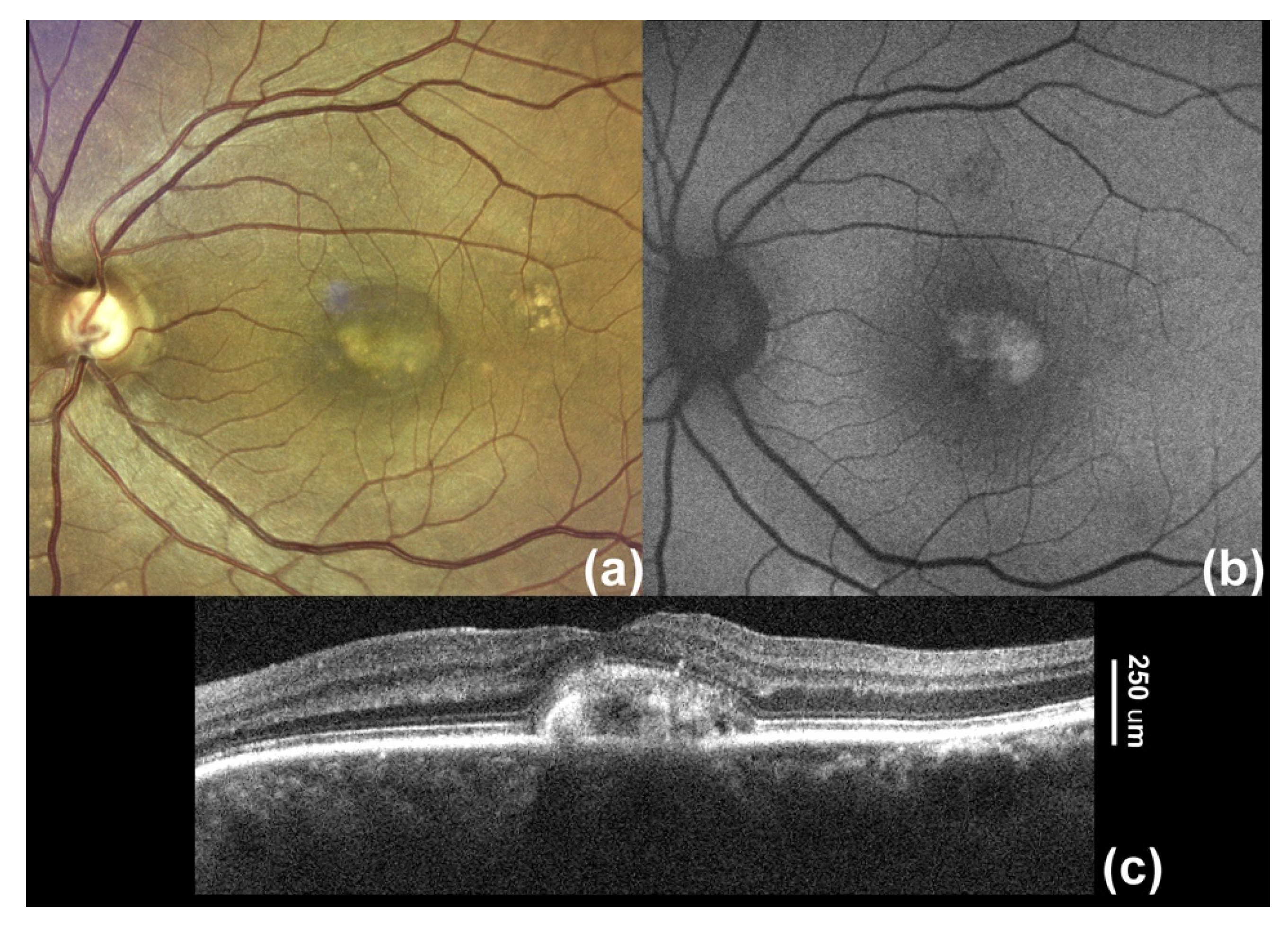
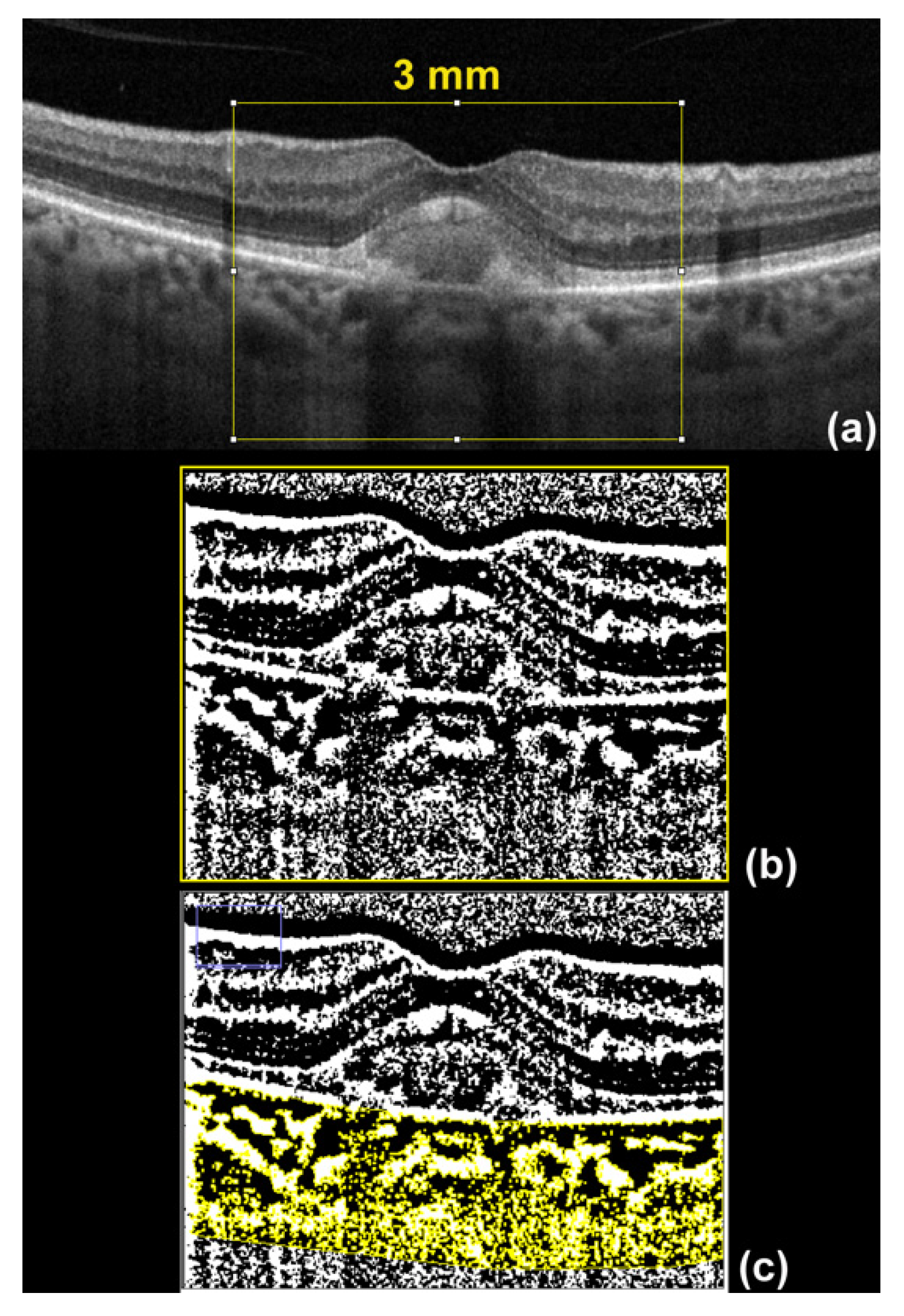
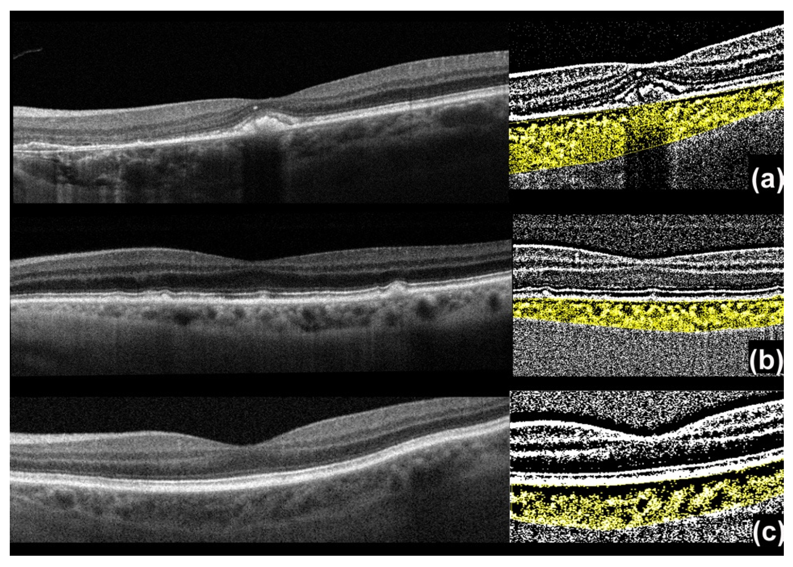
2.1. Imaging Processing

SDOCT images were obtained from RTVue XR (RTVue XR Avanti, Optovue, Inc., Fremont, CA) equipped with the AngioVue software (version 2017.1.0.151; Optovue Inc, Fremont, CA, US). The grid pattern, consisting of five vertical and horizontal b-scans centered on the fovea, was used in all cases. CT was measured manually using a digital caliper, from the outer border of the hyperreflective line corresponding to the outer border of the RPE to the choroidal/scleral boundary. CT was measured at the fovea, and 500 and 1500 µm from the nasal and temporal regions in the fovea.

2.2. Choroidal Vascularity Index (CVI) Assessment

CVI defines the proportion of the vascular component in the choroid through a ratio of luminal area compared to the total choroidal area [4,12]. This index quantitatively and precisely analyzes the choroidal vascular system, enabling quantification of the proportion of choroidal vasculature, which is useful in the evaluation and management of several retinal and choroidal disorders [13]. The CVI measurements are obtained through post-processing, using an open-source software ImageJ (distributed by Fiji, https://imagej.net/Fiji/Downloads (accessed on 1 September 2021)) through a procedure previously described by Sonoda et al. [11,14] that converts the luminal and interstitial areas of the choroid into binary images.
The horizontal B-scan centered on the fovea was exported into Fiji software for post-processing. To match the location covered by CT measurements (1500 µm from the nasal and temporal regions in the fovea), a 3 mm-wide area was selected for binarization (Figure 2). In brief, the region of interest (ROI) was selected using the polygon tool delimiting the outer boundary of the RPE (upper limit) and the choroid/scleral junction (lower limit). Three choroidal vessels larger than 100 microns were selected with the oval selection tool. Successively, binarization was carried out through Niblack’s auto local threshold technique in order to analyze the mean and standard deviation of dark and light pixels in the ROI. The Niblack method for binarization was considered the most suitable method amongst those available (i.e., Berson, Mean, Median, Midgrey, Sauvola) for differentiating the luminal and interstitial areas of the choroid [11,14]. Although other methods of binarization have been proposed, the gold standard for binarization is still unclear [15,16].
The software analysis systems then enabled identification of areas with dark pixels, the totality of which gave the luminal area (LA), while the totality of pixels in the ROI defined the total area (TA). The ratio of the TA to LA determines the CVI (Figure 3).

2.3. Statistical Analysis
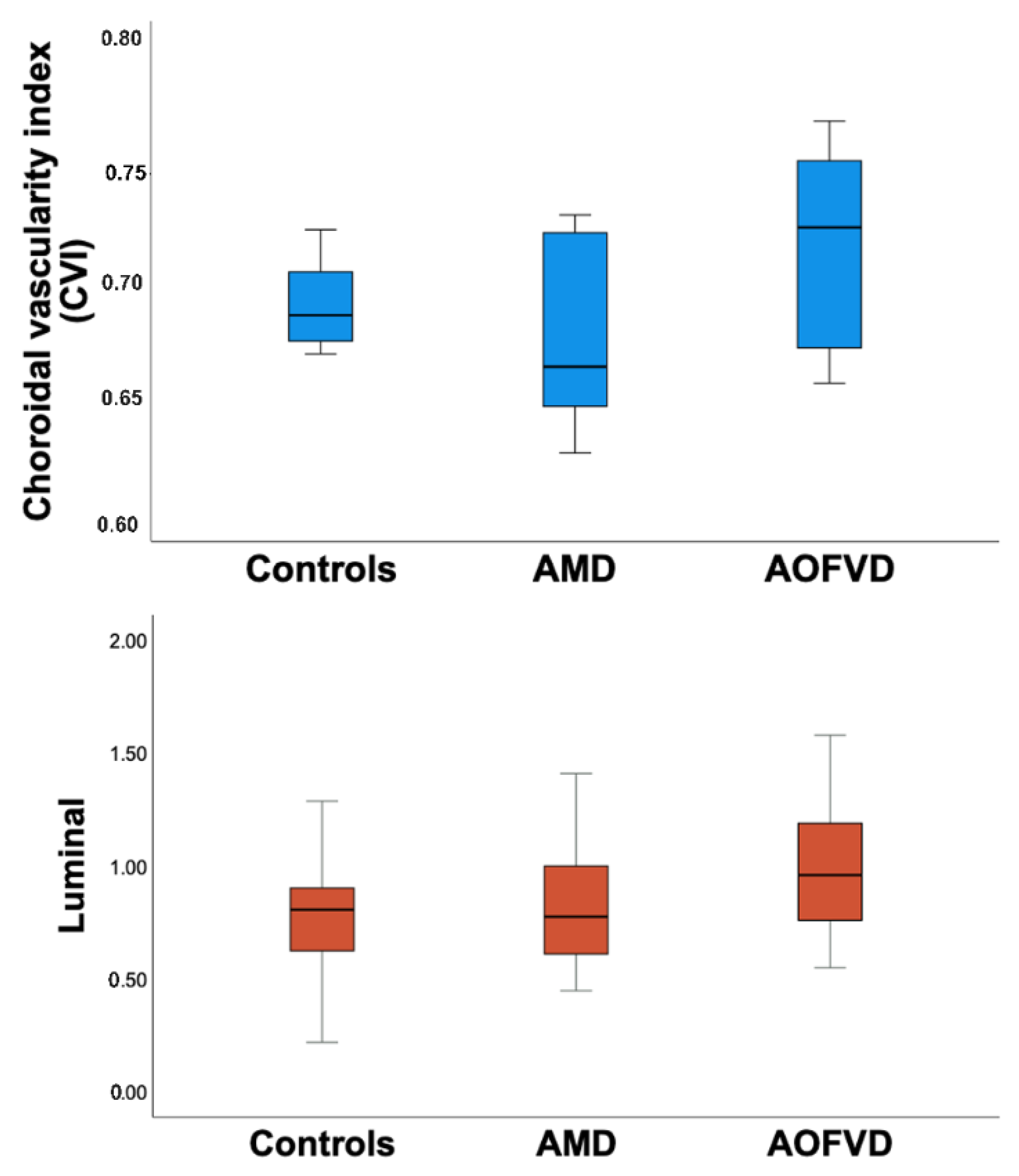
Normally distributed quantitative variables were reported as mean ± standard deviation unless otherwise specified, and distribution normality was verified through Shapiro–Wilk normality test (normal distribution, Shapiro–Wilk test p > 0.05). The chi-square test was used to compare categorical variables, whereas an unpaired t-test was adopted to evaluate quantitative variables between groups. Analysis of variance (ANOVA) was used to assess differences between groups. Spearman’s rank correlation was calculated for comparisons between variables. Statistical significance was set at p < 0.05. All calculations were carried out using SPSS statistical software (ver. 25; SPSS, Inc., Chicago, IL, USA).

3. Results

Fifty-four patient files were initially screened, and 18 patients (29.6%) were excluded from the analysis due to the presence of SDD, macular complications, unacceptable image quality, and vitelliruptive or pseudohypopyon stages. Baseline characteristics of the remaining 36 patients (36 eyes) and 16 healthy subjects (16 eyes) included in the analysis are summarized in Table 1. The groups were homogenous for gender (p = 0.06) and age (p = 0.80). Macular complications in the fellow eye were present in 3/18 AOFVD eyes (16.7%) and 4/18 CD eyes (22.2%) with no differences between groups (p = 0.68).

Analysis of Choroidal Vasculature

CVI was significantly different among groups (p = 0.004). On further analysis, the luminal area demonstrated the greatest difference between groups (p = 0.04). Box plots summarize the data distribution of the three different subgroups graphically (Figure 4). Choroidal vascular parameters are detailed in Table 2.
Post hoc analysis for the relevant choroidal parameters, including CVI and the luminal area, is reported in Table 3. On post hoc analysis, eyes with AOFVD presented a higher CVI than eyes with CD (+0.03 CI 95%: −0.57, −0.01, p = 0.001) and controls (+0.02, CI 95%: 0.0007, 0.04, p = 0.03). The luminal component was larger in AOFVD eyes when compared to controls (+0.26, CI95%: 0.05, 0.48, p = 0.02), and in CD eyes, this was close to statistical significance (+0.20, CI95%: −0.002, 0.41, p = 0.05). CT at various locations did not significantly differ among subgroups, as reported in Table 2.
In the AOFVD group, increasing age was associated with a thinner subfoveal CT (r = −0.50, p = 0.02), whereas CVI was not influenced by age (r = −0.33, p = 0.17). In the AMD group, neither subfoveal CT (r = −0.11, p = 0.66) nor CVI (r = −0.02, p = 0.33) were influenced by age.

4. Discussion

In this investigation we found that eyes with AOFVD presented a greater CVI than eyes with CD and controls. The major choroidal involvement was on the luminal component.
To evaluate the possible role of the choroid in the pathogenesis of AOFVD, some authors evaluated CT in AOFVD with contrasting results. Coscas et al. reported choroidal thickening in AOFVD and suggested a different pathogenetic mechanism with respect to advanced dry and neovascular AMD, where choroidal thinning was observed [7]. Similarly to these results, Cennamo et al. found increased subfoveal CT in patients with AOFVD compared to controls. These authors performed subgroup analysis of the different stages of disease (vitelliform, pseudohypopyon, and vitelliruptive) and did not find differences in CT between groups [10]. In contrast, Puche et al. and Grenga et al. did not report a difference in CT between eyes with AOFVD and control subjects [8,17]. However, these authors further analyzed CT in eyes with different stages of disease and found mean choroidal thickening in the vitelliruptive stage with respect to controls, indicating that CT changes may be stage-sensitive [8]. In order to account for any stage-sensitive bias, we only included the vitelliform type and, in accordance with Palacios et al. [9] and Grenga et al. [8], we did not find any difference in CT between eyes with AOFVD and control eyes.
Recent studies using OCTA have shown CC vessel density reduction in patients with AOFVD [10,18,19]. Cennamo et al. found reduced CC vessel density in the vitelliform stage and increased density in the vitelliruptive stage [10]. Querques et al. examined enface OCTA images and reported vascular network rarefaction with reduced CC [19], indicating stage-sensitive variations in line with the progressive nature of vitelliform deposits. OCTA technology presented several advantages, especially in terms of image quality, acquisition time, and microvascular resolution, which allow proper estimation of the choriocapillaris flow impairment [20,21,22]. To our knowledge, there are no previous studies in the literature that evaluate CVI in patients with AOFVD; therefore, we cannot compare our results with previous data. More importantly, our cohort of patients exclusively had AOFVD in the vitelliform stage. We found that eyes with AOFVD in comparison with control eyes exhibited a greater CVI due to a larger luminal area, indicating possible compensatory choroidal response mechanisms reflected in vessel dilatation. Indeed, it is well known that the choroid is prevalently vascular tissue that exhibits significant physiological modifications in thickness based on circadian rhythms, use of caffeine, and excessive liquid ingestion [23,24,25]. Retinal and choroidal disease mechanisms are probably not an exception in CT modifications.
Eyes with AOFVD presented a higher CVI than eyes with CD in our study. This could indicate CVI as a possible early biomarker heralding evolution to more advanced stages. We may speculate that choroidal vessels enlarge in an effort to clear vitelliform material. This could affect the normal equilibrium between the three vessel layers of the choroid, thus leading to a secondary CC impairment shown by OCTA reports of reduced CC vessel density [10,18,19].
Choroidal and outer retina alterations are shown even in later stages of AOFVD. Grenga et al. found discontinuity and the appearance of bumps in the RPE in pseudohypopyon and vitelliruptive stages [8]. It is known that vitelliform material, located between the RPE and outer segments of the photoreceptors, consists of photoreceptor outer segment debris, macrophages loaded with melanin/lipofuscin, and RPE cells [26,27]. In clinicopathological studies, the RPE at the base of the lesion was reported to be initially hypertrophic [26,27]. These could be compensatory attempts to remove metabolic waste products and restore retinal integrity. The choroid could manifest tentative repair mechanisms and the increased luminal area could reflect choroidal vasodilation in a compensatory attempt to overcome the damage inflicted due to a mechanical effect of vitelliform material on blood vessels, as postulated by Querques et al. [19].
The present study had some limitations: it was retrospective in nature; manual segmentation was used for CT measurement; image binarization is not corroborated by histopathological studies, and the analysis is based on the dark-pixel and light-pixel regions of the choroid that represent vasculature and stromal areas; calculations of CVI were performed on a single subfoveal B-scan owing to software limits; the sample size and standard deviation were small, but we included only patients in the vitelliform stage of the disease. Finally, this was a pilot investigation, so we could not directly compare our results with the existing literature.
Despite these drawbacks, our results revealed a greater CVI in AOFVD than in eyes with CD and in healthy eyes. This alteration was associated with preferential vasodilatation of the luminal component already evident in the vitelliform stage of AOFVD, indicating a possible role of choroidal vasculature in this condition. Moreover, CVI may represent a sensitive index that may have implications in predicting disease progression. Further prospective studies with larger patient populations and division in stages of vitelliform deposit progression are warranted to corroborate our findings.

Author Contributions

Conceptualization, S.A. and S.F.; methodology, S.F.; software, C.C.; validation, S.A., M.D.P. and G.S.; formal analysis, S.F.; investigation, S.F.; resources, S.A.; data curation, C.C.; writing—original draft preparation, S.F.; writing—review and editing, S.A.; visualization, S.A.; supervision, S.A.; project administration, G.S.; funding acquisition, S.A. All authors have read and agreed to the published version of the manuscript.

Funding

This research received no external funding.

Institutional Review Board Statement

The study was conducted according to the guidelines of the Declaration of Helsinki and approved by the Ethics Committee of Sapienza University of Rome, protocol number: CE 5749_2020 approved 1st June 2020.

Informed Consent Statement

Informed consent was obtained from all subjects involved in the study.

Data Availability Statement

The data presented in this study are available on request from the corresponding author. The data are not publicly available due to privacy protection laws.

Conflicts of Interest

The authors declare no conflict of interest.

References

  1. Gass, J.D. A clinicopathologic study of a peculiar foveomacular dystrophy. Trans. Am. Ophthalmol. Soc. 1974, 72, 139–156. [Google Scholar] [PubMed]
  2. Chowers, I.; Tiosano, L.; Audo, I.; Grunin, M.; Boon, C. Adult-onset foveomacular vitelliform dystrophy: A fresh perspective. Prog. Retin. Eye Res. 2015, 47, 64–85. [Google Scholar] [CrossRef] [PubMed]
  3. Querques, G.; Forte, R.; Querques, L.; Massamba, N.; Souied, E.H. Natural Course of Adult-Onset Foveomacular Vitelliform Dystrophy: A Spectral-Domain Optical Coherence Tomography Analysis. Am. J. Ophthalmol. 2011, 152, 304–313. [Google Scholar] [CrossRef]
  4. Ozcaliskan, S.; Balci, S.; Yenerel, N.M. Choroidal vascularity index determined by binarization of enhanced depth imaging optical coherence tomography images in eyes with intermediate age-related macular degeneration. Eur. J. Ophthalmol. 2020, 30, 1512–1518. [Google Scholar] [CrossRef]
  5. Querques, G.; Querques, L.; Forte, R.; Massamba, N.; Coscas, F.; Souied, E.H. Choroidal Changes Associated with Reticular Pseudodrusen. Investig. Opthalmol. Vis. Sci. 2012, 53, 1258–1263. [Google Scholar] [CrossRef] [Green Version]
  6. Agrawal, R.; Chhablani, J.; Chintan, S.; Shah, S.; Sarvaiya, C.; Banker, A. CHOROIDAL VASCULARITY INDEX IN CENTRAL SEROUS CHORIORETINOPATHY. Retina 2016, 36, 1646–1651. [Google Scholar] [CrossRef]
  7. Coscas, F.; Puche, N.; Coscas, G.; Srour, M.; Français, C.; Glacet-Bernard, A.; Querques, G.; Souied, E.H. Comparison of Macular Choroidal Thickness in Adult Onset Foveomacular Vitelliform Dystrophy and Age-Related Macular Degeneration. Investig. Opthalmol. Vis. Sci. 2014, 55, 64–69. [Google Scholar] [CrossRef] [PubMed] [Green Version]
  8. Grenga, P.L.; Fragiotta, S.; Cutini, A.; Meduri, A.; Vingolo, E.M. Enhanced Depth Imaging Optical Coherence Tomography in Adult-Onset Foveomacular Vitelliform Dystrophy. Eur. J. Ophthalmol. 2015, 26, 145–151. [Google Scholar] [CrossRef]
  9. Palácios, R.M.; Mendes, T.S.; Sano, R.Y.; Wu, D.C.; Aihara, T.; Manzano, R.P.D.A. Choroidal thickness using EDI-OCT in adult-onset vitelliform macular dystrophy. Int. J. Retin. Vitr. 2016, 2, 5. [Google Scholar] [CrossRef] [Green Version]
  10. Cennamo, G.; Montorio, D.; Mirra, F.; Comune, C.; D’Alessandro, A.; Tranfa, F. Study of vessel density in adult-onset foveomacular vitelliform dystrophy with optical coherence tomography angiography. Photodiagnosis Photodyn. Ther. 2020, 30, 101702. [Google Scholar] [CrossRef]
  11. Sonoda, S.; Sakamoto, T.; Yamashita, T.; Uchino, E.; Kawano, H.; Yoshihara, N.; Terasaki, H.; Shirasawa, M.; Tomita, M.; Ishibashi, T. Luminal and Stromal Areas of Choroid Determined by Binarization Method of Optical Coherence Tomographic Images. Am. J. Ophthalmol. 2015, 159, 1123–1131.e1. [Google Scholar] [CrossRef]
  12. Branchini, L.A.; Adhi, M.; Regatieri, C.; Nandakumar, N.; Liu, J.J.; Laver, N.; Fujimoto, J.G.; Duker, J.S. Analysis of Choroidal Morphologic Features and Vasculature in Healthy Eyes Using Spectral-Domain Optical Coherence Tomography. Ophthalmology 2013, 120, 1901–1908. [Google Scholar] [CrossRef]
  13. Iovino, C.; Pellegrini, M.; Bernabei, F.; Borrelli, E.; Sacconi, R.; Govetto, A.; Vagge, A.; Di Zazzo, A.; Forlini, M.; Finocchio, L.; et al. Choroidal Vascularity Index: An In-Depth Analysis of This Novel Optical Coherence Tomography Parameter. J. Clin. Med. 2020, 9, 595. [Google Scholar] [CrossRef] [Green Version]
  14. Sonoda, S.; Sakamoto, T.; Yamashita, T.; Shirasawa, M.; Uchino, E.; Terasaki, H.; Tomita, M. Choroidal Structure in Normal Eyes and After Photodynamic Therapy Determined by Binarization of Optical Coherence Tomographic Images. Investig. Opthalmol. Vis. Sci. 2014, 55, 3893–3899. [Google Scholar] [CrossRef] [PubMed]
  15. Agrawal, R.; Wei, X.; Goud, A.; Vupparaboina, K.K.; Jana, S.; Chhablani, J. Influence of scanning area on choroidal vascularity index measurement using optical coherence tomography. Acta Ophthalmol. 2017, 95, e770–e775. [Google Scholar] [CrossRef] [PubMed] [Green Version]
  16. Wei, X.; Sonoda, S.; Mishra, C.; Khandelwal, N.; Kim, R.; Sakamoto, T.; Agrawal, R. Comparison of Choroidal Vascularity Markers on Optical Coherence Tomography Using Two-Image Binarization Techniques. Investig. Opthalmol. Vis. Sci. 2018, 59, 1206–1211. [Google Scholar] [CrossRef] [Green Version]
  17. Puche, N.; Querques, G.; Blanco-Garavito, R.; Zerbib, J.; Gherdaoui, F.; Tilleul, J.; Coscas, F.; Glacet-Bernard, A.; Souied, E.H. En face enhanced depth imaging optical coherence tomography features in adult onset foveomacular vitelliform dystrophy. Graefe’s Arch. Clin. Exp. Ophthalmol. 2013, 252, 555–562. [Google Scholar] [CrossRef]
  18. Parodi, M.B.; Rabiolo, A.; Cicinelli, M.V.; Iacono, P.; Romano, F.; Bandello, F. Quantitative analysis of optical coherence tomography angiography in adult-onset foveomacular vitelliform dystrophy. Retina 2018, 38, 237–244. [Google Scholar] [CrossRef] [PubMed]
  19. Querques, G.; Zambrowski, O.; Corvi, F.; Miere, A.; Semoun, O.; Srour, M.; Souied, E.H. Optical coherence tomography angiography in adult-onset foveomacular vitelliform dystrophy. Br. J. Ophthalmol. 2016, 100, 1724–1730. [Google Scholar] [CrossRef]
  20. Carnevali, A.; Al-Dolat, W.; Sacconi, R.; Corbelli, E.; Querques, L.; Bandello, F.; Querques, G. Diagnosis, management and future treatment options for adult-onset foveomacular vitelliform dystrophy. Expert Rev. Ophthalmol. 2018, 13, 161–169. [Google Scholar] [CrossRef]
  21. Rabiolo, A.; Carnevali, A.; Bandello, F.; Querques, G. Optical Coherence Tomography Angiography: Evolution or Revolution? Expert Rev. Ophthalmol. 2016, 11, 243–245. [Google Scholar] [CrossRef] [Green Version]
  22. Carnevali, A.; Mastropasqua, R.; Gatti, V.; Vaccaro, S.; Mancini, A.; D’Aloisio, R.; Lupidi, M.; Cerquaglia, A.; Sacconi, R.; Borrelli, E.; et al. Optical Coherence Tomography Angiography in Intermediate and Late Age-Related Macular Degeneration: Review of Current Technical Aspects and Applications. Appl. Sci. 2020, 10, 8865. [Google Scholar] [CrossRef]
  23. Nickla, D.L.; Wildsoet, C.F.; Troilo, D. Diurnal rhythms in intraocular pressure, axial length, and choroidal thickness in a primate model of eye growth, the common marmoset. Investig. Ophthalmol. Vis. Sci. 2002, 43, 2519–2528. [Google Scholar]
  24. Altinkaynak, H.; Ceylan, E.; Kartal, B.; Keles, S.; Ekinci, M.; Olcaysü, O.O.; Altınkaynak, H. Measurement of Choroidal Thickness Following Caffeine Intake in Healthy Subjects. Curr. Eye Res. 2015, 41, 1–8. [Google Scholar] [CrossRef] [PubMed]
  25. Nagasato, D.; Mitamura, Y.; Egawa, M.; Kameoka, M.; Nagasawa, T.; Tabuchi, H.; Kinoshita, T.; Sonoda, S.; Sakamoto, T. Changes of choroidal structure and circulation after water drinking test in normal eyes. Graefe’s Arch. Clin. Exp. Ophthalmol. 2019, 257, 2391–2399. [Google Scholar] [CrossRef]
  26. Dubovy, S.R.; Hairston, R.J.; Schatz, H.; Schachat, A.P.; Bressler, N.M.; Finkelstein, D.; Green, W.R. Adult-onset foveomacular pigment epithelial dystrophy. Retina 2000, 20, 638–649. [Google Scholar] [CrossRef] [PubMed]
  27. Arnold, J.J.; Sarks, J.P.; Killingsworth, M.C.; Kettle, E.K.; Sarks, S.H. Adult vitelliform macular degeneration: A clinicopathological study. Eye 2003, 17, 717–726. [Google Scholar] [CrossRef] [Green Version]
Figure 1. Multimodal imaging in adult-onset foveomacular vitelliform dystrophy. (a) True color confocal fundus photograph (Compass, Centervue, Padova, Italy) shows a central roundish yellowish lesion. (b) Fundus autofluorescence (Spectralis HRA, Heidelberg Engineering, Germany) demonstrates hyperautofluorescent signal corresponding to the vitelliform lesion. (c) Spectral-domain optical coherence tomography b-scan obtained in the subfoveal region shows an accumulation of subretinal hyperreflective material with a dome-shaped configuration (Angiovue, Optovue, Inc, Fremont, CA, USA).
Applsci 11 10487 g001
Figure 2. Choroidal vascularity index processing. (a) A rectangle centered on the foveal region of 3 mm was selected, and then cropped for further analysis; (b) The image obtained was binarized using auto local threshold with Niblack method. (c) A polygonal region of interest was traced to delimitate the choroidal area used to calculate the choroidal vascularity index.
Applsci 11 10487 g002
Figure 3. Choroidal vascularity index performed in the three subgroups considered. (a) Adult-onset foveomacular vitelliform dystrophy; (b) early age-related macular degeneration with conventional drusen; (c) age-matched healthy control.
Applsci 11 10487 g003
Figure 4. Box plots for choroidal vascularity index (CVI) analysis (top, blue) and luminal component (bottom, orange) into the three different subgroups, including controls, age-related macular degeneration (AMD), and adult-onset foveomacular vitelliform dystrophy (AOFVD).
Applsci 11 10487 g004
Table 1. Baseline characteristics of the overall population.
FeatureAOFVDAMDControlsp Value
No. of eyes181816
No. of patients181816
Age (years)74.6 ± 9.276.1 ± 1376.4 ± 6.90.80
Gender (female)10 (55.5%)9 (50%)9 (56.2%)0.06
No.: number; %: percentage. AOFVD: adult-onset foveomacular vitelliform dystrophy; AMD: age-related macular degeneration.
Table 2. Clinical features in adult-onset foveomacular vitelliform dystrophy (AOFVD) and age-related macular degeneration with conventional drusen.
FeatureAOFVD N = 18AMD N = 18Controls N = 16p Value
Luminal Area1.05 ± 0.390.83 ± 0.270.77 ± 0.260.04
Total area1.46 ± 0.481.25 ± 0.431.12 ± 0.360.07
CVI0.71 ± 0.040.68 ± 0.030.69 ± 0.020.004
SFCT215.4 ± 52.2196.9 ± 64.3230.5 ± 68.40.29
CT N 500226.5 ± 61.3180. 5 ± 66.7213.6 ± 61.90.09
CT T 500221.8 ± 53.3202.4 ± 56.4207.1 ± 60.30.57
CT N 1500210.1 ± 68.9155.7 ± 70.9213.5 ± 74.60.04
CT T 1500206.1 ± 57.2206.7 ± 50.1191.3 ± 73.80.71
CVI: choroidal vascularity index; SFCT: subfoveal choroidal thickness; CT: choroidal thickness; N: nasal; T: temporal.
Table 3. Post- hoc analysis of the different subgroups; adult-onset foveomacular vitelliform dystrophy (AOFVD) and age-related macular degeneration with conventional drusen.
FeatureAOFVD
N = 18
AMD
N = 18
p value p value
Luminal AreaAMD0.05AOFVD:0.05
Control0.02Control:0.63
CVIAMD0.001AOFVD:0.001
Control0.03Control:0.25
Post-hoc test using least significant difference (LSD); AOFVD: adult-onset foveomacular vitelliform dystrophy; AMD: age-related macular degeneration.
Publisher’s Note: MDPI stays neutral with regard to jurisdictional claims in published maps and institutional affiliations.

