Article

Vitamin C in the Presence of Sub-Inhibitory Concentration of Aminoglycosides and Fluoroquinolones Alters Proteus mirabilis Biofilm Inhibitory Rate

by
Joanna Kwiecińska-Piróg
1,
Krzysztof Skowron
1,*,
Tomasz Bogiel
1,
Agata Białucha
1,
Jana Przekwas
2 and
Eugenia Gospodarek-Komkowska
1
1
Department of Microbiology, Nicolaus Copernicus University in Toruń, L. Rydygier Collegium Medicum in Bydgoszcz, 9 M. Skłodowska-Curie St., 85-094 Bydgoszcz, Poland
2
Department of Clinical Microbiology, Antoni Jurasz University Hospital No. 1, Bydgoszcz, 9 M. Skłodowska-Curie St., 85-094 Bydgoszcz, Poland
*
Author to whom correspondence should be addressed.
Submission received: 23 July 2019 / Revised: 6 August 2019 / Accepted: 8 August 2019 / Published: 11 August 2019

Abstract

:
Vitamin C has antimicrobial activity and is often used as an oral supplement accompanying antibiotic treatment in urinary tract infections (UTI). Proteus mirabilis is the third common species responsible for UTIs that are mostly treated with fluoroquinolones or aminoglycosides. Treatment of the UTI caused by P. mirabilis is problematic due to the ability to form biofilm on the urinary catheters. The aim of the study was to evaluate the influence of ascorbic acid in combination with antibiotics on P. mirabilis abilities to form biofilm. The susceptibility of P. mirabilis reference strain ATCC® 29906™ and four clinical strains isolated from the urine samples of patients with urinary catheter were evaluated according to EUCAST recommendations. The influence of ascorbic acid (0.4 mg × mL−1) in combination with antibiotics on biofilm formation was evaluated spectrophotometrically. Aminoglycosides at sub-inhibitory concentrations more successfully limited biofilm formation by P. mirabilis strains without ascorbic acid addition. Inhibition rate differences at the lowest concentrations of gentamicin and amikacin were statistically significant (p ≤ 0.05). Ascorbic acid addition to the culture medium limited the inhibitory effect of fluoroquinolones, facilitating biofilm formation by P. mirabilis strains. The addition of ascorbic acid during aminoglycosides therapy may disturb treatment of urinary tract infections related to the presence of P. mirabilis biofilm.

1. Introduction

Vitamin C (ascorbic acid, AA) was first identified in the 1920s by a Hungarian biochemist, Albert Szent-Györgyi [1]. This vitamin cannot be synthesized by humans, therefore dietary supplementation of about 75–1000 mg per day is needed [1]. Ascorbic acid (after oral intake) is absorbed from the small intestine and transported into cells by specific transporters: Sodium-depend Vitamin C Transporters (SVCTs)-1 or -2. After absorption, vitamin C is distributed by blood to the tissues, especially the liver, brain, and adrenals. The concentration of AA in human tissues (200–2300 µM, depending on the organ) is much higher than in body fluids (i.e., 50–70 µM in plasma) and depends strongly on dietary intake of vitamin C [2]. Daily intake of AA impacts also on its bioavailability—it is very high if AA is supplemented in small doses (15–30 mg/day) and decreases if high doses are provided [2]. Higher doses (>100 mg/day) increase plasma AA concentration, and are almost totally excreted in the urine [3]. The urine excretion of vitamin C is much higher in humans taking a dose of 2000 mg, when compared to 100 mg of AA per day [1,4].
Vitamin C has antioxidant properties, therefore any effects of its presence in the human body may be most prominent under conditions of the enhanced oxidative stress, i.e., during the process of inflammation [5]. Viral, bacterial or fungal infections cause reactive oxygen species (ROS) release by phagocytes. It is helpful in the limitation of infection through deactivation of viruses or bacteria killing. ROS may also cause damage to the host cells, therefore the level of ROS released by phagocytes should be reduced directly after infection [6]. Vitamin C is an essential co-enzyme in the oxidative stress pathways, capable of ROS removal [5].
This vitamin C has also direct antimicrobial activity [7]. The growth of strains belonging to the two most common bacterial uropathogens (Escherichia coli and Klebsiella pneumoniae) is inhibited at the vitamin C concentration of 10 mg × mL−1 and 20 mg × mL−1 [7].
In general, vitamins may alter antimicrobial activity of particular antibiotics [8]. Carlsson et al. (2005) [9] and Afzal et al. (2017) [10] showed an increased antimicrobial activity of fluoroquinolones and aminoglycosides during AA addition. Habash et al. (1999) [11] additionally proved that vitamin C decreases the adhesion and microorganisms colonization of the biomaterials used in diagnostic/treatment procedures involving the urinary tract. Bacteria derived from the biofilm structure formed on the biomaterials are strongly related with catheter-associated urinary tract infections (CAUTIs).
According to the literature, CAUTIs belong to one of the most frequent nosocomial infections that may result in increased mortality and prolong hospitalization time and their costs [12,13,14].
P. mirabilis is the third common (after E. coli and K. pneumoniae) species responsible for urinary tract infections (UTIs) and the second cause of the CAUTIs [15,16]. Bacteria of Proteus genus are Gram-negative motile rods, belonging to the Morganellaceae family [17]. The most important representative of the genus is P. mirabilis [18]. Depending on the environment conditions, P. mirabilis strains may form biofilm with typical nutritional channels or a rather plain structure without those channels but with characteristic swarming cells fraction [19]. P. mirabilis rods might form biofilm on the surfaces of the different polymers that the catheters are made from [20]. The most important therapeutic obstacle connected with biofilm-submerged cells is their changed susceptibility to antibiotics, compared to the cells of planktonic form. Biofilm resistance to antibiotics may result from different mechanisms, frequently co-existing in the same bacteria cell population [21].
Due to the presence of vitamin C in urine during dietary supplementation, often accompanying antibiotic treatment in urinary tract infections, we examined minimal inhibitory concentration (MIC) values of AA for P. mirabilis strains, and the impact of AA at the concentration obtained after oral supplementation of vitamin C (0.4 mg × mL−1) with aminoglycosides and fluoroquinolones antibiotics on biofilm formation by antimicrobial-sensitive P. mirabilis strains.

2. Results

2.1. Minimal Inhibitory Concentration of Antibiotics and Ascorbic Acid

All the strains included in the study were confirmed for the sensitivity to the used antibiotics. MIC values of the antibiotics and AA for each particular strain are set in Table 1.

2.2. Biofilm Formation

All the studied strains were able to form biofilm on the surface of polystyrene plates (Figure 1). The highest absorbance values of formazan were obtained for the R strain—the average value of absorbance was 0.4377. Amongst the clinical strains, the highest absorbance was found for the S4 strain (0.3427). That strain was isolated from the youngest patient (18 months old) without current antimicrobial therapy.

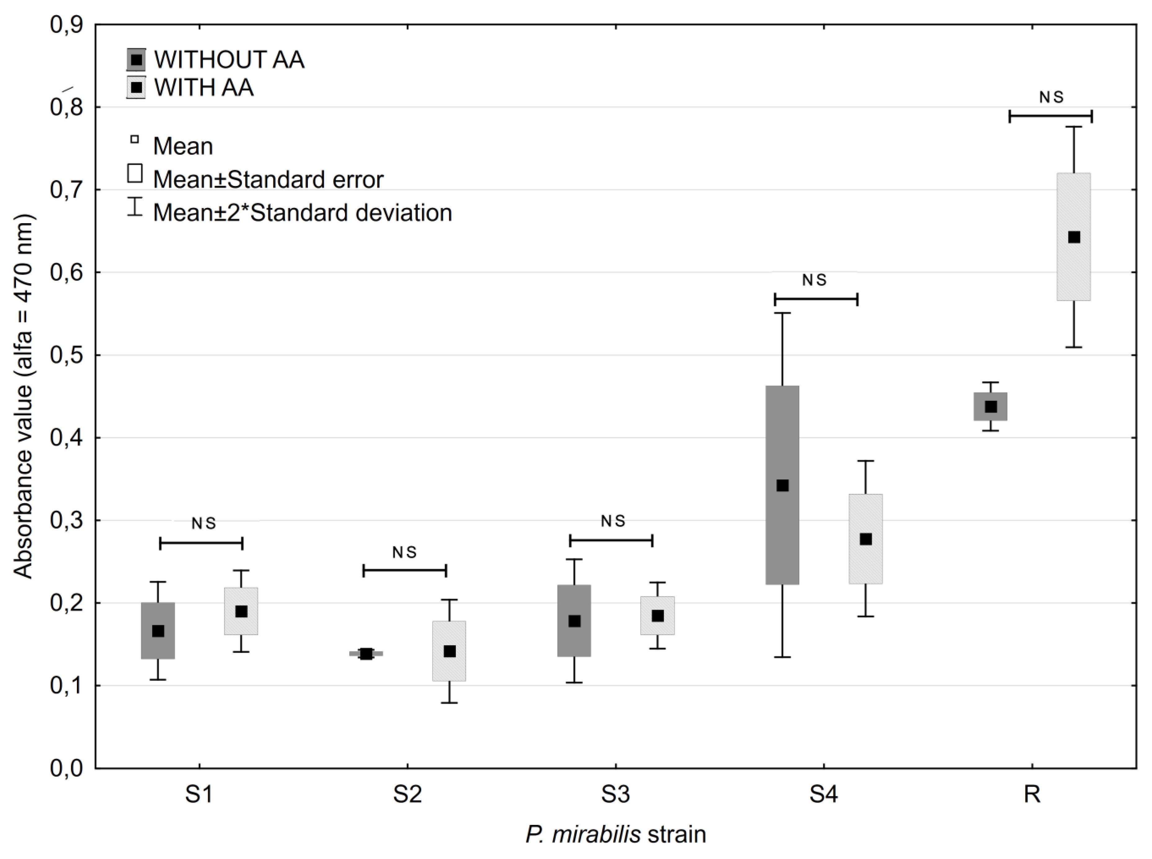
2.3. Ascorbic Acid Impact on Biofilm Formation

Generally, AA at the concentration of 0.4 mg × mL−1 did not change biofilm formation by P. mirabilis strains. The highest observed alteration was achieved for the R strain. The average absorbance values measured for the biofilm of P. mirabilis R strain in the medium without AA were 0.438, while the corresponding value for the AA-supplemented medium were generally higher, 0.643 (Figure 1). However, the differences were not statistically significant (p > 0.05).

2.4. Antibiotic Impact on Biofilm Formation

All examined concentrations of aminoglycosides were able to inhibit P. mirabilis biofilm formation. The highest values of the biofilm inhibitory rate (BIR) were observed at concentrations of studied antimicrobials corresponding to 1 MIC measured for planktonic cells (Figure 2 and Figure 3). Amikacin at the concentration corresponding to 0.5 MIC and 1 MIC, and 1 MIC gentamicin were able to inhibit biofilm formation stronger than 0.064 MIC and 0.125 MIC of gentamicin (Figure 2).
Amongst fluoroquinolones the highest values of BIR at all the examined concentrations of antimicrobials were obtained for norfloxacin (Figure 3). All studied concentrations of norfloxacin inhibited biofilm formation stronger than any sub-inhibitory concentration of ciprofloxacin (Figure 3). Favourable P. mirabilis biofilm formation at 0.064 and 0.25 MIC of ciprofloxacin was also noticed.

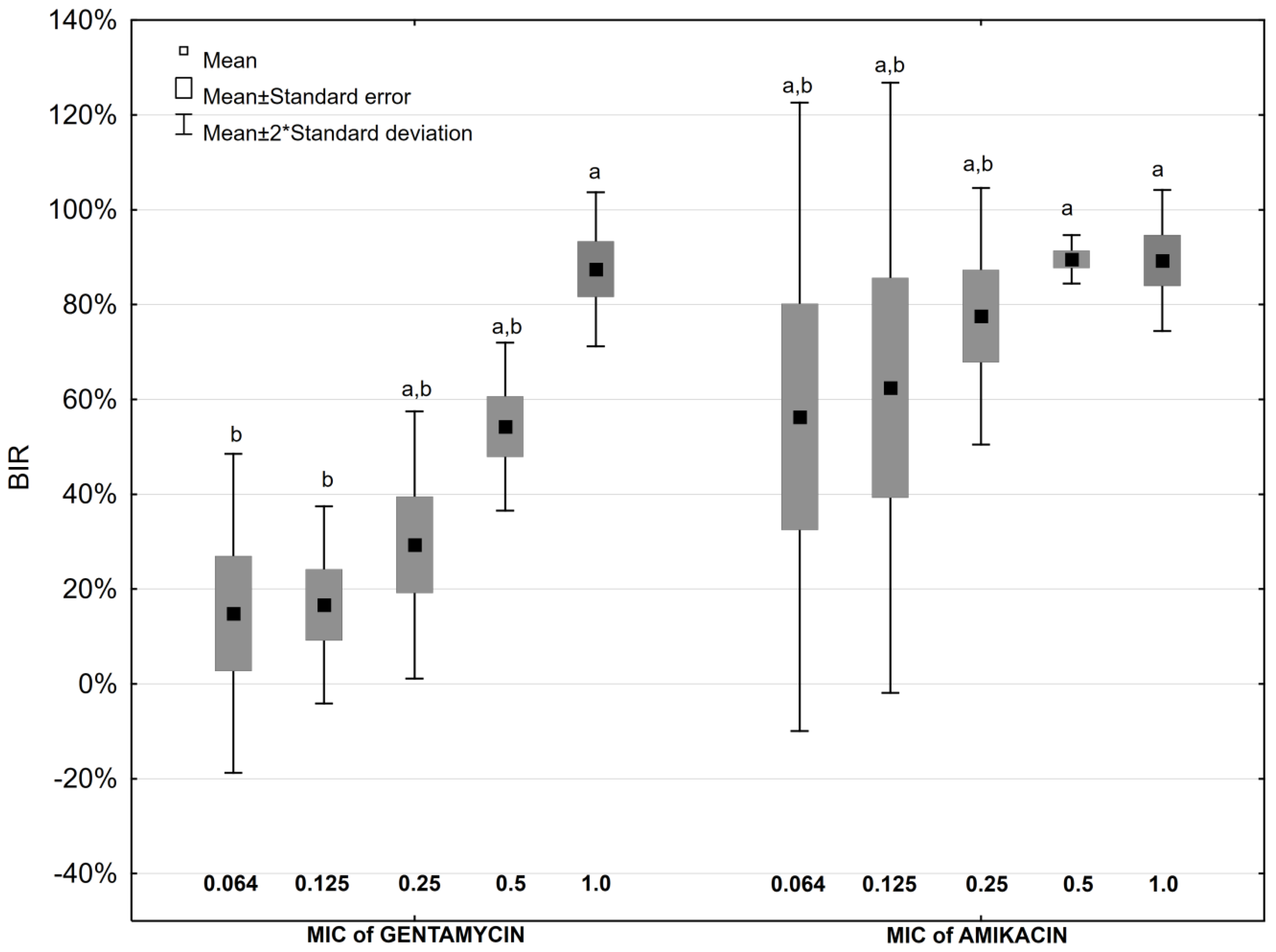
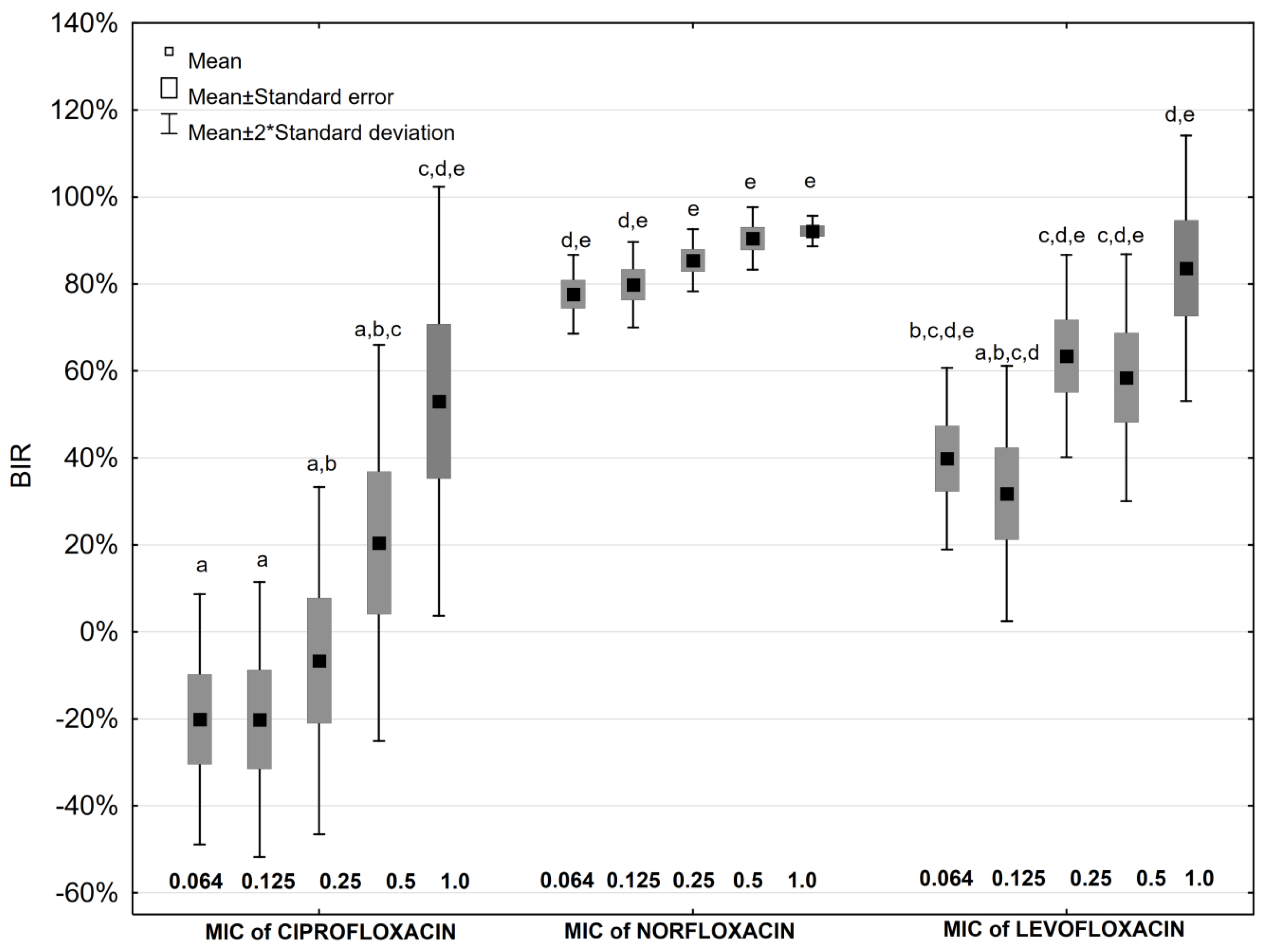
2.5. Antibiotic and Ascorbic Acid Impact on Biofilm Formation

Aminoglycosides limited P. mirabilis strains biofilm formation more successfully in the medium lacking AA addition (Table 2). The differences in BIR values for both, gentamicin at the highest concentrations applied and amikacin at all studied concentrations, were statistically significant (p ≤ 0.05). Surprisingly, simultaneous supplementation of the culture medium with AA and the aminoglycosides at concentrations equal to 0.064 MIC and 0.125 MIC increases P. mirabilis biofilm production, while biofilm formation is limited in the presence of the antibiotic itself (Table 2).
Similar results were observed for fluoroquinolones—supplementation of the culture medium with 0.4 mg × mL−1 AA decreases inhibitory activity of these antimicrobials, facilitating P. mirabilis biofilm formation. The exceptions were the ciprofloxacin concentration of 0.064–0.25 MIC value and levofloxacin concentration of 0.064–0.125 MIC value, where BIRs were higher in medium with AA than without, but the differences were not statistically significant. The presence of AA with 0.25 MIC and 0.5 MIC of norfloxacin decreased BIR. The differences observed, were statistically significant (p < 0.05).

3. Discussion

Proteus spp. biofilm synthesis is a significant clinical problem in terms of its eradication need and patients’ treatment applied. These bacteria are able to synthesize extracellular structures on the surfaces of all known types of catheters [22]. Biofilm constitution limits antimicrobial penetration and distribution through the whole structure, mostly to the bacteria submerged in its deeper layers, generating sub-inhibitory antibiotics concentrations. Moreover, biofilm-associated microorganisms express higher resistance levels to antimicrobials, resulting in bacteria multiplying although the drug is present in the environment [23]. That is the reason why antimicrobials concentrations active against planktonic forms of bacteria, frequently are unsuccessful for biofilms. They remain at too low concentration or for too short time to penetrate successfully biofilm structure. In general, with increasing antibiotic concentration its inhibitory influence on bacterial biofilm grows [24,25]. Similar conclusions were made in our study. Amongst the examined antibiotics that are most commonly used in urinary tract infection treatment, norfloxacin demonstrates the most effective antibiotic amongst those detected at the sub-inhibitory concentrations. We observed than even the highest studied concentration of antimicrobials capable of inhibiting the growth of planktonic forms of bacteria were unable to inhibit biofilm formation. It may cause a failure in the antimicrobial treatment applied. That is why searching for the alternative therapeutic solutions, disrupting antimicrobial biofilm activity at lower concentrations, is fully understood.
We found that 10 mg × mL−1 of AA inhibit growth of P. mirabilis planktonic cells. That confirms the results obtained by Verghese et al. (2017) [7]. They demonstrated antibacterial activity of 10 mg × mL−1 vitamin C on E. coli and K. pneumoniae strains isolated from urine samples. This activity is dose-depend and independent of the antimicrobial susceptibility patterns of the strains. Concentration of AA in urine in the bladder after oral-supplementation of vitamin C is much lower [4], therefore we examined the impact of a lower concentration of AA. In this study we found that 0.4 mg × mL−1 of AA, which is possible to obtain in the bladder, does not inhibit biofilm formation by P. mirabilis strains.
Similar results were obtained by Carlsson et al. (2005) [9]. They examined the influence of 10 mM AA solution on two E. coli reference strains growth on the surface of the urinary catheter and they noted 1000-fold (105 to 108) increase in the number of both bacteria strains after application of one vitamin C dosage to the culture medium for 24 h. El-Gebaly et al. (2012) [24] observed conflicting results: 65–78% and 85–90% biofilm formation inhibition while 80 and 100 mg × mL−1 of vitamin C were used, respectively. The concentrations of AA applied by them [5] were 8 and 10-fold higher than MIC values of vitamin C noted for the planktonic form of clinical P. mirabilis strains examined in our study, so it is very probably that concentrations higher than MIC values are able to inhibit biofilm formation. Habash et al. (1999) [11] showed that the presence of AA in the urine of patients inhibits E. coli and Enterococcus faecalis adhesion to biomaterials that the catheters are made from. This tendency was not observed for other urinary tract pathogens [11].
The strategy of our own study presented was to evaluate the influence of AA addition on antibiotics activity against P. mirabilis biofilm. Vitamin C is commonly prescribed with antibiotics during UTI treatment [26]. In our research, AA addition resulted in decreasing fluoroquinolones and aminoglycosides inhibitory effect on P. mirabilis strains biofilm formation. Similar results for the planktonic cells were obtained by Masadeh et al. (2012) [26] for ciprofloxacin. Pre-treatment of E. coli cells with 10 mM AA inhibits ciprofloxacin-induced hydrogen peroxide generation and reverse its antibacterial activity. Moreover, Aiassa et al. (2012) [27] investigated the decreased activity of ciprofloxacin on P. mirabilis strains when applied simultaneously with 10 mM of vitamin C. The observed decreased sensitivity to ciprofloxacin was explained by antioxidant activity of vitamin C, influencing the ciprofloxacin mode of action on bacteria cells, based on oxidative stress induction. The increase of MIC value for another fluoroquinolone (pefloxacin) was also detected for S. aureus in the presence of vitamin C in the study conducted by Awofisayo et al. (2012) [28].
The conflicting conclusions were obtained for higher concentrations of vitamin C (80 and 100 mg × mL−1) applied on biofilm examination by El-Gebaly et al. (2012) [24]. They detected synergistic activity of levofloxacin and vitamin C in biofilm reducing activity on the surface of the urinary catheter for various uropathogens, including P. mirabilis. The planktonic forms of both strains examined by them [24] were resistant to ciprofloxacin; while in the study presented, strains were sensitive to all the antibiotics applied. This, and 10-fold higher than MIC of AA concentrations used in the study, might have resulted in obtaining different results. Pandit et al. (2017) [29] demonstrated that low concentrations of vitamin C (10–30 mM of sodium ascorbate) also inhibits exopolysaccharide of Bacillus subtilis biofilms synthesis may led to the increase of bacteria susceptible to vitamin C-induced oxidative stress compounds or antimicrobials. Yassein (2004) [30] explains that higher activity of levofloxacin observed after environment pH growth by AA is more optimal level for this fluoroquinolone activity. Moreover, vitamin C plays a crucial role in the quorum sensing phenomenon inhibiting competitively autoinducer AI-2 [31].
The influence of antioxidants on aminoglycosides remains unknown. However, Cursino et al. (2005) [32] demonstrated a synergic effect of kanamycin or streptomycin and 1.0 mg × mL−1 of AA against multidrug resistance P. aeruginosa strains, but they did not find any interactions between tobramycin and the same concentration of AA. Goswami et al. (2007) [33] observed that AA and glutathione, both at the concentrations of 10 mM, decreased the sensitivity of the E. coli strain to streptomycin in a manner similar to fluoroquinolones. Andrade et al. (2014) [34] study obtained that antioxidants—alpha-tocopherol enhances the antibiotic activity of aminoglycosides against Gram-negative bacteria, which have a higher level of antimicrobial resistance. Shahzad et al. (2018) [8] also observed the synergistic effect of water-soluble and fat-soluble vitamins, including vitamin C, with beta-lactams against Acinetobacter baumannii and methicillin-resistant S. aureus (MRSA). They do not observed this effect for any of the examined aminoglycosides. In this study we demonstrated that AA strongly decreases the BIR value. Moreover, we observed the enhancement biofilm formation at the lowest sub-inhibitory concentrations of amikacin or gentamicin in the presence of AA while in the medium supplemented only with the antimicrobials—the inhibition of biofilm formation was demonstrated. Similar results were also proven by Wasfi et al. (2012) [25] for ceftriaxone applied at the concentration equal to 0.125 of MIC for this cephalosporine.
In the available literature, very few information on the influence of vitamin C supplementation on the CAUTIs treatment can be found. Thus, further studies of that issue are necessary, especially in terms of bacterial biofilm production.

4. Materials and Methods

4.1. Strains Origin

Proteus spp. strains were isolated from patients of the Antoni Jurasz University Hospital No 1 in Bydgoszcz, Poland. The examination included only strains obtained from urine samples collected using bladder catheters. Strains were identified during microbiological investigation at the Department of Microbiology.
The susceptibility of Proteus spp. strains was examined using BD Phoenix™ system (Becton Dickinson) and interpreted according to EUCAST recommendation [35]. Four randomly chosen clinical strain (named S1−S4), susceptible to the examined antimicrobials and one reference strain (R) purchased from American Type Culture Collection (ATCC® 29906™) were included into the study (Table 3). Strains identification was confirmed using MALDI-TOF MS technique (Microflex, Bruker). Investigation strains were stored in a Brain-Heart Infusion (BHI, Becton Dickinson) with 20.0% glycerol (Avantor) at −70 °C.

4.2. Minimal Inhibitory Concentration of Antibiotics and Ascorbic Acid

The MIC value for gentamicin (GEN), amikacin (AMK), ciprofloxacin (CIP), norfloxacin (NOR), levofloxacin (LEV), and ascorbic acid (AA) (all delivered by Sigma Aldrich) of P. mirabilis strains was examined by the microdilution method in the microtiter plates method according to the Clinical and Laboratory Standards Institute (CLSI) recommendation (2012) [36]. The growth of bacteria in the presence of examined antibiotic and AA at concentrations ranging from 0.001–128.0 μg × mL−1 was evaluated. Escherichia coli strain ATCC® 25922™ was used for the control assays for antimicrobials. The MIC value was read-out visually by the presence of turbidity of the suspension in the wells of microtiter plates at a given concentration of antibiotic or AA. Investigation was done in three independent replications.

4.3. Biofilm Formation

The ability to form biofilm by the studied strains was examined using 0.1% 2,3,5 triphenyltetrazolium chloride (TTC, Sigma Aldrich) by the microtiter methodology that was described previously [37]. That colorless substance is metabolized by living cells to red product-formazan. The investigation was done in three independent replications. Absorbance (A) read-outs were conducted with a spectrophotometer at the wavelength of 470 nm using KC4™ v3.4 and KC4™ Signature programs (BioTek).

4.4. Ascorbic Acid Impact on Biofilm Formation

The examined strains of P. mirabilis were cultivated on the Mueller-Hinton Agar II (MHA, Becton Dickinson) and cultured at 37 °C for 18 h. Next, the single colonies of each strain were inoculated into Tryptic Soy Bullion (TSB, Bio-Rad). After 18 h at 37 °C, each culture was centrifuged for 15 min at 4000 rpm and the supernatants was discarded. The remaining pellet was rinsed with 3 mL of phosphate buffered saline solution (pH = 7.2) (PBS, Avantor). Next, the bacterial suspension was centrifuged at 4000 rpm for 10 min and the pellet was used to make the suspension of 0.5 McFarland turbidity in Mueller-Hinton Broth (MHB, Becton Dickinson). Then, 100 μL of every suspension was placed in the wells of polystyrene 96-well plate, three repetitions each. The wells were filled with: (a) 100 μL of sterile MHB (biofilm formation evaluation/positive control), (b) 100 μL of AA (to the final concentration of 0.4 mg × mL−1) in MHB (AA impact assessment). The culture was incubated in a humid chamber at 35 ± 2 °C for 24 h. After incubation, the solutions were removed and the wells rinsed tree times with sterile PBS. Next, 100 μL of TSB and 100 µL of 0.1% TTC were added to each well. The plates were placed on a shaker at 250 rpm for 5 min at room temperature. Next, the plates were placed at 37 °C. After 3-h incubation, the TTC was removed and plates were rinsed three times with sterile water. Finally, 200 μL of 96% methanol (Avantor) were added to each well and left on a shaker at 400 rpm for 5 min at room temperature. Results were conducted with a spectrophotometer as described above.

4.5. Antibiotic Impact on Biofilm Formation

Based on MIC values results, the sub-inhibitory concentration of the studied antibiotics were prepared. The antibiotic concentrations were equivalent to 0.064, 0.125, 0.25, 0.5, and 1.0 of MIC values for particular antimicrobial. The preparation of examined strains inoculum was based on methods described in the previous section. Bacteria suspension (100 μL) was placed into the wells of polystyrene 96-well plate. The wells were filled with: (a) 100 μL of an antimicrobial at the concentration in final medium in range 0.064–1 MIC, diluted in MHB (sub-inhibitory antibiotic concentration impact assessment), (b) 100 μL of MHB only (biofilm formation evaluation/positive control). The culture of biofilm was conducted under conditions described above. Results were given as absorbance values.

4.6. Antibiotic and Ascorbic Acid Impact on Biofilm Formation

Preparation of the examined strains inoculum based on the methods was described in the previous section. Bacteria suspension (100 μL) was placed in the wells of polystyrene 96-well plate. Then, the wells were filled with: (a) 50 μL of an antibiotic at concentration in the final medium in the range 0.064–1 MIC, diluted in MHB, and 50 μL of sterile MHB (antibiotic impact evaluation), (b) 50 μL of an antibiotic at the same concentration as in the previous point, diluted in MHB, and 50 μL of AA (to final concentration 0.4 mg × mL−1) in MHB (antibiotic + AA impact estimation). The culture of biofilm was conducted under conditions described above. Results were given as absorbance values.

4.7. Biofilm Inhibition Rate

We introduced value described as Biofilm Inhibition Rate (BIR) to express our final results. It normalized the impact of TTC to formazan metabolism measurement time, regardless of the examined strain properties. BIR is based on absorbance values and is calculated according to the formula:
BIR = A x A y A x 100 %
where (Ax) stands for the absorbance value of positive control, and (Ay) for the absorbance value of the strain subjected defined concentration of antibiotics or/and ascorbic acid.

4.8. Statistical Analysis

Statistical analysis was performed using Statistica 12.5 PL (StatSoft) software. The confidence interval for all the tests applied was α = 0.05. The obtained results normality was evaluated with the Shapiro-Wilk test. Bonferroni test was applied to compare the impact of various concentrations of the studied antibiotics on biofilm formation (Figure 2, Figure 3), and U Mann-Whitney test was used to compare results obtained for AA (Figure 1), and AA with and without antibiotics (Table 2).

Author Contributions

Conceptualization, J.K.-P.; methodology, J.K.-P. and K.S.; validation, J.K.-P.; formal analysis, J.K.-P. and K.S.; investigation, J.K.-P. and A.B.; resources, J.K.-P. and J.P.; data creation, J.K.-P. and K.S.; writing—original draft preparation, A.B., J.P. and T.B.; writing—review and editing, J.K.-P. and T.B.; visualization, J.K.-P. and K.S.; supervision, J.K.-P. and E.G.-K.; project administration, J.K.-P.; funding acquisition, E.G.-K.

Funding

This research was funded by Collegium Medicum Nicolaus Copernicus University grant number 5/WF/2013 and Nicolaus Copernicus University funds from the maintenance of the research potential of the Department of Microbiology (DS-UPB no. 933).

Conflicts of Interest

The authors declare no conflict of interest.

References

  1. Pacier, C.; Martirosyan, D.M. Vitamin C: Optimal dosages, supplementation and use in disease prevention. FFHD 2015, 5, 89–107. [Google Scholar] [CrossRef]
  2. Padayatty, S.J.; Levine, M. Vitamin C: The known and the unknown and Goldilocks. Oral Dis. 2016, 22, 463–493. [Google Scholar] [CrossRef] [PubMed]
  3. Choi, J.; Kim, D.Y.; Choue, R.; Lim, H. Effects of vitamin C supplementation on plasma and urinary vitamin C concentration in Korean women. Clin. Nutr. Res. 2017, 6, 198–205. [Google Scholar] [CrossRef] [PubMed]
  4. Shibata, K.; Fukuwatari, T.; Ohta, M.; Okamoto, H.; Watanabe, T.; Fukui, T.; Nishimuta, M.; Totani, M.; Kimura, M.; Nakashima, M.; et al. Values of water-soluble vitamins in blood and urine of Japanese young men and women consuming a semi-purified diet based on the Japanese Dietary Reference Intakes. J. Nutr. Sci. Vitaminol. (Tokyo) 2005, 51, 319–328. [Google Scholar] [CrossRef] [PubMed]
  5. Liu, Z.; Ren, Z.; Zhang, J.; Chuang, C.C.; Kandaswamy, E.; Zhou, T.; Zuo, L. Role of ROS and nutritional antioxidants in human diseases. Front Physiol. 2018, 9, 477. [Google Scholar] [CrossRef] [PubMed]
  6. Hemilä, H. Vitamin C and infections. Nutrients 2017, 9, 339. [Google Scholar] [CrossRef] [PubMed]
  7. Verghese, R.J.; Mathew, S.K.; David, A. Antimicrobial activity of vitamin C demonstrated on pathogenic Escherichia coli and Klebsiella pneumoniae. J. Curr. Res. Sci. Med. 2017, 3, 88–93. [Google Scholar] [CrossRef]
  8. Shahzad, S.; Ashraf, M.A.; Sajid, M.; Shahzad, A.; Rafigue, A.; Mohmood, M.S. Evaluation of synergistic antimicrobial effect of vitamins (A, B1, B2, B6, B12, C, D, E and K) with antibiotics against resistant bacterial strains. J. Glob. Antimicrob. Resist. 2018, 13, 231–236. [Google Scholar] [CrossRef]
  9. Carlsson, S.; Weitzberg, E.; Wiklund, P.; Lundberg, J.O. Intravesical nitric oxide delivery for prevention of catheter-associated urinary tract infections. Antimicrob. Agents Chemother. 2005, 49, 2352–2355. [Google Scholar] [CrossRef]
  10. Afzal, S.; Ashraf, M.; Buksh, A.; Akhtar, A.; Rasheed, A.D. Efficacy of anti-microbial agents with ascorbic acid in catheter associated urinary tract infection. J. Infect. Dis. Prev. Med. 2017, 5, 1–6. [Google Scholar] [CrossRef]
  11. Habash, M.B.; Van der Mei, H.C.; Busscher, H.J.; Reid, G. The effect of water, ascorbic acid, and cranberry derived supplementation on human urine and uropathogen adhesion to silicone rubber. Can. J. Microbiol. 1999, 45, 691–694. [Google Scholar] [CrossRef] [PubMed]
  12. Tambyah, P.A.; Knasinski, V.; Maki, D.G. The direct costs of nosocomial catheter-associated urinary tract infection in the era of managed care. Infect. Control Hosp. Epidemiol. 2002, 23, 27–31. [Google Scholar] [CrossRef] [PubMed]
  13. Lee, M.K.; Chiu, C.S.; Chow, V.C.; Lam, R.K.; Lai, R.W. Prevalence of hospital infection and antibiotic use at a university medical center in Hong Kong. J. Hosp. Infect. 2007, 65, 341–347. [Google Scholar] [CrossRef] [PubMed]
  14. Barford, J.M.T.; Coates, A.R.M. The pathogenesis of catheter-associated urinary tract infection. J. Infect. Prev. 2009, 10, 50–56. [Google Scholar] [CrossRef]
  15. Coker, C.; Poore, C.A.; Li, X.; Mobley, H.L.T. Pathogenesis of Proteus mirabilis urinary tract infection. Microbes Infect. 2000, 2, 1497–1505. [Google Scholar] [CrossRef]
  16. Chen, C.Y.; Chen, Y.H.; Lu, P.; Lin, W.R.; Chen, T.C.; Lin, C.Y. Proteus mirabilis urinary tract infection and bacteremia: Risk factors, clinical presentation, and outcomes. J. Microbiol. Immunol. Infect. 2012, 45, 228–236. [Google Scholar] [CrossRef] [PubMed]
  17. National Center for Biotechnology Information. Available online: https://0-www-ncbi-nlm-nih-gov.brum.beds.ac.uk/ (accessed on 18 October 1996).
  18. Różalski, A.; Kwil, I.; Torzewska, A.; Baranowska, M.; Strączek, P. Proteus bacilli: Features and virulence factors. Post. Hig. Med. Dośw. 2007, 61, 204–219. [Google Scholar]
  19. Jones, S.M.; Yerly, J.; Hu, Y.; Ceri, H.; Martinuzzi, R. Structure of Proteus mirabilis biofilms grown in artificial urine and standard laboratory media. FEMS Microbiol. Lett. 2007, 268, 16–21. [Google Scholar] [CrossRef] [PubMed]
  20. Hawthorn, L.; Reid, G. The effect of protein and urine on uropathogen adhesion to polymer substrata. J. Biomed. Mater. Res. 1990, 24, 1325–1332. [Google Scholar] [CrossRef]
  21. Stewart, P.S. Mechanisms of antibiotic resistance in bacterial biofilms. Int. J. Med. Microbiol. 2002, 292, 107–113. [Google Scholar] [CrossRef]
  22. Morris, N.S.; Stickler, D.J.; Winters, C. Which indwelling urethral catheters resist encrustation by Proteus mirabilis biofilms? Br. J. Urol. 1997, 80, 58–63. [Google Scholar] [CrossRef] [PubMed]
  23. Iqbal, K.; Khan, A.; Khattak, M.M.A.K. Biological Significance of Ascorbic Acid (Vitamin C) in Human Health—A Review. Pak. J. Nutr. 2004, 3, 5–13. [Google Scholar] [CrossRef]
  24. El-Gebaly, E.; Essam, T.; Hashem, S.; El-Baky, R.A. Effect of levofloxacin and vitamin C on bacterial adherence and preformed biofilm on urethral catheter surfaces. J. MicrobBiochem. Technol. 2012, 4, 131–136. [Google Scholar] [CrossRef]
  25. Wasfi, R.; Abd El-Rahman, O.A.; Mansour, L.E.; Hanora, A.S.; Hashem, A.M.; Ashour, M.S. Antimicrobial activities against biofilm formed by Proteus mirabilis isolates from wound and urinary tract infections. Indian J. Med. Microbiol. 2012, 30, 76–80. [Google Scholar] [CrossRef] [PubMed]
  26. Masadeh, M.M.; Mhaidat, N.M.; Alzoubi, K.H.; Al-Azzam, S.I.; Shaweesh, A.I. Ciprofloxacin-induced antibacterial activity is reversed by vitamin E and vitamin C. Curr. Microbiol. 2012, 64, 457–462. [Google Scholar] [CrossRef] [PubMed]
  27. Aiassa, V.; Barnes, A.I.; Smania, A.M.; Albesa, I. Sublethal ciprofloxacin treatment leads to resistance via antioxidant systems in Proteus mirabilis. FEMS Microbiol. Lett. 2012, 327, 25–32. [Google Scholar] [CrossRef] [PubMed]
  28. Awofisayo, S.O.; Umoren, F.J.; Uwanta, E.J. The pharmacokinetic and biopharmaceutical effect of ascorbic acid (vitamin C) on pefloxacin on concurrent administration in human. J. Appl. Pharm. Sci. 2012, 2, 107–110. [Google Scholar] [CrossRef]
  29. Pandit, S.; Ravikumar, V.; Adbel-Haleem, A.M.; Derouiche, A.; Mokkapati, V.R.S.S.; Shibom, C.; Mineta, K.; Gojobori, T.; Gao, X.; Westerlund, F.; et al. Low concentration of vitamin C reduce the synthesis of extracellular polymers and destabilize bacterial biofilms. Front. Microbiol. 2017, 8, 2599. [Google Scholar] [CrossRef]
  30. Yassein, M.A.M. Studies on the Prevention of Adherence of Biofilm Associated Bacteria to Human Cells and Implanted Medical Devices. Available online: https://dsr.kau.edu.sa/Show_Res.aspx?Site_ID=305&LNG=EN&RN=53934 (accessed on 10 August 2019).
  31. Novak, J.S.; Fratamico, P.M. Evaluation of ascorbic acid as a quorum-sensing analogue to control growth, sporulation, and enterotoxin production in Clostridium perfringens. J. Food Sci. 2004, 69, 72–78. [Google Scholar] [CrossRef]
  32. Cursino, L.; Chartone-Souza, E.; Nascimento, A.M.A. Synergic interaction between ascorbic acid and antibiotics against Pseudomnas aeruginosa. Braz. Arch. Biol. Technol. 2005, 48, 379–384. [Google Scholar] [CrossRef]
  33. Goswami, M.; Mangoli, S.H.; Jawali, N. Effects of glutathione and ascorbic acid on streptomycin sensitivity of Escherichia coli. Antimicrob. Agents Chemother. 2007, 51, 1119–1122. [Google Scholar] [CrossRef]
  34. Andrade, J.C.; Morais-Braga, M.F.B.; Guedes, G.M.M.; Tintino, S.R.; Freitas, M.A.; Menezes, I.R.A.; Coutinho, H.D.M. Enhancement of the antibiotic activity of aminoglycosides by alpha-tocopherol and other cholesterol derivates. Biomed. Pharmacother. 2014, 68, 1065–1069. [Google Scholar] [CrossRef]
  35. European Committee on Antimicrobial Susceptibility Testing. Breakpoint Tables for Interpretation of MICs and Zone Diameters. Version 8.0. Available online: http://www.eucast.org (accessed on 3 September 2018).
  36. Clinical Laboratory Standards Institute. Available online: https://clsi.org/ (accessed on 30 November 2001).
  37. Kwiecińska-Piróg, J.; Skowron, K.; Zniszczol, K.; Gospodarek, E. The assessment of Proteus mirabilis susceptibility to ceftazidime and ciprofloxacin and the impact of these antibiotics at subinhibitory concentrations on Proteus mirabilis biofilms. BioMed Res. Int. 2013, 2013, 930876. [Google Scholar] [CrossRef]
Figure 1. Absorbance values corresponding to biofilm formation in the presence of ascorbic acid (0.4 mg × mL−1).
Antibiotics 08 00116 g001
Figure 2. Biofilm Inhibitory Rate values in the presence of aminoglycosides (a,b,c,…—values marked with different letters differ statistically significant at the significance level p ≤ 0.05).
Antibiotics 08 00116 g002
Figure 3. Biofilm Inhibitory Rate values in the presence of fluoroquinolones (a,b,c,…—values marked with different letters differ statistically significant at the significance level p ≤ 0.05).
Antibiotics 08 00116 g003
Table 1. The Minimal Inhibitory Concentration values of the examined antibiotics and ascorbic acid.
Strain No.GEN
[µg × mL−1]
AMK
[µg × mL−1]
CIP
[µg × mL−1]
NOR
[µg × mL−1]
LEV
[µg × mL−1]
AA
[µg × mL−1]
S10.5000.0160.0010.0040.01610.000
S20.2500.0160.0010.0020.00810.000
S30.0040.0020.0010.0020.00810.000
S40.5000.0080.0010.0080.01610.000
R0.2500.0160.0010.0040.01610.000
GEN—gentamicin, AMK—amikacin, CIP—ciprofloxacin, NOR—norfloxacin, LEV—levofloxacin, AA—ascorbic acid, S1-4—clinical strains, R—reference strain.
Table 2. Proteus mirabilis average Biofilm Inhibition Rate in terms of antibiotic MIC values with (+AA) and without ascorbic acid addition (0.4 mg × mL−1).
Antibiotic ConcentrationBiofilm Inhibition Rate [%]
AminoglycosidesFluoroquinolones
GENp *GEN + AAAMKp *AMK + AACIPp *CIP + AANORp *NOR + AALEVp *LEV+AA
0.064 MIC14.9NS−94.756.30.028−63.7−20.1NS−9.577.6NS68.439.8NS40.6
0.125 MIC16.7NS−50.562.50.047−6.0−20.2NS−11.179.8NS77.431.8NS42.3
0.25 MIC29.3NS0.777.60.0169.5−6.6NS−5.885.40.04773.163.4NS50.5
0.5 MIC54.3NS5.689.40.0098.820.5NS7.690.40.04775.758.5NS48.3
1 MIC87.50.02841.689.30.02854.353.0NS32.892.2NS91.183.6NS66.5
GEN—gentamicin, AMK—amikacin, CIP—ciprofloxacin, NOR—norfloxacin, LEV—levofloxacin, * p-value—statistically significant differences in the level of biofilm inhibition depending on the addition of ascorbic acid.
Table 3. Characteristics of P. mirabilis clinical isolates.
Strain NoDisease DiagnosisSexPatients AgeAntibiotic Treatment
S1UTIFemale85 YearsAmoxicillin/clavulanic acid
S2Abdominal hernia surgeryFemale59 YearsNone
S3UTI (urinary stones)Female86 YearsCiprofloxacin
S4UTIMale18 MonthsNone
UTI—Urinary Tract Infection.

