
New Glycosylated Dihydrochalcones Obtained by Biotransformation of 2′-Hydroxy-2-methylchalcone in Cultures of Entomopathogenic Filamentous Fungi


Abstract
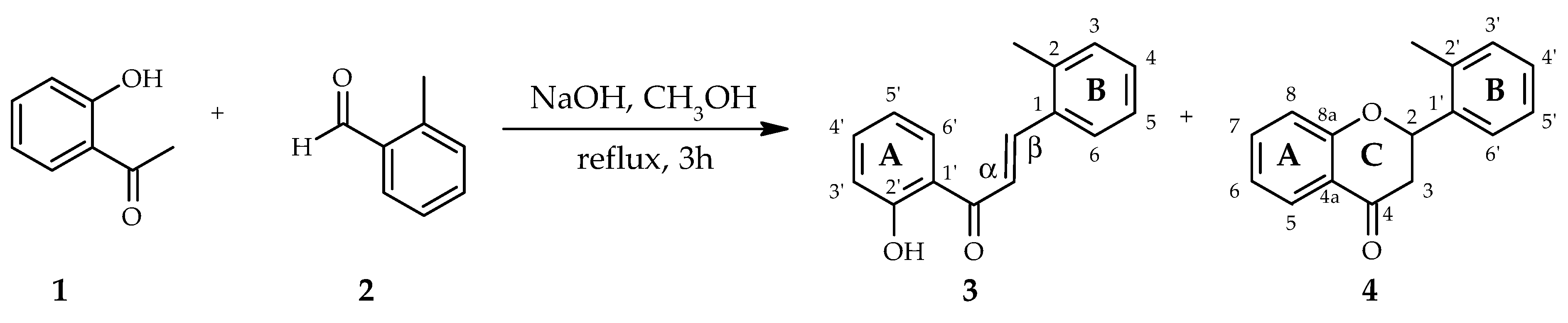
:1. Introduction
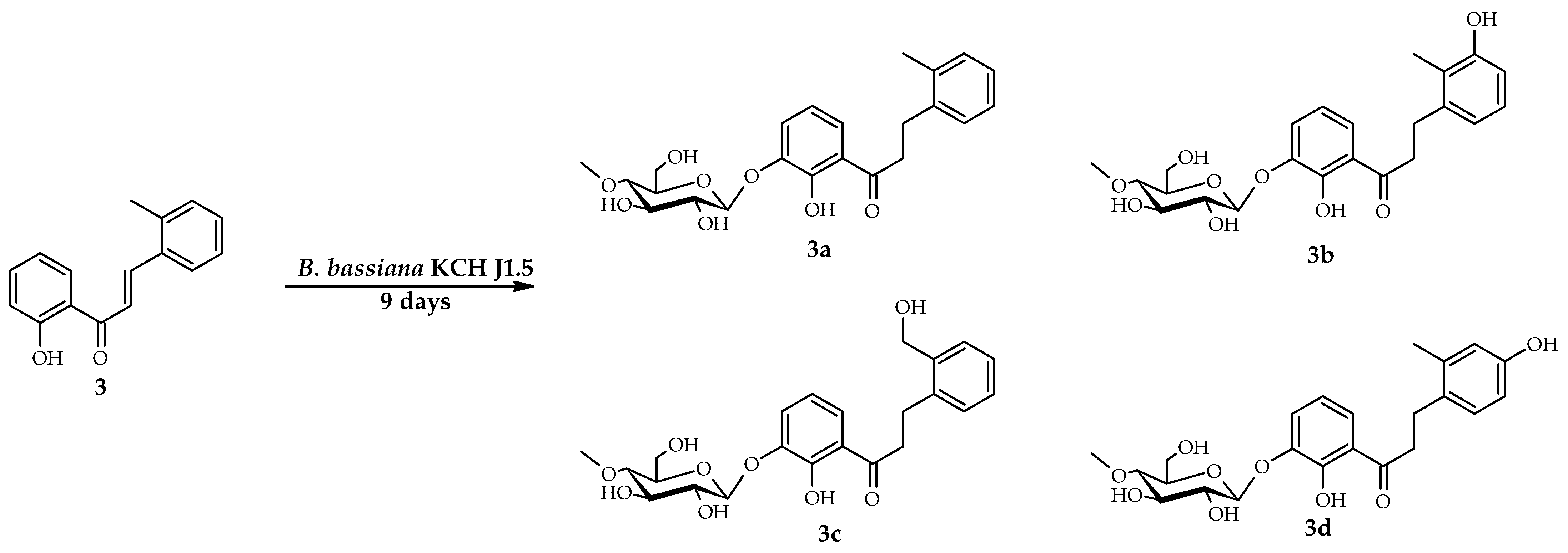
2. Results
2.1. Biotransformation of 2′-Hydroxy-2-methylchalcone (3) in the Culture of B.bassiana KCH J1.5
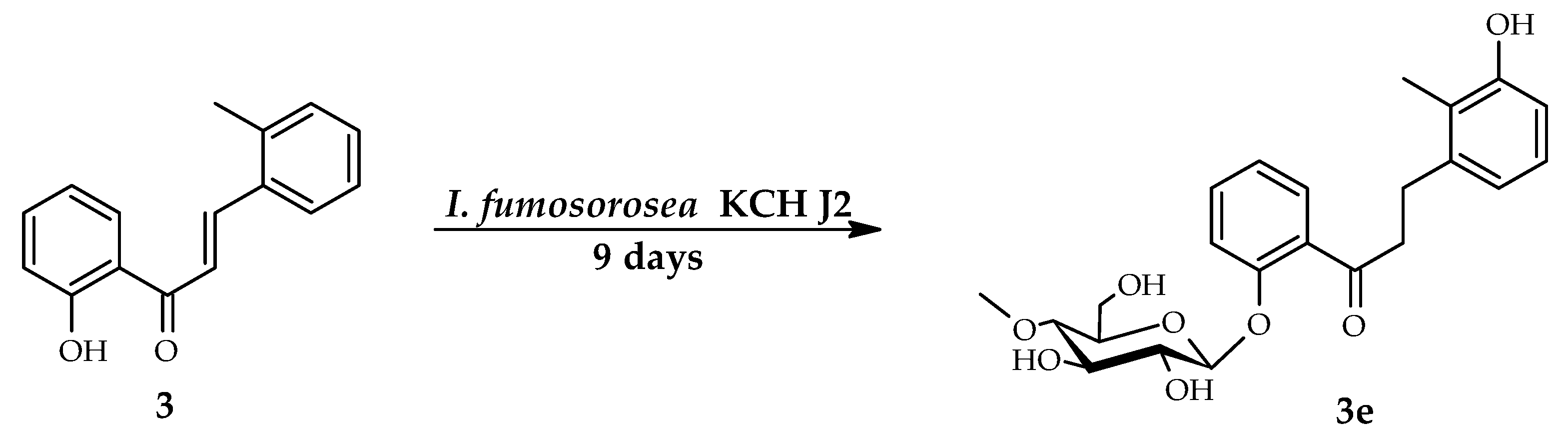
2.2. Biotransformation of 2′-Hydroxy-2-methylchalcone (3) in the Culture of I. fumosorosea KCH J2
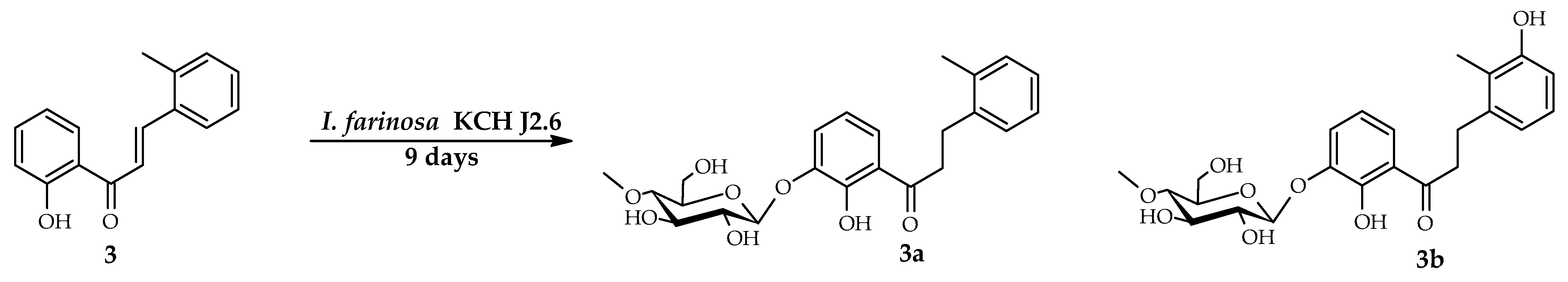
2.3. Biotransformations of 2′-Hydroxy-2-methylchalcone (3) in the Culture of I. farinosa KCH J2.6
3. Discussion
4. Materials and Methods
4.1. Substrate
2′-Hydroxy-2-methylchalcone (3)
4.2. Microorganisms
4.3. Analysis
4.4. Screening Procedure
4.5. The Semi-Preparative Biotransformations
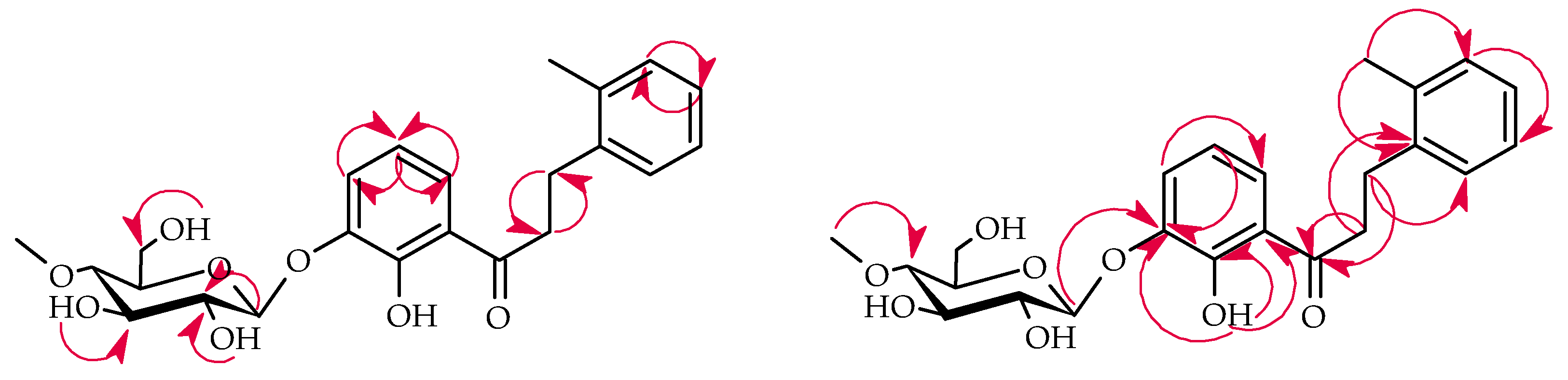
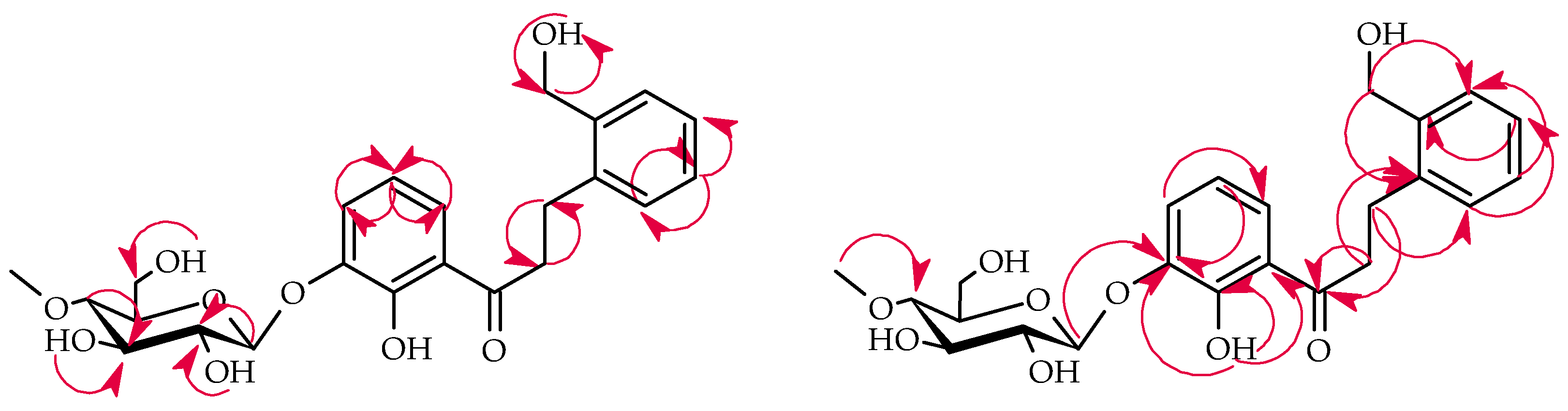
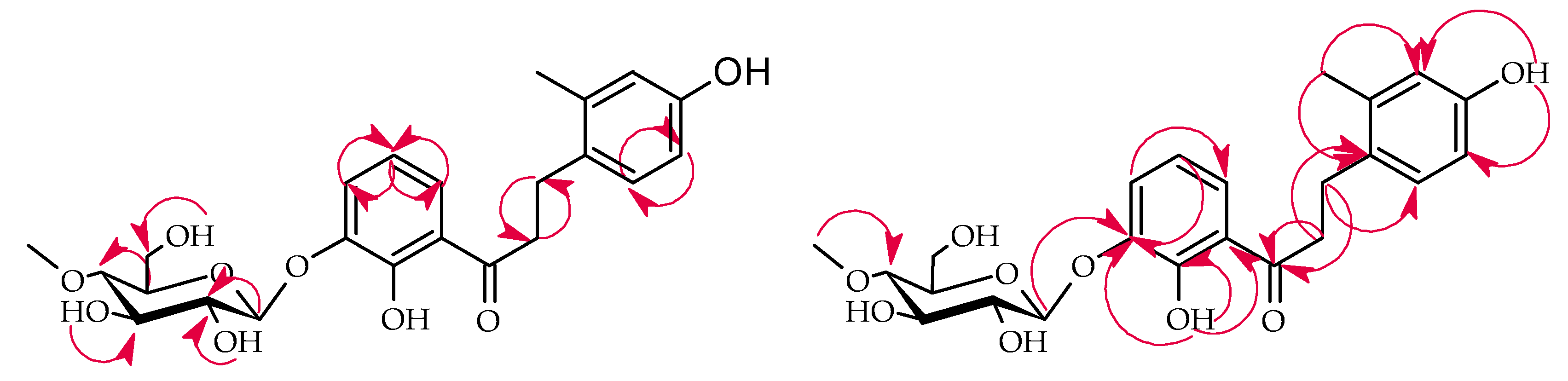
4.5.1. 2′-Hydroxy-2-methyldihydrochalcone 3′-O-β-d-(4″-O-Methyl)-glucopyranoside (3a)
4.5.2. 2′,3-Dihydroxy-2-methyldihydrochalcone 3′-O-β-d-(4″-O-Methyl)-glucopyranoside (3b)
4.5.3. 2′-Hydroxy-2-hydroxymethyldihydrochalcone 3′-O-β-d-(4″-O-Methyl)-glucopyranoside (3c)
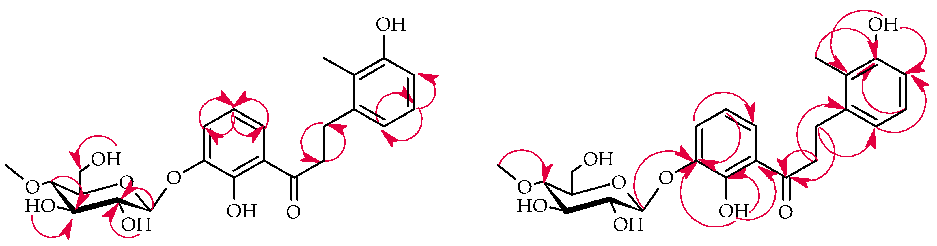
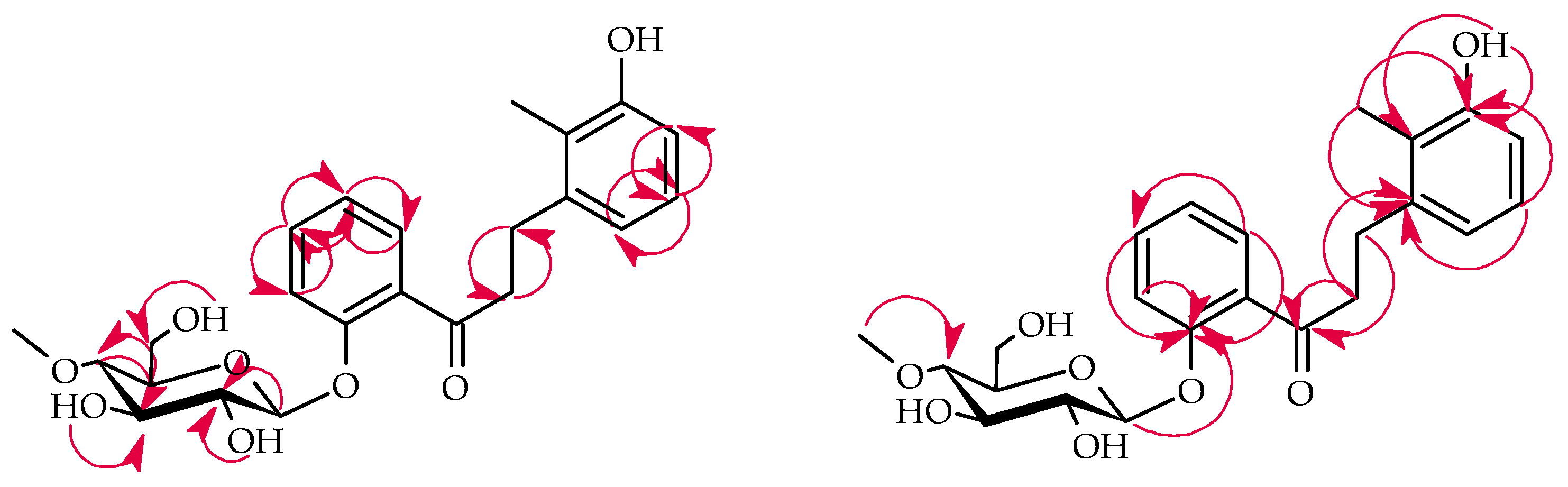
4.5.4. 2′,4-Dihydroxy-2-methyldihydrochalcone 3′-O-β-d-(4″-O-Methyl)-glucopyranoside (3d)
4.5.5. 3-Hydroxy-2-methyldihydrochalcone 2′-O-β-D-(4″-O-Methyl)-glucopyranoside (3e)
5. Conclusions
Supplementary Materials
Author Contributions
Funding
Data Availability Statement
Acknowledgments
Conflicts of Interest
References
- Crozier, A.; Jaganath, I.B.; Clifford, M.N. Dietary phenolics: Chemistry, bioavailability and effects on health. Nat. Prod. Rep. 2009, 26, 1001–1043. [Google Scholar] [CrossRef]
- Cassidy, A.; Minihane, A.M. The role of metabolism (and the microbiome) in defining the clinical efficacy of dietary flavonoids. Am. J. Clin. Nutr. 2017, 105, 10–22. [Google Scholar] [CrossRef] [Green Version]
- Perez-Vizcaino, F.; Fraga, C.G. Research trends in flavonoids and health. Arch. Biochem. Biophys. 2018, 646, 107–112. [Google Scholar] [CrossRef]
- Wang, T.Y.; Li, Q.; Bi, K. Bioactive flavonoids in medicinal plants: Structure, activity and biological fate. Asian J. Pharm. Sci. 2018, 13, 12–23. [Google Scholar] [CrossRef]
- Stompor, M.; Kałużny, M.; Żarowska, B. Biotechnological methods for chalcone reduction using whole cells of Lactobacillus, Rhodococcus and Rhodotorula strains as a way to produce new derivatives. Appl. Microbiol. Biotechnol. 2016, 100, 8371–8384. [Google Scholar] [CrossRef]
- Zhang, J.; Fu, X.L.; Yang, N.; Wang, Q.A. Synthesis and cytotoxicity of chalcones and 5-deoxyflavonoids. Sci. World J. 2013, 2013, 649485. [Google Scholar] [CrossRef] [PubMed] [Green Version]
- Nagula, R.L.; Wairkar, S. Recent advances in topical delivery of flavonoids: A review. J. Control. Release 2019, 296, 190–201. [Google Scholar] [CrossRef] [PubMed]
- Thilakarathna, S.H.; Rupasinghe, V.H.P. Flavonoid bioavailability and attempts for bioavailability enhancement. Nutrients 2013, 5, 3367–3387. [Google Scholar] [CrossRef]
- Koirala, N.; Thuan, N.H.; Ghimire, G.P.; Thang, D.V.; Sohng, J.K. Methylation of flavonoids: Chemical structures, bioactivities, progress and perspectives for biotechnological production. Enzym. Microb.Technol. 2016, 86, 103–116. [Google Scholar] [CrossRef]
- Wen, L.; Jiang, Y.; Yang, J.; Zhao, Y.; Tian, M.; Yang, B. Structure, bioactivity, and synthesis of methylated flavonoids. Ann. N. Y. Acad. Sci. 2017, 1398, 120–129. [Google Scholar] [CrossRef] [PubMed]
- Xiao, J.; Muzashvili, T.S.; Georgiev, M.I. Advances in the biotechnological glycosylation of valuable flavonoids. Biotechnol. Adv. 2014, 32, 1145–1156. [Google Scholar] [CrossRef]
- Dymarska, M.; Janeczko, T.; Kostrzewa-Susłow, E. Glycosylation of 3-hydroxyflavone, 3-methoxyflavone, quercetin and baicalein in fungal cultures of the genus Isaria. Molecules 2018, 23, 2477. [Google Scholar] [CrossRef] [PubMed] [Green Version]
- Xiao, J. Dietary flavonoid aglycones and their glycosides: Which show better biological significance? Crit. Rev. Food. Sci. Nutr. 2017, 57, 1874–1905. [Google Scholar] [CrossRef] [PubMed]
- Hostetler, G.L.; Ralston, R.A.; Schwartz, S.J. Flavones: Food sources, bioavailability, metabolism, and bioactivity. Adv. Nutr. 2017, 8, 423–435. [Google Scholar] [CrossRef] [PubMed] [Green Version]
- Janeczko, T.; Gładkowski, W.; Kostrzewa-Susłow, E. Microbial transformations of chalcones to produce food sweetener derivatives. J. Mol. Catal. B Enzym. 2013, 98, 55–61. [Google Scholar] [CrossRef]
- Ruiz-Ojeda, F.J.; Plaza-Díaz, J.; Sáez-Lara, M.J.; Gil, A. Effects of sweeteners on the gut microbiota: A review of experimental studies and clinical trials. Adv. Nutr. 2019, 10, S31–S48. [Google Scholar] [CrossRef] [PubMed] [Green Version]
- Yang, W.; Zhou, K.; Zhou, Y.; An, Y.; Hu, T.; Lu, J.; Huang, S.; Pei, G. Naringin dihydrochalcone ameliorates cognitive deficits and neuropathology in APP/PS1 transgenic mice. Front. Aging Neurosci. 2018, 10, 169. [Google Scholar] [CrossRef] [PubMed] [Green Version]
- Ehrenkranz, J.R.L.; Lewis, N.G.; Kahn, C.R.; Roth, J. Phlorizin: A review. Diabetes Metab. Res. Rev. 2005, 21, 31–38. [Google Scholar] [CrossRef]
- Londzin, P.; Siudak, S.; Cegieła, U.; Pytlik, M.; Janas, A.; Waligóra, A.; Folwarczna, J. Phloridzin, an apple polyphenol, exerted unfavorable effects on bone and muscle in an experimental model of type 2 diabetes in rats. Nutrients 2018, 10, 1701. [Google Scholar] [CrossRef] [Green Version]
- Payne, J.N.; Badwaik, V.D.; Waghwani, H.K.; Moolani, H.V.; Tockstein, S.; Thompson, D.H.; Dakshinamurthy, R. Development of dihydrochalcone-functionalized gold nanoparticles for augmented antineoplastic activity. Int. J. Nanomed. 2018, 13, 1917–1926. [Google Scholar] [CrossRef] [Green Version]
- Niederberger, K.E.; Tennant, D.R.; Bellion, P. Dietary intake of phloridzin from natural occurrence in foods. Br. J. Nutr. 2020, 123, 942–950. [Google Scholar] [CrossRef]
- Tian, L.; Cao, J.; Zhao, T.; Liu, Y.; Khan, A.; Cheng, G. The bioavailability, extraction, biosynthesis and distribution of natural dihydrochalcone: Phloridzin. Int. J. Mol. Sci. 2021, 22, 962. [Google Scholar] [CrossRef]
- Hofer, B. Recent developments in the enzymatic O-glycosylation of flavonoids. Appl. Microbiol. Biotechnol. 2016, 100, 4269–4281. [Google Scholar] [CrossRef] [PubMed]
- Nakamura, Y.; Watanabe, S.; Miyake, N.; Kohno, H.; Osawa, T. Dihydrochalcones: Evaluation as novel radical scavenging antioxidants. J. Agric. Food Chem. 2003, 51, 3309–3312. [Google Scholar] [CrossRef]
- Xiao, Z.; Wang, Y.; Wang, J.; Li, P.; Ma, F. Structure-antioxidant capacity relationship of dihydrochalcone compounds in Malus. Food Chem. 2019, 275, 354–360. [Google Scholar] [CrossRef] [PubMed]
- Wu, S.Y.; Fu, Y.H.; Zhou, Q.; Bai, M.; Chen, G.Y.; Han, C.R.; Song, X.P. A new dihydrochalcone glycoside from the stems of Homalium stenophyllum. Nat. Prod. Res. 2018, 32, 953–958. [Google Scholar] [CrossRef] [PubMed]
- Omar, A.M.; Dibwe, D.F.; Sun, S.; Tawila, A.M.; Kim, M.; Phrutivorapongkul, A.; Toyooka, N.; Awale, S. Fragranone C: A new dihydrochalcone glucopyranoside from Anneslea fragrans twigs. Nat. Prod. Res. 2020, 2, 1–6. [Google Scholar] [CrossRef]
- Bao, S.; Wang, Q.; Bao, W.; Ao, W. Structure elucidation and NMR assignments of a new dihydrochalcone from Empetrum nigrum subsp. asiaticum (Nakai ex H.Ito) Kuvaev. Nat. Prod. Res. 2020, 34, 930–934. [Google Scholar] [CrossRef]
- Pompermaier, L.; Heiss, E.H.; Alilou, M.; Mayr, F.; Monizi, M.; Lautenschlaeger, T.; Schuster, D.; Schwaiger, S.; Stuppner, H. Dihydrochalcone Glucosides from the Subaerial Parts of Thonningia sanguinea and Their in Vitro PTP1B Inhibitory Activities. J. Nat. Prod. 2018, 81, 2091–2100. [Google Scholar] [CrossRef] [PubMed]
- Lima, E.M.; Fernando, L.M.; Felix, L.P.; de Oliveira Filho, A.A.; Carneiro Neto, A.N.; Moura, R.T.; Teles, Y.C.F. First complete NMR data and theoretical study of an antimicrobial formylated dihydrochalcone from Psidium guineense Sw. Nat. Prod. Res. 2020, 1–5. [Google Scholar] [CrossRef]
- Dymarska, M.; Janeczko, T.; Kostrzewa-Susłow, E. Glycosylation of methoxylated flavonoids in the cultures of Isaria fumosorosea KCH J2. Molecules 2018, 23, 2578. [Google Scholar] [CrossRef] [PubMed] [Green Version]
- Dymarska, M.; Janeczko, T.; Kostrzewa-Susłow, E. Biotransformations of flavones and an isoflavone (daidzein) in cultures of entomopathogenic filamentous fungi. Molecules 2018, 23, 1356. [Google Scholar] [CrossRef] [Green Version]
- Dymarska, M.; Grzeszczuk, J.; Urbaniak, M.; Janeczko, T.; Pląskowska, E.; Stępień, Ł.; Kostrzewa-Susłow, E. Glycosylation of 6-methylflavone by the strain Isaria fumosorosea KCH J2. PLoS ONE 2017, 12, e0184885. [Google Scholar] [CrossRef]
- Krawczyk-Łebek, A.; Dymarska, M.; Janeczko, T.; Kostrzewa-Susłow, E. Entomopathogenic filamentous fungi as biocatalysts in glycosylation of methylflavonoids. Catalysts 2020, 10, 1148. [Google Scholar] [CrossRef]
- Dou, F.; Wang, Z.; Li, G.; Dun, B. Microbial transformation of flavonoids by Isaria fumosorosea ACCC 37814. Molecules 2019, 24, 1028. [Google Scholar] [CrossRef] [PubMed] [Green Version]
- Sordon, S.; Popłoński, J.; Tronina, T.; Huszcza, E. Regioselective O-glycosylation of flavonoids by fungi Beauveria bassiana, Absidia coerulea and Absidia glauca. Bioorg. Chem. 2019, 93, 102750. [Google Scholar] [CrossRef]
- Sordon, S.; Popłoński, J.; Tronina, T.; Huszcza, E. Microbial glycosylation of daidzein, genistein and biochanin a: Two new glucosides of biochanin A. Molecules 2017, 22, 81. [Google Scholar] [CrossRef] [Green Version]
- Strugała, P.; Tronina, T.; Huszcza, E.; Gabrielska, J. Bioactivity in vitro of quercetin glycoside obtained in Beauveria bassiana culture and its interaction with liposome membranes. Molecules 2017, 22, 1520. [Google Scholar] [CrossRef] [PubMed] [Green Version]
- Tronina, T.; Strugała, P.; Popłoński, J.; Włoch, A.; Sordon, S.; Bartmańska, A.; Huszcza, E. The Influence of Glycosylation of Natural and Synthetic Prenylated Flavonoids on Binding to Human Serum Albumin and Inhibition of Cyclooxygenases COX-1 and COX-2. Molecules 2017, 22, 1230. [Google Scholar] [CrossRef] [Green Version]
- Kozłowska, E.; Urbaniak, M.; Hoc, N.; Grzeszczuk, J.; Dymarska, M.; Stępień, Ł.; Pląskowska, E.; Kostrzewa-Susłow, E.; Janeczko, T. Cascade biotransformation of dehydroepiandrosterone (DHEA) by Beauveria species. Sci. Rep. 2018, 8, 13449. [Google Scholar] [CrossRef]
- Łużny, M.; Tronina, T.; Kozłowska, E.; Dymarska, M.; Popłoński, J.; Łyczko, J.; Kostrzewa-Susłow, E.; Janeczko, T. Biotransformation of methoxyflavones by selected entomopathogenic filamentous fungi. Int. J. Mol. Sci. 2020, 21, 6121. [Google Scholar] [CrossRef]
- Kim, H.J.; Lee, I.S. Microbial metabolism of the prenylated chalcone xanthohumol. J. Nat. Prod. 2006, 69, 1522–1524. [Google Scholar] [CrossRef]
- Tronina, T.; Bartmańska, A.; Milczarek, M.; Wietrzyk, J.; Popłoński, J.; Rój, E.; Huszcza, E. Antioxidant and antiproliferative activity of glycosides obtained by biotransformation of xanthohumol. Bioorg. Med. Chem. Lett. 2013, 23, 1957–1960. [Google Scholar] [CrossRef]
- Huszcza, E.; Bartmańska, A.; Tronina, T. Glycosylation of xanthohumol by fungi. Z. Naturforsch. C J. Biosci. 2008, 63, 557–560. [Google Scholar] [CrossRef] [Green Version]
- Overwin, H.; Wray, V.; Hofer, B. Biotransformation of phloretin by amylosucrase yields three novel dihydrochalcone glucosides. J. Biotechnol. 2015, 211, 103–106. [Google Scholar] [CrossRef] [PubMed]
- Kostrzewa-Susow, E.; Dymarska, M.; Guzik, U.; Wojcieszyńska, D.; Janeczko, T. Stenotrophomonas maltophilia: A gram-negative bacterium useful for transformations of flavanone and chalcone. Molecules 2017, 22, 1830. [Google Scholar] [CrossRef] [PubMed] [Green Version]
- Żyszka-Haberecht, B.; Poliwoda, A.; Lipok, J. Structural constraints in cyanobacteria-mediated whole-cell biotransformation of methoxylated and methylated derivatives of 2′-hydroxychalcone. J. Biotechnol. 2019, 293, 36–46. [Google Scholar] [CrossRef] [PubMed]
- Łużny, M.; Kozłowska, E.; Kostrzewa-Susłow, E.; Janeczko, T. Highly effective, regiospecific hydrogenation of methoxychalcone by Yarrowia lipolytica enables production of food sweeteners. Catalysts 2020, 10, 1135. [Google Scholar] [CrossRef]
- Silva, V.D.; Stambuk, B.U.; da Graca Nascimento, M. Efficient chemoselective biohydrogenation of 1,3-diaryl-2-propen-1-ones catalyzed by Saccharomyces cerevisiae yeasts in biphasic system. J. Mol. Catal. B Enzym. 2010, 63, 157–163. [Google Scholar] [CrossRef]
- Silva, A.M.S.; Tavares, H.R.; Barros, A.I.N.R.A.; Cavaleiro, J.A.S. NMR and structural and conformational features of 2′-hydroxychalcones and flavones. Spectrosc. Lett. 1997, 30, 1655–1667. [Google Scholar] [CrossRef]
- Yadav, N.; Dixit, S.K.; Bhattacharya, A.; Mishra, L.C.; Sharma, M.; Awasthi, S.K.; Bhasin, V.K. Antimalarial activity of newly synthesized chalcone derivatives in vitro. Chem. Biol. Drug Des. 2012, 80, 340–347. [Google Scholar] [CrossRef] [PubMed]


| Proton | Compound | |||||
|---|---|---|---|---|---|---|
| 3 | 3a | 3b | 3c | 3d | 3e | |
| H-α | 7.94 (d) | 3.40 (m) | 3.36 (m) | 3.46 (m) | 3.33 (m) | 3.34 (m) |
| J = 15.3 | ||||||
| H-β | 8.25 (d) | 3.03 (m) | 3.02 (m) | 3.10 (m) | 2.94 (m) | 2.95 (m) |
| J = 15.4 | ||||||
| H-3 | 7.30 (m) | 7.15 (dd) | - | 7.40 (m) | 6.65 (d) | - |
| J = 6.8 | J = 2.5 | |||||
| J = 1.7 | ||||||
| H-4 | 7.36 (m) | 7.10 (m) | 6.72 (m) | 7.20 (m) | - | 6.68 (d) |
| J = 8.0 | ||||||
| H-5 | 7.30 (m) | 7.10 (m) | 6.92 (t) | 7.20 (m) | 6.59 (dd) | 6.90 (t) |
| J = 7.8 | J = 8.2 | J = 7.8 | ||||
| J = 2.6 | ||||||
| H-6 | 7.97 (d) | 7.23 (m) | 6.72 (m) | 7.28 (m) | 7.03 (d) | 6.71 (d) |
| J = 7.8 | J = 8.2 | J = 7.5 | ||||
| H-3′ | 7.01 (m) | - | - | - | - | 7.30 (dd) |
| J = 8.4 | ||||||
| J = 0.5 | ||||||
| H-4′ | 7.58 (m) | 7.40 (dd) | 7.40 (dd) | 7.40 (m) | 7.39 (dd) | 7.47 (ddd) |
| J = 8.0 | J = 8.0 | J = 8.0 | J = 8.5 | |||
| J = 1.2 | J = 1.1 | J = 1.3 | J = 7.3 | |||
| J = 1.8 | ||||||
| H-5′ | 7.01 (m) | 6.86 (t) | 6.86 (t) | 6.85 (t) | 6.85 (t) | 7.11 (td) |
| J = 8.1 | J = 8.1 | J = 8.1 | J = 81 | J = 7.6 | ||
| J = 0.9 | ||||||
| H-6′ | 8.28 (dd) | 7.65 (dd) | 7.65 (dd) | 7.65 (dd) | 7.64 (dd) | 7.58 (dd) |
| J = 8.4 | J = 8.1 | J = 8.2 | J = 8.2 | J = 8.1 | J = 7.7 | |
| J = 1.1 | J = 1.3 | J = 1.4 | J = 1.3 | J = 1.4 | J = 1.8 | |
| H-1″ | - | 4.91 (d) | 4.91 (d) | 4.91 (d) | 4.91 (d) | 5.04 (d) |
| J = 7.8 | J = 7.8 | J = 7.8 | J = 7.8 | J = 7.8 | ||
| H-2″ | - | 3.50 (m) | 3.50 (ddd) | 3.49 (m) | 3.50 (m) | 3.53 (m) |
| J = 9.1 | ||||||
| J = 7.9 | ||||||
| J = 3.8 | ||||||
| H-3″ | - | 3.62 (td) | 3.62 (td) | 3.62 (m) | 3.62 (td) | 3.63 (td) |
| J = 9.1 | J = 9.0 | J = 9.0 | J = 9.0 | |||
| J = 3.8 | J = 3.9 | J = 3.9 | J = 4.3 | |||
| H-4″ | - | 3.22 (m) | 3.22 (m) | 3.22 (m) | 3.22 (m) | 3.20 (m) |
| H-5″ | - | 3.44 (ddd) | 3.44 (ddd) | 3.43 (m) | 3.44 (ddd) | 3.49 (ddd) |
| J = 9.7 | J = 9.7 | J = 9.6 | J = 9.7 | |||
| J = 4.8 | J = 4.8 | J = 4.7 | J = 4.6 | |||
| J = 2.1 | J = 2.2 | J = 2.1 | J = 2.0 | |||
| H-6″ | - | 3.81 (ddd) | 3.82 (ddd) | 3.81 (ddd) | 3.81 (ddd) | 3.83 (ddd) |
| J = 11.6 | J = 11.5 | J = 11.6 | J = 11.6 | J = 7.3 | ||
| J = 5.1 | J = 5.1 | J = 5.2 | J = 5.2 | J = 4.7 | ||
| J = 2.0 | J = 2.1 | J = 2.2 | J = 2.1 | J = 2.2 | ||
| 3.68 (ddd) | 3.68 (ddd) | 3.68 (ddd) | 3.68 (ddd) | 3.68 (m) | ||
| J = 11.6 | J = 11.6 | J = 11.8 | J = 11.7, | |||
| J = 6.9 | J = 6.8 | J = 7.3 | J = 7.0 | |||
| J = 4.9 | J = 4.9 | J = 4.8 | J = 4.8 | |||
| C4″-OCH3 | - | 3.56 (s) | 3.56 (s) | 3.56 (s) | 3.56 (s) | 3.55 (s) |
| C2′-OH | 12.88 (s) | 12.14 (s) | 12.18 (s) | 12.17 (s) | 12.20 (s) | - |
| C3-OH | - | - | 8.16 (s) | - | - | 8.08 (s) |
| C4-OH | - | - | - | - | 8.02 (s) | - |
| C2-CH3 | 2.51 (s) | 2.35 (s) | 2.21 (s) | - | 2.27 (s) | 2.19 (s) |
| 2″-OH | - | 4.68 (d) | 4.68 (d) | 4.59 (d) | 4.63 (d) | 4.65 (d) |
| J = 3.5 | J = 3.9 | J = 3.9 | J = 3.7 | J = 4.1 | ||
| 3″-OH | - | 4.42 (d) | 4.41 (d) | 4.39 (d) | 4.40 (d) | 4.48 (d) |
| J = 3.9 | J = 4.0 | J = 4.0 | J = 4.0 | J = 4.3 | ||
| 6″-OH | - | 3.76 (dd) | 3.76 (m) | 3.74 (dd) | 3.75 (dd) | 3.71 (dd) |
| J = 11.7 | J = 11.1 | J = 12.8 | J = 9.3 | |||
| J = 4.8 | J = 5.9 | J = 5.8 | J = 4.5 | |||
| 2-CH2- | - | - | - | 4.73 (d) | - | - |
| J = 5.5 | ||||||
| 2-CH2-OH | - | - | - | 4.15 (t) | - | - |
| J = 5.4 | ||||||
| Carbon | Compound | |||||
|---|---|---|---|---|---|---|
| 3 | 3a | 3b | 3c | 3d | 3e | |
| C-α | 122.3 | 40.2 | 40.5 | 41.3 | 40.6 | 44.7 |
| C-β | 143.4 | 28.0 | 28.5 | 27.3 | 27.4 | 28.6 |
| C-1 | 134.4 | 140.1 | 141.5 | 140.0 | 130.7 | 142.1 |
| C-2 | 139.5 | 136.8 | 123.2 | 140.7 | 137.9 | 123.2 |
| C-3 | 131.8 | 131.0 | 156.3 | 129.1 | 117.9 | 156.1 |
| C-4 | 131.6 | 127.1 | 113.7 | 127.0 | 156.6 | 113.4 |
| C-5 | 127.3 | 126.9 | 126.9 | 128.2 | 113.7 | 126.8 |
| C-6 | 127.8 | 129.6 | 121.1 | 130.0 | 130.6 | 121.0 |
| C-1′ | 120.9 | 121.5 | 121.5 | 121.5 | 121.5 | 131.0 |
| C-2′ | 164.5 | 153.5 | 153.6 | 153.7 | 153.6 | 157.1 |
| C-3′ | 119.0 | 147.2 | 147.2 | 147.2 | 147.2 | 117.3 |
| C-4′ | 137.5 | 123.9 | 123.9 | 124.0 | 123.9 | 133.9 |
| C-5′ | 119.9 | 119.3 | 119.3 | 119.3 | 119.3 | 123.1 |
| C-6′ | 131.4 | 124.8 | 124.8 | 124.8 | 124.8 | 130.4 |
| C-1″ | - | 102.6 | 102.6 | 102.6 | 102.6 | 102.4 |
| C-2″ | - | 74.9 | 74.9 | 74.9 | 74.9 | 75.0 |
| C-3″ | - | 77.9 | 77.9 | 77.9 | 77.9 | 78.1 |
| C-4″ | - | 80.0 | 80.0 | 80.0 | 80.0 | 80.1 |
| C-5″ | - | 77.2 | 77.1 | 77.2 | 77.2 | 77.3 |
| C-6″ | - | 62.1 | 62.1 | 62.1 | 62.1 | 62.1 |
| 4″-OCH3 | - | 60.6 | 60.5 | 60.5 | 60.5 | 60.6 |
| C=O | 195.0 | 206.8 | 206.9 | 207.0 | 207.2 | 202.6 |
| 2-CH3 | 19.8 | 19.4 | 11.4 | - | 19.5 | 11.5 |
| 2-CH2 | - | - | - | 62.9 | - | - |
Publisher’s Note: MDPI stays neutral with regard to jurisdictional claims in published maps and institutional affiliations. |
© 2021 by the authors. Licensee MDPI, Basel, Switzerland. This article is an open access article distributed under the terms and conditions of the Creative Commons Attribution (CC BY) license (https://creativecommons.org/licenses/by/4.0/).
Share and Cite
Krawczyk-Łebek, A.; Dymarska, M.; Janeczko, T.; Kostrzewa-Susłow, E. New Glycosylated Dihydrochalcones Obtained by Biotransformation of 2′-Hydroxy-2-methylchalcone in Cultures of Entomopathogenic Filamentous Fungi. Int. J. Mol. Sci. 2021, 22, 9619. https://0-doi-org.brum.beds.ac.uk/10.3390/ijms22179619
Krawczyk-Łebek A, Dymarska M, Janeczko T, Kostrzewa-Susłow E. New Glycosylated Dihydrochalcones Obtained by Biotransformation of 2′-Hydroxy-2-methylchalcone in Cultures of Entomopathogenic Filamentous Fungi. International Journal of Molecular Sciences. 2021; 22(17):9619. https://0-doi-org.brum.beds.ac.uk/10.3390/ijms22179619
Chicago/Turabian StyleKrawczyk-Łebek, Agnieszka, Monika Dymarska, Tomasz Janeczko, and Edyta Kostrzewa-Susłow. 2021. "New Glycosylated Dihydrochalcones Obtained by Biotransformation of 2′-Hydroxy-2-methylchalcone in Cultures of Entomopathogenic Filamentous Fungi" International Journal of Molecular Sciences 22, no. 17: 9619. https://0-doi-org.brum.beds.ac.uk/10.3390/ijms22179619