
The Influence of Different Guided Bone Regeneration Procedures on the Contour of Bone Graft after Wound Closure: A Retrospective Cohort Study


Abstract
:1. Introduction
2. Materials and Methods
2.1. Study Population
2.1.1. Inclusion Criteria
- Male or female patients aged 18 to 65 years (including 18 and 65 years).
- Presence of a three-wall or two-wall horizontal bone defect in the anterior region.
- Received one of the following GBR procedures: particulate bone substitute +CM (Group 1), particulate bone substitute + CM + healing caps (Group 2), sticky bone + CM + pins (Group 3), or sticky bone + CM + pins + surgical template (Group 4).
- Bone augmentation was applied > 3 months after tooth extraction.
- Good general health and absence of periodontal diseases.
- Patients were willing to participate in this study and signed the informed consent form.
2.1.2. Exclusion Criteria
- Uncontrolled systemic diseases.
- Heavy smokers (>20 cigarettes per day).
- Females in pregnancy or lactation.
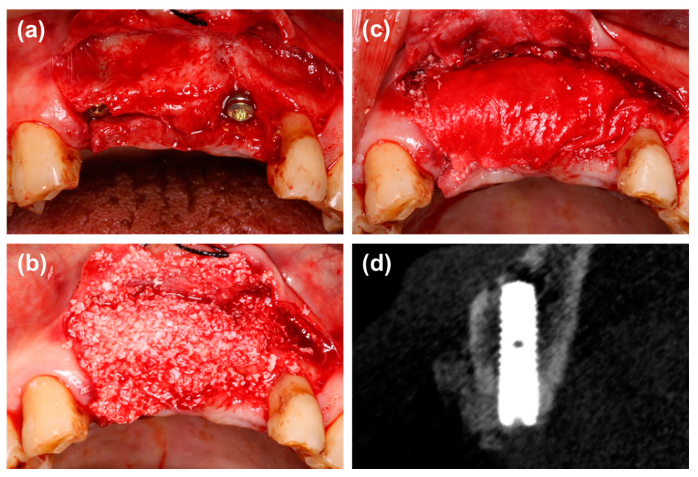
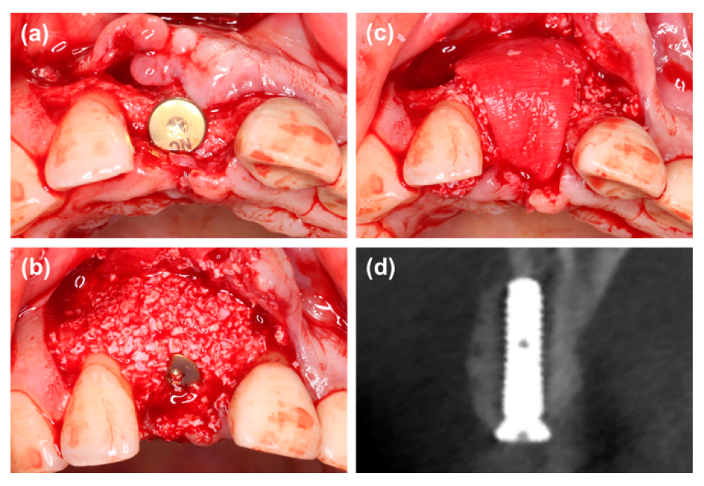
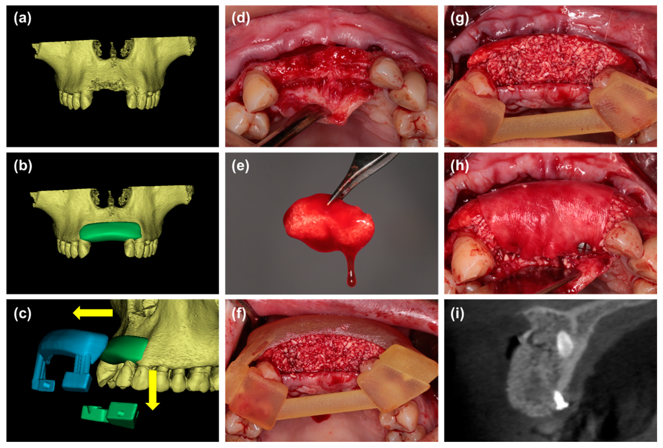
2.2. Surgical Procedures
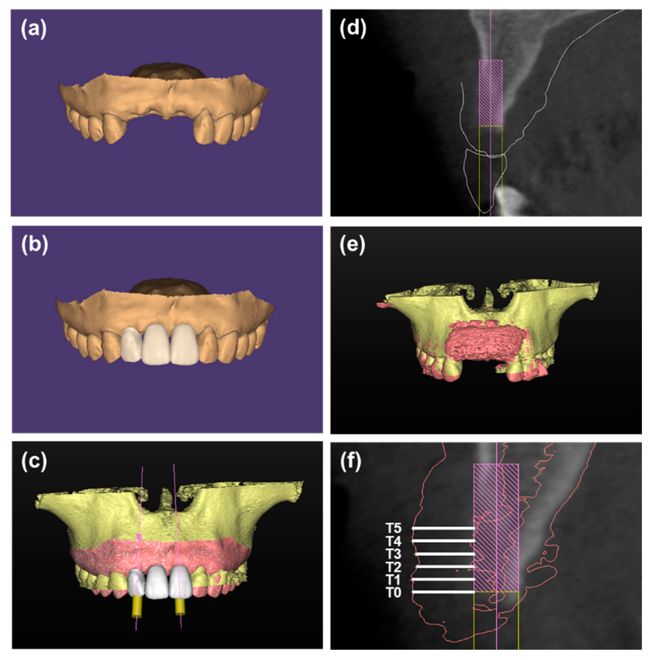
2.3. CBCT Analysis
2.4. Sample Size Calculation
2.5. Statistical Analysis
3. Results
4. Discussion
5. Conclusions
- GBR with customized sticky bone showed better results than GBR with particulate bone substitutes, GBR with particulate bone substitutes in combination with a wide healing cap and GBR with sticky bone with respect to the thickness of labial graft immediately after wound closure.
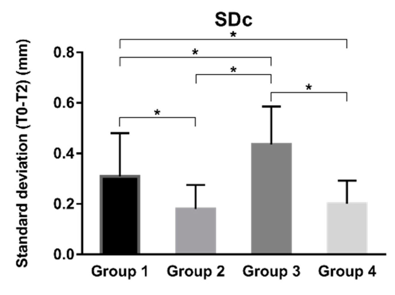
- The use of customized sticky bone and wide healing cap enhanced volume stability of bone grafts, especially in the coronal portion of bone grafts.
- The use of surgical templates contributed to an appropriate contour of bone grafts after wound closure.
Author Contributions
Funding
Institutional Review Board Statement
Informed Consent Statement
Data Availability Statement
Conflicts of Interest
References
- Araujo, M.G.; Lindhe, J. Dimensional ridge alterations following tooth extraction. An experimental study in the dog. J. Clin. Periodontol. 2005, 32, 212–218. [Google Scholar] [CrossRef]
- Chiapasco, M.; Zaniboni, M.; Boisco, M. Augmentation procedures for the rehabilitation of deficient edentulous ridges with oral implants. Clin. Oral Implants Res. 2006, 17, 136–159. [Google Scholar] [CrossRef]
- Donos, N.; Mardas, N.; Chadha, V. Clinical outcomes of implants following lateral bone augmentation: Systematic assessment of available options (barrier membranes, bone grafts, split osteotomy). J. Clin. Periodontol. 2008, 35, 173–202. [Google Scholar] [CrossRef] [PubMed]
- Benic, G.I.; Hammerle, C.H. Horizontal bone augmentation by means of guided bone regeneration. Periodontol. 2000 2014, 66, 13–40. [Google Scholar] [CrossRef] [PubMed]
- Troeltzsch, M.; Troeltzsch, M.; Kauffmann, P.; Gruber, R.; Brockmeyer, P.; Moser, N.; Rau, A.; Schliephake, H. Clinical efficacy of grafting materials in alveolar ridge augmentation: A systematic review. J. Craniomaxillofac. Surg. 2016, 44, 1618–1629. [Google Scholar] [CrossRef] [PubMed]
- Sanz-Sanchez, I.; Ortiz-Vigon, A.; Sanz-Martin, I.; Figuero, E.; Sanz, M. Effectiveness of lateral bone augmentation on the alveolar crest dimension: A systematic review and meta-analysis. J. Dent. Res. 2015, 94, 128S–142S. [Google Scholar] [CrossRef] [PubMed]
- Benic, G.I.; Bernasconi, M.; Jung, R.E.; Hammerle, C.H. Clinical and radiographic intra-subject comparison of implants placed with or without guided bone regeneration: 15-year results. J. Clin. Periodontol. 2017, 44, 315–325. [Google Scholar] [CrossRef]
- Urban, I.A.; Monje, A.; Lozada, J.L.; Wang, H.L. Long-term evaluation of peri-implant bone level after reconstruction of severely atrophic edentulous maxilla via vertical and horizontal guided bone regeneration in combination with sinus augmentation: A case series with 1 to 15 years of loading. Clin. Implant. Dent. Relat Res. 2017, 19, 46–55. [Google Scholar] [CrossRef] [PubMed] [Green Version]
- Haugen, H.J.; Lyngstadaas, S.P.; Rossi, F.; Perale, G. Bone grafts: Which is the ideal biomaterial? J. Clin. Periodontol. 2019, 46, 92–102. [Google Scholar] [CrossRef]
- Strietzel, F.P.; Khongkhunthian, P.; Khattiya, R.; Patchanee, P.; Reichart, P.A. Healing pattern of bone defects covered by different membrane types--a histologic study in the porcine mandible. J. Biomed. Mater. Res. B Appl. Biomater. 2006, 78, 35–46. [Google Scholar] [CrossRef]
- Schwarz, F.; Herten, M.; Ferrari, D.; Wieland, M.; Schmitz, L.; Engelhardt, E.; Becker, J. Guided bone regeneration at dehiscence-type defects using biphasic hydroxyapatite + beta tricalcium phosphate (Bone Ceramic) or a collagen-coated natural bone mineral (BioOss Collagen): An immunohistochemical study in dogs. Int. J. Oral Maxillofac. Surg. 2007, 36, 1198–1206. [Google Scholar] [CrossRef]
- Chiapasco, M.; Casentini, P. Horizontal bone-augmentation procedures in implant dentistry: Prosthetically guided regeneration. Periodontol. 2000 2018, 77, 213–240. [Google Scholar] [CrossRef] [PubMed]
- Shah, R.; Gowda, T.M.; Thomas, R.; Kumar, T.; Mehta, D.S. Biological activation of bone grafts using injectable platelet-rich fibrin. J. Prosthet. Dent. 2019, 121, 391–393. [Google Scholar] [CrossRef] [PubMed]
- Mir-Mari, J.; Wui, H.; Jung, R.E.; Hammerle, C.H.; Benic, G.I. Influence of blinded wound closure on the volume stability of different GBR materials: An in vitro cone-beam computed tomographic examination. Clin. Oral Implants Res. 2016, 27, 258–265. [Google Scholar] [CrossRef]
- Mertens, C.; Braun, S.; Krisam, J.; Hoffmann, J. The influence of wound closure on graft stability: An in vitro comparison of different bone grafting techniques for the treatment of one-wall horizontal bone defects. Clin. Implant. Dent. Relat Res. 2019, 21, 284–291. [Google Scholar] [CrossRef] [PubMed]
- Ciocca, L.; Fantini, M.; De Crescenzio, F.; Corinaldesi, G.; Scotti, R. Direct metal laser sintering (DMLS) of a customized titanium mesh for prosthetically guided bone regeneration of atrophic maxillary arches. Med. Biol. Eng. Comput. 2011, 49, 1347–1352. [Google Scholar] [CrossRef] [PubMed]
- Blume, O.; Hoffmann, L.; Donkiewicz, P.; Wenisch, S.; Back, M.; Franke, J.; Schnettler, R.; Barbeck, M. Treatment of severely resorbed maxilla due to peri-implantitis by guided bone regeneration using a customized allogenic bone block: A case report. Materials (Basel) 2017, 10, 1213. [Google Scholar] [CrossRef] [Green Version]
- Blume, O.; Back, M.; Born, T.; Smeets, R.; Jung, O.; Barbeck, M. Treatment of a bilaterally severely resorbed posterior mandible due to early tooth loss by Guided Bone Regeneration using customized allogeneic bone blocks: A case report with 24 months follow-up data. J. Esthet. Restor. Dent. 2018, 30, 474–479. [Google Scholar] [CrossRef]
- Buser, D.; Martin, W.; Belser, U.C. Optimizing esthetics for implant restorations in the anterior maxilla: Anatomic and surgical considerations. Int. J. Oral Maxillofac. Implants 2004, 19, 43–61. [Google Scholar]
- Rojas-Vizcaya, F. Biological aspects as a rule for single implant placement. The 3A-2B rule: A clinical report. J. Prosthodont. 2013, 22, 575–580. [Google Scholar] [CrossRef]
- Spray, J.R.; Black, C.G.; Morris, H.F.; Ochi, S. The influence of bone thickness on facial marginal bone response: Stage 1 placement through stage 2 uncovering. Ann. Periodontol. 2000, 5, 119–128. [Google Scholar] [CrossRef]
- Le, B.T.; Borzabadi-Farahani, A. Labial bone thickness in area of anterior maxillary implants associated with crestal labial soft tissue thickness. Implant. Dent. 2012, 21, 406–410. [Google Scholar] [CrossRef]
- Elnayef, B.; Porta, C.; Suarez-Lopez, D.A.F.; Mordini, L.; Gargallo-Albiol, J.; Hernandez-Alfaro, F. The Fate of Lateral Ridge Augmentation: A Systematic Review and Meta-Analysis. Int. J. Oral Maxillofac. Implants 2018, 33, 622–635. [Google Scholar] [CrossRef] [Green Version]
- Mir-Mari, J.; Benic, G.I.; Valmaseda-Castellon, E.; Hammerle, C.; Jung, R.E. Influence of wound closure on the volume stability of particulate and non-particulate GBR materials: An in vitro cone-beam computed tomographic examination. Part II. Clin. Oral Implants. Res. 2017, 28, 631–639. [Google Scholar] [CrossRef] [Green Version]
- Benic, G.I.; Eisner, B.M.; Jung, R.E.; Basler, T.; Schneider, D.; Hammerle, C. Hard tissue changes after guided bone regeneration of peri-implant defects comparing block versus particulate bone substitutes: 6-month results of a randomized controlled clinical trial. Clin. Oral Implants. Res. 2019. [Google Scholar] [CrossRef]
- Naenni, N.; Berner, T.; Waller, T.; Huesler, J.; Hammerle, C.; Thoma, D.S. Influence of wound closure on volume stability with the application of different GBR materials: An in vitro cone-beam computed tomographic study. J. Periodontal Implant. Sci. 2019, 49, 14–24. [Google Scholar] [CrossRef]
- Friedmann, A.; Strietzel, F.P.; Maretzki, B.; Pitaru, S.; Bernimoulin, J.P. Histological assessment of augmented jaw bone utilizing a new collagen barrier membrane compared to a standard barrier membrane to protect a granular bone substitute material. Clin. Oral Implants Res. 2002, 13, 587–594. [Google Scholar] [CrossRef]
- Soni, R.; Priya, A.; Yadav, H.; Mishra, N.; Kumar, L. Bone augmentation with sticky bone and platelet-rich fibrin by ridge-split technique and nasal floor engagement for immediate loading of dental implant after extracting impacted canine. Natl. J. Maxillofac. Surg. 2019, 10, 98–101. [Google Scholar] [CrossRef]
- Scarano, A.; Inchingolo, F.; Murmura, G.; Traini, T.; Piattelli, A.; Lorusso, F. Three-dimensional architecture and mechanical properties of bovine bone mixed with autologous platelet liquid, blood, or physiological water: An in vitro study. Int. J. Mol. Sci. 2018, 19, 1230. [Google Scholar] [CrossRef] [Green Version]
- Amorfini, L.; Migliorati, M.; Signori, A.; Silvestrini-Biavati, A.; Benedicenti, S. Block allograft technique versus standard guided bone regeneration: A randomized clinical trial. Clin. Implant. Dent. Relat. Res. 2014, 16, 655–667. [Google Scholar] [CrossRef]
- Miron, R.J.; Fujioka-Kobayashi, M.; Hernandez, M.; Kandalam, U.; Zhang, Y.; Ghanaati, S.; Choukroun, J. Injectable platelet rich fibrin (i-PRF): Opportunities in regenerative dentistry? Clin. Oral. Investig. 2017, 21, 2619–2627. [Google Scholar] [CrossRef]
- Wang, X.; Zhang, Y.; Choukroun, J.; Ghanaati, S.; Miron, R.J. Effects of an injectable platelet-rich fibrin on osteoblast behavior and bone tissue formation in comparison to platelet-rich plasma. Platelets 2018, 29, 48–55. [Google Scholar] [CrossRef]
- Varela, H.A.; Souza, J.; Nascimento, R.M.; Araujo, R.J.; Vasconcelos, R.C.; Cavalcante, R.S.; Guedes, P.M.; Araujo, A.A. Injectable platelet rich fibrin: Cell content, morphological, and protein characterization. Clin. Oral Investig. 2019, 23, 1309–1318. [Google Scholar] [CrossRef]
- Abd, E.R.M.; Wang, X.; Miusi, S.; Chai, J.; Mohamed, A.A.; Nefissa, H.M.; Ghanaati, S.; Choukroun, J.; Choukroun, E.; Zhang, Y.; et al. Injectable-platelet rich fibrin using the low speed centrifugation concept improves cartilage regeneration when compared to platelet-rich plasma. Platelets 2019, 30, 213–221. [Google Scholar] [CrossRef]
- Choukroun, J.; Ghanaati, S. Reduction of relative centrifugation force within injectable platelet-rich-fibrin (PRF) concentrates advances patients’ own inflammatory cells, platelets and growth factors: The first introduction to the low speed centrifugation concept. Eur. J. Trauma Emerg. Surg. 2018, 44, 87–95. [Google Scholar] [CrossRef] [Green Version]


| Granulate (Group 1) | Granulate + Healing Cap (Group 2) | Sticky Bone (Group 3) | Customized Sticky Bone (Group 4) | p Value | |
|---|---|---|---|---|---|
| Patient demographics | |||||
| Male/Female | 5/7 | 7/5 | 7/5 | 6/6 | 0.919 |
| Mean age ± SD | 35.8 ± 13.9 | 30.9 ± 7.13 | 40.2 ± 13.7 | 35.4 ± 13.9 | 0.361 |
| Information about augmented sites | |||||
| Number | 13 | 12 | 19 | 19 | |
| Location CI/LI/Canine | 11/2/0 | 12/0/0 | 10/8/1 | 9/9/1 | 0.010 * |
| Jaw Maxilla/Mandible | 13/0 | 11/1 | 17/2 | 15/4 | 0.342 |
| Three-wall defect/Two-wall defect | 9/4 | 12/0 | 14/5 | 11/8 | 0.062 |
| Implant | 0.000 * | ||||
| NobelActive | 10 | 4 | 11 | 5 | |
| Bone Level Titanium SLA | 3 | 7 | 1 | 2 | |
| Tapered Screw-Vent MTX | 0 | 0 | 0 | 1 | |
| xiom REG | 0 | 1 | 2 | 1 | |
| Staged implant placement | 0 | 0 | 5 | 10 | |
| Bone substitutes | 0.000 * | ||||
| Bio-Oss | 13 | 12 | 5 | 6 | |
| Bio-Gene | 0 | 0 | 14 | 13 |
| Factor | Parameter | ||||||
|---|---|---|---|---|---|---|---|
| T0 | T1 | T2 | T3 | T4 | T5 | SDC | |
| GBR procedures | 0.001 * | 0.001 * | 0.003 * | 0.001 * | 0.000 * | 0.000 * | 0.000 * |
| Location | 0.640 | 0.622 | 0.454 | 0.435 | 0.235 | 0.222 | 0.683 |
| Jaw | 0.289 | 0.433 | 0.449 | 0.627 | 0.677 | 0.723 | 0.074 |
| Defect type | 0.765 | 0.648 | 0.414 | 0.104 | 0.090 | 0.124 | 0.069 |
| Implant type | 0.888 | 0.816 | 0.684 | 0.514 | 0.142 | 0.091 | 0.588 |
| Bone substitutes | 0.213 | 0.367 | 0.364 | 0.461 | 0.214 | 0.038 * | 0.051 |
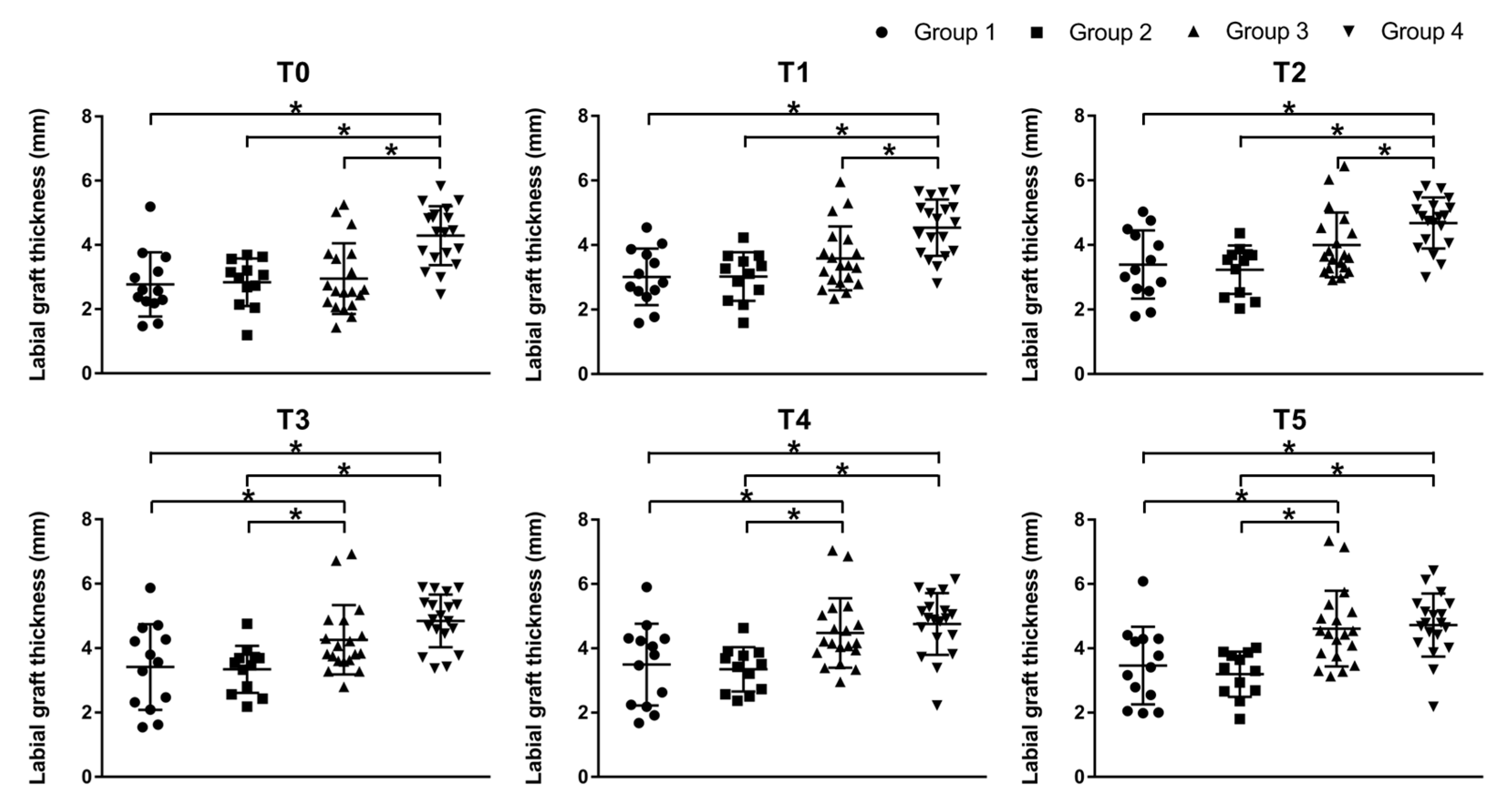
| Granulate (Group 1) | Granulate + Healing Cap (Group 2) | Sticky Bone (Group 3) | Customized Sticky Bone (Group 4) | Comparison between Groups | |
|---|---|---|---|---|---|
| Mean ± SD [95% CI] | |||||
| T0 | 2.77 ± 1.00 | 2.84 ± 0.74 | 2.95 ± 1.10 | 4.29 ± 0.92 | 1 vs. 2 |
| [2.16; 3.37] | [2.37; 3.31] | [2.42; 3.48] | [3.85; 4.73] | 1 vs. 3 | |
| 1 vs. 4 * | |||||
| 2 vs. 3 | |||||
| 2 vs. 4 * | |||||
| 3 vs. 4 * | |||||
| T1 | 3.01 ± 0.88 | 3.02 ± 0.75 | 3.59 ± 0.99 | 4.54 ± 0.88 | 1 vs. 2 |
| [2.48; 3.54] | [2.54; 3.50] | [3.11; 4.06] | [4.11; 4.96] | 1 vs. 3 | |
| 1 vs. 4 * | |||||
| 2 vs. 3 | |||||
| 2 vs. 4 * | |||||
| 3 vs. 4 * | |||||
| T2 | 3.39 ± 1.06 | 3.23 ± 0.75 | 4.00 ± 1.00 | 4.68 ± 0.79 | 1 vs. 2 |
| [2.75; 4.03] | [2.75; 3.71] | [3.52; 4.48] | [4.30; 5.06] | 1 vs. 3 | |
| 1 vs. 4 * | |||||
| 2 vs. 3 | |||||
| 2 vs. 4 * | |||||
| 3 vs. 4 * | |||||
| T3 | 3.41 ± 1.33 | 3.34 ± 0.73 | 4.26 ± 1.08 | 4.85 ± 0.82 | 1 vs. 2 |
| [2.61; 4.22] | [2.88; 3.81] | [3.74; 4.78] | [4.45; 5.24] | 1 vs. 3 * | |
| 1 vs. 4 * | |||||
| 2 vs. 3 * | |||||
| 2 vs. 4 * | |||||
| 3 vs. 4 | |||||
| T4 | 3.50 ± 1.27 | 3.35 ± 0.69 | 4.47 ± 1.08 | 4.76 ± 0.96 | 1 vs. 2 |
| [2.73; 4.26] | [2.91; 3.79] | [3.95; 4.99] | [4.29; 5.22] | 1 vs. 3 * | |
| 1 vs. 4 * | |||||
| 2 vs. 3 * | |||||
| 2 vs. 4 * | |||||
| 3 vs. 4 | |||||
| T5 | 3.46 ± 1.21 | 3.19 ± 0.70 | 4.61 ± 1.18 | 4.72 ± 0.98 | 1 vs. 2 |
| [2.73; 4.20] | [2.75; 3.64] | [4.05; 5.18] | [4.25; 5.19] | 1 vs. 3 * | |
| 1 vs. 4 * | |||||
| 2 vs. 3 * | |||||
| 2 vs. 4 * | |||||
| 3 vs. 4 | |||||
| SDc | 0.31 ± 0.17 | 0.18 ± 0.09 | 0.44 ± 0.15 | 0.20 ± 0.09 | 1 vs. 2 * |
| [0.21; 0.41] | [0.12; 0.24] | [0.36; 0.51] | [0.16; 0.25] | 1 vs. 3 * | |
| 1 vs. 4 * | |||||
| 2 vs. 3 * | |||||
| 2 vs. 4 | |||||
| 3 vs. 4 * |
Publisher’s Note: MDPI stays neutral with regard to jurisdictional claims in published maps and institutional affiliations. |
© 2021 by the authors. Licensee MDPI, Basel, Switzerland. This article is an open access article distributed under the terms and conditions of the Creative Commons Attribution (CC BY) license (http://creativecommons.org/licenses/by/4.0/).
Share and Cite
Wang, M.; Zhang, X.; Li, Y.; Mo, A. The Influence of Different Guided Bone Regeneration Procedures on the Contour of Bone Graft after Wound Closure: A Retrospective Cohort Study. Materials 2021, 14, 583. https://0-doi-org.brum.beds.ac.uk/10.3390/ma14030583
Wang M, Zhang X, Li Y, Mo A. The Influence of Different Guided Bone Regeneration Procedures on the Contour of Bone Graft after Wound Closure: A Retrospective Cohort Study. Materials. 2021; 14(3):583. https://0-doi-org.brum.beds.ac.uk/10.3390/ma14030583
Chicago/Turabian StyleWang, Maoxia, Xiaoqing Zhang, Yazhen Li, and Anchun Mo. 2021. "The Influence of Different Guided Bone Regeneration Procedures on the Contour of Bone Graft after Wound Closure: A Retrospective Cohort Study" Materials 14, no. 3: 583. https://0-doi-org.brum.beds.ac.uk/10.3390/ma14030583