Recent Advances in Cell-Based Therapies for Ischemic Stroke

 ,
,
Abstract
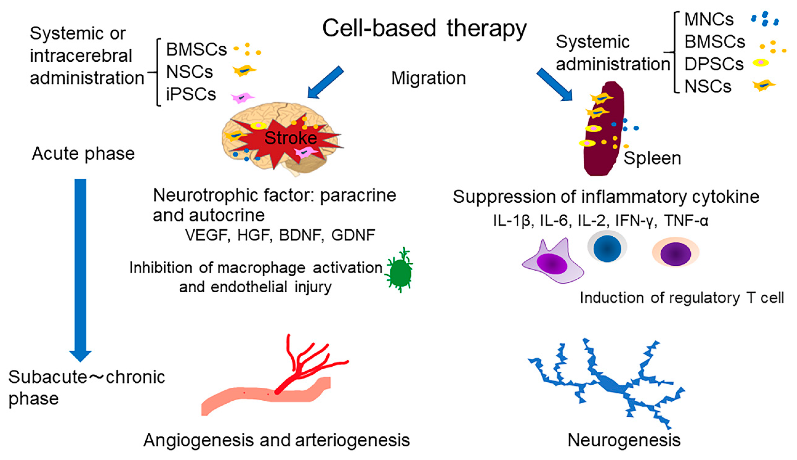
:1. Introduction
2. Bone Marrow Mononuclear Cells
2.1. Protective Mechanisms of Bone Marrow Mononuclear Cells against Stroke
2.2. Clinical Studies of Bone Marrow Mononuclear Cells Efficacy Following a Stroke
3. Bone Marrow Mesenchymal Stem Cells
3.1. Protective Mechanisms of Bone Marrow Mesenchymal Stem Cells against Stroke
3.2. Efficacy and Safety of Bone Marrow Mesenchymal Stem Cells in Clinical Trials
4. Dental Pulp Stem Cells
4.1. Protective Effects of Dental Pulp Stem Cells after Ischemic Stroke In Vivo
4.2. Clinical Studies of Dental Pulp Stem Cells Administration after a Stroke
5. Neural Stem Cells
5.1. Experimental Studies Investigating Neural Stem Cells Transplantation to Treat Stroke
5.2. Clinical Studies Investigating Neural Stem Cells Transplantation to Treat Stroke
6. Induced Pluripotent Stem Cells
6.1. Applying Induced Pluripotent Stem Cells in Animal Models of Stroke
6.2. Merits and Hurdles of Using Induced Pluripotent Stem Cells for Cell Therapy to Treat Stroke
7. Gene Modifications in Stem Cell Therapy to Repair Stroke-Induced Damage
7.1. Brain-Derived Neurotrophic Factor
7.2. Glial-Derived Neurotrophic Factor
7.3. Nerve Growth Factor
7.4. Vascular Endothelial Growth Factor
7.5. Hepatocyte Growth Factor
7.6. Placenta Growth Factor
7.7. Angiopoietin-1
7.8. Erythropoietin
7.9. Interleukin-10
7.10. Noggin
8. Perspectives
Funding
Conflicts of Interest
Abbreviations
| MNCs | Mononuclear cells |
| BMSCs | Bone marrow mesenchymal stem cells |
| DPSCs | Dental pulp stem cells |
| NSCs | Neural stem cells |
| iPSCs | Inducible pluripotent stem cells |
| VEGF | Vascular endothelial growth factor |
| HGF | Hepatocyte growth factor |
| BDNF | Brain-derived neurotrophic factor |
| GDNF | Glial-derived neurotrophic factor |
| Il-1β | Interleukin 1 beta |
| Il-6 | Interleukin 6 |
| Il-2 | Interleukin 2 |
| IFN-γ | Interferon gamma |
| TNF-α | Tumor necrosis factor alpha |
| ECs | Endothelial cells |
| SMCs | Smooth muscle cells |
| MCAO | Middle cerebral artery occlusion |
| CBF | Cerebral blood flow |
| NIHSS | National Institutes of Health Stroke Scale |
| HLA-DR | Human leukocyte antigen DR isotype |
| Treg | Regulatory T cell |
| mRS | Modified Rankin Scale |
| ALDH | Aldehyde dehydrogenase |
| EQ-50 | European Quality of Life- 5 dimension |
| MSCs | Mesenchymal stem cells |
| CNS | Central nervous system |
| EGF | Epidermal growth factor |
| SVZ | Sub-ventricular zone |
| SGZ | Sub-granular zone |
| FGF | Fibroblast growth factor |
| ANG-1 | Angiopoietin 1 |
| EO | Erythropoietin |
| AAV | Adeno-associated virus |
| Bcl-2 | B cell lymphoma 2 |
| Bax | Bcl-2 associated X |
| ICH | Intracerebral hemorrhage |
| BCP | Bone morphogenetic protein |
References
- Bang, O.Y.; Lee, J.S.; Lee, P.H.; Lee, G. Autologous mesenchymal stem cell transplantation in stroke patients. Ann. Neurol. 2005, 57, 874–882. [Google Scholar] [CrossRef]
- Bhatia, V.; Gupta, V.; Khurana, D.; Sharma, R.R.; Khandelwal, N. Randomized Assessment of the Safety and Efficacy of Intra-Arterial Infusion of Autologous Stem Cells in Subacute Ischemic Stroke. AJNR Am. J. Neuroradiol. 2018, 39, 899–904. [Google Scholar] [CrossRef] [PubMed] [Green Version]
- Hess, D.C.; Wechsler, L.R.; Clark, W.M.; Savitz, S.I.; Ford, G.A.; Chiu, D.; Yavagal, D.R.; Uchino, K.; Liebeskind, D.S.; Auchus, A.P.; et al. Safety and efficacy of multipotent adult progenitor cells in acute ischaemic stroke (MASTERS): A randomised, double-blind, placebo-controlled, phase 2 trial. Lancet Neurol. 2017, 16, 360–368. [Google Scholar] [CrossRef]
- Honmou, O.; Houkin, K.; Matsunaga, T.; Niitsu, Y.; Ishiai, S.; Onodera, R.; Waxman, S.G.; Kocsis, J.D. Intravenous administration of auto serum-expanded autologous mesenchymal stem cells in stroke. Brain 2011, 134, 1790–1807. [Google Scholar] [CrossRef] [PubMed] [Green Version]
- Levy, M.L.; Crawford, J.R.; Dib, N.; Verkh, L.; Tankovich, N.; Cramer, S.C. Phase I/II Study of Safety and Preliminary Efficacy of Intravenous Allogeneic Mesenchymal Stem Cells in Chronic Stroke. Stroke 2019, 50, 2835–2841. [Google Scholar] [CrossRef] [PubMed]
- Savitz, S.I.; Yavagal, D.; Rappard, G.; Likosky, W.; Rutledge, N.; Graffagnino, C.; Alderazi, Y.; Elder, J.A.; Chen, P.R.; Budzik, R.F., Jr.; et al. A Phase 2 Randomized, Sham-Controlled Trial of Internal Carotid Artery Infusion of Autologous Bone Marrow-Derived ALD-401 Cells in Patients With Recent Stable Ischemic Stroke (RECOVER-Stroke). Circulation 2019, 139, 192–205. [Google Scholar] [CrossRef]
- Shichinohe, H.; Kawabori, M.; Iijima, H.; Teramoto, T.; Abumiya, T.; Nakayama, N.; Kazumata, K.; Terasaka, S.; Arato, T.; Houkin, K. Research on advanced intervention using novel bone marrOW stem cell (RAINBOW): A study protocol for a phase I, open-label, uncontrolled, dose-response trial of autologous bone marrow stromal cell transplantation in patients with acute ischemic stroke. BMC Neurol. 2017, 17, 179. [Google Scholar] [CrossRef]
- Steinberg, G.K.; Kondziolka, D.; Wechsler, L.R.; Lunsford, L.D.; Coburn, M.L.; Billigen, J.B.; Kim, A.S.; Johnson, J.N.; Bates, D.; King, B.; et al. Clinical Outcomes of Transplanted Modified Bone Marrow-Derived Mesenchymal Stem Cells in Stroke: A Phase 1/2a Study. Stroke 2016, 47, 1817–1824. [Google Scholar] [CrossRef] [Green Version]
- Savitz, S.I.; Parsha, K. Enhancing Stroke Recovery with Cellular Therapies. In Stroke, 6th ed.; Grotta, J.C., Albers, G.W., Broderick, J.P., Kasner, S.E., Lo, E.H., Mendelow, A.D., Sacco, R.L., Wong, L.K.S., Eds.; Elsevier: London, UK, 2016; pp. 981–991. [Google Scholar]
- Suda, S.; Yang, B.; Schaar, K.; Xi, X.; Pido, J.; Parsha, K.; Aronowski, J.; Savitz, S.I. Autologous Bone Marrow Mononuclear Cells Exert Broad Effects on Short- and Long-Term Biological and Functional Outcomes in Rodents with Intracerebral Hemorrhage. Stem Cells Dev. 2015, 24, 2756–2766. [Google Scholar] [CrossRef] [Green Version]
- Kamiya, F.; Ueda, M.; Nito, C.; Kamiya, N.; Inaba, T.; Suda, S.; Saito, T.; Muraga, K.; Katayama, Y. Effect of repeated allogeneic bone marrow mononuclear cell transplantation on brain injury following transient focal cerebral ischemia in rats. Life Sci. 2014, 95, 22–28. [Google Scholar] [CrossRef]
- Kamiya, N.; Ueda, M.; Igarashi, H.; Nishiyama, Y.; Suda, S.; Inaba, T.; Katayama, Y. Intra-arterial transplantation of bone marrow mononuclear cells immediately after reperfusion decreases brain injury after focal ischemia in rats. Life Sci. 2008, 83, 433–437. [Google Scholar] [CrossRef] [PubMed]
- Higashi, Y.; Kimura, M.; Hara, K.; Noma, K.; Jitsuiki, D.; Nakagawa, K.; Oshima, T.; Chayama, K.; Sueda, T.; Goto, C.; et al. Autologous bone-marrow mononuclear cell implantation improves endothelium-dependent vasodilation in patients with limb ischemia. Circulation 2004, 109, 1215–1218. [Google Scholar] [CrossRef] [Green Version]
- Penicka, M.; Lang, O.; Widimsky, P.; Kobylka, P.; Kozak, T.; Vanek, T.; Dvorak, J.; Tintera, J.; Bartunek, J. One-day kinetics of myocardial engraftment after intracoronary injection of bone marrow mononuclear cells in patients with acute and chronic myocardial infarction. Heart 2007, 93, 837–841. [Google Scholar] [CrossRef] [PubMed] [Green Version]
- Burchfield, J.S.; Iwasaki, M.; Koyanagi, M.; Urbich, C.; Rosenthal, N.; Zeiher, A.M.; Dimmeler, S. Interleukin-10 from transplanted bone marrow mononuclear cells contributes to cardiac protection after myocardial infarction. Circ. Res. 2008, 103, 203–211. [Google Scholar] [CrossRef] [PubMed] [Green Version]
- Pokushalov, E.; Romanov, A.; Chernyavsky, A.; Larionov, P.; Terekhov, I.; Artyomenko, S.; Poveshenko, O.; Kliver, E.; Shirokova, N.; Karaskov, A.; et al. Efficiency of intramyocardial injections of autologous bone marrow mononuclear cells in patients with ischemic heart failure: A randomized study. J. Cardiovasc. Transl. Res. 2010, 3, 160–168. [Google Scholar] [CrossRef]
- Troidl, K.; Schaper, W. Arteriogenesis versus angiogenesis in peripheral artery disease. Diabetes/Metab. Res. Rev. 2012, 28 (Suppl. 1), 27–29. [Google Scholar] [CrossRef]
- Raval, Z.; Losordo, D.W. Cell therapy of peripheral arterial disease: From experimental findings to clinical trials. Circ. Res. 2013, 112, 1288–1302. [Google Scholar] [CrossRef]
- Terry, T.; Chen, Z.; Dixon, R.A.; Vanderslice, P.; Zoldhelyi, P.; Willerson, J.T.; Liu, Q. CD34(+)/M-cadherin(+) bone marrow progenitor cells promote arteriogenesis in ischemic hindlimbs of ApoE(-)/(-) mice. PLoS ONE 2011, 6, e20673. [Google Scholar] [CrossRef] [Green Version]
- Wang, J.; Yu, L.; Jiang, C.; Chen, M.; Ou, C.; Wang, J. Bone marrow mononuclear cells exert long-term neuroprotection in a rat model of ischemic stroke by promoting arteriogenesis and angiogenesis. Brain Behav. Immun. 2013, 34, 56–66. [Google Scholar] [CrossRef] [Green Version]
- Wang, J.; Fu, X.; Jiang, C.; Yu, L.; Wang, M.; Han, W.; Liu, L.; Wang, J. Bone marrow mononuclear cell transplantation promotes therapeutic angiogenesis via upregulation of the VEGF-VEGFR2 signaling pathway in a rat model of vascular dementia. Behav. Brain Res. 2014, 265, 171–180. [Google Scholar] [CrossRef] [Green Version]
- Fujita, Y.; Ihara, M.; Ushiki, T.; Hirai, H.; Kizaka-Kondoh, S.; Hiraoka, M.; Ito, H.; Takahashi, R. Early protective effect of bone marrow mononuclear cells against ischemic white matter damage through augmentation of cerebral blood flow. Stroke 2010, 41, 2938–2943. [Google Scholar] [CrossRef] [PubMed] [Green Version]
- Wang, J.; Liu, X.; Lu, H.; Jiang, C.; Cui, X.; Yu, L.; Fu, X.; Li, Q.; Wang, J. CXCR4(+)CD45(-) BMMNC subpopulation is superior to unfractionated BMMNCs for protection after ischemic stroke in mice. Brain Behav. Immun. 2015, 45, 98–108. [Google Scholar] [CrossRef] [PubMed] [Green Version]
- Chen, J.; Zhang, Z.G.; Li, Y.; Wang, L.; Xu, Y.X.; Gautam, S.C.; Lu, M.; Zhu, Z.; Chopp, M. Intravenous administration of human bone marrow stromal cells induces angiogenesis in the ischemic boundary zone after stroke in rats. Circ. Res. 2003, 92, 692–699. [Google Scholar] [CrossRef] [PubMed] [Green Version]
- Kikuchi-Taura, A.; Okinaka, Y.; Takeuchi, Y.; Ogawa, Y.; Maeda, M.; Kataoka, Y.; Yasui, T.; Kimura, T.; Gul, S.; Claussen, C.; et al. Bone Marrow Mononuclear Cells Activate Angiogenesis via Gap Junction-Mediated Cell-Cell Interaction. Stroke 2020, 51, 1279–1289. [Google Scholar] [CrossRef] [PubMed]
- Okinaka, Y.; Kikuchi-Taura, A.; Takeuchi, Y.; Ogawa, Y.; Boltze, J.; Gul, S.; Claussen, C.; Taguchi, A. Clot-Derived Contaminants in Transplanted Bone Marrow Mononuclear Cells Impair the Therapeutic Effect in Stroke. Stroke 2019, 50, 2883–2891. [Google Scholar] [CrossRef] [PubMed] [Green Version]
- Yang, B.; Li, W.; Satani, N.; Nghiem, D.M.; Xi, X.; Aronowski, J.; Savitz, S.I. Protective Effects of Autologous Bone Marrow Mononuclear Cells After Administering t-PA in an Embolic Stroke Model. Transl. Stroke Res. 2018, 9, 135–145. [Google Scholar] [CrossRef] [PubMed]
- Li, Y.; Mao, W.W.; Zhang, C.G.; Wan, L.; Jing, C.H.; Hua, X.M.; Li, S.T.; Cheng, J. Neuroprotective effects of intravenous transplantation of bone marrow mononuclear cells from 5-fluorouracil pre-treated rats on ischemic stroke. Behav. Brain Res. 2016, 301, 287–292. [Google Scholar] [CrossRef]
- Yang, B.; Migliati, E.; Parsha, K.; Schaar, K.; Xi, X.; Aronowski, J.; Savitz, S.I. Intra-arterial delivery is not superior to intravenous delivery of autologous bone marrow mononuclear cells in acute ischemic stroke. Stroke 2013, 44, 3463–3472. [Google Scholar] [CrossRef] [Green Version]
- Nakano-Doi, A.; Nakagomi, T.; Fujikawa, M.; Nakagomi, N.; Kubo, S.; Lu, S.; Yoshikawa, H.; Soma, T.; Taguchi, A.; Matsuyama, T. Bone marrow mononuclear cells promote proliferation of endogenous neural stem cells through vascular niches after cerebral infarction. Stem Cells 2010, 28, 1292–1302. [Google Scholar] [CrossRef]
- Savitz, S.I.; Misra, V.; Kasam, M.; Juneja, H.; Cox, C.S., Jr.; Alderman, S.; Aisiku, I.; Kar, S.; Gee, A.; Grotta, J.C. Intravenous autologous bone marrow mononuclear cells for ischemic stroke. Ann. Neurol. 2011, 70, 59–69. [Google Scholar] [CrossRef]
- Taguchi, A.; Sakai, C.; Soma, T.; Kasahara, Y.; Stern, D.M.; Kajimoto, K.; Ihara, M.; Daimon, T.; Yamahara, K.; Doi, K.; et al. Intravenous Autologous Bone Marrow Mononuclear Cell Transplantation for Stroke: Phase1/2a Clinical Trial in a Homogeneous Group of Stroke Patients. Stem Cells Dev. 2015, 24, 2207–2218. [Google Scholar] [CrossRef] [PubMed] [Green Version]
- Prasad, K.; Sharma, A.; Garg, A.; Mohanty, S.; Bhatnagar, S.; Johri, S.; Singh, K.K.; Nair, V.; Sarkar, R.S.; Gorthi, S.P.; et al. Intravenous autologous bone marrow mononuclear stem cell therapy for ischemic stroke: A multicentric, randomized trial. Stroke 2014, 45, 3618–3624. [Google Scholar] [CrossRef] [PubMed] [Green Version]
- Boltze, J.; Modo, M.M.; Mays, R.W.; Taguchi, A.; Jolkkonen, J.; Savitz, S.I. Stem Cells as an Emerging Paradigm in Stroke 4: Advancing and Accelerating Preclinical Research. Stroke 2019, 50, 3299–3306. [Google Scholar] [CrossRef] [PubMed]
- Javazon, E.H.; Beggs, K.J.; Flake, A.W. Mesenchymal stem cells: Paradoxes of passaging. Exp. Hematol. 2004, 32, 414–425. [Google Scholar] [CrossRef] [PubMed]
- Gnecchi, M.; Melo, L.G. Bone marrow-derived mesenchymal stem cells: Isolation, expansion, characterization, viral transduction, and production of conditioned medium. Methods Mol. Biol. 2009, 482, 281–294. [Google Scholar] [PubMed]
- Kuroda, S.; Houkin, K. Translational challenge for bone marrow stroma cell therapy after stroke. Front. Neurol. Neurosci. 2013, 32, 62–68. [Google Scholar] [PubMed]
- Suda, S.; Shimazaki, K.; Ueda, M.; Inaba, T.; Kamiya, N.; Katsura, K.; Katayama, Y. Combination therapy with bone marrow stromal cells and FK506 enhanced amelioration of ischemic brain damage in rats. Life Sci. 2011, 89, 50–56. [Google Scholar] [CrossRef]
- Dulamea, A.O. The potential use of mesenchymal stem cells in stroke therapy—From bench to bedside. J. Neurol. Sci. 2015, 352, 1–11. [Google Scholar] [CrossRef]
- Li, G.; Yu, F.; Lei, T.; Gao, H.; Li, P.; Sun, Y.; Huang, H.; Mu, Q. Bone marrow mesenchymal stem cell therapy in ischemic stroke: Mechanisms of action and treatment optimization strategies. Neural. Regen. Res. 2016, 11, 1015–1024. [Google Scholar] [CrossRef]
- Wang, Z.; He, D.; Zeng, Y.Y.; Zhu, L.; Yang, C.; Lu, Y.J.; Huang, J.Q.; Cheng, X.Y.; Huang, X.H.; Tan, X.J. The spleen may be an important target of stem cell therapy for stroke. J. Neuroinflamm. 2019, 16, 20. [Google Scholar] [CrossRef]
- Tobin, M.K.; Stephen, T.K.L.; Lopez, K.L.; Pergande, M.R.; Bartholomew, A.M.; Cologna, S.M.; Lazarov, O. Activated Mesenchymal Stem Cells Induce Recovery Following Stroke Via Regulation of Inflammation and Oligodendrogenesis. J. Am. Heart Assoc. 2020, 9, e013583. [Google Scholar] [CrossRef]
- Nakajima, M.; Nito, C.; Sowa, K.; Suda, S.; Nishiyama, Y.; Nakamura-Takahashi, A.; Nitahara-Kasahara, Y.; Imagawa, K.; Hirato, T.; Ueda, M.; et al. Mesenchymal Stem Cells Overexpressing Interleukin-10 Promote Neuroprotection in Experimental Acute Ischemic Stroke. Mol. Ther. Methods Clin. Dev. 2017, 6, 102–111. [Google Scholar] [CrossRef] [PubMed] [Green Version]
- Kawabori, M.; Kuroda, S.; Ito, M.; Shichinohe, H.; Houkin, K.; Kuge, Y.; Tamaki, N. Timing and cell dose determine therapeutic effects of bone marrow stromal cell transplantation in rat model of cerebral infarct. Neuropathology 2013, 33, 140–148. [Google Scholar] [CrossRef]
- Toyoshima, A.; Yasuhara, T.; Kameda, M.; Morimoto, J.; Takeuchi, H.; Wang, F.; Sasaki, T.; Sasada, S.; Shinko, A.; Wakamori, T.; et al. Intra-Arterial Transplantation of Allogeneic Mesenchymal Stem Cells Mounts Neuroprotective Effects in a Transient Ischemic Stroke Model in Rats: Analyses of Therapeutic Time Window and Its Mechanisms. PLoS ONE 2015, 10, e0127302. [Google Scholar] [CrossRef] [PubMed]
- Nakazaki, M.; Sasaki, M.; Kataoka-Sasaki, Y.; Oka, S.; Suzuki, J.; Sasaki, Y.; Nagahama, H.; Hashi, K.; Kocsis, J.D.; Honmou, O. Intravenous infusion of mesenchymal stem cells improves impaired cognitive function in a cerebral small vessel disease model. Neuroscience 2019, 408, 361–377. [Google Scholar] [CrossRef] [PubMed]
- Wang, L.Q.; Lin, Z.Z.; Zhang, H.X.; Shao, B.; Xiao, L.; Jiang, H.G.; Zhuge, Q.C.; Xie, L.K.; Wang, B.; Su, D.M.; et al. Timing and dose regimens of marrow mesenchymal stem cell transplantation affect the outcomes and neuroinflammatory response after ischemic stroke. CNS Neurosci. Ther. 2014, 20, 317–326. [Google Scholar] [CrossRef]
- Steinberg, G.K.; Kondziolka, D.; Wechsler, L.R.; Lunsford, L.D.; Kim, A.S.; Johnson, J.N.; Bates, D.; Poggio, G.; Case, C.; McGrogan, M.; et al. Two-year safety and clinical outcomes in chronic ischemic stroke patients after implantation of modified bone marrow-derived mesenchymal stem cells (SB623): A phase 1/2a study. J. Neurosurg. 2018, 131, 1462–1472. [Google Scholar] [CrossRef]
- Kawabori, M.; Tanimori, A.; Kitta, S.; Shichinohe, H.; Houkin, K. Evaluation of Novel Stereotactic Cannula for Stem Cell Transplantation against Central Nervous System Disease. Stem Cells Int. 2020, 2020, 4085617. [Google Scholar] [CrossRef] [Green Version]
- Nosrat, I.V.; Smith, C.A.; Mullally, P.; Olson, L.; Nosrat, C.A. Dental pulp cells provide neurotrophic support for dopaminergic neurons and differentiate into neurons in vitro; implications for tissue engineering and repair in the nervous system. Eur. J. Neurosci. 2004, 19, 2388–2398. [Google Scholar] [CrossRef] [Green Version]
- Pierdomenico, L.; Bonsi, L.; Calvitti, M.; Rondelli, D.; Arpinati, M.; Chirumbolo, G.; Becchetti, E.; Marchionni, C.; Alviano, F.; Fossati, V.; et al. Multipotent mesenchymal stem cells with immunosuppressive activity can be easily isolated from dental pulp. Transplantation 2005, 80, 836–842. [Google Scholar] [CrossRef] [Green Version]
- Gronthos, S.; Mankani, M.; Brahim, J.; Robey, P.G.; Shi, S. Postnatal human dental pulp stem cells (DPSCs) in vitro and in vivo. Proc. Natl. Acad. Sci. USA 2000, 97, 13625–13630. [Google Scholar] [CrossRef] [PubMed] [Green Version]
- Ponnaiyan, D.; Jegadeesan, V. Comparison of phenotype and differentiation marker gene expression profiles in human dental pulp and bone marrow mesenchymal stem cells. Eur. J. Dent. 2014, 8, 307–313. [Google Scholar] [CrossRef] [PubMed]
- Rajendran, R.; Gopal, S.; Masood, H.; Vivek, P.; Deb, K. Regenerative potential of dental pulp mesenchymal stem cells harvested from high caries patient’s teeth. J. Stem Cells 2013, 8, 25–41. [Google Scholar] [PubMed]
- Zhang, X.; Zhou, Y.; Li, H.; Wang, R.; Yang, D.; Li, B.; Fu, J. Intravenous administration of DPSCs and BDNF improves neurological performance in rats with focal cerebral ischemia. Int. J. Mol. Med. 2018, 41, 3185–3194. [Google Scholar] [CrossRef] [PubMed] [Green Version]
- Inoue, T.; Sugiyama, M.; Hattori, H.; Wakita, H.; Wakabayashi, T.; Ueda, M. Stem cells from human exfoliated deciduous tooth-derived conditioned medium enhance recovery of focal cerebral ischemia in rats. Tissue Eng. Part A 2013, 19, 24–29. [Google Scholar] [CrossRef] [PubMed] [Green Version]
- Leong, W.K.; Lewis, M.D.; Koblar, S.A. Concise review: Preclinical studies on human cell-based therapy in rodent ischemic stroke models: Where are we now after a decade? Stem Cells 2013, 31, 1040–1043. [Google Scholar] [CrossRef] [PubMed]
- Sakai, K.; Yamamoto, A.; Matsubara, K.; Nakamura, S.; Naruse, M.; Yamagata, M.; Sakamoto, K.; Tauchi, R.; Wakao, N.; Imagama, S.; et al. Human dental pulp-derived stem cells promote locomotor recovery after complete transection of the rat spinal cord by multiple neuro-regenerative mechanisms. J. Clin. Investig. 2012, 122, 80–90. [Google Scholar] [CrossRef]
- Kumasaka, A.; Kanazawa, K.; Ohke, H.; Miura, I.; Miura, Y. Post-ischemic Intravenous Administration of Allogeneic Dental Pulp-Derived Neurosphere Cells Ameliorated Outcomes of Severe Forebrain Ischemia in Rats. Neurocritical Care 2017, 26, 133–142. [Google Scholar] [CrossRef]
- Leong, W.K.; Henshall, T.L.; Arthur, A.; Kremer, K.L.; Lewis, M.D.; Helps, S.C.; Field, J.; Hamilton-Bruce, M.A.; Warming, S.; Manavis, J.; et al. Human adult dental pulp stem cells enhance poststroke functional recovery through non-neural replacement mechanisms. Stem Cells Transl. Med. 2012, 1, 177–187. [Google Scholar] [CrossRef]
- Song, M.; Lee, J.H.; Bae, J.; Bu, Y.; Kim, E.C. Human Dental Pulp Stem Cells Are More Effective than Human Bone Marrow-Derived Mesenchymal Stem Cells in Cerebral Ischemic Injury. Cell Transplant. 2017, 26, 1001–1016. [Google Scholar] [CrossRef]
- Nito, C.; Sowa, K.; Nakajima, M.; Sakamoto, Y.; Suda, S.; Nishiyama, Y.; Nakamura-Takahashi, A.; Nitahara-Kasahara, Y.; Ueda, M.; Okada, T.; et al. Transplantation of human dental pulp stem cells ameliorates brain damage following acute cerebral ischemia. Biomed. Pharm. 2018, 108, 1005–1014. [Google Scholar] [CrossRef] [PubMed]
- Sowa, K.; Nito, C.; Nakajima, M.; Suda, S.; Nishiyama, Y.; Sakamoto, Y.; Nitahara-Kasahara, Y.; Nakamura-Takahashi, A.; Ueda, M.; Kimura, K.; et al. Impact of Dental Pulp Stem Cells Overexpressing Hepatocyte Growth Factor after Cerebral Ischemia/Reperfusion in Rats. Mol. Ther. Methods Clin. Dev. 2018, 10, 281–290. [Google Scholar] [CrossRef] [Green Version]
- Zhang, X.; Zhou, Y.; Li, H.; Wang, R.; Yang, D.; Li, B.; Cao, X.; Fu, J. Transplanted Dental Pulp Stem Cells Migrate to Injured Area and Express Neural Markers in a Rat Model of Cerebral Ischemia. Cell Physiol. Biochem. 2018, 45, 258–266. [Google Scholar] [CrossRef]
- Savitz, S.I. Developing Cellular Therapies for Stroke. Stroke 2015, 46, 2026–2031. [Google Scholar] [CrossRef] [Green Version]
- Kalladka, D.; Muir, K.W. Stem cell therapy in stroke: Designing clinical trials. Neurochem. Int. 2011, 59, 367–370. [Google Scholar] [CrossRef]
- Ribeiro, T.B.; Duarte, A.S.; Longhini, A.L.; Pradella, F.; Farias, A.S.; Luzo, A.C.; Oliveira, A.L.; Olalla Saad, S.T. Neuroprotection and immunomodulation by xenografted human mesenchymal stem cells following spinal cord ventral root avulsion. Sci. Rep. 2015, 5, 16167. [Google Scholar] [CrossRef] [PubMed]
- Lee, J.A.; Kim, B.I.; Jo, C.H.; Choi, C.W.; Kim, E.K.; Kim, H.S.; Yoon, K.S.; Choi, J.H. Mesenchymal stem-cell transplantation for hypoxic-ischemic brain injury in neonatal rat model. Pediatr. Res. 2010, 67, 42–46. [Google Scholar] [CrossRef] [PubMed] [Green Version]
- Martire, A.; Bedada, F.B.; Uchida, S.; Poling, J.; Kruger, M.; Warnecke, H.; Richter, M.; Kubin, T.; Herold, S.; Braun, T. Mesenchymal stem cells attenuate inflammatory processes in the heart and lung via inhibition of TNF signaling. Basic Res. Cardiol. 2016, 111, 54. [Google Scholar] [CrossRef] [PubMed] [Green Version]
- Shichita, T.; Ago, T.; Kamouchi, M.; Kitazono, T.; Yoshimura, A.; Ooboshi, H. Novel therapeutic strategies targeting innate immune responses and early inflammation after stroke. J. Neurochem. 2012, 123 (Suppl. 2), 29–38. [Google Scholar] [CrossRef]
- Nosrat, I.V.; Widenfalk, J.; Olson, L.; Nosrat, C.A. Dental pulp cells produce neurotrophic factors, interact with trigeminal neurons in vitro, and rescue motoneurons after spinal cord injury. Dev. Biol. 2001, 238, 120–132. [Google Scholar] [CrossRef] [PubMed] [Green Version]
- Matsushita, K.; Motani, R.; Sakuta, T.; Yamaguchi, N.; Koga, T.; Matsuo, K.; Nagaoka, S.; Abeyama, K.; Maruyama, I.; Torii, M. The role of vascular endothelial growth factor in human dental pulp cells: Induction of chemotaxis, proliferation, and differentiation and activation of the AP-1-dependent signaling pathway. J. Dent. Res. 2000, 79, 1596–1603. [Google Scholar] [CrossRef]
- Liu, X.; Ye, R.; Yan, T.; Yu, S.P.; Wei, L.; Xu, G.; Fan, X.; Jiang, Y.; Stetler, R.A.; Liu, G.; et al. Cell based therapies for ischemic stroke: From basic science to bedside. Prog. Neurobiol. 2014, 115, 92–115. [Google Scholar] [CrossRef] [PubMed] [Green Version]
- Liesz, A.; Bauer, A.; Hoheisel, J.D.; Veltkamp, R. Intracerebral interleukin-10 injection modulates post-ischemic neuroinflammation: An experimental microarray study. Neurosci. Lett. 2014, 579, 18–23. [Google Scholar] [CrossRef] [PubMed]
- Zhang, Z.G.; Zhang, L.; Jiang, Q.; Zhang, R.; Davies, K.; Powers, C.; Bruggen, N.; Chopp, M. VEGF enhances angiogenesis and promotes blood-brain barrier leakage in the ischemic brain. J. Clin. Investig. 2000, 106, 829–838. [Google Scholar] [CrossRef] [PubMed] [Green Version]
- Király, M.; Kádár, K.; Horváthy, D.B.; Nardai, P.; Rácz, G.Z.; Lacza, Z.; Varga, G.; Gerber, G. Integration of neuronally predifferentiated human dental pulp stem cells into rat brain in vivo. Neurochem. Int. 2011, 59, 371–381. [Google Scholar] [CrossRef] [PubMed]
- Jin, K.; Sun, Y.; Xie, L.; Mao, X.O.; Childs, J.; Peel, A.; Logvinova, A.; Banwait, S.; Greenberg, D.A. Comparison of ischemia-directed migration of neural precursor cells after intrastriatal, intraventricular, or intravenous transplantation in the rat. Neurobiol. Dis. 2005, 18, 366–374. [Google Scholar] [CrossRef] [PubMed]
- Walczak, P.; Zhang, J.; Gilad, A.A.; Kedziorek, D.A.; Ruiz-Cabello, J.; Young, R.G.; Pittenger, M.F.; van Zijl, P.C.; Huang, J.; Bulte, J.W. Dual-modality monitoring of targeted intraarterial delivery of mesenchymal stem cells after transient ischemia. Stroke 2008, 39, 1569–1574. [Google Scholar] [CrossRef]
- Ge, J.; Guo, L.; Wang, S.; Zhang, Y.; Cai, T.; Zhao, R.C.; Wu, Y. The size of mesenchymal stem cells is a significant cause of vascular obstructions and stroke. Stem Cell Rev. 2014, 10, 295–303. [Google Scholar] [CrossRef]
- Lee, J.S.; Hong, J.M.; Moon, G.J.; Lee, P.H.; Ahn, Y.H.; Bang, O.Y. A long-term follow-up study of intravenous autologous mesenchymal stem cell transplantation in patients with ischemic stroke. Stem Cells 2010, 28, 1099–1106. [Google Scholar] [CrossRef]
- Altman, J. Are new neurons formed in the brains of adult mammals? Science 1962, 135, 1127–1128. [Google Scholar] [CrossRef] [Green Version]
- Reynolds, B.A.; Tetzlaff, W.; Weiss, S. A multipotent EGF-responsive striatal embryonic progenitor cell produces neurons and astrocytes. J. Neurosci. 1992, 12, 4565–4574. [Google Scholar] [CrossRef] [Green Version]
- Reynolds, B.A.; Weiss, S. Generation of neurons and astrocytes from isolated cells of the adult mammalian central nervous system. Science 1992, 255, 1707–1710. [Google Scholar] [CrossRef] [PubMed] [Green Version]
- Gritti, A.; Bonfanti, L.; Doetsch, F.; Caille, I.; Alvarez-Buylla, A.; Lim, D.A.; Galli, R.; Verdugo, J.M.; Herrera, D.G.; Vescovi, A.L. Multipotent neural stem cells reside into the rostral extension and olfactory bulb of adult rodents. J. Neurosci. 2002, 22, 437–445. [Google Scholar] [CrossRef] [PubMed] [Green Version]
- Zhang, G.L.; Zhu, Z.H.; Wang, Y.Z. Neural stem cell transplantation therapy for brain ischemic stroke: Review and perspectives. World J. Stem Cells 2019, 11, 817–830. [Google Scholar] [CrossRef] [PubMed]
- Liao, L.Y.; Lau, B.W.; Sanchez-Vidana, D.I.; Gao, Q. Exogenous neural stem cell transplantation for cerebral ischemia. Neural. Regen. Res. 2019, 14, 1129–1137. [Google Scholar] [PubMed]
- Faigle, R.; Song, H. Signaling mechanisms regulating adult neural stem cells and neurogenesis. Biochim. Biophys. Acta 2013, 1830, 2435–2448. [Google Scholar]
- Sirko, S.; von Holst, A.; Weber, A.; Wizenmann, A.; Theocharidis, U.; Gotz, M.; Faissner, A. Chondroitin sulfates are required for fibroblast growth factor-2-dependent proliferation and maintenance in neural stem cells and for epidermal growth factor-dependent migration of their progeny. Stem Cells 2010, 28, 775–787. [Google Scholar] [CrossRef]
- Cooke, M.J.; Wang, Y.; Morshead, C.M.; Shoichet, M.S. Controlled epi-cortical delivery of epidermal growth factor for the stimulation of endogenous neural stem cell proliferation in stroke-injured brain. Biomaterials 2011, 32, 5688–5697. [Google Scholar] [CrossRef]
- Bath, K.G.; Akins, M.R.; Lee, F.S. BDNF control of adult SVZ neurogenesis. Dev. Psychobiol. 2012, 54, 578–589. [Google Scholar] [CrossRef] [Green Version]
- Xu, G.; Shen, J.; Ishii, Y.; Fukuchi, M.; Dang, T.C.; Zheng, Y.; Hamashima, T.; Fujimori, T.; Tsuda, M.; Funa, K.; et al. Functional analysis of platelet-derived growth factor receptor-beta in neural stem/progenitor cells. Neuroscience 2013, 238, 195–208. [Google Scholar] [CrossRef]
- Yokobori, S.; Saito, K.; Sasaki, K.; Kanaya, T.; Fujiki, Y.; Yamaguchi, M.; Satoh, S.; Watanabe, A.; Igarashi, Y.; Suzuki, G.; et al. Treatment for Geriatric Traumatic Brain Injury: A Nationwide Cohort Study. J. Nippon Med. Sch. 2020. [Google Scholar] [CrossRef] [PubMed]
- Horie, N.; Hiu, T.; Nagata, I. Stem cell transplantation enhances endogenous brain repair after experimental stroke. Neurol. Med. Chir. 2015, 55, 107–112. [Google Scholar] [CrossRef] [PubMed] [Green Version]
- Song, M.; Kim, Y.J.; Kim, Y.H.; Roh, J.; Kim, E.C.; Lee, H.J.; Kim, S.U.; Yoon, B.W. Long-term effects of magnetically targeted ferumoxide-labeled human neural stem cells in focal cerebral ischemia. Cell Transplant. 2015, 24, 183–190. [Google Scholar] [CrossRef] [PubMed] [Green Version]
- Rosenblum, S.; Smith, T.N.; Wang, N.; Chua, J.Y.; Westbroek, E.; Wang, K.; Guzman, R. BDNF Pretreatment of Human Embryonic-Derived Neural Stem Cells Improves Cell Survival and Functional Recovery After Transplantation in Hypoxic-Ischemic Stroke. Cell Transplant. 2015, 24, 2449–2461. [Google Scholar] [CrossRef] [PubMed] [Green Version]
- Cheng, Y.; Zhang, J.; Deng, L.; Johnson, N.R.; Yu, X.; Zhang, N.; Lou, T.; Zhang, Y.; Wei, X.; Chen, Z.; et al. Intravenously delivered neural stem cells migrate into ischemic brain, differentiate and improve functional recovery after transient ischemic stroke in adult rats. Int. J. Clin. Exp. Pathol. 2015, 8, 2928–2936. [Google Scholar] [PubMed]
- Yao, H.; Gao, M.; Ma, J.; Zhang, M.; Li, S.; Wu, B.; Nie, X.; Jiao, J.; Zhao, H.; Wang, S.; et al. Transdifferentiation-Induced Neural Stem Cells Promote Recovery of Middle Cerebral Artery Stroke Rats. PLoS ONE 2015, 10, e0137211. [Google Scholar] [CrossRef]
- Abeysinghe, H.C.; Bokhari, L.; Quigley, A.; Choolani, M.; Chan, J.; Dusting, G.J.; Crook, J.M.; Kobayashi, N.R.; Roulston, C.L. Pre-differentiation of human neural stem cells into GABAergic neurons prior to transplant results in greater repopulation of the damaged brain and accelerates functional recovery after transient ischemic stroke. Stem Cell Res. Ther. 2015, 6, 186. [Google Scholar] [CrossRef] [Green Version]
- Bacigaluppi, M.; Russo, G.L.; Peruzzotti-Jametti, L.; Rossi, S.; Sandrone, S.; Butti, E.; De Ceglia, R.; Bergamaschi, A.; Motta, C.; Gallizioli, M.; et al. Neural Stem Cell Transplantation Induces Stroke Recovery by Upregulating Glutamate Transporter GLT-1 in Astrocytes. J. Neurosci. 2016, 36, 10529–10544. [Google Scholar] [CrossRef]
- Zhu, J.D.; Wang, J.J.; Ge, G.; Kang, C.S. Effects of Noggin-Transfected Neural Stem Cells on Neural Functional Recovery and Underlying Mechanism in Rats with Cerebral Ischemia Reperfusion Injury. J. Stroke Cerebrovasc. Dis. 2017, 26, 1547–1559. [Google Scholar] [CrossRef]
- Hou, B.; Ma, J.; Guo, X.; Ju, F.; Gao, J.; Wang, D.; Liu, J.; Li, X.; Zhang, S.; Ren, H. Exogenous Neural Stem Cells Transplantation as a Potential Therapy for Photothrombotic Ischemia Stroke in Kunming Mice Model. Mol. Neurobiol. 2017, 54, 1254–1262. [Google Scholar] [CrossRef]
- George, P.M.; Bliss, T.M.; Hua, T.; Lee, A.; Oh, B.; Levinson, A.; Mehta, S.; Sun, G.; Steinberg, G.K. Electrical preconditioning of stem cells with a conductive polymer scaffold enhances stroke recovery. Biomaterials 2017, 142, 31–40. [Google Scholar] [CrossRef] [PubMed]
- Tian, L.; Zhu, W.; Liu, Y.; Gong, Y.; Lv, A.; Wang, Z.; Ding, X.; Li, S.; Fu, Y.; Lin, Y.; et al. Neural Stem Cells Transfected with Leukemia Inhibitory Factor Promote Neuroprotection in a Rat Model of Cerebral Ischemia. Neurosci. Bull. 2019, 35, 901–908. [Google Scholar] [CrossRef] [PubMed]
- Kim, J.; Shin, K.; Cha, Y.; Ban, Y.H.; Park, S.K.; Jeong, H.S.; Park, D.; Choi, E.K.; Kim, Y.B. Neuroprotective effects of human neural stem cells over-expressing choline acetyltransferase in a middle cerebral artery occlusion model. J. Chem. Neuroanat. 2020, 103, 101730. [Google Scholar] [CrossRef] [PubMed]
- Kondori, B.J.; Asadi, M.H.; Bahadoran, H.; Yari, A.; Sarshoori, J.R. Intra-arterial transplantation of neural stem cells improve functional recovery after transient ischemic stroke in adult rats. Bratisl. Lek. Listy 2020, 121, 8–13. [Google Scholar] [CrossRef] [PubMed] [Green Version]
- Wang, G.; Han, B.; Shen, L.; Wu, S.; Yang, L.; Liao, J.; Wu, F.; Li, M.; Leng, S.; Zang, F.; et al. Silencing of circular RNA HIPK2 in neural stem cells enhances functional recovery following ischaemic stroke. EBioMedicine 2020, 52, 102660. [Google Scholar] [CrossRef] [Green Version]
- Daadi, M.M.; Li, Z.; Arac, A.; Grueter, B.A.; Sofilos, M.; Malenka, R.C.; Wu, J.C.; Steinberg, G.K. Molecular and magnetic resonance imaging of human embryonic stem cell-derived neural stem cell grafts in ischemic rat brain. Mol. Ther. 2009, 17, 1282–1291. [Google Scholar] [CrossRef]
- Yokobori, S.; Sasaki, K.; Kanaya, T.; Igarashi, Y.; Nakae, R.; Onda, H.; Masuno, T.; Suda, S.; Sowa, K.; Nakajima, M.; et al. Feasibility of Human Neural Stem Cell Transplantation for the Treatment of Acute Subdural Hematoma in a Rat Model: A Pilot Study. Front. Neurol. 2019, 10, 82. [Google Scholar] [CrossRef]
- Horie, N.; Pereira, M.P.; Niizuma, K.; Sun, G.; Keren-Gill, H.; Encarnacion, A.; Shamloo, M.; Hamilton, S.A.; Jiang, K.; Huhn, S.; et al. Transplanted stem cell-secreted vascular endothelial growth factor effects poststroke recovery, inflammation, and vascular repair. Stem Cells 2011, 29, 274–285. [Google Scholar] [CrossRef] [Green Version]
- Lee, S.T.; Chu, K.; Jung, K.H.; Kim, S.J.; Kim, D.H.; Kang, K.M.; Hong, N.H.; Kim, J.H.; Ban, J.J.; Park, H.K.; et al. Anti-inflammatory mechanism of intravascular neural stem cell transplantation in haemorrhagic stroke. Brain 2008, 131, 616–629. [Google Scholar] [CrossRef] [Green Version]
- Savitz, S.I.; Dinsmore, J.; Wu, J.; Henderson, G.V.; Stieg, P.; Caplan, L.R. Neurotransplantation of fetal porcine cells in patients with basal ganglia infarcts: A preliminary safety and feasibility study. Cerebrovasc. Dis. 2005, 20, 101–107. [Google Scholar] [CrossRef]
- Kalladka, D.; Sinden, J.; Pollock, K.; Haig, C.; McLean, J.; Smith, W.; McConnachie, A.; Santosh, C.; Bath, P.M.; Dunn, L.; et al. Human neural stem cells in patients with chronic ischaemic stroke (PISCES): A phase 1, first-in-man study. Lancet 2016, 388, 787–796. [Google Scholar] [CrossRef] [Green Version]
- Wechsler, L.R.; Bates, D.; Stroemer, P.; Andrews-Zwilling, Y.S.; Aizman, I. Cell Therapy for Chronic Stroke. Stroke 2018, 49, 1066–1074. [Google Scholar] [CrossRef] [PubMed]
- Muir, K.W.; Bulters, D.; Willmot, M.; Sprigg, N.; Dixit, A.; Ward, N.; Tyrrell, P.; Majid, A.; Dunn, L.; Bath, P.; et al. Intracerebral implantation of human neural stem cells and motor recovery after stroke: Multicentre prospective single-arm study (PISCES-2). J. Neurol. Neurosurg. Psychiatr. 2020, 91, 396–401. [Google Scholar] [CrossRef] [PubMed] [Green Version]
- Takahashi, K.; Yamanaka, S. Induction of pluripotent stem cells from mouse embryonic and adult fibroblast cultures by defined factors. Cell 2006, 126, 663–676. [Google Scholar] [CrossRef] [Green Version]
- Takahashi, K.; Tanabe, K.; Ohnuki, M.; Narita, M.; Ichisaka, T.; Tomoda, K.; Yamanaka, S. Induction of pluripotent stem cells from adult human fibroblasts by defined factors. Cell 2007, 131, 861–872. [Google Scholar] [CrossRef] [PubMed] [Green Version]
- Chen, S.J.; Chang, C.M.; Tsai, S.K.; Chang, Y.L.; Chou, S.J.; Huang, S.S.; Tai, L.K.; Chen, Y.C.; Ku, H.H.; Li, H.Y.; et al. Functional improvement of focal cerebral ischemia injury by subdural transplantation of induced pluripotent stem cells with fibrin glue. Stem Cells Dev. 2010, 19, 1757–1767. [Google Scholar] [CrossRef]
- Jiang, M.; Lv, L.; Ji, H.; Yang, X.; Zhu, W.; Cai, L.; Gu, X.; Chai, C.; Huang, S.; Sun, J.; et al. Induction of pluripotent stem cells transplantation therapy for ischemic stroke. Mol. Cell Biochem. 2011, 354, 67–75. [Google Scholar] [CrossRef]
- Kawai, H.; Yamashita, T.; Ohta, Y.; Deguchi, K.; Nagotani, S.; Zhang, X.; Ikeda, Y.; Matsuura, T.; Abe, K. Tridermal tumorigenesis of induced pluripotent stem cells transplanted in ischemic brain. J. Cereb. Blood Flow Metab. 2010, 30, 1487–1493. [Google Scholar] [CrossRef]
- Yamashita, T.; Kawai, H.; Tian, F.; Ohta, Y.; Abe, K. Tumorigenic development of induced pluripotent stem cells in ischemic mouse brain. Cell Transplant. 2011, 20, 883–891. [Google Scholar] [CrossRef] [Green Version]
- Gomi, M.; Takagi, Y.; Morizane, A.; Doi, D.; Nishimura, M.; Miyamoto, S.; Takahashi, J. Functional recovery of the murine brain ischemia model using human induced pluripotent stem cell-derived telencephalic progenitors. Brain Res. 2012, 1459, 52–60. [Google Scholar] [CrossRef]
- Oki, K.; Tatarishvili, J.; Wood, J.; Koch, P.; Wattananit, S.; Mine, Y.; Monni, E.; Tornero, D.; Ahlenius, H.; Ladewig, J.; et al. Human-induced pluripotent stem cells form functional neurons and improve recovery after grafting in stroke-damaged brain. Stem Cells 2012, 30, 1120–1133. [Google Scholar] [CrossRef] [PubMed]
- Polentes, J.; Jendelova, P.; Cailleret, M.; Braun, H.; Romanyuk, N.; Tropel, P.; Brenot, M.; Itier, V.; Seminatore, C.; Baldauf, K.; et al. Human induced pluripotent stem cells improve stroke outcome and reduce secondary degeneration in the recipient brain. Cell Transplant. 2012, 21, 2587–2602. [Google Scholar] [CrossRef] [PubMed] [Green Version]
- Chang, D.J.; Lee, N.; Park, I.H.; Choi, C.; Jeon, I.; Kwon, J.; Oh, S.H.; Shin, D.A.; Do, J.T.; Lee, D.R.; et al. Therapeutic potential of human induced pluripotent stem cells in experimental stroke. Cell Transplant. 2013, 22, 1427–1440. [Google Scholar] [CrossRef]
- Jensen, M.B.; Yan, H.; Krishnaney-Davison, R.; Al Sawaf, A.; Zhang, S.C. Survival and differentiation of transplanted neural stem cells derived from human induced pluripotent stem cells in a rat stroke model. J. Stroke Cerebrovasc. Dis. 2013, 22, 304–308. [Google Scholar] [CrossRef] [Green Version]
- Mohamad, O.; Drury-Stewart, D.; Song, M.; Faulkner, B.; Chen, D.; Yu, S.P.; Wei, L. Vector-free and transgene-free human iPS cells differentiate into functional neurons and enhance functional recovery after ischemic stroke in mice. PLoS ONE 2013, 8, e64160. [Google Scholar] [CrossRef] [Green Version]
- Yuan, T.; Liao, W.; Feng, N.H.; Lou, Y.L.; Niu, X.; Zhang, A.J.; Wang, Y.; Deng, Z.F. Human induced pluripotent stem cell-derived neural stem cells survive, migrate, differentiate, and improve neurologic function in a rat model of middle cerebral artery occlusion. Stem Cell Res. Ther. 2013, 4, 73. [Google Scholar] [CrossRef] [PubMed] [Green Version]
- Tornero, D.; Wattananit, S.; Grønning Madsen, M.; Koch, P.; Wood, J.; Tatarishvili, J.; Mine, Y.; Ge, R.; Monni, E.; Devaraju, K.; et al. Human induced pluripotent stem cell-derived cortical neurons integrate in stroke-injured cortex and improve functional recovery. Brain 2013, 136, 3561–3577. [Google Scholar] [CrossRef] [PubMed] [Green Version]
- Tornero, D.; Tsupykov, O.; Granmo, M.; Rodriguez, C.; Grønning-Hansen, M.; Thelin, J.; Smozhanik, E.; Laterza, C.; Wattananit, S.; Ge, R.; et al. Synaptic inputs from stroke-injured brain to grafted human stem cell-derived neurons activated by sensory stimuli. Brain 2017, 140, 692–706. [Google Scholar] [CrossRef] [Green Version]
- Tatarishvili, J.; Oki, K.; Monni, E.; Koch, P.; Memanishvili, T.; Buga, A.M.; Verma, V.; Popa-Wagner, A.; Brüstle, O.; Lindvall, O.; et al. Human induced pluripotent stem cells improve recovery in stroke-injured aged rats. Restor. Neurol. Neurosci. 2014, 32, 547–558. [Google Scholar] [CrossRef]
- Payne, S.L.; Anandakumaran, P.N.; Varga, B.V.; Morshead, C.M.; Nagy, A.; Shoichet, M.S. In Vitro Maturation of Human iPSC-Derived Neuroepithelial Cells Influences Transplant Survival in the Stroke-Injured Rat Brain. Tissue Eng. Part A 2018, 24, 351–360. [Google Scholar] [CrossRef]
- Jensen, M.B.; Jager, L.D.; Cohen, L.K.; Kwok, S.S.; Kwon, J.M.; Hall, C.A.; Heilingoetter, C. Effects of neural differentiation maturity status of human induced pluripotent stem cells prior to grafting in a subcortical ischemic stroke model. Neurol. Psychiatr. Brain Res. 2016, 22, 178–182. [Google Scholar] [CrossRef] [PubMed] [Green Version]
- Laterza, C.; Uoshima, N.; Tornero, D.; Wilhelmsson, U.; Stokowska, A.; Ge, R.; Pekny, M.; Lindvall, O.; Kokaia, Z. Attenuation of reactive gliosis in stroke-injured mouse brain does not affect neurogenesis from grafted human iPSC-derived neural progenitors. PLoS ONE 2018, 13, e0192118. [Google Scholar] [CrossRef] [PubMed] [Green Version]
- Baker, E.W.; Platt, S.R.; Lau, V.W.; Grace, H.E.; Holmes, S.P.; Wang, L.; Duberstein, K.J.; Howerth, E.W.; Kinder, H.A.; Stice, S.L.; et al. Induced Pluripotent Stem Cell-Derived Neural Stem Cell Therapy Enhances Recovery in an Ischemic Stroke Pig Model. Sci. Rep. 2017, 7, 10075. [Google Scholar] [CrossRef] [Green Version]
- Lau, V.W.; Platt, S.R.; Grace, H.E.; Baker, E.W.; West, F.D. Human iNPC therapy leads to improvement in functional neurologic outcomes in a pig ischemic stroke model. Brain Behav. 2018, 8, e00972. [Google Scholar] [CrossRef]
- Ortuno-Costela, M.D.C.; Cerrada, V.; Garcia-Lopez, M.; Gallardo, M.E. The Challenge of Bringing iPSCs to the Patient. Int. J. Mol. Sci. 2019, 20, 6305. [Google Scholar] [CrossRef] [PubMed] [Green Version]
- de Rham, C.; Villard, J. Potential and limitation of HLA-based banking of human pluripotent stem cells for cell therapy. J. Immunol. Res. 2014, 2014, 518135. [Google Scholar] [CrossRef] [PubMed] [Green Version]
- Kurozumi, K.; Nakamura, K.; Tamiya, T.; Kawano, Y.; Ishii, K.; Kobune, M.; Hirai, S.; Uchida, H.; Sasaki, K.; Ito, Y.; et al. Mesenchymal stem cells that produce neurotrophic factors reduce ischemic damage in the rat middle cerebral artery occlusion model. Mol. Ther. 2005, 11, 96–104. [Google Scholar] [CrossRef] [PubMed]
- Schabitz, W.R.; Sommer, C.; Zoder, W.; Kiessling, M.; Schwaninger, M.; Schwab, S. Intravenous Brain-Derived Neurotrophic Factor Reduces Infarct Size and Counterregulates Bax and Bcl-2 Expression After Temporary Focal Cerebral Ischemia. Stroke 2000, 31, 2212–2217. [Google Scholar] [CrossRef] [Green Version]
- Ding, H.; Chen, J.; Su, M.; Lin, Z.; Zhan, H.; Yang, F.; Li, W.; Xie, J.; Huang, Y.; Liu, X.; et al. BDNF promotes activation of astrocytes and microglia contributing to neuroinflammation and mechanical allodynia in cyclophosphamide-induced cystitis. J. Neuroinflamm. 2020, 17, 19. [Google Scholar] [CrossRef] [Green Version]
- Hira, K.; Ueno, Y.; Tanaka, R.; Miyamoto, N.; Yamashiro, K.; Inaba, T.; Urabe, T.; Okano, H.; Hattori, N. Astrocyte-Derived Exosomes Treated With a Semaphorin 3A Inhibitor Enhance Stroke Recovery via Prostaglandin D2 Synthase. Stroke 2018, 49, 2483–2494. [Google Scholar] [CrossRef]
- Curcio, M.; Salazar, I.L.; Inacio, A.R.; Duarte, E.P.; Canzoniero, L.M.; Duarte, C.B. Brain ischemia downregulates the neuroprotective GDNF-Ret signaling by a calpain-dependent mechanism in cultured hippocampal neurons. Cell Death Dis. 2015, 6, e1645. [Google Scholar] [CrossRef]
- Ding, J.; Cheng, Y.; Gao, S.; Chen, J. Effects of nerve growth factor and Noggin-modified bone marrow stromal cells on stroke in rats. J. Neurosci. Res. 2011, 89, 222–230. [Google Scholar] [CrossRef] [PubMed]
- Tondreau, T.; Dejeneffe, M.; Meuleman, N.; Stamatopoulos, B.; Delforge, A.; Martiat, P.; Bron, D.; Lagneaux, L. Gene expression pattern of functional neuronal cells derived from human bone marrow mesenchymal stromal cells. BMC Genom. 2008, 9, 166. [Google Scholar] [CrossRef] [PubMed] [Green Version]
- Lee, H.J.; Kim, K.S.; Park, I.H.; Kim, S.U. Human neural stem cells over-expressing VEGF provide neuroprotection, angiogenesis and functional recovery in mouse stroke model. PLoS ONE 2007, 2, e156. [Google Scholar] [CrossRef] [PubMed] [Green Version]
- Zhao, M.Z.; Nonoguchi, N.; Ikeda, N.; Watanabe, T.; Furutama, D.; Miyazawa, D.; Funakoshi, H.; Kajimoto, Y.; Nakamura, T.; Dezawa, M.; et al. Novel therapeutic strategy for stroke in rats by bone marrow stromal cells and ex vivo HGF gene transfer with HSV-1 vector. J. Cereb. Blood Flow Metab. 2006, 26, 1176–1188. [Google Scholar] [CrossRef] [PubMed] [Green Version]
- Liu, H.; Honmou, O.; Harada, K.; Nakamura, K.; Houkin, K.; Hamada, H.; Kocsis, J.D. Neuroprotection by PlGF gene-modified human mesenchymal stem cells after cerebral ischaemia. Brain 2006, 129, 2734–2745. [Google Scholar] [CrossRef] [PubMed]
- Toyama, K.; Honmou, O.; Harada, K.; Suzuki, J.; Houkin, K.; Hamada, H.; Kocsis, J.D. Therapeutic benefits of angiogenetic gene-modified human mesenchymal stem cells after cerebral ischemia. Exp. Neurol. 2009, 216, 47–55. [Google Scholar] [CrossRef]
- Ruan, L.; Wang, B.; ZhuGe, Q.; Jin, K. Coupling of neurogenesis and angiogenesis after ischemic stroke. Brain Res. 2015, 1623, 166–173. [Google Scholar] [CrossRef] [Green Version]
- Cho, G.W.; Koh, S.H.; Kim, M.H.; Yoo, A.R.; Noh, M.Y.; Oh, S.; Kim, S.H. The neuroprotective effect of erythropoietin-transduced human mesenchymal stromal cells in an animal model of ischemic stroke. Brain Res. 2010, 1353, 1–13. [Google Scholar] [CrossRef]
- Brines, M.L.; Ghezzi, P.; Keenan, S.; Agnello, D.; de Lanerolle, N.C.; Cerami, C.; Itri, L.M.; Cerami, A. Erythropoietin Crosses the Blood-Brain Barrier to Protect Against Experimental Brain Injury. Proc. Natl. Acad. Sci. USA 2000, 97, 10526–10531. [Google Scholar] [CrossRef] [Green Version]
- Zhou, Z.; Peng, X.; Insolera, R.; Fink, D.J.; Mata, M. Interleukin-10 provides direct trophic support to neurons. J. Neurochem. 2009, 110, 1617–1627. [Google Scholar] [CrossRef] [PubMed] [Green Version]
- Nomoto, T.; Okada, T.; Shimazaki, K.; Yoshioka, T.; Nonaka-Sarukawa, M.; Ito, T.; Takeuchi, K.; Katsura, K.I.; Mizukami, H.; Kume, A.; et al. Systemic delivery of IL-10 by an AAV vector prevents vascular remodeling and end-organ damage in stroke-prone spontaneously hypertensive rat. Gene Ther. 2009, 16, 383–391. [Google Scholar] [CrossRef] [PubMed]
- Lu, H.; Liu, X.; Zhang, N.; Zhu, X.; Liang, H.; Sun, L.; Cheng, Y. Neuroprotective Effects of Brain-Derived Neurotrophic Factor and Noggin-Modified Bone Mesenchymal Stem Cells in Focal Cerebral Ischemia in Rats. J. Stroke Cerebrovasc. Dis. 2016, 25, 410–418. [Google Scholar] [CrossRef] [PubMed]


| Authors, Year | Cell Type | Number of Cells | Animal Model | Delivery Method | Delivery Timing | Results | Reference |
|---|---|---|---|---|---|---|---|
| Okinaka, Y. et al. 2019 | Human clot-free MNCs | 1 × 10⁵ | Mice permanent MCAO | Intravenous | 48 h post-ischemia induction | Brain atrophy ↓ | [26] |
| Yang, B. et al. 2017 | Rat MNCs | 1 × 10⁷ | Rat embolic stroke model with recombinant tissue plasminogen activator | Intravenous (femoral vein) | 3 h post-ischemia induction | Infarct volume → Hemorrhage transformation ↓ BBB permeability ↓ Inflammation modulation | [27] |
| Li, Y. et al. 2016 | MNCs from 5-fluorouracil pre-treated rats | 1 × 10⁷ | Rat MCAO (120 min) | Intravenous (tail vein) | 24 h post-ischemia induction |
Infarct volume
↓
Neurological outcome ↑ growth factors ↑ | [28] |
| Suda, S. et al. 2015 | Rat MNCs | 1 × 10⁷ | Rat ICH model | Intravenous (tail vein) | 24 h post-ICH induction | Brain edema↓ Brain atrophy↓ Cognitive functional recovery ↑Inflammation modulation Angiogenesis ↑ | [10] |
| Yang, B. et al. 2013 | Rat MNCs | 1 × 10⁷ | Rat MCAO (90 min) | Intravenous and intra-arterial | 24h post-ischemia induction |
Neurological outcome
↑
Inflammation modulation Neurogenesis ↑ | [29] |
| Nakano-Doi, A. et al. 2010 | Mice MNCs | 1 × 10⁶ | Mice permanent MCAO | Intravenous (tail vein) | 24h post-ischemia |
Neurological outcome
↑
Cerebral blood flow ↑ Endothelial proliferation ↑ Proliferation of neural stem/progenitor cells ↑ | [30] |
| Authors, Year | Cell Type | Number of Cells | Animal Model | Delivery Method | Delivery Timing | Results | Reference |
|---|---|---|---|---|---|---|---|
| Tobin, M.K. et al. 2020 | Interferon-γ-activated BMSCs | 5 × 10⁶/kg | Rat MCAO (90 min) | Intravenous (retro-orbital sinus) | 4.5 h post-ischemia induction | Infarct volume ↓ Neurological outcome ↑ Cerebral blood flow ↑ Oligodendrogenesis ↑ Inflammation modulation | [42] |
| Nakajima, M. et al. 2017 | Human BMSCs Interleukin-10-transfected BMSCs | 1 × 10⁶ | Rat MCAO (90 min) | Intravenous | 0 or 3 h after ischemia reperfusion | Infarct volume ↓ Neurological outcome↑ Inflammation modulation | [43] |
| Kawabori, M. et al. 2016 | Rat BMSCs | 1 × 10⁵ or 1 × 10⁶ | Rat permanent MCAO | Ipsilateral striatum | 1 or 4 weeks post-ischemia induction | Neurological outcome ↑ Differentiation of MSCs | [44] |
| Toyoshima, A. et al. 2015 | Rat BMSCs | 1 × 10⁶ | Rat MCAO (90 min) | Intra-arterial | 1, 6, 24, 48 h after ischemia induction | Infarct volume ↓ Neurological outcome ↑ neurotrophic factor ↑ | [45] |
| Nakazaki, M. et al. 2015 | Rat BMSCs | 1 × 10⁷ | Spontaneously hypertensive rat (stroke-prone) (SHRSP) model | Intravenous | 21 weeks of age | Disruption of blood brain barrier ↓ Brain atrophy ↓ Amyloid β accumulation ↓ Cognitive functional recovery ↑ | [46] |
| Wang, L.Q. et al. 2014 | Rat BMSCs | 1 × 10⁴~7 | Rat permanent MCAO | Intravenous (tail vein) | 3 and 24 h and 7 days post-ischemia induction | Infarct volume ↓ Neurological outcome ↑ Inflammation modulation | [47] |
| Authors, Year | Cell Type | Number of Cells | Animal Model | Delivery Method | Delivery Timing | Results | Reference |
|---|---|---|---|---|---|---|---|
| Leong, W.K. et al. 2012 | Human DPSCs | 6 × 10⁵ | Rat MCAO (2 h) | Intracerebral (striatum and cortex) | 24 h post-ischemia induction | Differentiation into astrocytes Neuroprotection Functional outcomes ↑ | [60] |
| Song, M. et al. 2017 | Human DPSCs | 4 × 10⁶ | Rat MCAO (2 h) | Intravenous (tail vein) | 24 h post-ischemia induction | Infarct volume ↓ Neurological outcome ↑ Differentiation into astrocytes and neuron-like cells Promoted angiogenesis and inhibited astrocytes | [61] |
| Kumasaka, A. et al. 2017 | Rat DPSCs (dental pulp-derived neurospheres) | 1 × 10⁶ | Rat severe forebrain ischemia (11 min) | Intravenous (tail vein) | 3 h post-ischemia induction | Survival rate ↑ Cognitive functional recovery ↑Reduced the dead neurons of hippocampus CA1 | [59] |
| Nito, C. et al. 2018 | Human DPSCs | 1 × 10⁶ | Rat MCAO (90 min) | Intravenous (tail vein) | Immediately or 3 h post-ischemia |
Infarct volume
↓
Neurological outcome ↑ Inflammation modulation | [62] |
| Sowa, K. et al. 2018 | Human DPSCs HGF-transfected DPSCs | 1 × 10⁶ | Rat MCAO (90 min) | Intravenous (tail vein) | Immediately post-ischemia |
Infarct volume
↓
Neurological outcome ↑ Inflammation modulation Promoted angiogenesis | [63] |
| Zhang, X. et al. 2018 | Rat DPSCs | 1 × 10⁶ | Rat MCAO (2 h) | Intravenous (tail vein) | 24 h post-ischemia |
Infarct volume
↓
Edema volume ↓ Differentiation into neuron-like cells | [64] |
| Authors, Year | Cell Type | Experimental Model | Procedure of Transplantation, Timing | Results | Reference |
|---|---|---|---|---|---|
| Wang, G. et al. 2020 | NSCs (transducted with circHIPK2 siRNA) | Mice MCAO | Intracerebral, 7 days post-ischemia induction | Neural differentiation ↑Neuronal plasticity in the ischemic brain ↑ Long-lasting neuroprotection Functional deficits ↓ | [93] |
| Kondori, B.J. et al. 2020 | NSCs isolated from rat SVZ | Rat MCAO | Intra-arterial, 1 day post -ischemia | Infarct size and volume ↓Neurological outcome ↑ | [92] |
| Kim et al. 2020 | human neural stem cells (NSCs) encoding gene of choline acetyltransferase (F3.ChAT), an acetylcholine-synthesizing enzyme | Rat MCAO | Intravenous, 2 h post-ischemia | Infarction volume ↓ Cognitive dysfunction ↓Behavioral deficits ↓ | [91] |
| Tian et al. 2019 | Leukemia inhibitory factor (LIF)-transfected NSCs | Rat MCAO | Intravenous, 6 h post-ischemia |
Infarction volume
↓
Neurological recovery ↑ Glial cell regeneration ↑ White matter injury ↓ | [90] |
| George et al. 2017 | Electrically preconditioned hNPCs | Rat MCAO | Intracerebral, 7 days post-ischemia induction | Functional outcomes ↑ | [89] |
| Hou et al. 2017 | NSCs | Mice photothromboticischemia stroke model | Intracerebral, 2 days post-ischemia induction | Infarct size and volume ↑ Functional recovery ↓ Neurogenesis ↑ | [88] |
| Zhu et al. 2017 | NSCs (Noggin-transfected) | Rat MCAO | Intracerebral, 3 days post-ischemia induction | Neurological scores ↑ Apoptotic neurons ↓ Neuronal morphological damage ↓ | [87] |
| Bacigaluppi et al. 2016 | Neural precursor cells | Mice MCAO | Intracerebral, 3 days post-MCAO | Synaptic strength ↑ Functional recovery ↑ VEGF ↑ | [86] |
| Abeysinghe et al. 2015 | Pre-differentiation of NSCs into GABAergic neurons | Rat MCAO | Intracerebral, 7 days post-MCAO | Motor function ↑ Proliferation ↑ Neurogenesis ↑ | [85] |
| Yao et al. 2015 | Induced NSCs and NSCs | Rat MCAO | Intracerebral, 2 days post-MCAO | Intracerebral lesion size ↓Functional recovery ↑ | [84] |
| Cheng et al. 2015 | NSCs | Rat MCAO | Intravenous injection, 1 day post-MCAO | Functional recovery ↑Neurogenesis ↑ | [83] |
| Rosenblum et al. 2015 | Brain-derived neurotrophic factor pretreatment of human embryo-derived NSCs | Mice hypoxia-ischemia model | Intra-arterial injection, 3 d post-hypoxia-ischemia | Neuroprotection ↑ Survival ↑ Functional recovery ↑ | [82] |
| Song et al. 2015 | Ferumoxide-labeled hNSCs | Rat MCAO | Intravenous injection, 1 day post-MCAO | Infarct volume ↓ Functional recovery ↑Neurogenesis ↑ | [81] |
© 2020 by the authors. Licensee MDPI, Basel, Switzerland. This article is an open access article distributed under the terms and conditions of the Creative Commons Attribution (CC BY) license (http://creativecommons.org/licenses/by/4.0/).
Share and Cite
Suda, S.; Nito, C.; Yokobori, S.; Sakamoto, Y.; Nakajima, M.; Sowa, K.; Obinata, H.; Sasaki, K.; Savitz, S.I.; Kimura, K. Recent Advances in Cell-Based Therapies for Ischemic Stroke. Int. J. Mol. Sci. 2020, 21, 6718. https://0-doi-org.brum.beds.ac.uk/10.3390/ijms21186718
Suda S, Nito C, Yokobori S, Sakamoto Y, Nakajima M, Sowa K, Obinata H, Sasaki K, Savitz SI, Kimura K. Recent Advances in Cell-Based Therapies for Ischemic Stroke. International Journal of Molecular Sciences. 2020; 21(18):6718. https://0-doi-org.brum.beds.ac.uk/10.3390/ijms21186718
Chicago/Turabian StyleSuda, Satoshi, Chikako Nito, Shoji Yokobori, Yuki Sakamoto, Masataka Nakajima, Kota Sowa, Hirofumi Obinata, Kazuma Sasaki, Sean I. Savitz, and Kazumi Kimura. 2020. "Recent Advances in Cell-Based Therapies for Ischemic Stroke" International Journal of Molecular Sciences 21, no. 18: 6718. https://0-doi-org.brum.beds.ac.uk/10.3390/ijms21186718