Next Issue
Volume 11, January
Previous Issue
Volume 10, November


Biosensors, Volume 10, Issue 12 (December 2020) – 25 articles

Cover Story (view full-size image): 
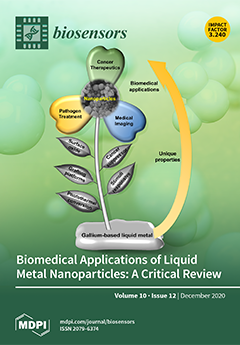
Gallium-based liquid metal alloys, as metallic fluids at ambient temperature, can be readily processed into nanoparticles of a size ranging from tens to hundreds of nanometers. These nanoparticles acquire many unique but useful properties that are beneficial in biomedical applications. This review seeks to highlight exciting advances in the use of the gallium-based liquid metal nanoparticles for biomedical applications, including cancer therapy, medical imaging, and pathogen treatment. We also offer a foresight to exemplify underexplored knowledge and highlighting the research challenges faced by LMNP science and technology in expanding into applications potentially yielding clinical advances. View this paper.
  • Issues are regarded as officially published after their release is announced to the table of contents alert mailing list.
  • You may sign up for e-mail alerts to receive table of contents of newly released issues.
  • PDF is the official format for papers published in both, html and pdf forms. To view the papers in pdf format, click on the "PDF Full-text" link, and use the free Adobe Reader to open them.
Order results
Result details
Section
Select all
Export citation of selected articles as:
16 pages, 5087 KiB  
Article
Silicon Nanowire Field-Effect Transistor as Biosensing Platforms for Post-Translational Modification
by Ping-Chia Su, Bo-Han Chen, Yi-Chan Lee and Yuh-Shyong Yang
Biosensors 2020, 10(12), 213; https://0-doi-org.brum.beds.ac.uk/10.3390/bios10120213 - 21 Dec 2020
Cited by 10 | Viewed by 2960
Abstract
Protein tyrosine sulfation (PTS), a vital post-translational modification, facilitates protein–protein interactions and regulates many physiological and pathological responses. Monitoring PTS has been difficult owing to the instability of sulfated proteins and the lack of a suitable method for detecting the protein sulfate ester. [...] Read more.
Protein tyrosine sulfation (PTS), a vital post-translational modification, facilitates protein–protein interactions and regulates many physiological and pathological responses. Monitoring PTS has been difficult owing to the instability of sulfated proteins and the lack of a suitable method for detecting the protein sulfate ester. In this study, we combined an in situ PTS system with a high-sensitivity polysilicon nanowire field-effect transistor (pSNWFET)-based sensor to directly monitor PTS formation. A peptide containing the tyrosine sulfation site of P-selectin glycoprotein ligand (PSGL)-1 was immobilized onto the surface of the pSNWFET by using 3-aminopropyltriethoxysilane and glutaraldehyde as linker molecules. A coupled enzyme sulfation system consisting of tyrosylprotein sulfotransferase and phenol sulfotransferase was used to catalyze PTS of the immobilized PSGL-1 peptide. Enzyme-catalyzed sulfation of the immobilized peptide was readily observed through the shift of the drain current–gate voltage curves of the pSNWFET before and after PTS. We expect that this approach can be developed as a next generation biochip for biomedical research and industries. Full article
(This article belongs to the Special Issue Semiconductor-Based Biosensors)
Show Figures

Graphical abstract

15 pages, 1931 KiB  
Article
Rapid Multianalyte Microfluidic Homogeneous Immunoassay on Electrokinetically Driven Beads
by Pierre-Emmanuel Thiriet, Danashi Medagoda, Gloria Porro and Carlotta Guiducci
Biosensors 2020, 10(12), 212; https://0-doi-org.brum.beds.ac.uk/10.3390/bios10120212 - 21 Dec 2020
Cited by 3 | Viewed by 3366
Abstract
The simplicity of homogeneous immunoassays makes them suitable for diagnostics of acute conditions. Indeed, the absence of washing steps reduces the binding reaction duration and favors a rapid and compact device, a critical asset for patients experiencing life-threatening diseases. In order to maximize [...] Read more.
The simplicity of homogeneous immunoassays makes them suitable for diagnostics of acute conditions. Indeed, the absence of washing steps reduces the binding reaction duration and favors a rapid and compact device, a critical asset for patients experiencing life-threatening diseases. In order to maximize analytical performance, standard systems employed in clinical laboratories rely largely on the use of high surface-to-volume ratio suspended moieties, such as microbeads, which provide at the same time a fast and efficient collection of analytes from the sample and controlled aggregation of collected material for improved readout. Here, we introduce an integrated microfluidic system that can perform analyte detection on antibody-decorated beads and their accumulation in confined regions within 15 min. We employed the system to the concomitant analysis of clinical concentrations of Neutrophil Gelatinase-Associated Lipocalin (NGAL) and Cystatin C in serum, two acute kidney injury (AKI) biomarkers. To this end, high-aspect-ratio, three-dimensional electrodes were integrated within a microfluidic channel to impart a controlled trajectory to antibody-decorated microbeads through the application of dielectrophoretic (DEP) forces. Beads were efficiently retained against the fluid flow of reagents, granting an efficient on-chip analyte-to-bead binding. Electrokinetic forces specific to the beads’ size were generated in the same channel, leading differently decorated beads to different readout regions of the chip. Therefore, this microfluidic multianalyte immunoassay was demonstrated as a powerful tool for the rapid detection of acute life-threatening conditions. Full article
(This article belongs to the Special Issue Microfluidic Bio-Sensors and Their Applications)
Show Figures

Figure 1

9 pages, 2472 KiB  
Article
DNA–Gold Nanozyme-Modified Paper Device for Enhanced Colorimetric Detection of Mercury Ions
by Min-Xin Mao, Rong Zheng, Chi-Fang Peng and Xin-Lin Wei
Biosensors 2020, 10(12), 211; https://0-doi-org.brum.beds.ac.uk/10.3390/bios10120211 - 18 Dec 2020
Cited by 20 | Viewed by 3094
Abstract
In this work, a paper device consisted of a patterned paper chip, wicking pads, and a base was fabricated. On the paper chip, DNA–gold nanoparticles (DNA–AuNPs) were deposited and Hg2+ ions could be adsorbed by the DNA–AuNPs. The formed DNA–AuNP/Hg2+ nanozyme [...] Read more.
In this work, a paper device consisted of a patterned paper chip, wicking pads, and a base was fabricated. On the paper chip, DNA–gold nanoparticles (DNA–AuNPs) were deposited and Hg2+ ions could be adsorbed by the DNA–AuNPs. The formed DNA–AuNP/Hg2+ nanozyme could catalyze the tetramethylbenzidine (TMB)–H2O2 chromogenic reaction. Due to the wicking pads, a larger volume of Hg2+ sample could be applied to the paper device for Hg2+ detection and therefore the color response could be enhanced. The paper device achieved a cut-off value of 50 nM by the naked eye for Hg2+ under optimized conditions. Moreover, quantitative measurements could be implemented by using a desktop scanner and extracting grayscale values. A linear range of 50–2000 nM Hg2+ was obtained with a detection limit of 10 nM. In addition, the paper device could be applied in the detection of environmental water samples with high recoveries ranging from 85.7% to 105.6%. The paper-device-based colorimetric detection was low-cost, simple, and demonstrated high potential in real-sample applications. Full article
(This article belongs to the Special Issue Cellulose-Based Biosensing Platforms)
Show Figures

Figure 1

8 pages, 1129 KiB  
Communication
Highly Sensitive Detection of CA 125 Protein with the Use of an n-Type Nanowire Biosensor
by Kristina A. Malsagova, Tatyana O. Pleshakova, Rafael A. Galiullin, Andrey F. Kozlov, Ivan D. Shumov, Vladimir P. Popov, Fedor V. Tikhonenko, Alexander V. Glukhov, Vadim S. Ziborov, Oleg F. Petrov, Vladimir E. Fortov, Alexander I. Archakov and Yuri D. Ivanov
Biosensors 2020, 10(12), 210; https://0-doi-org.brum.beds.ac.uk/10.3390/bios10120210 - 18 Dec 2020
Cited by 12 | Viewed by 2740
Abstract
The detection of CA 125 protein in a solution using a silicon-on-insulator (SOI)-nanowire biosensor with n-type chip has been experimentally demonstrated. The surface of nanowires was modified by covalent immobilization of antibodies against CA 125 in order to provide the biospecificity of the [...] Read more.
The detection of CA 125 protein in a solution using a silicon-on-insulator (SOI)-nanowire biosensor with n-type chip has been experimentally demonstrated. The surface of nanowires was modified by covalent immobilization of antibodies against CA 125 in order to provide the biospecificity of the target protein detection. We have demonstrated that the biosensor signal, which results from the biospecific interaction between CA 125 and the covalently immobilized antibodies, increases with the increase in the protein concentration. At that, the minimum concentration, at which the target protein was detectable with the SOI-nanowire biosensor, amounted to 1.5 × 10⁻¹⁶ M. Full article
(This article belongs to the Special Issue Electrical and Electro-Optical Biosensors)
Show Figures

Figure 1

22 pages, 68913 KiB  
Review
Review of Integrated Optical Biosensors for Point-of-Care Applications
by Yung-Tsan Chen, Ya-Chu Lee, Yao-Hsuan Lai, Jin-Chun Lim, Nien-Tsu Huang, Chih-Ting Lin and Jian-Jang Huang
Biosensors 2020, 10(12), 209; https://0-doi-org.brum.beds.ac.uk/10.3390/bios10120209 - 18 Dec 2020
Cited by 87 | Viewed by 8507
Abstract
This article reviews optical biosensors and their integration with microfluidic channels. The integrated biosensors have the advantages of higher accuracy and sensitivity because they can simultaneously monitor two or more parameters. They can further incorporate many functionalities such as electrical control and signal [...] Read more.
This article reviews optical biosensors and their integration with microfluidic channels. The integrated biosensors have the advantages of higher accuracy and sensitivity because they can simultaneously monitor two or more parameters. They can further incorporate many functionalities such as electrical control and signal readout monolithically in a single semiconductor chip, making them ideal candidates for point-of-care testing. In this article, we discuss the applications by specifically looking into point-of-care testing (POCT) using integrated optical sensors. The requirement and future perspective of integrated optical biosensors for POC is addressed. Full article
(This article belongs to the Special Issue Last Advances in Optical Biosensors)
Show Figures

Figure 1

12 pages, 13324 KiB  
Article
Interdigitated Sensor Optimization for Blood Sample Analysis
by Julien Claudel, Thanh-Tuan Ngo, Djilali Kourtiche and Mustapha Nadi
Biosensors 2020, 10(12), 208; https://0-doi-org.brum.beds.ac.uk/10.3390/bios10120208 - 16 Dec 2020
Cited by 10 | Viewed by 3129
Abstract
Interdigitated (ITD) sensors are specially adapted for the bioimpedance analysis (BIA) of low-volume (microliter scale) biological samples. Impedance spectroscopy is a fast method involving simple and easy biological sample preparation. The geometry of an ITD sensor makes it easier to deposit a sample [...] Read more.
Interdigitated (ITD) sensors are specially adapted for the bioimpedance analysis (BIA) of low-volume (microliter scale) biological samples. Impedance spectroscopy is a fast method involving simple and easy biological sample preparation. The geometry of an ITD sensor makes it easier to deposit a sample at the microscopic scale of the electrodes. At this scale, the electrode size induces an increase in the double-layer effect, which may completely limit interesting bandwidths in the impedance measurements. This work focuses on ITD sensor frequency band optimization via an original study of the impact of the metalization ratio α. An electrical sensor model was studied to determine the best α ratio. A ratio of 0.6 was able to improve the low-frequency cutoff by a factor of up to 2.5. This theoretical approach was confirmed by measurements of blood samples with three sensors. The optimized sensor was able to extract the intrinsic electrical properties of blood in the frequency band of interest. Full article
(This article belongs to the Special Issue Biosensors for Diagnosis and Monitoring)
Show Figures

Figure 1

21 pages, 2268 KiB  
Article
Advances in the Detection of Toxic Algae Using Electrochemical Biosensors
by Linda K. Medlin, Maria Gamella, Gerardo Mengs, Verónica Serafín, Susana Campuzano and José M. Pingarrón
Biosensors 2020, 10(12), 207; https://0-doi-org.brum.beds.ac.uk/10.3390/bios10120207 - 16 Dec 2020
Cited by 9 | Viewed by 3203
Abstract
Harmful algal blooms (HABs) are more frequent as climate changes and tropical toxic species move northward, especially along the Iberian Peninsula, a rich aquaculture area. Monitoring programs, detecting the presence of toxic algae before they bloom, are of paramount importance to protect ecosystems, [...] Read more.
Harmful algal blooms (HABs) are more frequent as climate changes and tropical toxic species move northward, especially along the Iberian Peninsula, a rich aquaculture area. Monitoring programs, detecting the presence of toxic algae before they bloom, are of paramount importance to protect ecosystems, aquaculture, human health and local economies. Rapid, reliable species identification methods using molecular barcodes coupled to biosensor detection tools have received increasing attention as an alternative to the legally required but impractical microscopic counting-based techniques. Our electrochemical detection system has improved, moving from conventional sandwich hybridization protocols using different redox mediators and signal probes with different labels to a novel strategy involving the recognition of RNA heteroduplexes by antibodies further labelled with bacterial antibody binding proteins conjugated with multiple enzyme molecules. Each change has increased sensitivity. A 150-fold signal increase has been produced with our newest protocol using magnetic microbeads (MBs) and amperometric detection at screen-printed carbon electrodes (SPCEs) to detect the target RNA of toxic species. We can detect as few as 10 cells L−1 for some species by using a fast (~2 h), simple (PCR-free) and cheap methodology (~2 EUR/determination) that will allow this methodology to be integrated into easy-to-use portable systems. Full article
(This article belongs to the Special Issue Sensors for Environmental Monitoring and Food Safety)
Show Figures

Graphical abstract

9 pages, 2891 KiB  
Communication
Development of a Graphene-Based Biosensor for Detecting Recombinant Cyanovirin-N
by Pedro Rodrigues de Almeida III, André Melro Murad, Luciano Paulino Silva, Elibio Leopoldo Rech and Elmo Salomão Alves
Biosensors 2020, 10(12), 206; https://0-doi-org.brum.beds.ac.uk/10.3390/bios10120206 - 16 Dec 2020
Cited by 9 | Viewed by 2905
Abstract
We present a graphene-based biosensor selective to recombinant cyanovirin-N (rCV-N), an antiviral protein that has proven to be an effective microbicide to inhibit HIV replication. We modified the graphene monolayer devices with 1-pyrenebutanoic acid succinimidyl ester, which interacts with both graphene and the [...] Read more.
We present a graphene-based biosensor selective to recombinant cyanovirin-N (rCV-N), an antiviral protein that has proven to be an effective microbicide to inhibit HIV replication. We modified the graphene monolayer devices with 1-pyrenebutanoic acid succinimidyl ester, which interacts with both graphene and the primary and secondary amines of antibodies. By monitoring the change in the electrical resistance of the device, we were able to detect rCV-N in solutions in the range of 0.01 to 10 ng/mL, and found that the detection limit was 0.45 pg/mL, which is much smaller than that obtained with currently available techniques. This is important for applications of this microbicide against HIV, since it may be produced at a large scale from soya bean seeds processed using the available industrial processing technologies. The sensor showed high sensitivity, selectivity, and reproducibility. Full article
(This article belongs to the Special Issue Field-Effect Transistors for Biosensing Applications)
Show Figures

Graphical abstract

34 pages, 51903 KiB  
Review
Recent Progress in Wearable Biosensors: From Healthcare Monitoring to Sports Analytics
by Shun Ye, Shilun Feng, Liang Huang and Shengtai Bian
Biosensors 2020, 10(12), 205; https://0-doi-org.brum.beds.ac.uk/10.3390/bios10120205 - 15 Dec 2020
Cited by 58 | Viewed by 15321
Abstract
Recent advances in lab-on-a-chip technology establish solid foundations for wearable biosensors. These newly emerging wearable biosensors are capable of non-invasive, continuous monitoring by miniaturization of electronics and integration with microfluidics. The advent of flexible electronics, biochemical sensors, soft microfluidics, and pain-free microneedles have [...] Read more.
Recent advances in lab-on-a-chip technology establish solid foundations for wearable biosensors. These newly emerging wearable biosensors are capable of non-invasive, continuous monitoring by miniaturization of electronics and integration with microfluidics. The advent of flexible electronics, biochemical sensors, soft microfluidics, and pain-free microneedles have created new generations of wearable biosensors that explore brand-new avenues to interface with the human epidermis for monitoring physiological status. However, these devices are relatively underexplored for sports monitoring and analytics, which may be largely facilitated by the recent emergence of wearable biosensors characterized by real-time, non-invasive, and non-irritating sensing capacities. Here, we present a systematic review of wearable biosensing technologies with a focus on materials and fabrication strategies, sampling modalities, sensing modalities, as well as key analytes and wearable biosensing platforms for healthcare and sports monitoring with an emphasis on sweat and interstitial fluid biosensing. This review concludes with a summary of unresolved challenges and opportunities for future researchers interested in these technologies. With an in-depth understanding of the state-of-the-art wearable biosensing technologies, wearable biosensors for sports analytics would have a significant impact on the rapidly growing field—microfluidics for biosensing. Full article
(This article belongs to the Special Issue Microfluidics for Biosensing)
Show Figures

Figure 1

18 pages, 4974 KiB  
Article
The Incorporation of Amplified Metal-Enhanced Fluorescence in a CMOS-Based Biosensor Increased the Detection Sensitivity of a DNA Marker of the Pathogenic Fungus Colletotrichum gloeosporioides
by Dorin Harpaz, Noam Alkan and Evgeni Eltzov
Biosensors 2020, 10(12), 204; https://0-doi-org.brum.beds.ac.uk/10.3390/bios10120204 - 13 Dec 2020
Cited by 8 | Viewed by 3054
Abstract
Half of the global agricultural fresh produce is lost, mainly because of rots that are caused by various pathogenic fungi. In this study, a complementary metal-oxide-semiconductor (CMOS)-based biosensor was developed, which integrates specific DNA strands that allow the detection of enoyl-CoA-hydratase/isomerase, which is [...] Read more.
Half of the global agricultural fresh produce is lost, mainly because of rots that are caused by various pathogenic fungi. In this study, a complementary metal-oxide-semiconductor (CMOS)-based biosensor was developed, which integrates specific DNA strands that allow the detection of enoyl-CoA-hydratase/isomerase, which is a quiescent marker of Colletotrichum gloeosporioides fungi. The developed biosensor mechanism is based on the metal-enhanced fluorescence (MEF) phenomenon, which is amplified by depositing silver onto a glass surface. A surface DNA strand is then immobilized on the surface, and in the presence of the target mRNA within the sample, the reporter DNA strand that is linked to horseradish peroxidase (HRP) enzyme will also bind to it. The light signal that is later produced from the HRP enzyme and its substrate is enhanced and detected by the coupled CMOS sensor. Several parameters that affect the silver-deposition procedure were examined, including silver solution temperature and volume, heating mode, and the tank material. Moreover, the effect of blocking treatment (skim milk or bovine serum albumin (BSA)) on the silver-layer stability and nonspecific DNA absorption was tested. Most importantly, the effect of the deposition reaction duration on the silver-layer formation and the MEF amplification was also investigated. In the study findings a preferred silver-deposition reaction duration was identified as 5–8 min, which increased the deposition of silver on the glass surface up to 13-times, and also resulted in the amplification of the MEF phenomenon with a maximum light signal of 50 relative light units (RLU). It was found that MEF can be amplified by a customized silver-deposition procedure that results in increased detection sensitivity. The implementation of the improved conditions increased the biosensor sensitivity to 3.3 nM (4500 RLU) with a higher detected light signal as compared to the initial protocol (400 RLU). Moreover, the light signal was amplified 18.75-, 11.11-, 5.5-, 11.25-, and 3.75-times in the improved protocol for all the tested concentrations of the target DNA strand of 1000, 100, 10, 3.3, and 2 nM, respectively. The developed biosensor system may allow the detection of the pathogenic fungus in postharvest produce and determine its pathogenicity state. Full article
(This article belongs to the Special Issue Fluorescence Biosensors 2020)
Show Figures

Figure 1

10 pages, 2582 KiB  
Article
A CRISPR/Cas12a Based Universal Lateral Flow Biosensor for the Sensitive and Specific Detection of African Swine-Fever Viruses in Whole Blood
by Jinghua Wu, Omar Mukama, Wei Wu, Zhiyuan Li, Jean De Dieu Habimana, Yinghui Zhang, Rong Zeng, Chengrong Nie and Lingwen Zeng
Biosensors 2020, 10(12), 203; https://0-doi-org.brum.beds.ac.uk/10.3390/bios10120203 - 10 Dec 2020
Cited by 31 | Viewed by 4624
Abstract
Cross-border pathogens such as the African swine fever virus (ASFV) still pose a socio-economic threat. Cheaper, faster, and accurate diagnostics are imperative for healthcare and food safety applications. Currently, the discovery of the Clustered Regularly Interspaced Short Palindromic Repeats (CRISPR) has paved the [...] Read more.
Cross-border pathogens such as the African swine fever virus (ASFV) still pose a socio-economic threat. Cheaper, faster, and accurate diagnostics are imperative for healthcare and food safety applications. Currently, the discovery of the Clustered Regularly Interspaced Short Palindromic Repeats (CRISPR) has paved the way for the diagnostics based on Cas13 and Cas12/14 that exhibit collateral cleavage of target and single-stranded DNA (ssDNA) reporter. The reporter is fluorescently labeled to report the presence of a target. These methods are powerful; however, fluorescence-based approaches require expensive apparatuses, complicate results readout, and exhibit high-fluorescence background. Here, we present a new CRISPR–Cas-based approach that combines polymerase chain reaction (PCR) amplification, Cas12a, and a probe-based lateral flow biosensor (LFB) for the simultaneous detection of seven types of ASFV. In the presence of ASFVs, the LFB responded to reporter trans-cleavage by naked eyes and achieved a sensitivity of 2.5 × 10⁻¹⁵ M within 2 h, and unambiguously identified ASFV from swine blood. This system uses less time for PCR pre-amplification and requires cheaper devices; thus, it can be applied to virus monitoring and food samples detection. Full article
(This article belongs to the Special Issue Biosensors for Body Fluid Analysis)
Show Figures

Figure 1

13 pages, 2532 KiB  
Article
Development of a Pharmacogenetic Lab-on-Chip Assay Based on the In-Check Technology to Screen for Genetic Variations Associated to Adverse Drug Reactions to Common Chemotherapeutic Agents
by Rosario Iemmolo, Valentina La Cognata, Giovanna Morello, Maria Guarnaccia, Mariamena Arbitrio, Enrico Alessi and Sebastiano Cavallaro
Biosensors 2020, 10(12), 202; https://0-doi-org.brum.beds.ac.uk/10.3390/bios10120202 - 09 Dec 2020
Cited by 2 | Viewed by 2558
Abstract
Background: Antineoplastic agents represent the most common class of drugs causing Adverse Drug Reactions (ADRs). Mutant alleles of genes coding for drug-metabolizing enzymes are the best studied individual risk factors for these ADRs. Although the correlation between genetic polymorphisms and ADRs is well-known, [...] Read more.
Background: Antineoplastic agents represent the most common class of drugs causing Adverse Drug Reactions (ADRs). Mutant alleles of genes coding for drug-metabolizing enzymes are the best studied individual risk factors for these ADRs. Although the correlation between genetic polymorphisms and ADRs is well-known, pharmacogenetic tests are limited to centralized laboratories with expensive or dedicated instrumentation used by specialized personnel. Nowadays, DNA chips have overcome the major limitations in terms of sensibility, specificity or small molecular detection, allowing the simultaneous detection of several genetic polymorphisms with time and costs-effective advantages. In this work, we describe the design of a novel silicon-based lab-on-chip assay able to perform low-density and high-resolution multi-assay analysis (amplification and hybridization reactions) on the In-Check platform. Methods: The novel lab-on-chip was used to screen 17 allelic variants of three genes associated with adverse reactions to common chemotherapeutic agents: DPYD (Dihydropyrimidine dehydrogenase), MTHFR (5,10-Methylenetetrahydrofolate reductase) and TPMT (Thiopurine S-methyltransferase). Results: Inter- and intra assay variability were performed to assess the specificity and sensibility of the chip. Linear regression was used to assess the optimal hybridization temperature set at 52 °C (R2 ≈ 0.97). Limit of detection was 50 nM. Conclusions: The high performance in terms of sensibility and specificity of this lab-on-chip supports its further translation to clinical diagnostics, where it may effectively promote precision medicine. Full article
(This article belongs to the Special Issue Microfluidic Bio-Sensors and Their Applications)
Show Figures

Figure 1

10 pages, 3290 KiB  
Article
Wearable Laser Doppler Flowmetry Sensor: A Feasibility Study with Smoker and Non-Smoker Volunteers
by Mou Saha, Viktor Dremin, Ilya Rafailov, Andrey Dunaev, Sergei Sokolovski and Edik Rafailov
Biosensors 2020, 10(12), 201; https://0-doi-org.brum.beds.ac.uk/10.3390/bios10120201 - 07 Dec 2020
Cited by 17 | Viewed by 4241
Abstract
Novel, non-invasive wearable laser Doppler flowmetry (LDF) devices measure real-time blood circulation of the left middle fingertip and the topside of the wrist of the left hand. The LDF signals are simultaneously recorded for fingertip and wrist. The amplitude of blood flow signals [...] Read more.
Novel, non-invasive wearable laser Doppler flowmetry (LDF) devices measure real-time blood circulation of the left middle fingertip and the topside of the wrist of the left hand. The LDF signals are simultaneously recorded for fingertip and wrist. The amplitude of blood flow signals and wavelet analysis of the signal are used for the analysis of blood perfusion parameters. The aim of this pilot study is to validate the accuracy of blood circulation measurements recorded by one such non-invasive wearable LDF device for healthy young non-smokers and smokers. This study reveals a higher level of blood perfusion in the non-smoker group compared to the smoker group and vice-versa for the variation of pulse frequency. This result can be useful to assess the sensitivity of the wearable LDF sensor in determining the effect of nicotine for smokers as compared to non-smokers and also the blood microcirculation in smokers with different pathologies. Full article
(This article belongs to the Special Issue Photonic Biosensors: Detection, Analysis and Medical Diagnostics)
Show Figures

Figure 1

11 pages, 2659 KiB  
Article
A Dual Immunological Raman-Enabled Crosschecking Test (DIRECT) for Detection of Bacteria in Low Moisture Food
by Cheng Pan, Binbin Zhu and Chenxu Yu
Biosensors 2020, 10(12), 200; https://0-doi-org.brum.beds.ac.uk/10.3390/bios10120200 - 04 Dec 2020
Cited by 7 | Viewed by 2272
Abstract
Among the physical, chemical and biological hazards that could arise with respect to food safety, bacterial contamination has been one of the main concerns in recent years. Bacterial contamination in low moisture foods (LMFs) was an emerging threat which used to draw less [...] Read more.
Among the physical, chemical and biological hazards that could arise with respect to food safety, bacterial contamination has been one of the main concerns in recent years. Bacterial contamination in low moisture foods (LMFs) was an emerging threat which used to draw less attention as LMFs were considered at low risk of such a hazard. Bacteria can survive in low moisture environments and cause foodborne diseases once they enter the digestive system. Common detection methods such as ELISA and PCR are not well suited to LMFs, as most of them operate under aqueous environments. In this study, a Dual Immunological Raman-Enabled Crosschecking Test (DIRECT) was developed for LMFs using a nano-scaled surface enhanced Raman scattering (SERS) biosensor platform and multivariate discriminant analysis with a portable Raman spectrometer. It could provide a limit of detection (LOD) of 102 CFU/g of bacteria in model LMFs, with a detection time of 30–45 min. It has the potential to become a quick screening method for on-site bacteria detection for LMFs to identify food safety risks in real time. Full article
(This article belongs to the Special Issue Biosensors for Food and Agricultural Research)
Show Figures

Figure 1

24 pages, 2179 KiB  
Review
Recent Progress in Manufacturing Techniques of Printed and Flexible Sensors: A Review
by Dinesh Maddipatla, Binu B. Narakathu and Massood Atashbar
Biosensors 2020, 10(12), 199; https://0-doi-org.brum.beds.ac.uk/10.3390/bios10120199 - 03 Dec 2020
Cited by 85 | Viewed by 7437
Abstract
This review provides an outlook on some of the significant research work done on printed and flexible sensors. Printed sensors fabricated on flexible platforms such as paper, plastic and textiles have been implemented for wearable applications in the biomedical, defense, food, and environmental [...] Read more.
This review provides an outlook on some of the significant research work done on printed and flexible sensors. Printed sensors fabricated on flexible platforms such as paper, plastic and textiles have been implemented for wearable applications in the biomedical, defense, food, and environmental industries. This review discusses the materials, characterization methods, and fabrication methods implemented for the development of the printed and flexible sensors. The applications, challenges faced and future opportunities for the printed and flexible sensors are also presented in this review. Full article
(This article belongs to the Section Biosensor and Bioelectronic Devices)
Show Figures

Figure 1

15 pages, 2831 KiB  
Article
A Comparative Study of Approaches to Improve the Sensitivity of Lateral Flow Immunoassay of the Antibiotic Lincomycin
by Kseniya V. Serebrennikova, Olga D. Hendrickson, Elena A. Zvereva, Demid S. Popravko, Anatoly V. Zherdev, Chuanlai Xu and Boris B. Dzantiev
Biosensors 2020, 10(12), 198; https://0-doi-org.brum.beds.ac.uk/10.3390/bios10120198 - 03 Dec 2020
Cited by 9 | Viewed by 2923
Abstract
This study provides a comparative assessment of the various nanodispersed markers and related detection techniques used in the immunochromatographic detection of an antibiotic lincomycin (LIN). Improving the sensitivity of the competitive lateral flow immunoassay is important, given the increasing demands for the monitoring [...] Read more.
This study provides a comparative assessment of the various nanodispersed markers and related detection techniques used in the immunochromatographic detection of an antibiotic lincomycin (LIN). Improving the sensitivity of the competitive lateral flow immunoassay is important, given the increasing demands for the monitoring of chemical contaminants in food. Gold nanoparticles (AuNPs) and CdSe/ZnS quantum dots (QDs) were used for the development and comparison of three approaches for the lateral flow immunoassay (LFIA) of LIN, namely, colorimetric, fluorescence, and surface-enhanced Raman spectroscopy (SERS)-based LFIAs. It was demonstrated that, for colorimetric and fluorescence analysis, the detection limits were comparable at 0.4 and 0.2 ng/mL, respectively. A SERS-based method allowed achieving the gain of five orders of magnitude in the assay sensitivity (1.4 fg/mL) compared to conventional LFIAs. Therefore, an integration of a SERS reporter into the LFIA is a promising tool for extremely sensitive quantitative detection of target analytes. However, implementation of this time-consuming technique requires expensive equipment and skilled personnel. In contrast, conventional AuNP- and QD-based LFIAs can provide simple, rapid, and inexpensive point-of-care testing for practical use. Full article
Show Figures

Figure 1

13 pages, 1732 KiB  
Article
An Improved HRPE-Based Transcriptional Output Reporter to Detect Hypoxia and Anoxia in Plant Tissue
by Gabriele Panicucci, Sergio Iacopino, Elisa De Meo, Pierdomenico Perata and Daan A. Weits
Biosensors 2020, 10(12), 197; https://0-doi-org.brum.beds.ac.uk/10.3390/bios10120197 - 03 Dec 2020
Cited by 12 | Viewed by 5526
Abstract
Oxygen levels in plant tissues may vary, depending on metabolism, diffusion barriers, and environmental availability. Current techniques to assess the oxic status of plant cells rely primarily on invasive microoptodes or Clark-type electrodes, which are not optimally suited for experiments that require high [...] Read more.
Oxygen levels in plant tissues may vary, depending on metabolism, diffusion barriers, and environmental availability. Current techniques to assess the oxic status of plant cells rely primarily on invasive microoptodes or Clark-type electrodes, which are not optimally suited for experiments that require high spatial and temporal resolution. In this case, a genetically encoded oxygen biosensor is required instead. This article reports the design, test, and optimization of a hypoxia-signaling reporter, based on five-time repeated hypoxia-responsive promoter elements (HRPE) driving the expression of different reporter proteins. Specifically, this study aimed to improve its performance as a reporter of hypoxic conditions by testing the effect of different untranslated regions (UTRs) at the 5′ end of the reporter coding sequence. Next, we characterized an optimized version of the HRPE promoter (HRPE-Ω) in terms of hypoxia sensitivity and time responsiveness. We also observed that severe oxygen deficiency counteracted the reporter activity due to inhibition of GFP maturation, which requires molecular oxygen. To overcome this limitation, we therefore employed an oxygen-independent UnaG fluorescent protein-coupled to an O2-dependent mCherry fluorophore under the control of the optimized HRPE-Ω promoter. Remarkably, this sensor, provided a different mCherry/UnaG ratiometric output depending on the externally imposed oxygen concentration, providing a solution to distinguish between different degrees of tissue hypoxia. Moreover, a ubiquitously expressed UnaG-mCherry fusion could be used to image oxygen concentrations directly, albeit at a narrow range. The luminescent and fluorescent hypoxia-reporters described here can readily be used to conduct studies that involve anaerobiosis in plants. Full article
Show Figures

Figure 1

21 pages, 7253 KiB  
Review
Biomedical Applications of Liquid Metal Nanoparticles: A Critical Review
by Haiyue Li, Ruirui Qiao, Thomas P. Davis and Shi-Yang Tang
Biosensors 2020, 10(12), 196; https://0-doi-org.brum.beds.ac.uk/10.3390/bios10120196 - 30 Nov 2020
Cited by 60 | Viewed by 6451
Abstract
This review is focused on the basic properties, production, functionalization, cytotoxicity, and biomedical applications of liquid metal nanoparticles (LMNPs), with a focus on particles of the size ranging from tens to hundreds of nanometers. Applications, including cancer therapy, medical imaging, and pathogen treatment [...] Read more.
This review is focused on the basic properties, production, functionalization, cytotoxicity, and biomedical applications of liquid metal nanoparticles (LMNPs), with a focus on particles of the size ranging from tens to hundreds of nanometers. Applications, including cancer therapy, medical imaging, and pathogen treatment are discussed. LMNPs share similar properties to other metals, such as photothermal conversion ability and a propensity to form surface oxides. Compared to many other metals, especially mercury, the cytotoxicity of gallium is low and is considered by many reports to be safe when applied in vivo. Recent advances in exploring different grafting molecules are reported herein, as surface functionalization is essential to enhance photothermal therapeutic effects of LMNPs or to facilitate drug delivery. This review also outlines properties of LMNPs that can be exploited in making medical imaging contrast agents, ion channel regulators, and anti-pathogenic agents. Finally, a foresight is offered, exemplifying underexplored knowledge and highlighting the research challenges faced by LMNP science and technology in expanding into applications potentially yielding clinical advances. Full article
(This article belongs to the Special Issue Liquid Metal Enabled Bio-Related Applications)
Show Figures

Figure 1

11 pages, 2860 KiB  
Article
Label-Free and Sensitive Determination of Cadmium Ions Using a Ti-Modified Co3O4-Based Electrochemical Aptasensor
by Yang Liu, Dongwei Zhang, Jina Ding, Kashif Hayat, Xijia Yang, Xuejia Zhan, Dan Zhang, Yitong Lu and Pei Zhou
Biosensors 2020, 10(12), 195; https://0-doi-org.brum.beds.ac.uk/10.3390/bios10120195 - 30 Nov 2020
Cited by 11 | Viewed by 2330
Abstract
The current work demonstrates an electrochemical aptasensor for sensitive determination of Cd2+ based on the Ti-modified Co3O4 nanoparticles. In this unlabeled system, Ti-modified Co3O4 nanoparticles act as current signal amplifiers modified on the screen-printed carbon electrode [...] Read more.
The current work demonstrates an electrochemical aptasensor for sensitive determination of Cd2+ based on the Ti-modified Co3O4 nanoparticles. In this unlabeled system, Ti-modified Co3O4 nanoparticles act as current signal amplifiers modified on the screen-printed carbon electrode (SPCE) surface, while the derivative aptamer of Cd2+ works as a target recognizer. In addition, the sensing is based on the increase in electrochemical probe thionine current signal due to the binding of aptamer to Cd2+ via specific recognition. In the current study, key parameters, including aptamer concentration, pH, and incubation time were optimized, respectively, to ensure sensing performance. Cyclic voltammetry was used not only to characterize each preparation and optimization step, but also to profile the bindings of aptamer to Cd2+. Under optimal conditions, Cd2+ can be determined in a linear range of 0.20 to 15 ng/mL, with a detection limit of 0.49 ng/mL, significantly below the maximum concentration limit set by the U.S. Environmental Protection Agency. Based on comparative analysis and the results of recovery test with real samples, this simple, label-free but highly selective method has considerable potential and thus can be used as an in-situ environmental monitoring platform for Cd2+ testing. Full article
(This article belongs to the Special Issue New Developments for Efficient Rapid Bioassays)
Show Figures

Figure 1

19 pages, 6181 KiB  
Review
A Systematic Review of Food Allergy: Nanobiosensor and Food Allergen Detection
by Adriano Aquino and Carlos Adam Conte-Junior
Biosensors 2020, 10(12), 194; https://0-doi-org.brum.beds.ac.uk/10.3390/bios10120194 - 29 Nov 2020
Cited by 35 | Viewed by 5713
Abstract
Several individuals will experience accidental exposure to an allergen. In this sense, the industry has invested in the processes of removing allergenic compounds in food. However, accidental exposure to allergenic proteins can result from allergenic substances not specified on labels. Analysis of allergenic [...] Read more.
Several individuals will experience accidental exposure to an allergen. In this sense, the industry has invested in the processes of removing allergenic compounds in food. However, accidental exposure to allergenic proteins can result from allergenic substances not specified on labels. Analysis of allergenic foods is involved in methods based on immunological, genetic, and mass spectrometry. The traditional methods have some limitations, such as high cost. In recent years, biosensor and nanoparticles combined have emerged as sensitive, selective, low-cost, and time-consuming techniques that can replace classic techniques. Nevertheless, each nanomaterial has shown a different potential to specific allergens or classes. This review used Preferred Reporting Items for Systematic Reviews and the Meta-Analysis guidelines (PRISMA) to approach these issues. A total of 104 articles were retrieved from a standardized search on three databases (PubMed, Scopus and Web of Science). The systematic review article is organized by the category of allergen detection and nanoparticle detection. This review addresses the relevant biosensors and nanoparticles as gold, carbon, graphene, quantum dots to allergen protein detection. Among the selected articles it was possible to notice a greater potential application on the allergic proteins Ah, in peanuts and gold nanoparticle-base as a biosensor. We envision that in our review, the association between biosensor and nanoparticles has shown promise in the analysis of allergenic proteins present in different food samples. Full article
(This article belongs to the Special Issue Nanoparticles-Based Biosensors)
Show Figures

Figure 1

27 pages, 4462 KiB  
Review
Proximal Methods for Plant Stress Detection Using Optical Sensors and Machine Learning
by Alanna V. Zubler and Jeong-Yeol Yoon
Biosensors 2020, 10(12), 193; https://0-doi-org.brum.beds.ac.uk/10.3390/bios10120193 - 29 Nov 2020
Cited by 38 | Viewed by 8443
Abstract
Plant stresses have been monitored using the imaging or spectrometry of plant leaves in the visible (red-green-blue or RGB), near-infrared (NIR), infrared (IR), and ultraviolet (UV) wavebands, often augmented by fluorescence imaging or fluorescence spectrometry. Imaging at multiple specific wavelengths (multi-spectral imaging) or [...] Read more.
Plant stresses have been monitored using the imaging or spectrometry of plant leaves in the visible (red-green-blue or RGB), near-infrared (NIR), infrared (IR), and ultraviolet (UV) wavebands, often augmented by fluorescence imaging or fluorescence spectrometry. Imaging at multiple specific wavelengths (multi-spectral imaging) or across a wide range of wavelengths (hyperspectral imaging) can provide exceptional information on plant stress and subsequent diseases. Digital cameras, thermal cameras, and optical filters have become available at a low cost in recent years, while hyperspectral cameras have become increasingly more compact and portable. Furthermore, smartphone cameras have dramatically improved in quality, making them a viable option for rapid, on-site stress detection. Due to these developments in imaging technology, plant stresses can be monitored more easily using handheld and field-deployable methods. Recent advances in machine learning algorithms have allowed for images and spectra to be analyzed and classified in a fully automated and reproducible manner, without the need for complicated image or spectrum analysis methods. This review will highlight recent advances in portable (including smartphone-based) detection methods for biotic and abiotic stresses, discuss data processing and machine learning techniques that can produce results for stress identification and classification, and suggest future directions towards the successful translation of these methods into practical use. Full article
(This article belongs to the Special Issue Biosensors for Food and Agricultural Research)
Show Figures

Figure 1

19 pages, 5162 KiB  
Review
BODIPY Dyes as Probes and Sensors to Study Amyloid-β-Related Processes
by Sergei V. Dzyuba
Biosensors 2020, 10(12), 192; https://0-doi-org.brum.beds.ac.uk/10.3390/bios10120192 - 27 Nov 2020
Cited by 23 | Viewed by 4962
Abstract
Amyloid formation plays a major role in a number of neurodegenerative diseases, including Alzheimer’s disease. Amyloid-β peptides (Aβ) are one of the primary markers associated with this pathology. Aβ aggregates exhibit a diverse range of morphologies with distinct pathological activities. Recognition of the [...] Read more.
Amyloid formation plays a major role in a number of neurodegenerative diseases, including Alzheimer’s disease. Amyloid-β peptides (Aβ) are one of the primary markers associated with this pathology. Aβ aggregates exhibit a diverse range of morphologies with distinct pathological activities. Recognition of the Aβ aggregates by using small molecule-based probes and sensors should not only enhance understanding of the underlying mechanisms of amyloid formation, but also facilitate the development of therapeutic strategies to interfere with amyloid neurotoxicity. BODIPY (boron dipyrrin) dyes are among the most versatile small molecule fluorophores. BODIPY scaffolds could be functionalized to tune their photophysical properties to the desired ranges as well as to adapt these dyes to various types of conditions and environments. Thus, BODIPY dyes could be viewed as unique platforms for the design of probes and sensors that are capable of detecting and tracking structural changes of various Aβ aggregates. This review summarizes currently available examples of BODIPY dyes that have been used to investigate conformational changes of Aβ peptides, self-assembly processes of Aβ, as well as Aβ interactions with various molecules. Full article
(This article belongs to the Special Issue Last Advances in Optical Biosensors)
Show Figures

Figure 1

11 pages, 1664 KiB  
Letter
Waveguide-Based Fluorescent Immunosensor for the Simultaneous Detection of Carbofuran and 3-Hydroxy-Carbofuran
by Weiming Sun, Lanhua Liu, Abdul Ghaffar Memon, Xiaohong Zhou and Hongwei Zhao
Biosensors 2020, 10(12), 191; https://0-doi-org.brum.beds.ac.uk/10.3390/bios10120191 - 27 Nov 2020
Cited by 10 | Viewed by 2271
Abstract
Carbofuran (CBF) is an efficient and broad-spectrum insecticide. As testing indicators for water quality and agricultural products, CBF and its metabolite 3-hydroxy-carbofuran (3-OH-CBF) are regulated by many countries. The detection of CBF and 3-OH-CBF is of great importance for the environment and human [...] Read more.
Carbofuran (CBF) is an efficient and broad-spectrum insecticide. As testing indicators for water quality and agricultural products, CBF and its metabolite 3-hydroxy-carbofuran (3-OH-CBF) are regulated by many countries. The detection of CBF and 3-OH-CBF is of great importance for the environment and human health. However, an immunosensor detection method for the simultaneous analysis of CBF and 3-OH-CBF remains unavailable. Herein, we report a waveguide-based fluorescent immunosensor for detecting CBF and 3-OH-CBF, synchronously. The immunosensor is based on a broad-spectrum monoclonal antibody with high binding affinity against CBF and 3-OH-CBF. The linear detection ranges for CBF and 3-OH-CBF are 0.29–2.69 and 0.12–4.59 μg/L, with limits of detection of 0.13 μg/L for CBF and 0.06 μg/L for 3-OH-CBF, respectively. The whole detection process for each cycle is approximately 30 min. The results show a good application prospect for the rapid detection of CBF and 3-OH-CBF in water or agricultural products. Full article
(This article belongs to the Special Issue Last Advances in Optical Biosensors)
Show Figures

Figure 1

8 pages, 12041 KiB  
Communication
A Rapid Colorimetric Assay for On-Site Authentication of Cephalopod Species
by Giuseppina Tatulli, Paola Cecere, Davide Maggioni, Andrea Galimberti and Pier Paolo Pompa
Biosensors 2020, 10(12), 190; https://0-doi-org.brum.beds.ac.uk/10.3390/bios10120190 - 24 Nov 2020
Cited by 8 | Viewed by 2953
Abstract
A colorimetric assay, exploiting the combination of loop-mediated isothermal amplification (LAMP) with DNA barcoding, was developed to address the authentication of some cephalopod species, a relevant group in the context of seafood traceability, due to the intensive processing from the fishing sites to [...] Read more.
A colorimetric assay, exploiting the combination of loop-mediated isothermal amplification (LAMP) with DNA barcoding, was developed to address the authentication of some cephalopod species, a relevant group in the context of seafood traceability, due to the intensive processing from the fishing sites to the shelf. The discriminating strategy relies on accurate design of species-specific LAMP primers within the conventional 5’ end of the mitochondrial COI DNA barcode region and allows for the identification of Loligo vulgaris among two closely related and less valuable species. The assay, coupled to rapid genomic DNA extraction, is suitable for large-scale screenings and on-site applications due to its easy procedures, with fast (30 min) and visual readout. Full article
(This article belongs to the Section Nano- and Micro-Technologies in Biosensors)
Show Figures

Figure 1

13 pages, 2783 KiB  
Article
A Multichannel Microfluidic Sensing Cartridge for Bioanalytical Applications of Monolithic Quartz Crystal Microbalance
by María Calero, Román Fernández, Pablo García, José Vicente García, María García, Esther Gamero-Sandemetrio, Ilya Reviakine, Antonio Arnau and Yolanda Jiménez
Biosensors 2020, 10(12), 189; https://0-doi-org.brum.beds.ac.uk/10.3390/bios10120189 - 24 Nov 2020
Cited by 8 | Viewed by 3128
Abstract
Integrating acoustic wave sensors into lab-on-a-chip (LoC) devices is a well-known challenge. We address this challenge by designing a microfluidic device housing a monolithic array of 24 high-fundamental frequency quartz crystal microbalance with dissipation (HFF-QCMD) sensors. The device features six 6-µL channels of [...] Read more.
Integrating acoustic wave sensors into lab-on-a-chip (LoC) devices is a well-known challenge. We address this challenge by designing a microfluidic device housing a monolithic array of 24 high-fundamental frequency quartz crystal microbalance with dissipation (HFF-QCMD) sensors. The device features six 6-µL channels of four sensors each for low-volume parallel measurements, a sealing mechanism that provides appropriate pressure control while assuring liquid confinement and maintaining good stability, and provides a mechanical, electrical, and thermal interface with the characterization electronics. We validate the device by measuring the response of the HFF-QCMD sensors to the air-to-liquid transition, for which the robust Kanazawa–Gordon–Mason theory exists, and then by studying the adsorption of model bioanalytes (neutravidin and biotinylated albumin). With these experiments, we show how the effects of the protein–surface interactions propagate within adsorbed protein multilayers, offering essentially new insight into the design of affinity-based bioanalytical sensors. Full article
(This article belongs to the Special Issue Microfluidics for Biosensing)
Show Figures

Graphical abstract

Previous Issue
Next Issue
Back to TopTop